Roles of mucus adhesion and cohesion in
cough clearance
Brian Buttona,b,c, Henry P. Goodella,c, Eyad Atieha, Yu-Cheng Chena, Robert Williamsa, Siddharth Shenoya,c,
Elijah Lackeya, Nathan T. Shenkutea, Li-Heng Caid,e, Robert G. Dennisc, Richard C. Bouchera, and Michael Rubinsteina,f,g,h,i,1 aMarsico Lung Institute, University of North Carolina at Chapel Hill, Chapel Hill, NC 27599-7248;bDepartment of Biochemistry and Biophysics, University of North Carolina at Chapel Hill, Chapel Hill, NC 27599-7260;cDepartment of Biomedical Engineering, University of North Carolina at Chapel Hill, Chapel Hill, NC 27599-7575;dDepartment of Materials Science and Engineering, University of Virginia, Charlottesville, VA 22904;eDepartment of Chemical Engineering, University of Virginia, Charlottesville, VA 22904;fDepartment of Mechanical Engineering and Materials Science, Duke University, Durham, NC 27708;gDepartment of Biomedical Engineering, Duke University, Durham, NC 27708;hDepartment of Physics, Duke University, Durham, NC 27708; andiDepartment of Chemistry, Duke University, Durham, NC 27708
Edited by Peter Agre, Johns Hopkins Bloomberg School of Public Health, Baltimore, MD, and approved September 28, 2018 (received for review July 9, 2018)
Clearance of intrapulmonary mucus by the high-velocity airflow generated by cough is the major rescue clearance mechanism in subjects with mucoobstructive diseases and failed cilial-dependent mucus clearance, e.g., subjects with cystic fibrosis (CF) or chronic obstructive pulmonary disease (COPD). Previous studies have in-vestigated the mechanical forces generated at airway surfaces by cough but have not considered the effects of mucus biophysical properties on cough efficacy. Theoretically, mucus can be cleared by cough from the lung by an adhesive failure, i.e., breaking mucus-cell surface adhesive bonds and/or by cohesive failure, i.e., directly fracturing mucus. Utilizing peel-testing technologies, mucus-epithelial surface adhesive and mucus cohesive strengths were mea-sured. Because both mucus concentration and pH have been reported to alter mucus biophysical properties in disease, the effects of mucus concentration and pH on adhesion and cohesion were com-pared. Both adhesive and cohesive strengths depended on mucus concentration, but neither on physiologically relevant changes in pH nor bicarbonate concentration. Mucus from bronchial epithelial cultures and patient sputum samples exhibited similar adhesive and cohesive properties. Notably, the magnitudes of both adhesive and cohesive strength exhibited similar velocity and concentration de-pendencies, suggesting that viscous dissipation of energy within mucus during cough determines the efficiency of cough clearance of diseased, hyperconcentrated, mucus. Calculations of airflow-induced shear forces on airway mucus related to mucus concentra-tion predicted substantially reduced cough clearance in small versus large airways. Studies designed to improve cough clearance in sub-jects with mucoobstructive diseases identified reductions of mucus concentration and viscous dissipation as key therapeutic strategies.
airway physiology
|
lung disease|
mucus clearance|
cough|
cystic fibrosisT
he pulmonary mucus clearance system represents a keyin-nate host defense system that has evolved to protect the lung from inhaled pathogens and particulates. A principal component of the mucus clearance system is the mucin-rich mucus layer that is responsible for binding inhaled foreign materials and pathogens. In health, the mucus layer is a viscoelastic reversible gel, composed of:
(i)∼1.1% (0.01 g/mL) organic content, including∼0.5 wt % mucins
and∼0.6% globular proteins; (ii) 0.9% salt; and (iii) 98% water (1).
Upon release into this dilute (watery) milieu, the mucin oligomers,
which are stored in intracellular granules as compact (∼350 nm
di-ameter) structures, swell and unfold into the linear strands which form the structure of transportable airway mucus (2, 3). Conse-quently, efficient cilia-dependent mucus clearance in health requires a balance of ion and water transport, mucin secretion, and ciliary beat. Progress in understanding how cilia-dependent mucus transport is successful in health and how it fails in disease, producing intrapulmonary mucus accumulation, has emerged from a novel
description of the mucus transport system (4). This“gel-on-brush”
model describes how concentration-dependent osmotic moduli distribute water between the mucus layer and the periciliary layer
(PCL). In diseases like cystic fibrosis (CF), abnormal ion transport produces a liquid-depleted airway surface (5) with a more con-centrated than normal mucus, increasing from 0.01 g/mL organic content (2% solids) up to 0.2 g/mL organic content (21% solids), with the proportional (20-fold) increase in mucin concentration (6). As a reflection of the increased mucus concentration in CF, the osmotic modulus of the mucus layer exceeds the osmotic modulus of the PCL, resulting in osmotic compression of the PCL by the mucus layer, failure of cilia-mediated clearance, and ulti-mately, mucus layer adherence to the airway surfaces (4). This scenario is consistent with reports of increased mucin concentra-tions in CF airway secreconcentra-tions and scanning EM images of mucus on the airways of lungs excised from CF patients (6, 7).
There has been less progress in understanding how cough can clear accumulated, typically hyperconcentrated mucus in disease. During the expiratory phase of cough, high-speed airflow results in momentum transfer to mucus accumulated on airway surfaces, propelling the mucus toward the larynx. Previous analyses of cough have focused on airflow/shear-induced flow of non-Newtonian mucus mimics (8, 9) or airway samples from patients with bronchiectasis in plastic tubes (10). These studies demon-strated that increases in sample viscosity and elasticity produced slower airflow-mediated transport. However, to fully elucidate
Significance
Mucoobstructive lung diseases, including chronic obstructive pulmonary disease, asthma, and cystic fibrosis, are character-ized by intrapulmonary accumulations of hyperconcentrated mucus. Ultimately, mucus accumulation in disease reflects the failure of the major rescue mucus clearance pathway, i.e., cough. Studies were performed to understand how abnormal mucus and its interactions with the cell surface produce a failure of cough clearance. These studies identified mucus concentration-dependent cohesive and adhesive properties, governed by mucus viscous energy dissipation, as rate limiting for the efficiency of cough clearance. Parallel studies designed to restore mucus cough clearability identified reduction of mucus concentration (rehydration) and use of mucolytics as additive and promising therapeutic strategies.
Author contributions: B.B., R.C.B., and M.R. designed research; B.B., H.P.G., E.A., Y.-C.C., R.W., S.S., E.L., N.T.S., and L.-H.C. performed research; B.B., H.P.G., L.-H.C., and R.G.D. contributed new reagents/analytic tools; B.B., E.A., Y.-C.C., R.W., S.S., E.L., and N.T.S. analyzed data; and B.B., R.C.B., and M.R. wrote the paper.
The authors declare no conflict of interest.
This article is a PNAS Direct Submission.
Published under thePNAS license.
See Commentary on page 12340.
1To whom correspondence should be addressed. Email: [email protected].
This article contains supporting information online atwww.pnas.org/lookup/suppl/doi:10. 1073/pnas.1811787115/-/DCSupplemental.
Published online November 12, 2018.
MEDICAL
SCIENCES
SEE
COM
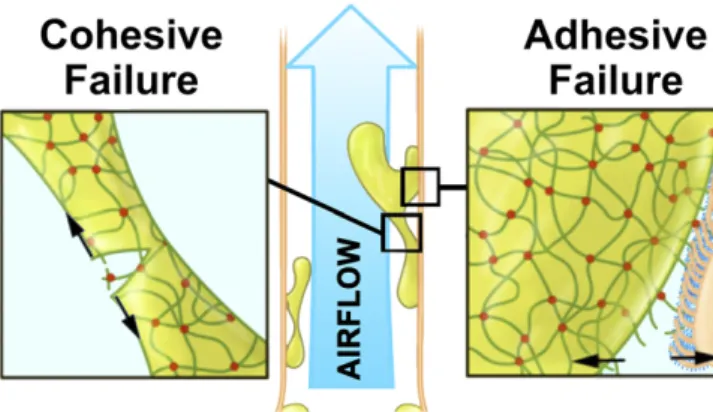
how accumulated mucus responds to cough-induced shear forces, it is necessary to understand the biophysical interactions between airway mucus and airway surfaces. We postulated that two mechanisms may participate in high-speed airflow removal of
mucus from airway surfaces with cough: (i) “disadhesion,” i.e.,
overcoming the“adhesive”interactions/bonds between mucus and
the airway cell surface, whereby accumulated mucus is physically
stripped off the airway surface; and (ii) “tearing,”i.e., breaking
mucus–mucus cohesive bonds, resulting in portions of mucus
breaking off adherent mucus masses (Fig. 1).
In this study, we investigated the properties of mucus that
govern: (i) the adhesive strength of mucus to the cell surface; and
(ii) mucus cohesive strength. Because of the conflicting notions
favoring concentration versus pH in producing changes in the biophysical properties of mucus pertinent to cough efficiency, the roles of mucus concentration versus mucus pH/bicarbonate on adhesion and cohesion were compared (11). These studies were performed using in vitro peel-test systems to directly measure the adhesive and cohesive forces of mucus produced by human bronchial epithelial (HBE) cultures from normal (i.e., nondiseased) and CF individuals and sputum from individuals with mucoob-structive lung disease. Finally, studies were performed to identify single or combination therapies that reduced the adhesive and/or cohesive forces of concentrated mucus, to improve mucus clearance in patients with mucoobstructive lung diseases.
Results
Concentration and Velocity Dependence of Mucus/PCL Adhesive
Strength. Mucus adhesion strength (i.e., fracture toughness) is
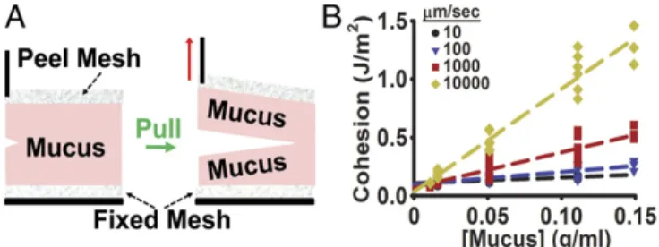
defined as the energy per unit area required to separate mucus from the PCL (Fig. 1). Mucus-airway surface adhesive strength was
assessed using a peel-testing device (Fig. 2A) that measured the force
required to“peel”the mucus layer off the surface of the epithelium
of well-differentiated HBE cultures covered by an endogenous
mu-cus layer (seeSI Appendixfor more details about this system).
The goal of these studies was to assess the effects of mucus concentration and peeling velocity on the strength of adhesion. To vary concentration, normal HBE cultures were generated with a wide range of mucus concentrations, spanning normal (2% solids, 0.01 g/mL organic content) to severe CF-like ranges (up to 21% solids, 0.2 g/mL organic content) (6, 12). The peeling velocity is predicted to be important for adhesion because energy is dissi-pated both at the mucus/PCL interface and within the mucus layer as it deforms and ultimately separates from the PCL (13). The higher the velocity of separation, the more energy must be dissi-pated. Here, studies were performed over a range of peeling
ve-locities, from 10 to 5,000μm/s, as the crack propagation velocity of
mucus upon disadhesion will likely be considerably smaller than
the airflow velocity, which can reach 300 m/s in the largest airways (14). The magnitude of adhesive strength was both
concentration-and velocity dependent (Fig. 2B).
Relationship of Proposed CF-Specific pH/HCO3− Abnormalities on
Mucus Adhesive Strength. Studies were conducted to compare
effects of mucus concentration versus reduced mucus pH or bi-carbonate levels on adhesive strength. Airway epithelia-mucus ad-hesive strength was measured in non-CF HBE cultures with mucus
produced under three conditions: (i) normal pH (7.4) and normal
bicarbonate (25 mM HCO3); (ii) 7.4 pH and 0 mM HCO3; and
(iii) reduced pH (6.6) and 25 mM HCO3. The linear fits of the
concentration dependence of the strength of adhesion for each
pH/HCO3−condition were indistinguishable (Fig. 3A). This result
demonstrates that adhesion strength was controlled by concentra-tion and not by changes in either mucus pH or bicarbonate levels over the tested concentration ranges. Taken together, these data suggest that mucus concentration, and not pH/bicarbonate, is the dominant factor controlling mucus adhesion strength.
We next asked whether mucus produced by CF versus normal (non-CF) HBE cultures had properties other than increased con-centration that made CF mucus more or less adherent to the cell surface. Accordingly, these experiments directly compared the adhesive strength of mucus produced by CF vs. non-CF airway cultures over a range of defined mucus concentrations. Both CF (red inverted triangles) and non-CF (black circles) cultures ex-hibited a similar concentration dependence on adhesion strength
(Fig. 3B). Collectively, these data demonstrate that there was no
difference in the adhesive strength at the interfaces between CF and non-CF HBE mucus and their respective epithelial surfaces when compared at the same concentration over a range of mucus concentrations. However, as the mucus concentration is reported to be significantly higher in CF individuals, the magnitude of
Fig. 1. Conceptual model of airflow-mediated mucus clearance from airways. Clearance of airway mucus is mediated by: (i) cohesive failure involving fracture of the mucus layer by tearing mucin strands (dark-green lines); or (ii) adhesive failure requiring disruption of mucus–PCL layer (dark-blue strands surrounding the cilia) interactions, stripping the mucus layer off the cell surface.
Fig. 2. Peel test of mucus–PCL adhesive strength. (A) Schematic diagram of airway epithelia in profile detailing the embedded mesh in the mucus con-nected by a silk thread to a motor and force sensor. (B) Data showing the effect of mucus concentration on adhesive strength at various peeling ve-locities: 10μm/s (black circles), 100μm/s (blue inverted triangles), 1 mm/s (red squares), and 5 mm/s (gold diamonds).
adhesive strength is predicted to be correspondingly higher in CF airways, compared with nondiseased individuals (6).
Concentration and Crack Propagation Velocity Dependence of
Cohesive Strength.As shown in Fig. 1, the high-velocity airflow
associated with cough may accelerate the clearance of accumu-lated, thickened mucus from airway surfaces by tearing discrete mucus masses off adherent mucus plaques. As part of this process,
it is necessary to physically tear apart the mucin–mucin (and/or
other protein–protein) bonds/associations that hold mucus
to-gether. The goal of these studies was to measure the magnitude of the cohesive strength of mucus and investigate the effect of mucus concentration and crack propagation velocity.
For cohesion studies, a modified version of the peel tester was
used to measure the force required to tear mucus apart (Fig. 4A).
The cohesive strength of mucus was measured in a series of studies using mucus isolated from normal (non-CF) HBE cultures. As with adhesive strength, the cohesive strength of normal HBE mucus was dependent on the tearing velocity over a range of mucus concen-trations spanning from normal (0.1 g/mL) to severe CF lung disease
(0.11 g/mL) (Fig. 4B). This result indicates that CF-like concentrated
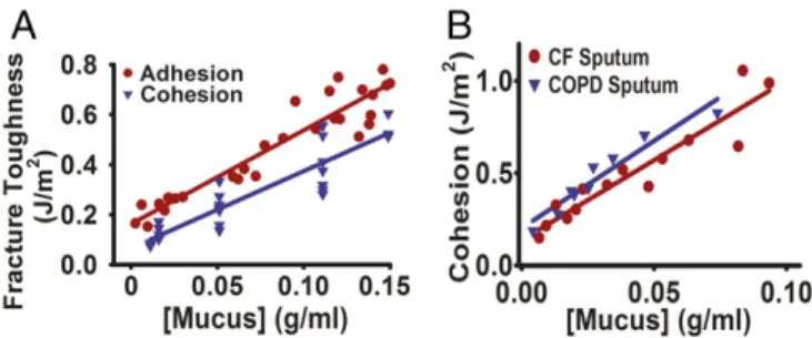
mucus will require more force to tear at all velocities compared with mucus at normal (nondiseased) concentrations. Of particular in-terest is that our data demonstrate that mucus adhesion and co-hesion exhibited a similar dependence on mucus concentration,
over a range spanning from normal to CF (Fig. 5A).
An advantage of the cohesive peel tester is the ability to in-vestigate the properties of samples derived in vivo. Consequently, potential CF-specific pH or other effects could be identified by comparison with sputum from subjects with other mucoobstructive lung diseases with normal cystic fibrosis transmembrane conductance regulator (CFTR) function. As a mucoobstructive disease control, samples from chronic obstructive pulmonary disease (COPD) sub-jects, who also produce sputum (expectorated mucus) with increased concentrations, were studied (15). Importantly, measurements of the cohesive strength of sputum from subjects with a wide array of dis-ease severity were included. CF and COPD sputum samples exhibited cohesive strengths that were strongly correlated with
mucus concentration, but not disease type (Fig. 5B).
Reduction of Adhesive and Cohesive Strength with Therapeutic
Agents.The finding that mucus adhesive and cohesive strengths
increased with increased mucus concentration suggests that therapies directed at decreasing mucus concentration with agents which hydrate the airways would be effective in reducing adhesive/cohesive fracture toughness. Studies were performed to compare the effect of hydrating agents (i.e., saline) versus more classic mucolytic agents on the adhesive and cohesive strengths of mucus. To mimic in vivo delivery and minimize effects on concentration, each mucolytic compound was nebulized in small volumes (nl) onto the surface of normal HBE cultures (16).
Data in Fig. 6A demonstrate that the adhesive strength of
concentrated HBE mucus (0.16 g/mL) was significantly reduced by the addition of saline to reduce mucus concentration by half (i.e., 0.08 g/mL). In addition to reducing concentration, we tested the hypothesis that reducing energy dissipation during mucus
adhesive fracture at the mucus layer–cell surface interface would
also be effective. One approach was to reduce mucin polymer
length with a dithiol reducing agent [N-acetylcysteine (NAC),
100 mM]. NAC was quite effective in reducing adhesive strength of concentrated (0.16 g/mL final) mucus. As a second approach, we
tested the hypothesis that disruption of mucin–mucin hydrophobic
interactions and/or surface tension at the mucus layer–cell
sur-face intersur-face would reduce adhesive strength. A surfactant (Nonidet P-40, 0.01%) (17) produced a significant decrease in the adhesion strength of concentrated mucus (at 0.16 g/mL fi-nal). The relationships between adhesive strength (shown as the reciprocal), mucus concentration, therapeutic maneuvers, and
improvement in cough clearance are depicted in Fig. 6B.
Similar studies were performed to characterize strategies to
reduce mucus cohesive strength (Fig. 6CandD). In these studies,
cohesive strength was measured before and after the addition of a surfactant (Nonidet P-40, 0.01% final) and a reducing agent (DTT, 20 mM final) in the absence of mucus concentration changes (i.e., all performed at the same 0.12-g/mL mucus con-centration as the control). Both agents were effective in reducing the magnitude of mucus cohesive strength in the absence of a change in mucus concentration. To test the effect of mucus hy-dration, mucus concentration was reduced by half (from 0.12 to 0.06 g/mL) with the addition of saline. A substantial reduction in cohesion was observed with dilution, i.e., hydration. Finally, to test whether the cohesive strength of partially rehydrated mucus (at 0.06 g/mL) could be further reduced with the addition of reducing agents, studies were conducted with the combination of saline and DTT. The combination produced a further decrease in co-hesion strength compared with saline alone.
Discussion
Cough constitutes an important backup mechanism to remove mucus from the lungs of subjects with lung disease. After acute or chronic accumulation of mucus in the lung, clearance of mucus by the high-velocity airflow associated with cough often becomes the sole mechanism for mucus clearance. Our model of cough (Fig. 1) suggests that there are at least two modes by which mucus can be
cleared by cough from the lungs, including: (i) overcoming adhesive
interactions between the mucus and cell surface to peel mucus off
airway surfaces; and/or (ii) fracturing mucus itself, i.e., overcoming
mucus cohesive interactions, to clear mucus in fragments.
The energy per unit area needed to separate mucus from the cell surface/PCL defines the adhesion strength (i.e., adhesive
fracture toughness) of the mucus–PCL interface. The minimum
energy needed to separate two surfaces in contact is called the
work of adhesion (Wa), which defines the fracture toughness at
zero velocity. In this study, we developed a peel-testing device capable of measuring the adhesive strength between the mucus layer and the cell surface. An important observation from our
studies was that at normal mucus concentrations (∼0.01 g/mL, 2%
total solids) the work of adhesion at zero velocity was only about three times the surface tension of mucus (18). Therefore, despite
mucus being characterized as“sticky,”the low concentrations of
mucins in the mucus layer in health produced only a small
con-tribution to mucus–PCL adhesion above the water–water surface
tension forces generated at the mucus–PCL layer interface.
However, when mucus becomes more concentrated, as in CF, the strength of adhesion between mucus and the PCL increased. The simplest interpretation of these findings is that when mucus con-centration and, hence, mucin concon-centration (6) is increased, ad-ditional connections between the mucus and cell surface produce an increase in adhesion strength.
A key variable that governed the magnitude of the mucus
layer–cell surface adhesive strength was the rate by which the
mucus layer was peeled off the epithelial surface. The higher the rate of peeling, i.e., peeling velocity, the greater the force re-quired to separate the mucus from the airway surface. This
Fig. 4. Mucus cohesion. (A) Schematic representation of dual-mesh peel test with mucus positioned between the two meshes. (B) Data showing the effect of non-CF HBE mucus concentration on cohesive strength at various peeling velocities: 10μm/s (black circles), 100μm/s (blue triangles), 1 mm/s (red squares), and 10 mm/s (gold diamonds).
MEDICAL
SCIENCES
SEE
COM
higher force reflects the higher energy dissipation both at the crack and in the bulk mucus layer (19). The dissipative
compo-nent of fracture toughness (Gv) at a given crack propagation
velocity (v) is proportional to the thermodynamic work of
ad-hesion (Wa), reflecting the fact that the energy loss increases
when the interface is stressed (20). The dissipative component of
the fracture toughness can be expressed as: Gv = Γ − Wa =
Waϕ(v), where Γ is the fracture toughness and ϕ is a
di-mensionless function of crack propagation velocity (v) (21). This
function is often found to increase as a power of crack
propa-gation velocity ϕ(v)∼ vβ(22). Consistent with this notion, our
data demonstrated that the adhesion strength was dependent not only on the concentration of mucus but also on peeling rate.
In addition to stripping mucus off the airway surface (i.e., disadhesion), we hypothesized that airflow might tear fragments off adherent mucus masses and carry them out of the airway to the larynx. This type of clearance requires that the force of air-flow cohesively breaks mucus. Unlike disadhesion, this mode of failure results in the airway surface still being covered by a layer of adherent mucus. Like adhesion, there is a work of cohesion
(Wc), which is the energy per unit area required to produce two
new surfaces when a material is divided into two parts at very low crack propagation velocities.
To investigate cohesive failure in cough clearance, we measured the force required to pull mucus apart. The measured force per unit area required to tear mucus apart describes the cohesive strength of mucus. Our studies revealed that the cohesive strength of airway mucus was linearly dependent on the concentration of the mucus layer. As with adhesion, the cohesive strength of healthy mucus was found to be very low, i.e., slightly above twice the surface tension of mucus (18). However, when HBE mucus became more concentrated, it became increasingly difficult to pull
it apart. A similar cohesion–concentration relationship for sputum
samples from CF and non-CF subjects was observed (Fig. 5B),
suggesting that concentration is the common variable dominating cohesive strength. Importantly, the mucus cohesive strength again was highly dependent on the rate of tearing. The faster the mucus was pulled, the harder it was to pull mucus apart, owing to the increase in energy dissipated at higher velocities.
An unexpected finding was that the adhesive and cohesive fracture toughness had similar dependencies on mucus
concen-tration and peeling velocity (Fig. 5A), suggesting that a common
dominant mechanism controlled this mucus property. The
magnitude of both adhesive and cohesive fracture toughness (Γ)
and their dependence on mucus concentration (c) and crack
propagation velocity (v) can be written as:
Γ=2γ
"
1+c
co+
c coβ3.3
v vo
β#
, [1]
whereγis the surface tension of mucus (18),cois the
character-istic concentration at which work of adhesion (Wa)/cohesion
(Wc) doubles (atv=0),v0is the characteristic peeling velocity
at which the dissipative component of fracture toughness is
com-parable to the work of adhesion/cohesion (atc=co), andβis the
dynamic exponent. The first term in Eq.1, (2γ), represents the
contribution of surface tension to the work of adhesion/cohesion.
The second term, (2γc/co), represents the contribution to the
work of cohesion/adhesion (atv=0) from intermolecular bond
breaking and/or mucin polymers being pulled out from the
op-posite sides of the crack. Based on data in Figs. 2Band 4B, this
term has a linear dependence on the mucus concentration, as the quantity of mucus/mucins and the corresponding number of in-terfacial bonds/associations increase linearly with concentration.
The third term, [2γc/(coβ3.3)](v/vo)β, represents velocity-dependent
viscous dissipation, which has been proposed to be related to the dynamic moduli of polymers (23).
Since both adhesion and cohesion are dependent on the vis-cous dissipation, we conjectured that this term dominates at high velocities and produces the similar concentration and velocity dependencies in both adhesion and cohesion. To test this
as-sumption, Eq.1was rearranged to the form:
ðΓ−2γÞ
2γc =
1
co+ 1
β3.3
co
v vo
β
, [2]
and the term of (Γ−2γ)/(2γc) for both adhesion and cohesion
was plotted as a function of peel velocity (Fig. 7A). Fitting the
peeling velocity dependence of (Γ−2γ)/(2γc) to a constant plus a
power law (Eq.2) for the adhesion data, we obtainedβ=0.43±
0.04,co=0.078±0.020 g/mL, andcovoβ=7.1±1.1 g/mL(m/s)0.43.
For the cohesion data, the best fit resulted inβ=0.38±0.11,co=
0.49±2.03 g/mL, andcovoβ=10.6±1.6 g/mL(m/s)0.38. The large
error in the estimation ofcoreflects the relatively small
contribu-tion of the c/co term compared with the other terms in Eq. 1.
Fig. 6. Therapeutic treatments to reduce mucus adhesion/cohesive interac-tions. (A) Adhesive strength of mucus (0.16 g/mL) before (Ctrl) and after ad-dition of a surfactant (Nonidet P-40; 0.01%) or reducing agent (NAC; final concentration ∼100 mM). For comparison, a mucus hydrator (saline) was added to reduce final mucus concentration to∼0.08 g/mL. (B) The relationship between changes in reciprocal adhesive strength (black line; plotted as the inverse fracture toughness) over the range of mucus concentrations (in Fig. 2B) showing the effect of mucolytics (NAC and Nonidet P-40) and rehydration. A larger value (i.e., lower fracture toughness) is expected to result in an im-provement of cough clearance (right axis). Red dashed line denotes the in-verse of two times the mucus surface tension (γ). (C) Mucus cohesive strength of non-CF HBE mucus (0.12 g/mL) before (Ctrl) and after addition of a sur-factant (Nonidet P-40; 0.01% final), a reducing agent (DTT; 20 mM final), and mucus hydrator (saline to reduce mucus concentration to 0.06 g/mL). Also shown is the effect of combining a hydrator and reducing agent (saline+DTT, at 0.06 g/mL). (*P<0.05 vs. control. All data are presented as mean±SD at 1 mm/s). (D) Plot showing the relationship between changes in reciprocal cohesive strength in response to hydrators and mucolytics, similar toB.
Therefore, in the range of concentrations employed in this study
(0.01–0.19 g/mL), the work of adhesion (Wa) was similar to the
work of cohesion (Wc). Importantly, the dynamic exponent (β) of
the dissipative component of fracture toughness,Gv∼ϕ(v)∼vβ,
obtained from the fits of adhesion and cohesion data were similar, i.e., within uncertainty of each other, and close to the exponent of
the frequency dependence of the mucus loss modulus,G′′(ω)∼ωβ.
This result suggests that viscous dissipation represents the common major contributor to both adhesive and cohesive fracture toughness at high velocities, consistent with the dependence of both on crack
propagation velocity (Fig. 7A) (13). Since the work of adhesion and
cohesion are similar and viscous dissipation within mucus in both adhesion and cohesion processes are also similar, both concentra-tion and peel velocity dependencies of mucus adhesive and cohe-sive strengths were similar to each other. Notably, the observation that all mucus was removed from the airway surface in the peel assay suggests adhesive failure dominates in mucus expectoration during cough.
Studies of cohesive strength after treatment with mucolytics,
such as the reducing agent DTT (Fig. 6C), demonstrated that
cohesive strength of mucus can be reduced without altering mucus concentration. To elucidate which properties of mucus as described
in Eq.1were altered when mucus was treated with DTT, the
co-hesive strength of HBE mucus before and after reduction with DTT
over a range of velocities was fitted to Eq.2(Fig. 7B). When treated
with DTT, the fit resulted in similar values ofβ(0.44±0.12 for DTT
vs. 0.43±0.08 for control) andco(0.51±0.89 g/mL for DTT vs.
0.83±0.52 g/mL for control). However, the termcovoβwas
signif-icantly different between the two groups, 38.8±0.7 g/mL(m/s)0.44
for DTT vs. 11.5 ± 0.6 g/mL(m/s)0.43 for control. The
inter-pretation of this finding is that treating mucus with DTT increased its relaxation rate (decreases its viscous dissipation) and,
there-fore, increased the characteristic velocity (vo) at which fracture
toughness became significant. This change of mucus properties in response to DTT was predictable, i.e., lower molecular weight mucins produced by reduction have shorter relaxation times and, thus, higher relaxation rates, consistent with the theory of entangled polymer solutions (24, 25).
Cough efficiency is determined by both the forces applied to mucus by airflow and mucus-airway surface properties. During cough, air flows through the proximal airways at very high
ve-locities (vair), reaching hundreds of meters per second in large
airways (14). This high airflow velocity creates large shear
stresses at the air–mucus interface. The stress imparted by air (σ)
can be estimated from the dynamic pressure (pd =ρ·vair2/2) as
σ=f·pd/4, wherefis the Darcy friction factor (26) andρis the
density of air. Dynamic pressures vary from∼1 Pa forvair≈1 m/s
during tidal breathing to∼104Pa forvair≈100 m/s during cough
(27). The Darcy friction factor f decreases with an increasing
Reynolds numberRe=vairD/μin an airway with diameterDand
a kinematic viscosity of air (μ) of≈10−5m2/s. For a laminar flow,
f=64/Re, whereas for turbulent flow, (f) decreases more slowly
and saturates at a valuef≈0.02–0.04 depending on the
rough-ness of the mucus surface (28). Accordingly, the shear stress at
mucus surfaces reachesσ≈100 Pa at high velocities, e.g.,vair≈
100 m/s (airway generations 0–3), but shear stress is onlyσ≈1 Pa,
or even lower, for smaller cough velocities (vair≈10 m/s) in
smaller airways (generations>7) (27, 29).
The effects of forces applied by airflow to mucus to produce expectoration also require analyses of mucus mass (height). The
force per unit length applied on adherent mucus of thickness (L)
on an airway surface is proportional to the product ofLand
sur-face shear stress. Thus, for shear stresses ofσ≈100 Pa, the force
per unit length applied on mucus of heightL≈1 cm is∼1.0 J/m2,
while for a smaller accumulated mass of sizeL≈1 mm, the force
per unit length is∼0.1 J/m2. The latter value is below the adhesive
fracture toughness of mucus at any concentration (Figs. 2Band
4B). Therefore, high airflow-induced stresses in larger airways
would be capable of peeling off larger mucus accumulations with
anLof∼1 cm. This prediction is supported by the observation that
the mass of sputum expectorated by cough in subjects with
mucoobstructive lung disease averages∼1 g (corresponding to the
volume ∼1 cm3) (30). In contrast, in smaller airways (<2 mm,
generations 7 and greater), mucus cannot reach a mass (height) that would permit shear stress forces to exceed adhesive fracture toughness at any concentration. This prediction is consistent with a recent study by Dunican et al. (31) demonstrating a failure of distal airways to clear adherent mucus plugs over time in subjects with severe asthma.
Our studies were designed to also ask whether reported
CFTR-mediated defects in HCO3−secretion/airway surface acidification
(11) produced abnormalities in mucus adhesive/cohesive proper-ties in addition to, or instead of, changes in mucus concentration. Specifically, studies were performed to investigate whether de-creases in mucus bicarbonate and/or pH resulted in the pro-duction of a mucus that was more adherent to the cell surface. Studies of non-CF HBE cultures, in which mucus was produced in the presence or absence of bicarbonate, coupled with comparisons to CF cultures, demonstrated that neither low airway surface bi-carbonate concentration, lower pH, nor potentially altered mucin glycosylation patterns (32) contributed to the adhesive interac-tions of the mucus layer to the PCL. These findings are consistent with recent data suggesting that pH/bicarbonate had little effect on the viscoelastic properties of airway mucus, whereas concen-tration effects were large (33).
Notably, our analyses of the adhesive/cohesive properties of HBE mucus in vitro appear relevant to studies of sputum produced by patients. CF sputum exhibited a concentration dependence of cohesion similar to that predicted from HBE mucus studies. Sputum was also obtained from subjects with COPD, another mucoobstructive lung disease with high mucus/sputum mucin concentrations (15). Like CF, COPD mucus cohesive properties
were highly correlated with mucus concentration (Fig. 5A).
One of the goals of therapeutics for mucoobstructive lung dis-eases is to mobilize nonclearable mucus from the surfaces of the lung. Our data suggest that use of mucus hydrators, such as saline or hypertonic saline, to reduce the concentration of the mucus represents the simplest way to reduce adhesive and cohesive forces. However, our studies also suggest that reducing agents and surfactants that break or disrupt interactions that contribute to the viscous dissipation in mucus may also provide significant benefit to patients interdependent of mucus concentration. Indeed, our data demonstrate that hydration and mucolytics exhibit additive activ-ities on mucus adhesion and cohesion strength and suggest that combination therapies may be most effective.
In summary, a common property of mucus, i.e., viscous dissi-pation, dominates the adhesive and cohesive properties that govern the efficiency of cough clearance. Both adhesive and cohesive strengths were strongly correlated with changes in mucus concentration but not pH. Analyses of airflow shear forces juxtaposed to mucus properties predict that disease-like hyperconcentrated mucus cannot be coughed out of small airways and clearance from large airways requires high airflows/shear Fig. 7. Energy dissipation in adhesive and cohesive fracture toughness.
(A) Adhesive (red circles) and cohesive (blue inverted triangles) fracture toughness data plotted using Eq.2. (B) Cohesive strength of non-CF HBE mucus before (red circles) and after (blue inverted triangles) treatment with DTT (20 mM), over a range of peeling velocities fitted to Eq.2.
MEDICAL
SCIENCES
SEE
COM
applied to an accumulation of relatively large masses (1 cm) of mucus. The concentration dependence of cohesive and adhesive properties of mucus suggests that the failure to effectively clear the lung of accumulated mucus by cough in CF reflects a con-centration-, not pH-, dependent airway surface defect. Resto-ration of cough efficacy may be most effectively provided by restoring mucus concentrations to normal ranges with hydrating agents coupled with viscosity-lowering agents.
Materials and Methods
Primary Cell Culture.HBE cells from normal donors and CF patients were obtained from the University of North Carolina Cystic Fibrosis Tissue Culture Core under the auspices of protocols approved by the UNC Institutional Review Board using previously reported methodologies (34).
Adhesion Peel Test.To measure the adhesion between the mucus and PCL layers exhibited by cell cultures, a peel-test system (35) was constructed (Fig. 2A). To peel the mucus layer from the underlying epithelial layer, a laser-cut 0.3×7.0-mm porous cellulose mesh (Kimberly Clark) was UV sterilized and carefully positioned on the apical surface of HBE cultures with endogenous followed by incubation for 12–16 h within a tissue culture incubator (seeSI Appendixfor additional details).
Cohesion Peel Test.The cohesive strength of mucus was measured in a device similar to that used to measure adhesive strength. Here, a thin layer of mucus was positioned between two laser-cut peel meshes; one (0.5×15.0 mm) was connected to the force sensor and the other (0.7×18 mm) was affixed to the bottom of a glass recording chamber via double-sided medical tape (#1522; 3M) (Fig. 4A). Mucus samples were incubated for 15 min before peeling. (The calibration procedures for both adhesion and cohesion assays are described in theSI Appendix).
Mucus Harvesting.Mucus for the cohesion peel experiments was harvested from well-differentiated non-CF HBE cultures as previously described (36). Briefly, a large number of HBE cultures (between 24 and 96) were allowed to accumulate mucus for up to 4 wk. On the day of the cohesive measurements, the mucus
was lavaged by incubating the apical surface with a small volume of saline (50μl/cm2) for 30 min at 37 °C. Mucus samples were then carefully removed from the culture using a positive-displacement pipetter (Gilson) and pooled. The dilute mucus samples were pooled and spin-concentrated (Ultra 10K; Amicon) at 4,000 g (4 °C) to the desired mucus concentration. Mucus concentration at each step was determined as previously described (37). In these studies, mucus samples were used on the same day as prepared and never frozen.
Sputum Collection.Spontaneous sputum samples were collected as detailed previously (6). Approval was received from the UNC Institutional Review Board for use of excess human tissue specimens for studies described here. Samples from anonymized donors with CF and COPD were collected on ice and assayed on the day of collection.
Delivery of Test Agents.In studies measuring mucus adhesion of endogenous mucus, each test solution (DTT, NAC at 10×stocks in PBS) was nebulized to the luminal surface of HBE cultures using a specially modified ultrasonic nebulizer (38) (Aeroneb Pro; Aerogen) at a rate of∼200 nl/min. The volume of nebulization (hence delivery time) was 100 nl drug/μl mucus, which was estimated by XZ-confocal microscopy in parallel cultures (39). For the co-hesion studies, 1μl of each agent (100×stock) was added directly to 100μl of mucus and mixed for 15 s by careful stirring, avoiding shearing and bubble formation.
Statistics.The means were analyzed using ANOVA for multiple comparisons. Two-tailed Student’sttests were used for analyzing all other experiments. A two-tailedztest was used to compare the linear fits of two populations of data. Sigma Plot (Systat) was used was used for all analysis. APvalue less than 0.05 was considered significant for all statistical analysis.
ACKNOWLEDGMENTS.We thank Dr. Scott Donaldson for assistance with sputum sample collection and Eric Roe for editorial assistance. We also acknowledge the UNC Marsico Lung Institute Tissue Core for cell culture. This project was funded by the Cystic Fibrosis Foundation (Grants BUTTON07XX0, RUBINS09XX0, and BOUCHE15R0), NIH (Grants R01HL125280, R01HL136961, P01HL108808, and P30DK065988), and NSF (Grants EFMA-1830957 and DMR-1121107).
1. Sheehan JK, Kesimer M, Pickles R (2006) Innate immunity and mucus structure and function.Novartis Found Symp279:155–166, discussion 167–169, 216–219. 2. Verdugo P, Deyrup-Olsen I, Martin AW, Luchtel DL (1992) Polymer gel phase
transi-tion: The molecular mechanism of product release in mucin secretion?Mechanics of Swelling(Springer, Berlin), pp 671–681.
3. Verdugo P (1990) Goblet cells secretion and mucogenesis.Annu Rev Physiol52: 157–176.
4. Button B, et al. (2012) A periciliary brush promotes the lung health by separating the mucus layer from airway epithelia.Science337:937–941.
5. Boucher RC (2007) Cystic fibrosis: A disease of vulnerability to airway surface de-hydration.Trends Mol Med13:231–240.
6. Henderson AG, et al. (2014) Cystic fibrosis airway secretions exhibit mucin hyper-concentration and increased osmotic pressure.J Clin Invest124:3047–3060. 7. Lopez-Vidriero MT, Reid L (1976) Pathophysiology of mucus secretion in cystic fibrosis.
Mod Probl Paediatr19:120–128.
8. Zahm JM, et al. (1991) Role of simulated repetitive coughing in mucus clearance.Eur Respir J4:311–315.
9. King M, Brock G, Lundell C (1985) Clearance of mucus by simulated cough.J Appl Physiol (1985)58:1776–1782.
10. Tambascio J, et al. (2013) The influence of purulence on ciliary and cough transport in bronchiectasis.Respir Care58:2101–2106.
11. Tang XX, et al. (2016) Acidic pH increases airway surface liquid viscosity in cystic fi-brosis.J Clin Invest126:879–891.
12. Anderson WH, et al. (2015) The relationship of mucus concentration (hydration) to mucus osmotic pressure and transport in chronic bronchitis.Am J Respir Crit Care Med
192:182–190.
13. Gent AN, Lai S-M (1994) Interfacial bonding, energy dissipation, and adhesion.
J Polym Sci Part B Polym Phys32:1543–1555.
14. Farzan S (1990)Cough and Sputum Production(Butterworths, Oxford).
15. Kesimer M, et al. (2017) Airway mucin concentration as a marker of chronic bron-chitis.N Engl J Med377:911–922.
16. Button B, Okada SF, Frederick CB, Thelin WR, Boucher RC (2013) Mechanosensitive ATP release maintains proper mucus hydration of airways.Sci Signal6:ra46. 17. Rubin BK (2002) Physiology of airway mucus clearance.Respir Care47:761–768. 18. Tarran R, Grubb BR, Gatzy JT, Davis CW, Boucher RC (2001) The relative roles of
passive surface forces and active ion transport in the modulation of airway surface liquid volume and composition.J Gen Physiol118:223–236.
19. Creton C, Ciccotti M (2016) Fracture and adhesion of soft materials: A review.Rep Prog Phys79:046601.
20. Maugis D, Barquins M (1978) Fracture mechanics and the adherence of viscoelastic bodies.J Phys D Appl Phys11:1989–2023.
21. Brown HR (1991) The adhesion between polymers.Annu Rev Mater Sci21:463–489. 22. Maugis D (1985) Subcritical crack growth, surface energy, fracture toughness,
stick-slip and embrittlement.J Mater Sci20:3041–3073.
23. Zosel A (1985) Adhesion and tack of polymers: Influence of mechanical properties and surface tensions.Colloid Polym Sci263:541–553.
24. Doi M, Edwards SF (1986)The Theory of Polymer Dynamics(Clarendon, Oxford). 25. de Gennes PG, Leger L (1982) Dynamics of entangled polymer chains.Annu Rev Phys
Chem33:49–61.
26. Massey BS, Ward-Smith AJ (2012)Mechanics of Fluids(Spon Press, London). 27. Leith DE (1968) Cough.Phys Ther48:439–447.
28. Haaland SE (1983) Simple and explicit formulas for the friction factor in turbulent pipe flow.J Fluids Eng105:89.
29. Leith DE (1985) The development of cough.Am Rev Respir Dis131:S39–S42. 30. Alexis NE, et al. (2016) Baseline sputum parameters in normals, asthmatics, COPD,
atopics, smokers and ex-smokers.J Allergy Clin Immunol137:AB208.
31. Dunican EM, et al.; National Heart Lung and Blood Institute (NHLBI) Severe Asthma Research Program (SARP) (2018) Mucus plugs in patients with asthma linked to eo-sinophilia and airflow obstruction.J Clin Invest128:997–1009.
32. Xia B, Royall JA, Damera G, Sachdev GP, Cummings RD (2005) Altered O-glycosylation and sulfation of airway mucins associated with cystic fibrosis.Glycobiology15: 747–775.
33. Long R, et al. (2017) The effects of weight percent solids and hydrogen ion activity on three common mucus systems.Pediatr Pulmonol52:S279.
34. Fulcher ML, Gabriel S, Burns KA, Yankaskas JR, Randell SH (2005) Well-differentiated human airway epithelial cell cultures.Methods Mol Med107:183–206.
35. Bundy K, Schlegel U, Rahn B, Geret V, Perren S (2000) An improved peel test method for measurement of adhesion to biomaterials.J Mater Sci Mater Med11:517–521. 36. Hill DB, et al. (2014) A biophysical basis for mucus solids concentration as a candidate
biomarker for airways disease.PLoS One9:e87681.
37. Hill DB, Button B (2012) Establishment of respiratory air-liquid interface cultures and their use in studying mucin production, secretion, and function.Methods Mol Biol
842:245–258.