R E S E A R C H
Open Access
Effect of transient scrotal hyperthermia on
human sperm: an iTRAQ-based proteomic
analysis
Yan-Qing Wu
1†, Meng Rao
2†, Shi-Fu Hu
1, Dan-Dan Ke
3, Chang-Hong Zhu
1,4and Wei Xia
1,4*Abstract
Background:Through this prospective study, we aimed to explore the change of molecular modification after the transient scrotal hyperthermia on human sperm.
Methods:Ten healthy subjects selected with strict screening criteria underwent testicular warming in a 43 °C water bath for 30 min a day for 10 consecutive days. Semen samples were collected 2 weeks before the first heat treatment and 6 weeks after the first heat treatment. Proteins from the samples were labeled with isobaric tags for relative and
absolute quantitation and analyzed by two-dimensional liquid chromatography–tandem mass spectrometry.
Results:In contrast to the control, of the 3446 proteins identified, 61 proteins were deregulated: 28 were up-regulated and 33 were down-regulated. Approximately 95% of the differentially expressed proteins were found to participate in spermatogenesis, fertilization, or other aspects of reproduction. In particular, the expression of sperm motility and energy metabolism-related proteins AKAP4, SPESP1, ODF1, ODF2, GAPDHS, and ACTRT2, validated by western blotting of the proteins obtained from human and mouse samples, tended to be reduced under scrotal hyperthermia. Conclusions:The results indicated that the proteins AKAP4, ODF1, ODF2, GAPDHS, SPESP1, and ACTRT2, play an important role in the heat-induced reversible reduction in sperm concentration and motility and have the potential to be the biomarkers and clinical targets for scrotal heat treatment induced male infertility.
Keywords:Sperm, Proteomics, ITRAQ, Hyperthermia
Introduction
In mammals, including humans, the scrotal temperature required for normal spermatogenesis within the testes is 2 °C to 8 °C lower than the core body temperature. Some patients with varicocele and people in certain occupations, such as boilermakers, cab drivers, and sedentary people, may develop scrotal hyperthermia, resulting in low sperm
concentration and mobility and, consequently, infertility. However, once the influencing factor disappears and the scrotal temperature returns to 33 °C–35 °C, spermatogen-esis, and sperm parameters revert to normal after one or two spermatogenic cycles [1–3].
Exposing the testes of mice [4], rats [5,6], monkeys [7], bulls [8], sheep [9], and humans [10,11] to high tempera-tures induced germ cell apoptosis in spermatogenesis [12]. Heat stress in the scrotum induced the apoptosis of germ cells [13–16], imbalances in antioxidant levels leading to oxidative stress [17–19], and deregulation in the gene expression pattern [20]. Comparative proteomics of testis biopsy specimens of men subjected to transient scrotal hyperthermia has revealed that these deregulated proteins
© The Author(s). 2020Open AccessThis article is licensed under a Creative Commons Attribution 4.0 International License, which permits use, sharing, adaptation, distribution and reproduction in any medium or format, as long as you give appropriate credit to the original author(s) and the source, provide a link to the Creative Commons licence, and indicate if changes were made. The images or other third party material in this article are included in the article's Creative Commons licence, unless indicated otherwise in a credit line to the material. If material is not included in the article's Creative Commons licence and your intended use is not permitted by statutory regulation or exceeds the permitted use, you will need to obtain permission directly from the copyright holder. To view a copy of this licence, visithttp://creativecommons.org/licenses/by/4.0/. The Creative Commons Public Domain Dedication waiver (http://creativecommons.org/publicdomain/zero/1.0/) applies to the data made available in this article, unless otherwise stated in a credit line to the data.
* Correspondence:[email protected]
†Yan-Qing Wu and Meng Rao contributed equally to this work.
1Institute of Reproductive Health, Tongji Medical College, Huazhong
University of Science and Technology, Wuhan, People’s Republic of China 4Reproductive Medicine Center, Tongji Medical College, Huazhong University
were associated with germ cell proliferation and apoptosis [21]. Heat stress not only destroys spermatocytes but also affects sperm maturation in the caput epididymis [22]. Ex-posing sperm to hyperthermia in vitro has been shown to reduce total sperm motility and metabolic activity [23]. In a previous study, we found that oxidative stress, mito-chondrial dysfunction, and tight packaging of sperm DNA could induce reversible impairment of spermatogenesis [24]. We also found that sperm concentration and motility in human subjects who experienced scrotal warming in a 43 °C water bath for 30 min a day for 10 consecutive days reduced to the lowest levels (30 and 65%, respectively) in the sixth week after treatment [24, 25]. Although many studies have evaluated the effect of scrotal hyperthermia, the molecular change underlying the oligospermia or asth-enospermia induced by heat stress remains unclear.
To understand this molecular modification, we used iso-baric tags for relative and absolute quantitation (iTRAQ)-based proteomic analysis to explore the potential effects of scrotal temperatures on male reproductive function. Here we show that these deregulated proteins mainly partici-pate in spermatogenesis, fertilization, and reproduction, of which these expressions of down-regulated proteins AKAP4, ODF1, ODF2, GAPDHS, SPESP1 and, ACTRT2 were validated by western blotting of proteins obtained from human and mouse sperm samples.
Material and methods
Subjects and human sperm specimens
This study was approved by the Ethics Committee of Tongji Medical College, Huazhong University of Science and Technology. Subjects were recruited using the same method and following the same exclusion criteria as in our previous study [25]. Briefly, men aged between 22 and 50 years, who had at least one child were included, and those with cryptorchidism or varicocele, without children, or with severe heart, brain, or renal disease or with sexu-ally transmitted infections, epididymitis, epididymo-orchitis in anamnesis were excluded [26]. In addition, par-ticipants were instructed do not to keep in trousers pockets cell phones during the study because of their radi-ation negative influence on semen quality [27]. Ten sub-jects met our criteria, of which the subsub-jects underwent testicular warming in a 43 °C water bath for 30 min a day for 10 consecutive days. The lower half body of each sub-ject was immersed in the bathtub with the water regulated to 43 °C. To keep the water temperature constant, we con-tinuously added the adjusted hot water (43 °C) into the bathtub. Meanwhile, we also drained the water from the bathtub by the same flow rate. Sperm specimens were col-lected 2 weeks before and 6 weeks after the first heat treat-ment and were prepared for proteomic analysis by iTRAQ and two-dimensional liquid chromatography–tandem
mass spectrometry (2D-LC–MS/MS) to identify differen-tially expressed proteins.
Heat treatment in mice
Fifty adult male ICR mice (8–10 weeks old) were ob-tained from the Center for Disease Control and Preven-tion of Hubei Province in China (Grade SPF, Certificate No. SCXK 2015–0018). All mice were fed and main-tained under a controlled environment of 20 °C–22 °C, 12/12-h light/dark cycle, and 60% humidity, with ad libi-tum access to food and water. All animal procedures were approved by the Institutional Review Board of Tongji Medical College and were performed by the Na-tional Institutes of Health Guiding Principles on the Care and Use of Animals. The mice were anesthetized with intraperitoneal injections of sodium pentobarbital at 30–50 mg/kg body weight. As described previously [28], the anesthetized mice were placed on a handmade raft in a posture that kept the scrotum immersed in water for 30 min at 43 °C (heat treatment group) or 33 °C (control). The mice were then transferred to a warm room until recovery from anesthesia.
Mature sperm samples were obtained from the epi-didymis of the mice as previously described [28]. Briefly, sperm samples were obtained by the oil drop method 21 days after the heat treatment, which is when the sperm concentration and motility after hyperthermia were con-sidered to be lowest.
Protein sample preparation and iTRAQ labeling
HPLC System (Agilent) using the following parameters: Poly-SEA, 5μ, 300 Å, 2.0 × 150 mm, 0.5-ml/min flow, and UV detection at 215 nm and 280 nm. Mixed labeled proteins were separated into 12 segments by liquid chro-matography. The data were acquired using a Triple TOF 5600 System (SCIEX, USA) together with a Nanospray III source (SCIEX, USA) and a pulled quartz tip as the emitter (New Objectives, USA), with the ion spray volt-age at 2.5 kV, the curtain gas pressure at 30 psi, the nebulizer gas pressure at 5 psi, and the interface heater temperature at 150 °C.
Protein identification and quantification
Protein data were analyzed using ProteinPilot Software v.4.5 (SCIEX, USA), which uses the Paragon algorithm for database searching against the human database. The parameters were as follows: TripleTOF 5600, iTRAQ 4-plex quantification, cysteine modified with iodoaceta-mide, biological modification selected as ID focus, and trypsin digestion. Using an automated bait database search strategy, the false discovery rate (FDR; i.e., false-positive matches divided by total matches) was evaluated using Proteomics System Performance Evaluation Pipe-line Software integrated with ProteinPilot Software. The iTRAQ 4-plex was then selected for protein quantifica-tion with unique peptides during the search. The pep-tides of the sperm samples before and after heat treatment were labeled with isobaric tags 113/115 and 114/116, respectively. One biological replicate of each before and after heat treatment samples was again la-beled with isobaric tags 115 and 116. The isobaric tag-labeled samples were then pooled. All proteins with FDR < 1% and the number of peptides > 1 were further ana-lyzed. The differentially expressed proteins were deter-mined by p-values based on the ratios of different protein markers. The proteins that conformed to the re-quirements of a mean fold change of > 1.5 or < 0.67 and a p-value of < 0.05 were considered to be differentially expressed.
GO and KEGG pathway enrichment analysis
All proteins of interest were matched by a homology search against the human sperm protein database for gene ontology (GO) and KEGG pathway enrichment analysis. The linkages between the protein ID and GO information were established using the public databases NCBI, KEGG, and GO. GO analysis was performed using the Quick GO database, and statistical methods, including the Fisher’s exact test, were used to obtain p
values. The annotations for pathways of enriched pro-teins were used as query data for KEGG pathway enrich-ment analysis. The protein-protein interactions (PPIs) were identified from the String database. All annotations were associated with their information code.
Western blotting
Sperm samples were homogenized in 80μl of an ice-cold lysis buffer containing a cocktail of protease inhibi-tors and lysed by sonication several times. These homog-enates were centrifuged at 12,000×g for 15 min at 4 °C, and the supernatant was collected for the quantification of total proteins. Samples with equal protein quantity
(50μg/lane) were electrophoresed on a 10% SDS-PAGE
gel and transferred to polyvinylidene difluoride mem-branes (Beyotime, China). The memmem-branes were blocked with 5% milk for 1 h at room temperature and then in-cubated with primary antibodies overnight at 4 °C. After washing with Tris-buffered saline with Tween 20, the membranes were incubated with secondary antibodies for 1 h at room temperature. Antibodies are listed in Supplementary Table1. The band intensities were quan-tified by densitometry using ImageJ analysis software (Research Services Branch).
Statistical analysis
Data were analyzed using the Student’s t-test or the Mann–WhitneyU test in case of differences in variance (Fisher’s exact test). All analyses were performed using SPSS 19.0 and GraphPad Prism 7.0. The results are pre-sented as the mean ± SEM and were considered signifi-cant forpvalues of < 0.05. Asterisks indicate: *,p< 0.05.
Results
Experimental design and workflow for the identification of altered proteins in human sperm before and after heat treatment
To identify the heat stress-response proteins in human sperm and achieve a better understanding of the under-lying metabolic process and the mechanism of spermato-genesis, we collected sperms from human sperm samples obtained 2 weeks before and 6 weeks after the first treatment and analyzed using an iTRAQ-based quantitative proteomic approach combined with 2D-LC–MS/MS (Fig. 1). The proteins in the sperm samples were quantitatively identified in two biological replicates. In total, 3446 proteins with an FDR of < 1% and a p -value of < 0.05 were identified (Supplementary Table2).
Functional characterization and annotation of the differentially expressed proteins
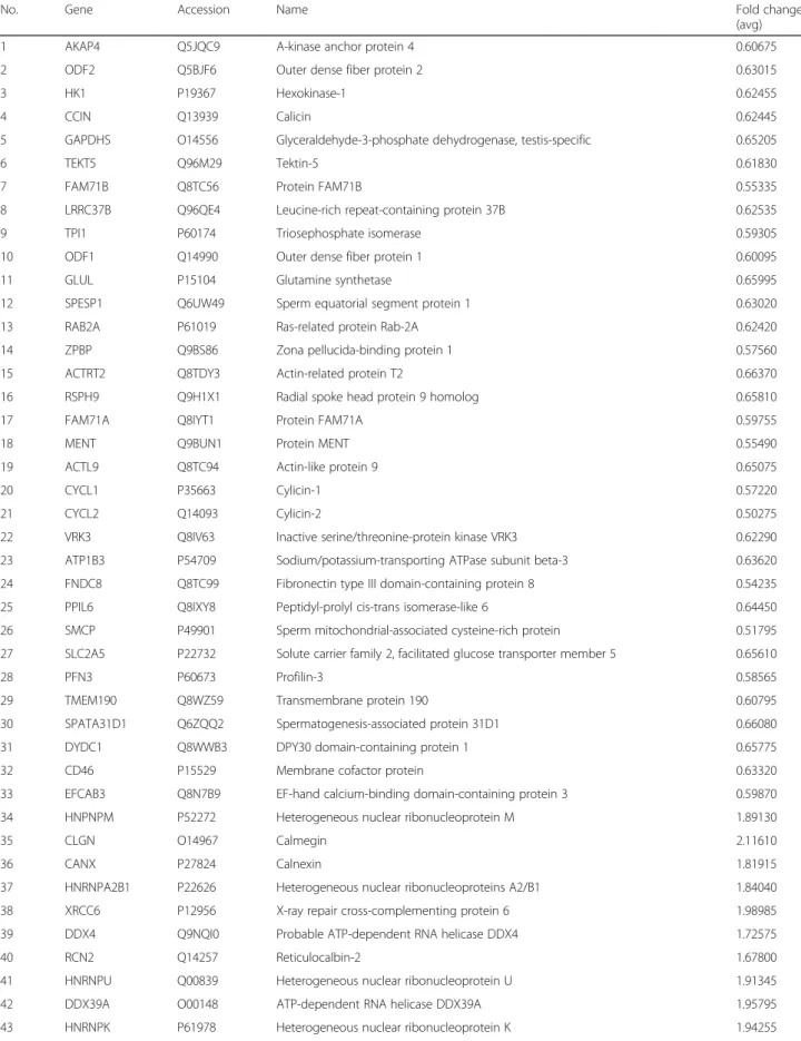
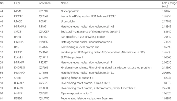
proteins were found to be significantly upregulated (28 proteins) or downregulated (33 proteins) in the heat-treated group 6 weeks after treatment compared with the control group (Table1).
To understand the functions and pathways of the differen-tially expressed proteins, we used the network tools provided with the DAVID, String, and KEGG databases. The identified proteins were analyzed through GO enrichment analysis
Table 1Differentially expressed proteins in the sixth week after heat-treated group compared with those in the control group
No. Gene Accession Name Fold change
(avg)
1 AKAP4 Q5JQC9 A-kinase anchor protein 4 0.60675
2 ODF2 Q5BJF6 Outer dense fiber protein 2 0.63015
3 HK1 P19367 Hexokinase-1 0.62455
4 CCIN Q13939 Calicin 0.62445
5 GAPDHS O14556 Glyceraldehyde-3-phosphate dehydrogenase, testis-specific 0.65205
6 TEKT5 Q96M29 Tektin-5 0.61830
7 FAM71B Q8TC56 Protein FAM71B 0.55335
8 LRRC37B Q96QE4 Leucine-rich repeat-containing protein 37B 0.62535
9 TPI1 P60174 Triosephosphate isomerase 0.59305
10 ODF1 Q14990 Outer dense fiber protein 1 0.60095
11 GLUL P15104 Glutamine synthetase 0.65995
12 SPESP1 Q6UW49 Sperm equatorial segment protein 1 0.63020
13 RAB2A P61019 Ras-related protein Rab-2A 0.62420
14 ZPBP Q9BS86 Zona pellucida-binding protein 1 0.57560
15 ACTRT2 Q8TDY3 Actin-related protein T2 0.66370
16 RSPH9 Q9H1X1 Radial spoke head protein 9 homolog 0.65810
17 FAM71A Q8IYT1 Protein FAM71A 0.59755
18 MENT Q9BUN1 Protein MENT 0.55490
19 ACTL9 Q8TC94 Actin-like protein 9 0.65075
20 CYCL1 P35663 Cylicin-1 0.57220
21 CYCL2 Q14093 Cylicin-2 0.50275
22 VRK3 Q8IV63 Inactive serine/threonine-protein kinase VRK3 0.62290
23 ATP1B3 P54709 Sodium/potassium-transporting ATPase subunit beta-3 0.63620
24 FNDC8 Q8TC99 Fibronectin type III domain-containing protein 8 0.54235
25 PPIL6 Q8IXY8 Peptidyl-prolyl cis-trans isomerase-like 6 0.64450
26 SMCP P49901 Sperm mitochondrial-associated cysteine-rich protein 0.51795
27 SLC2A5 P22732 Solute carrier family 2, facilitated glucose transporter member 5 0.65610
28 PFN3 P60673 Profilin-3 0.58565
29 TMEM190 Q8WZ59 Transmembrane protein 190 0.60795
30 SPATA31D1 Q6ZQQ2 Spermatogenesis-associated protein 31D1 0.66080
31 DYDC1 Q8WWB3 DPY30 domain-containing protein 1 0.65775
32 CD46 P15529 Membrane cofactor protein 0.63320
33 EFCAB3 Q8N7B9 EF-hand calcium-binding domain-containing protein 3 0.59870
34 HNPNPM P52272 Heterogeneous nuclear ribonucleoprotein M 1.89130
35 CLGN O14967 Calmegin 2.11610
36 CANX P27824 Calnexin 1.81915
37 HNRNPA2B1 P22626 Heterogeneous nuclear ribonucleoproteins A2/B1 1.84040
38 XRCC6 P12956 X-ray repair cross-complementing protein 6 1.98985
39 DDX4 Q9NQI0 Probable ATP-dependent RNA helicase DDX4 1.72575
40 RCN2 Q14257 Reticulocalbin-2 1.67800
41 HNRNPU Q00839 Heterogeneous nuclear ribonucleoprotein U 1.91345
42 DDX39A O00148 ATP-dependent RNA helicase DDX39A 1.95795
KEGG analysis revealed that the most active pathways (p≤
0.05) involving the differentially expressed proteins included the splicing process, carbohydrate digestion and absorption, glycolysis/gluconeogenesis, and fructose and mannose metab-olism (Fig.2d). Probable interaction analysis of the differen-tially expressed proteins against the String database revealed two independent PPI networks, one containing 19 proteins and the other containing 7 proteins. The PPI networks with the top nine KEGG enriched pathways are shown in Fig.3.
Confirmation of heat stress-induced proteins in mouse sperm by western blotting
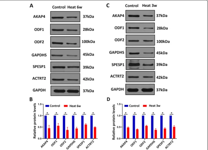
To validate the proteomics finding, the proteins related to sperm motility and energy metabolism AKAP4, SPESP1, ODF1, ODF2, GAPDHS, and ACTRT2 were evaluated in human sperms by western blotting. In line with our prote-omics results, the proteins levels of sperm motility and en-ergy metabolism-related proteins AKAP4, SPESP1, ODF1, ODF2, GAPDHS, and ACTRT2 were all significantly de-creased in human sperms after heat treatment in compari-son with control sperms (Fig. 4a-b), indicating that the proteomic reported here was reliable. Meanwhile, we op-erated mice subjected to testicular warming in a 43 °C or 33 °C water bath for 30 min once time. After 21 days, epi-didymis sperm samples were collected and used for west-ern blotting. Meaningfully, these proteins AKAP4, ODF1, ODF2, GAPDHS, SPESP1, and ACTRT2 were down-regulated in mouse sperms of heat treatment compared
with control (Fig. 4c-d). Collectively, these results indi-cated that these proteins AKAP4, ODF1, ODF2, GAPD HS, SPESP1, and ACTRT2 were significantly down-regulated after heat treatment of scrotum, suggesting that these proteins may serve as the biomarkers for scrotal heat treatment-induced male infertility.
Discussion
In the study, we identified 61 proteins significantly deregulated in human sperms of 6 weeks after first heat treatment of the scrotum compared with control using iTRAQ and 2D-LC–MS/MS and selected some proteins related to sperm motility and energy metabolism for veri-fication by western blotting in human and mouse sperms.
These deregulated proteins were found to be involved in several biological processes, including sexual reproduction, fertilization, gametogenesis, sperm motility, cell develop-ment, and sperm differentiation. The MF enrichment ana-lysis suggests that the identified differentially expressed proteins are mainly involved in nucleotide binding and nu-cleoside phosphate binding, consistent with the main func-tions of these significantly change proteins reported in the previous proteomics study of local heat shock to the scro-tum [21]. Our results revealed some new proteins associ-ated with sperm motility, that might be transcribed completely before the metaphase of spermatogenesis and translated in spermatozoa, for example, AKAP4 and mitochondrial-encoded proteins [29].
Table 1Differentially expressed proteins in the sixth week after heat-treated group compared with those in the control group
(Continued)
No. Gene Accession Name Fold change
(avg)
44 NPM1 P06748 Nucleophosmin 1.80460
45 DDX17 Q92841 Probable ATP-dependent RNA helicase DDX17 1.76955
46 UMOD P07911 Uromodulin 2.17185
47 HNRNPA3 P51991 Heterogeneous nuclear ribonucleoprotein A3 2.18345
48 SMC3 Q9UQE7 Structural maintenance of chromosomes protein 3 1.63640
49 RANBP1 P43487 Ran-specific GTPase-activating protein 1.70640
50 HNRNPL P14866 Heterogeneous nuclear ribonucleoprotein L 1.74625
51 RAN P62826 GTP-binding nuclear protein Ran 1.85995
52 DHX15 O43143 Putative pre-mRNA-splicing factor ATP-dependent RNA helicase DHX15 1.76235
53 ELAVL1 Q15717 ELAV-like protein 1 1.66060
54 HNRNPF P52597 Heterogeneous nuclear ribonucleoprotein F 2.04530
55 KHDRBS1 Q07666 KH domain-containing, RNA-binding, signal transduction-associated protein 1 2.13890
56 HNRNPD Q14103 Heterogeneous nuclear ribonucleoprotein D0 2.00500
57 SF3B3 Q15393 Splicing factor 3B subunit 3 1.82035
58 RBMXL2 O75526 RNA-binding motif protein, X-linked-like-2 1.60315
59 RBMY1C P0DJD4 RNA-binding motif protein, Y chromosome, family 1 member C 2.45005
60 MYEF2 Q9P2K5 Myelin expression factor 2 1.66025
It takes approximately 64 days in humans and 34.5 days in mice from the first spermatogenesis wave to sperm, and both require four spermatogenic cycles, of which latter 3/ 4 interval of the four spermatogenic cycles prepares the stage VII spermatids to form mature spermatozoa, which move to the epididymis [28, 30]. In humans, transient scrotal hyperthermia induced the lowest sperm concentra-tion and motility in the latter 3/4 cycle of the four sper-matogenic cycles, i.e., at 6 to 8 weeks after heat shock [25]. The sperm concentration and motility of mice after brief scrotal heat treatment have been reported to be lowest at
approximately 21 days after transient scrotal heat [28]. The time of lowest sperm concentration and motility after transient scrotal hyperthermia in humans can be consid-ered equivalent to that in mice.
down-regulated proteins, such as AKAP4, GAPDHS, SPESP1, ACTRT2, ODF2, and ODF1, play an important role in spermatogenesis. AKAP4 was mainly expressed in the tail of spermatozoa [31] and is involved in sperm mo-tility. It acted as a signaling molecule to regulate sperm-atozoa and mature sperm acrosome activation by stimulating a signaling cascade [32–34], which was essen-tial for sperm motility and fertility [35]. Heat treatment in-duces sperm DNA fragmentation significantly increasing and spermatozoa apoptosis [25, 36, 37]. Furthermore, knockdown of AKAP4 led to increased DNA fragmenta-tion and apoptosis [38]. That indicated AKAP4 downregu-lation is associated with human sperm damage after the transient scrotal hyperthermia. GAPDHS was a testis-specific enzyme expressed at the tubular fibrous sheath of the main segment of the sperm flagellum. It participates in glycolysis and glycogen metabolism pathways and the synthesis of ATP, which is important for sperm motility.
Gapdhs deficient mice exhibited non-motile sperm and male sterility [39,40]. Abnormal GAPDHS expression led to non-obstructive azoospermia [41]. SPESP1 is involved in the fusion of sperm and egg cells and its abnormal ex-pression leads to abnormal distribution of other proteins in spermatozoa. Meanwhile, Spesp1 mutant mice have been shown to low sperm viability and male infertility [42, 43]. The cytoskeletal protein ACTRT2 is expressed at the post-acrosomal region and middle part of human sperm and plays a role in sperm motility. ACTRT2 expression in the mature spermatozoa of patients with varicocele was
previously shown to be up-regulated [44], in contrast to the results of the ACTRT2 expression obtained in our study. This difference suggests that the short-term heat shock of scrotum causes its upregulation for compensa-tion. ACTRT2 expression has not been evaluated in mice or other species. Our PPI analysis revealed an interaction between ACTRT2 and GAPDHS, but the specific func-tions of these proteins remain to be clarified. Abnormal ODF1 expression has been shown to cause abnormal sperm morphology and low sperm motility [45]. ODF1, also known as HSP10, plays an important role in sperm head and neck connections and Odf1 defect mice have been shown to exhibit impairment in the mitochondrial sheath movement of spermatozoa [46,47]. Previous stud-ies have demonstrated that ODF2 transcription was mainly initiated in primary spermatocyte and, in addition to ODF2 post-processing, combination, and storage is reg-ulated by the hnRNP (A, M, U, K) family proteins [48,49]. It is then expressed in round spermatids, which interacts with Tssk4 to regulate sperm movement [50]. Thus, these deregulated proteins are closely associated with low sperm viability and concentration after the heat treatment of scrotum and reversible male infertility.
proteins were mainly involved in sexual reproduction, fertilization, sperm motility, and cellular development. Be-sides, these proteins AKAP4, ODF1, ODF2, GAPDHS, SPESP1, and ACTRT2 were examined to better verify these results in mice after heat treatment. Further studies of the relationship between these proteins and heat treat-ment of scrotum are needed to confirm and to further dis-sect the mechanisms underlying, which are promising to be the biomarkers and clinical targets for scrotal heat treatment-induced male infertility.
Supplementary information
Supplementary informationaccompanies this paper athttps://doi.org/10. 1186/s12958-020-00640-w.
Additional file 1.
Additional file 2.
Abbreviations
iTRAQ:isobaric Tags for Relative and Absolute Quantitation; 2D-LC-MS/ MS: Two-dimensional liquid chromatography–tandem mass spectrometry; AKAP4: A-Kinase anchor protein 4; ODF2: Outer dense fiber protein 2; ODF1: Outer dense fiber protein 1; SPESP1: Sperm equatorial segment protein 1; ACTRT2: Actin-related protein t2; GO: Gene ontology;
hnRNP: Heterogeneous Nuclear Ribonucleoprotein GAPDHS, Glyceraldehyde-3-Phosphate dehydrogenase, testis-specific; FDR: False discovery rate
Acknowledgments
Not applicable.
Authors’contributions
WX conceived and direct the study. QW and WX wrote the manuscript. QW, MR, SH, and DK performed all experiments. WX and CZ supervised the project. All authors read and approved the final manuscript.
Funding
This work was supported by the National Natural Science Foundation of China (grant no. 81671507), Fundamental Research Funds for the Central Universities (HUST; grant no. 2015ZDTD050), and National Key Research and Development Program (grant no. 2016YFC1000903).
Availability of data and materials
The datasets used and/or analyzed during the current study are available from the corresponding author on reasonable request.
Ethics approval and consent to participate
All the human procedures were approved by the Ethics Committee of Tongji Medical College, Huazhong University of Science and Technology. All the animal operations were permitted by the Institutional Animal Care and Use Committee (IACUC) of Tongji Medical College, Huazhong University of Science and Technology.
Consent for publication
Not applicable.
Competing interests
The authors declare no conflicts of interest.
Author details
1Institute of Reproductive Health, Tongji Medical College, Huazhong
University of Science and Technology, Wuhan, People’s Republic of China. 2Department of reproduction and genetics, the first affiliated hospital of
Kunming medical university, Kunming, People’s Republic of China. 3Department of Obstetrics and Gynecological Ultrasound Imaging, Renmin
Hospital of Wuhan University, Wuhan, People’s Republic of China.
4Reproductive Medicine Center, Tongji Medical College, Huazhong University
of Science and Technology, Wuhan, People’s Republic of China.
Received: 3 June 2020 Accepted: 29 July 2020
References
1. Thonneau P, Ducot B, Bujan L, Mieusset R, Spira A. Effect of male occupational heat exposure on time to pregnancy. Int J Androl. 1997;20(5): 274–8.
2. Hjollund NH, Bonde JP, Jensen TK, Olsen J. Diurnal scrotal skin temperature and semen quality. The Danish first pregnancy planner study team. Int J Androl. 2000;23(5):309–18.
3. Garolla A, Torino M, Sartini B, Cosci I, Patassini C, Carraro U, Foresta C. Seminal and molecular evidence that sauna exposure affects human spermatogenesis. Hum Reprod. 2013;28(4):877–85.
4. Rockett JC, Mapp FL, Garges JB, Luft JC, Mori C, Dix DJ. Effects of hyperthermia on spermatogenesis, apoptosis, gene expression, and fertility in adult male mice. Biol Reprod. 2001;65(1):229–39.
5. CHOWDHURY AK, STEINBERGER E. A quantitative study of the effect of heat on germinal epithelium of rat testes. Am J Anat. 1964;115:509–24. 6. Lue YH, Sinha HA, Swerdloff RS, Im P, Taing KS, Bui T, Leung A, Wang C.
Single exposure to heat induces stage-specific germ cell apoptosis in rats: role of Intratesticular testosterone on stage specificity. Endocrinology. 1999; 140(4):1709–17.
7. Lue Y, Wang C, Liu YX, Hikim AP, Zhang XS, Ng CM, Hu ZY, Li YC, Leung A, Swerdloff RS. Transient testicular warming enhances the suppressive effect of testosterone on spermatogenesis in adult cynomolgus monkeys (Macaca fascicularis). J Clin Endocrinol Metab. 2006;91(2):539–45.
8. Barth AD, Bowman PA. The sequential appearance of sperm abnormalities after scrotal insulation or dexamethasone treatment in bulls. Can Vet J. 1994;35(2):93–102.
9. Mieusset R, Quintana CP, Sanchez PL, Sowerbutts SF, Zupp JL, Setchell BP. Effects of heating the testes and epididymides of rams by scrotal insulation on fertility and embryonic mortality in ewes inseminated with frozen semen. J Reprod Fertil. 1992;94(2):337–43.
10. Mieusset R, Bujan L. The potential of mild testicular heating as a safe, effective and reversible contraceptive method for men. Int J Androl. 1994; 17(4):186–91.
11. Kandeel FR, Swerdloff RS. Role of temperature in regulation of spermatogenesis and the use of heating as a method for contraception. Fertil Steril. 1988;49(1):1–23.
12. Mieusset R, Bujan L. Testicular heating and its possible contributions to male infertility: a review. Int J Androl. 1995;18(4):169–84.
13. Goldman A, Rodriguez-Casuriaga R, Gonzalez-Lopez E, Capoano CA, Santinaque FF, Geisinger A. MTCH2 is differentially expressed in rat testis
and mainly related to apoptosis of spermatocytes. Cell Tissue Res. 2015; 361(3):869–83.
14. Durairajanayagam D, Agarwal A, Ong C. Causes, effects and molecular mechanisms of testicular heat stress. Reprod BioMed Online. 2015;30(1):14–27. 15. Kim JH, Park SJ, Kim TS, Park HJ, Park J, Kim BK, Kim GR, Kim JM, Huang SM, Chae JI, et al. Testicular hyperthermia induces unfolded protein response signaling activation in spermatocyte. Biochem Biophys Res Commun. 2013; 434(4):861–6.
16. Kanter M, Aktas C, Erboga M. Heat stress decreases testicular germ cell proliferation and increases apoptosis in short term: an
immunohistochemical and ultrastructural study. Toxicol Ind Health. 2013; 29(2):99–113.
17. Paul C, Teng S, Saunders PT. A single, mild, transient scrotal heat stress causes hypoxia and oxidative stress in mouse testes, which induces germ cell death. Biol Reprod. 2009;80(5):913–9.
18. Lin C, Shin DG, Park SG, Chu SB, Gwon LW, Lee JG, Yon JM, Baek IJ, Nam SY. Curcumin dose-dependently improves spermatogenic disorders induced by scrotal heat stress in mice. Food Funct. 2015;6(12):3770–7.
19. Kaur S, Bansal MP. Protective role of dietary-supplemented selenium and vitamin E in heat-induced apoptosis and oxidative stress in mice testes. ANDROLOGIA. 2015;47(10):1109–19.
20. Li YC, Hu XQ, Xiao LJ, Hu ZY, Guo J, Zhang KY, Song XX, Liu YX. An oligonucleotide microarray study on gene expression profile in mouse testis of experimental cryptorchidism. Front Biosci. 2006;11:2465–82.
21. Zhu H, Cui Y, Xie J, Chen L, Chen X, Guo X, Zhu Y, Wang X, Tong J, Zhou Z, et al. Proteomic analysis of testis biopsies in men treated with transient scrotal hyperthermia reveals the potential targets for contraceptive development. PROTEOMICS. 2010;10(19):3480–93.
22. Wang X, Liu F, Gao X, Liu X, Kong X, Wang H, Li J. Comparative proteomic analysis of heat stress proteins associated with rat sperm maturation. Mol Med Rep. 2016;13(4):3547–52.
23. Sabes-Alsina M, Tallo-Parra O, Mogas MT, Morrell JM, Lopez-Bejar M. Heat stress has an effect on motility and metabolic activity of rabbit spermatozoa. Anim Reprod Sci. 2016;173:18–23.
24. Rao M, Xia W, Yang J, Hu LX, Hu SF, Lei H, Wu YQ, Zhu CH. Transient scrotal hyperthermia affects human sperm DNA integrity, sperm apoptosis, and sperm protein expression. Andrology-US. 2016;4(6):1054–63.
25. Rao M, Zhao XL, Yang J, Hu SF, Lei H, Xia W, Zhu CH: Effect of transient scrotal hyperthermia on sperm parameters, seminal plasma biochemical markers, and oxidative stress in men. In., vol. 17; 2015: 668–675. 26. Banyra O, Nikitin O, Ventskivska I. Acute epididymo-orchitis: relevance of
local classification and partner's follow-up. Cent European J Urol. 2019;72(3): 324–9.
27. Gorpinchenko I, Nikitin O, Banyra O, Shulyak A. The influence of direct mobile phone radiation on sperm quality. Cent European J Urol. 2014;67(1):65–71. 28. Perez-Crespo M, Pintado B, Gutierrez-Adan A. Scrotal heat stress effects on
sperm viability, sperm DNA integrity, and the offspring sex ratio in mice. Mol Reprod Dev. 2008;75(1):40–7.
29. Amaral A, Castillo J, Ramalho-Santos J, Oliva R. The combined human sperm proteome: cellular pathways and implications for basic and clinical science. Hum Reprod Update. 2014;20(1):40–62.
30. OAKBERG EF. Duration of spermatogenesis in the mouse and timing of stages of the cycle of the seminiferous epithelium. Am J Anat. 1956;99(3): 507–16.
31. Moretti E, Baccetti B, Scapigliati G, Collodel G. Transmission electron microscopy, immunocytochemical and fluorescence in situ hybridisation studies in a case of 100% necrozoospermia: case report. ANDROLOGIA. 2006;38(6):233–8.
32. Yang SM, Li HB, Wang JX, Shi YC, Cheng HB, Wang W, Li H, Hou JQ, Wen DG. Morphological characteristics and initial genetic study of multiple morphological anomalies of the flagella in China. Asian J Androl. 2015;17(3): 513–5.
33. Teijeiro JM, Marini PE. The effect of oviductal deleted in malignant brain tumor 1 over porcine sperm is mediated by a signal transduction pathway that involves pro-AKAP4 phosphorylation. Reproduction. 2012;143(6):773–85. 34. Krisfalusi M, Miki K, Magyar PL, O'Brien DA. Multiple glycolytic enzymes are
tightly bound to the fibrous sheath of mouse spermatozoa. Biol Reprod. 2006;75(2):270–8.
36. Hamilton T, Siqueira A, de Castro LS, Mendes CM, Delgado JC, de Assis PM, Mesquita LP, Maiorka PC, Nichi M, Goissis MD, et al. Effect of heat stress on sperm DNA: protamine assessment in ram spermatozoa and testicle. Oxidative Med Cell Longev. 2018;2018:5413056.
37. Baskaran S, Agarwal A, Panner SM, Finelli R, Robert KA, Iovine C, Pushparaj PN, Samanta L, Harlev A, Henkel R. Tracking research trends and hotspots in sperm DNA fragmentation testing for the evaluation of male infertility: a scientometric analysis. Reprod Biol Endocrinol. 2019;17(1):110.
38. Jagadish N, Devi S, Gupta N, Suri V, Suri A. Knockdown of A-kinase anchor protein 4 inhibits proliferation of triple-negative breast cancer cells in vitro and in vivo. Tumour Biol. 2020;42(4):1390175187.
39. Miki K, Qu W, Goulding EH, Willis WD, Bunch DO, Strader LF, Perreault SD, Eddy EM, O'Brien DA. Glyceraldehyde 3-phosphate dehydrogenase-S, a sperm-specific glycolytic enzyme, is required for sperm motility and male fertility. Proc Natl Acad Sci U S A. 2004;101(47):16501–6.
40. Margaryan H, Dorosh A, Capkova J, Manaskova-Postlerova P, Philimonenko A, Hozak P, Peknicova J. Characterization and possible function of glyceraldehyde-3-phosphate dehydrogenase-spermatogenic protein GAPD HS in mammalian sperm. Reprod Biol Endocrinol. 2015;13:15.
41. Dorosh A, Tepla O, Zatecka E, Ded L, Koci K, Peknicova J. Expression analysis of MND1/GAJ, SPATA22, GAPDHS and ACR genes in testicular biopsies from non-obstructive azoospermia (NOA) patients. Reprod Biol Endocrinol. 2013;11:42. 42. Fujihara Y, Murakami M, Inoue N, Satouh Y, Kaseda K, Ikawa M, Okabe M.
Sperm equatorial segment protein 1, SPESP1, is required for fully fertile sperm in mouse. J Cell Sci. 2010;123(Pt 9):1531–6.
43. McCarthy NS, Melton PE, Cadby G, Yazar S, Franchina M, Moses EK, Mackey DA, Hewitt AW. Meta-analysis of human methylation data for evidence of sex-specific autosomal patterns. BMC Genomics. 2014;15:981.
44. Liu Y, Guo Y, Song N, Fan Y, Li K, Teng X, Guo Q, Ding Z. Proteomic pattern changes associated with obesity-induced asthenozoospermia. Andrology-US. 2015;3(2):247–59.
45. Chen J, Wang Y, Xu X, Yu Z, Gui YT, Cai ZM. Differential expression of ODF1 in human ejaculated spermatozoa and its clinical significance. Zhonghua Nan Ke Xue. 2009;15(10):891–4.
46. Yang K, Grzmil P, Meinhardt A, Hoyer-Fender S. Haplo-deficiency of ODF1/ HSPB10 in mouse sperm causes relaxation of head-to-tail linkage. Reproduction. 2014;148(5):499–506.
47. Yang K, Meinhardt A, Zhang B, Grzmil P, Adham IM, Hoyer-Fender S. The small heat shock protein ODF1/HSPB10 is essential for tight linkage of sperm head to tail and male fertility in mice. Mol Cell Biol. 2012;32(1):216–25.
48. Pletz N, Medack A, Riess EM, Yang K, Kazerouni ZB, Huber D, Hoyer-Fender S. Transcriptional activation of Odf2/Cenexin by cell cycle arrest and the stress activated signaling pathway (JNK pathway). Biochim Biophys Acta. 2013;1833(6):1338–46.
49. Wang X, Wei Y, Fu G, Li H, Saiyin H, Lin G, Wang Z, Chen S, Yu L. Tssk4 is essential for maintaining the structural integrity of sperm flagellum. Mol Hum Reprod. 2015;21(2):136–45.
50. Wang X, Li H, Fu G, Wang Y, Du S, Yu L, Wei Y, Chen S. Testis-specific serine/threonine protein kinase 4 (Tssk4) phosphorylates Odf2 at Ser-76. Sci Rep. 2016;6:22861.
Publisher’s Note