Open Access
Research
Expression pattern and regulation of genes differ between
fibroblasts of adhesion and normal human peritoneum
Ujjwal K Rout*
1,2, Ghassan M Saed
1and Michael P Diamond
1Address: 1Division of Reproduction Endocrinology and Infertility, Department of Obstetrics and Gynecology, Wayne State University, School of
Medicine, Detroit, MI 48201, USA and 2Division of Pediatric Surgery, Department of Surgery, University of Mississippi Medical Center, Jackson,
MS 39216, USA
Email: Ujjwal K Rout* - [email protected]; Ghassan M Saed - [email protected]; Michael P Diamond - [email protected] * Corresponding author
Abstract
Background: Injury to the peritoneum during surgery is followed by a healing process that frequently results in the attachment of adjacent organs by a fibrous mass, referred commonly as adhesions. Because injuries to the peritoneum during surgery are inevitable, it is imperative that we understand the mechanisms of adhesion formation to prevent its occurrence. This requires thorough understanding of the molecular sequence that results in the attachment of injured peritoneum and the development of fibrous tissue. Recent data show that fibroblasts from the injured peritoneum may play a critical role in the formation of adhesion tissues. Therefore, identifying changes in gene expression pattern in the peritoneal fibroblasts during the process may provide clues to the mechanisms by which adhesion develop.
Methods: In this study, we compared expression patterns of larger number of genes in the fibroblasts isolated from adhesion and normal human peritoneum using gene filters. Contributions of TGF-beta1 and hypoxia in the altered expression of specific genes were also examined using a semiquantitative RT-PCR technique.
Results: Results show that several genes are differentially expressed between fibroblasts of normal and adhesion peritoneum and that the peritoneal fibroblast may acquire a different phenotype during adhesion formation. Genes that are differentially expressed between normal and adhesion fibroblasts encode molecules involved in cell adhesion, proliferation, differentiation, migration and factors regulating cytokines, transcription, translation and protein/vesicle trafficking.
Conclusions: Our data substantiate that adhesion formation is a multigenic phenomenon and not all changes in gene expression pattern between normal and adhesion fibroblasts are the function of TGF-beta1 and hypoxia that are known to influence adhesion formation. Analysis of the gene expression data in the perspective of known functions of genes connote to additional targets that may be manipulated to inhibit adhesion development.
Background
Peritoneal adhesions resulting from surgical injury are often associated with pelvic pain, bowel obstruction and
infertility [1]. Epidemiological studies conclude that 30 to 35% of all hospital readmissions are associated with adhesion associated complications, of which 4.5 to 5.1%
Published: 10 January 2005
Reproductive Biology and Endocrinology 2005, 3:1 doi:10.1186/1477-7827-3-1
Received: 30 August 2004 Accepted: 10 January 2005
This article is available from: http://www.rbej.com/content/3/1/1 © 2005 Rout et al; licensee BioMed Central Ltd.
are directly related to adhesions [2]. Mechanisms of adhe-sion formations are not completely known. It is also not clear why adhesion form in some patients and not in oth-ers. Therefore, deciphering genetic components that sig-nal adhesion formation may help diagnose adhesion-prone patients prior to surgery. Needless to mention that such information will facilitate finding ways to prevent post-surgical adhesion formation.
Parietal and visceral peritoneum that surfaces the intra-peritoneal organs is covered by a layer of squamous epi-thelial cells, the mesothelium. The submesoepi-thelial layer consists of fibroblasts, macrophages and blood vessels. Surgical abrasion to the peritoneum releases mesothelial cells, macrophages, fibroblasts, and blood containing cytokines and several cell types at the site of injury. Coag-ulation of blood creates a fibrinous mass between injured surfaces. In some patients fibrinolysis of clot followed by proliferation of mesothelial cells covers the wound. In others, failure of fibrinolysis followed by proliferation and migration of fibroblasts into the proteinous mass generates fibrous tissues of adhesion. Consequently, the process of adhesion formation include inflammatory response, fibrin deposition, cell-proliferation, -differenti-ation, -migr-differenti-ation, -death, angiogenesis, extra cellular matrix (ECM) turnover regulated by cytokines, hypoxia, genetic and epigenetic factors [3].
Recent studies illustrate roles of peritoneal fibroblasts in adhesion development [4-10]. It is also proposed that fibroblasts from the chronic wounds migrate into the fibrin deposit; secrete ECM proteins causing wound con-traction and scar formation [11]. The migration of fibrob-lasts may be coordinated by TGF-β1 mediated interactions of integrin receptors [10] with the RGD sequence of the fibrin, fibrinogen and fibronectin at the fibrin clot [12]. Additional cytokines and the hypoxic condition at the site of injury may also influence peritoneal fibroblasts to attain a phenotype supporting formation of adhesion tis-sue. This change in the phenotype of fibroblasts may be induced by changes in expression pattern of several genes during the process of adhesion development. Therefore, identifying differences in the global gene expression pat-tern between normal and adhesion fibroblasts may pro-vide additional clues to the mechanisms by which normal fibroblasts attain the adhesive, proliferating and migra-tory phenotype required for the formation of fibrous tis-sues of adhesions. In the present study, we compared gene expression patterns between adhesion and normal perito-neal fibroblasts using GF211 gene filters (Research genet-ics) containing 4325 randomly selected known genes. Furthermore, we confirmed the expression pattern of genes of interest by a semiquantitative RT-PCR method and examined possible contribution/s of TGF-β1 and
hypoxia in the transformation of normal peritoneal fibroblasts into an adhesion phenotype.
Methods
Peritoneal-tissue collection, fibroblast-isolation and culture
Tissues were collected at the initiation of surgery and after the entry into the abdominal cavity of female patients (25–50 years) undergoing laparatomy for pelvic pain as described earlier [4]. All patients gave informed written consent for the tissue collection, which was conducted under a protocol approved by the Wayne State University Institutional Review Board. Normal parietal peritoneal tissues were collected from these patients from the ante-rior abdominal wall, approximately midway between the umbilicus and symphyses pubis, and lateral to the mid-line incision. Peritoneal tissues from adhesions, that were at least 3 inches away from the site of normal tissue col-lections, were also collected from the same patient. The peritoneal fibroblasts were isolated and separated from mesothelial cells by a differential centrifugation proce-dure that is briefly described earlier [4]. The isolation of fibroblasts from mesothelial cells were also verified by the RT-PCR detection of Collagen type I, Matrix metallopro-teinase-2 (MMP-2) and Transforming growth factor-β3 (TGF-β3) [13-15].
The primary cultures were maintained in a humidified incubator (37°C, 5% CO2) for 3 days in DMEM (Life Tech.) supplemented with 10% fetal bovine serum (Life Tech.) and antibiotics (Penicillin and Streptomycin 50 U/ ml; Life Tech.). The monolayer of cells were passaged in trypsin-EDTA solution (Life Tech.). Cells at 3–5 passages were cultured in serum free medium in 75 cm2 flasks
(Fisher Scientific, Pittsburgh, PA) to 75% confluency prior to studies.
Gene expression pattern in the fibroblasts from adhesion and normal peritoneum
Total RNA was isolated from monolayer of fibroblasts at 12 h of culture in serum free medium using Trizol reagent (Invitrogen Inc.). Human Gene Filters (GF211; Research Genetics, Inc., Huntsville, AL) containing 4325 known human cDNA spots were used for the identification of dif-ferentially expressed genes between adhesion and normal fibroblasts from human peritoneum. Method suggested by the manufacturer was strictly followed. In brief, 10 µg of total RNA from monolayer cultures of fibroblasts were subjected to cDNA synthesis in presence of 10 µl 33dCTP
was transferred to separate roller tubes of the hybridiza-tion oven (Fisher Scientific, Inc., Pittsburg, PA), each con-taining MicroHyb hybridization solution (Research Genetics) supplemented with Human Cot-1 DNA (Life Technology) and Poly dA (Research Genetics). The mem-branes were rotated at 10 RPM and at 42°C for 2 h. Radio-labeled cDNA prepared from adhesion and normal fibroblasts total RNA was denatured by heating in a boil-ing water bath for 3 min. The denatured probes were then injected into the prehybridization solution containing respective membrane. The membranes were hybridized with respective probe for 18 h at 42°C. The hybridization solution was then replaced with washing solution (2 ×
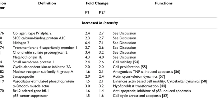
SSC containing 1% SDS). The temperature of the oven was raised to 50°C and RPM of rotors was increased to 15. Membranes were washed for 20 minutes when washing solution was replaced with a batch of fresh and pre-warmed (50°C) washing solution. Washing was contin-ued for additional 20 min. A third wash was performed with 0.5 × SSC solution containing 1% SDS at 55°C for 15 minutes. Membranes with cDNA spots facing up were covered with Saran wrap and exposed to phosphor screen (Kodak) for overnight. The screen was scanned with a Phosphor Imager (Storm System; Amersham Biosciences Corp., Piscataway, NJ). After acquisition of signal intensi-ties from the normal and adhesion fibroblasts of one Table 2: Genes differentially expressed in the adhesion fibroblasts and known to have roles in cell-adhesion, -proliferation, -migration, -differentiation and -death.
Accession Number
Definition Fold Change Functions
P1 P2*
Increased in Intensity
gi:17986276 Collagen, type IV alpha 2 2.4 2.7 See Discussion gi:4506760 S100 calcium-binding protein A10 2.3 2.7 See Discussion gi:6679055 Nidogen 2 6.4 7.1 See Discussion gi:14250074 Transmembrane 4 superfamily member 1 3.7 2.6 See Discussion gi:4758081 Chondroitin sulfate proteoglycan 2 3.4 3.2 See Discussion gi:187538 Metallothionein 1E 4.3 4.0 See Discussion gi:4336324 Small membrane protein 1 2.4 2.6 Cell viability [54] gi:17738299 Cyclin-dependent kinase inhibitor 2A 2.0 2.0 Cell proliferation [55]
gi:16359382 Nuclear receptor subfamily 4, group A 1.6 2.1 Antagonizes TNF-α induced apoptosis [56] gi:40353726 Synaptopodin 2.9 2.4 Actin cytoskeleton dynamics [57]
gi:23398519 Vasodilator-stimulated phosphoprotein 1.5 2.1 Enhances actin based cell motility, Cytoskeltal dynamics [58] gi:28329 α-Smooth muscle actin 3.0 3.2 Myofibroblast transformation [44]
gi:14574570 Bcl-2 related gene bfl-1 1.6 1.4 Anti apoptotic; inhibitor of p53 induced apoptosis gi:796812 p53 tumor suppressor 1.5 1.6 Cell cycle arrest and apoptosis [52]
Decreased in Intensity
gi:184522 Insulin-like growth factor binding protein 3 3.2 2.3 See Discussion
gi:4504618 Insulin-like growth factor binding protein 7 2.3 2.0 Growth suppressing factor [59]
gi:28610153 Interleukin 8 3.2 2.6 Inhibits fibroblast migration, delays wound healing, reduces wound contraction [60]
gi:4504982 Lectin, galactoside-binding, soluble 3 [galectin)
3.0 3.0 Tumor-suppressive and pro apoptotic [61] gi:12803916 Gap junction protein, beta 1, [Connexin 32) 1.8 2.2 Tumor suppressive and Proapoptotic [62]
gi:14589894 Cadherin 5, type 2, VE-cadherin [vascular] 2.3 1.7 Down regulation associates with tumor metastasis, Initiates endothelial-mesenchymal transdifferentiation [63]
gi: 16198356 Lactotransferrin 2.2 2.1 Inhibits growth of malignant tumors. Elevated by high level of estrogen [64]
gi:21619838 Lipocalin 2, Oncogene 24p3 3.3 2.5 Proapoptotic [65]
gi: 23273645 Calponin 1, basic, Smooth muscle cell 1.7 2.5 Inhibits smooth muscle cell contraction and Tumor Suppressive [66]
patient, filters were stripped according to protocol and subjected to gene filter experiments with the RNA samples from a second patient and images were scanned. Tiff images obtained from normal and adhesion fibroblasts of two patients were analyzed using Pathway 4 software (Research Genetics) for the identification of differentially expressed genes between the normal and adhesion fibrob-lasts of each patient.
Relative abundance of selected genes in the fibroblasts from adhesion and normal peritoneum
Steady-state levels of mRNA of selected genes that are known to have a role in cellular adhesion, proliferation, migration, apoptosis and demonstrating different expres-sion levels between adheexpres-sion and normal fibroblasts in the gene filter experiments were verified further by a pre-viously described semiquantitative RT-PCR method [16]. Total RNA (1 µg) from the monolayer culture of adhesion or normal fibroblasts was subjected to reverse transcrip-tion as described earlier. Complementary DNA (100 ng) was subjected to PCR amplification of the cDNA of inter-ests in a 25 µl reaction mixture containing 50 mM Tris-HCl (pH 8.4), 50 mM KCl, 2.5 mM MgCl2, 0.2 mM dNTP, 0.5 U Taq Polymerase (all from Life Technology, BRL) and 1 µM each of sense and antisense primers. Primer sequences were determined using Primer3 software from the Internet http://frodo.wi.mit.edu/cgi-bin/primer3/ primer3_www.cgi. The control primers (sense 5'-ggaggttc-gaagacgatcag-3' and antisense 5'-cgctgagccagtcagtgtag-3') were expected to provide an amplicon of 509 bp from
human 18S ribosomal subunit cDNA (gi: 337376). Acces-sion numbers of genes of interests are provided in Table 2 and nucleotide sequences of primers and expected size amplicons are provided in the Table 3. Each PCR cycle consisted of a hot start at 95°C for 1 min, followed by melting at 95°C for 30 sec, annealing at 58°C for 1 min and extension at 72°C for 1 min. At the end an extension reaction at 72°C for 10 min was performed.
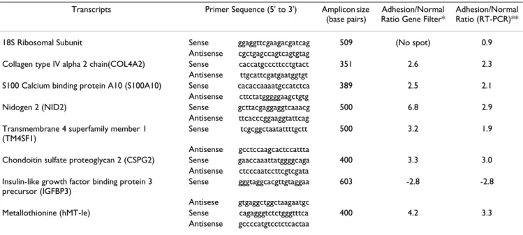
Initially cDNA of interests were amplified from normal peritoneal fibroblasts at different (25 to 35) PCR cycles. PCR products were subjected to agarose gel electrophore-sis. Molecular weight marker (100 bp DNA ladder; Life Technology) were also loaded in adjacent lanes. DNA in the gel were stained with 1:10,000 dilution of SYBR Green I dye (Molecular Probes, Inc., Eugene, OR) and photo-graphed using a DC 120 Kodak digital camera (Eastman Kodak, Rochester, NY) for the verification of size and analysis of band intensity using Image J software http:// rsb.info.nih.gov/ij/. Band intensities were plotted to determine the linearity of PCR reactions for the amplifica-tion of target transcripts. Target cDNA were amplified by PCR from normal and peritoneal fibroblasts at specific PCR cycle within its linear range of amplification. Total RNA samples from normal and adhesion fibroblasts of 4 patients (included RNA from normal and adhesion fibroblasts of two patients used for the gene filter experi-ments) were used for the RT-PCR experiments. Optical densities obtained from amplicons of 4 patients (1 nor-mal and 1 adhesion fibroblast RNA sample per patient) Table 3: PCR primers, amplicon size and expression ratios of genes between adhesion and normal peritoneal fibroblasts
Transcripts Primer Sequence (5' to 3') Amplicon size (base pairs)
Adhesion/Normal Ratio Gene Filter*
Adhesion/Normal Ratio (RT-PCR)** 18S Ribosomal Subunit Sense ggaggttcgaagacgatcag 509 (No spot) 0.9
Antisense cgctgagccagtcagtgtag
Collagen type IV alpha 2 chain(COL4A2) Sense caccatgcccttcctgtact 351 2.6 2.3 Antisense ttgcattcgatgaatggtgt
S100 Calcium binding protein A10 (S100A10) Sense cacaccaaaatgccatctca 389 2.5 2.1 Antisense cttctatgggggaagctgtg
Nidogen 2 (NID2) Sense gcttacgaggaggtcaaacg 500 6.8 2.9 Antisense ttcacccggaaggtattcag
Transmembrane 4 superfamily member 1 (TM4SF1)
Sense tcgcggctaatattttgctt 500 3.2 1.9 Antisense gcctccaagcactccattta
Chondoitin sulfate proteoglycan 2 (CSPG2) Sense gaaccaaattatggggcaga 400 3.3 3.0 Antisense ctcccaatccttcgtcgata
Insulin-like growth factor binding protein 3 precursor (IGFBP3)
Sense gggtaggcacgttgtaggaa 603 -2.8 -2.8 Antisese gtgaggctggctaagaatgc
Metallothionine (hMT-Ie) Sense cagagggtctctgggtttca 400 4.2 3.3 Antisense gccccatgtcctctcactaa
were used to derive mean ± standard error of mean values representing relative levels of each mRNA species in nor-mal and adhesion fibroblasts.
Effects of TGF-β1 or hypoxia on gene expression pattern
Effects of TGF-β1 or hypoxic conditions on the steady state levels of specific gene transcripts in the normal peri-toneal fibroblasts were also studied to examine the possi-bility of adhesion causing factors potentiating the gene expression pattern in the normal fibroblasts similar to adhesion fibroblasts. Normal peritoneal fibroblasts were cultured in six well culture plates (FALCON). When confluent, monolayer of cells in culture were exposed to 1 ng/ml TGF-β1 (Sigma Chemical Company, St. Louis, MO) or hypoxia (2% Oxygen) for 24 h. Control plates were cul-tured for the same duration in absence of TGF-β1 or hypoxia. Total RNA was isolated from the control, TGF-β1 and hypoxia treated cells and subjected to RT-PCR reactions as described above to determine relative levels of 18S ribosomal subunit and gene specific transcripts in the control and treated cells. RT-PCR experiments were con-ducted twice with the normal peritoneal fibroblasts iso-lated from 3 patients to have six control, six TGF-β1 treated and six hypoxia exposed amplicons. This included normal fibroblasts from one new patient and two patients that were used exclusively for RT-PCR experiments for the confirmation of gene array data. Optical densities of amplicons from six control or treated cells per mRNA spe-cies were used to derive the mean ± standard error of mean values for comparison.
Statistical analysis
Band intensity value of each RT-PCR experiment (normal, adhesion or treated fibroblasts) was used to derive Mean ± Standard error of Mean using Statview 4.5 software (Abacus Concepts, Berkley, CA). Differences between Means were tested for significance by one-way analysis of variance with the specific post hoc test using the same
software to compare differences in the steady state levels of different mRNA species.
Results
Expression pattern of genes between adhesion and normal peritoneal fibroblasts
Hybridization intensities of radio labeled cDNA from nor-mal or adhesion fibroblasts from both the patients were different when analyzed using Pathway software. Com-parison of hybridization intensities from individual gene spots between normal and adhesion fibroblast RNA (Fig-ure 1) demonstrated that the expression levels of ~4% genes were >1.5 fold different. BLAST search of the acces-sion number of genes from the list provided by the man-ufacturer showed that genes with altered expression level between normal and adhesion fibroblasts are reported to be involved in cell adhesion and migration; transforma-tion, transcriptransforma-tion, translation and growth factors as well as cytokines and signaling molecules.
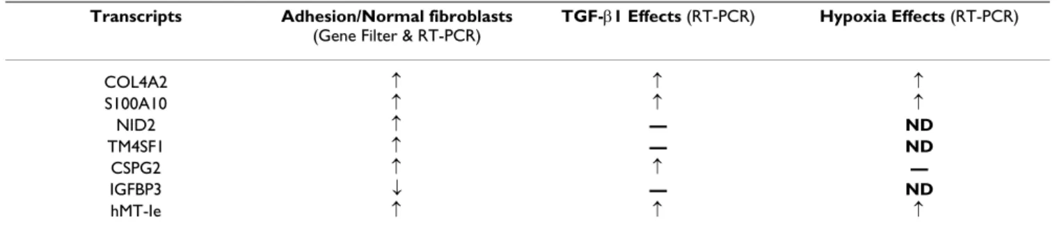
Gene filter data from two patients showed similar expres-sion pattern of collagen type 1 (alpha 2), Collagen type III (alpha 1), fibronectin 1, Matrix metalloproteinase-1 (MMP-1), Transforming Growth Factor beta-1 (TGF-β1), TGF-β2 and tissue plasminogen activator as reported ear-lier using multiplex PCR technique (Table-1). Signal intensities representing TGF-β3 (gi:22531293), TGF-β III Receptor (gi:26251745), VEGF-A (gi:2565322), VEGF-B (gi:39725673) and VEGF-C (gi:19924300) expression lev-els were respectively 1.6, 1.5, 1.9, 1.3 and 1.3 fold (aver-age values from two patients) lower in the adhesion compared to normal fibroblasts. No spots for antiapop-totic bcl-2 and proapopantiapop-totic bax were present in GF211 filters. Signal intensities representing anti apoptotic mole-cule bcl-2 related gene bfl-1 (gi: 14574570) and pro-apoptotic molecule p53 (gi:796812) were higher (Table 2) in adhesion compared to normal fibroblasts. Expres-sion levels of proapoptotic molecule bad (gi: 14670387) and bak1 (gi: 33457353) were not different between nor-Table 4: Expression profiles of genes in the adhesion vs. normal peritoneal fibroblasts and the effects of TGF-β1 or hypoxia on the expression level of genes in the normal peritoneal fibroblasts
Transcripts Adhesion/Normal fibroblasts
(Gene Filter & RT-PCR)
TGF-β1 Effects (RT-PCR) Hypoxia Effects (RT-PCR)
COL4A2 ↑ ↑ ↑
S100A10 ↑ ↑ ↑
NID2 ↑ — ND
TM4SF1 ↑ — ND
CSPG2 ↑ ↑ —
IGFBP3 ↓ — ND
hMT-Ie ↑ ↑ ↑
Images depicting radioactive signals from GF211 filters hybridized with radiolabeled cDNA
Figure 1
Images depicting radioactive signals from GF211 filters hybridized with radiolabeled cDNA. Gene filters were hybridized with 33P labeled cDNA from normal peritoneal fibroblasts or fibroblasts from adhesion tissue. Unbound signals were washed and relative radioactive signal intensities were detected using a Phosphoroimager as described in the Methods. A. Tiff images of radioactive signals from individual gene spots of filters hybridized with normal (above) and adhesion fibrob-lasts, both isolated from Patient 1. B. Scatter plot showing signal intensities from normal peritoneal (Intensity I) and adhesion (Intensity II) fibroblasts. Dotted lines indicate a two fold changes in hybridization intensities from the median (solid line).
A
mal and adhesion fibroblasts. A list of additional genes that are differentially expressed between normal and adhesion fibroblasts and known to be involved in apop-tosis as well as cell adhesion, proliferation and migration are listed in Table 2.
Semiquantitative RT-PCR experiments (Figure 2) con-ducted to verify expression pattern of specific genes from the gene filter experiments that were not studied earlier in the peritoneal fibroblasts confirmed higher expression (p < 0.05) of Collagen type IV (alpha 2) chain (COL4A2), S100 Calcium binding protein A10 (S100A10), Nidogen 2 (NID2), Transmembrane 4 superfamily member 1 (TM4SF1), Chondroitin sulfate proteoglycan 2 (CSPG2) and Metallothioneine (hMT-Ie) in adhesion compared to normal fibroblasts. The semiquantitative RT-PCR experi-ments also confirmed lower expression levels of Insulin-like growth factor binding protein 3 precursor (IGFBP3) mRNA in the adhesion compared to normal peritoneal fibroblasts. Transcript levels of 18S ribosomal subunit estimated by RT-PCR method was not significantly differ-ent between fibroblasts isolated from normal and adhe-sion peritoneum (Figure 2 and Table 3).
Effects of TGF-β1 or hypoxia on the expression levels of specific genes in the normal peritoneal fibroblasts
Exposure to TGF-β1 or hypoxic conditions for 24 h altered
expression levels of specific genes in the normal perito-neal fibroblasts as evidenced by semiquantitative RT-PCR. Transcript levels of COL4A2, S100A10, CSPG2 and hMT-Ie were up regulated by TGF-β1 in the normal peritoneal fibroblasts (Figure 3), whereas transcript levels of NID2, TM4SF1, and IGFBP3 were not altered by TGF-β1 treat-ment. Hypoxic conditions elevated expression levels of COL4A2, S100A10 and hMT-Ie transcripts in the normal peritoneal fibroblasts (Figure 4). Transcript levels of CSPG2 were not significantly altered by hypoxia.
Discussion
We present evidence that the expression pattern of large number of genes differ between the fibroblasts isolated from adhesion tissues and normal human peritoneal sup-porting the notion that adhesion fibroblasts may attain a different phenotype following peritoneal injury. Genes that displayed altered expression levels in this transition included those involved in cell proliferation, differentia-tion, signaling molecules, transcription and translation factors, proteolysis and cytokines. Results indicate that fibroblasts from adhesion tissue may perceive and respond to external and internal cues differently than those residing in normal human peritoneum. We attempted to decipher the functional consequences of altered gene expression pattern in the adhesion fibroblast to further elucidate the mechanism of adhesion formation and point to additional ways adhesion development may be restrained.
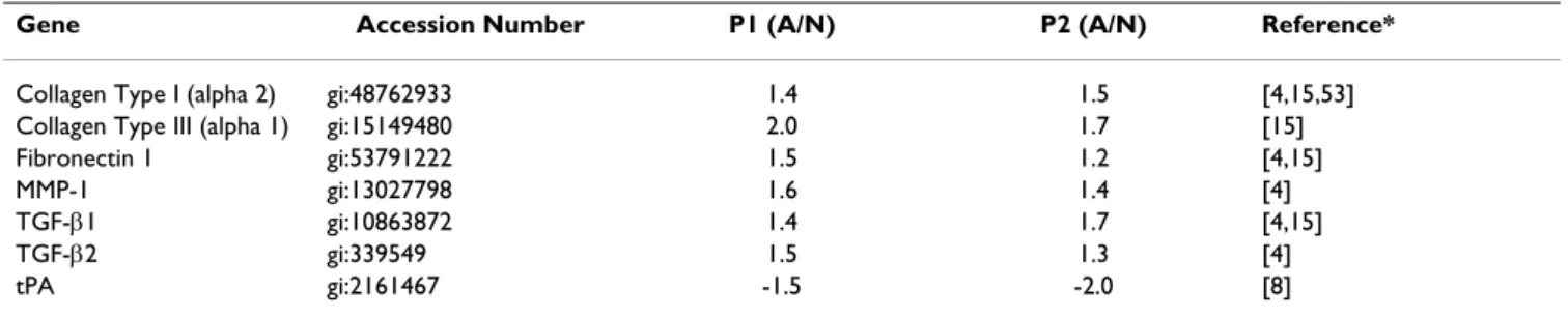
Expression pattern of genes in the fibroblasts from normal and pathological sites are shown to be different also in earlier studies [17]. More relevant to the present study are the reports [4,8] on the mRNA levels of human type I col-lagen (alpha 2), fibronectin 1, MMP-1, TIMP-1, TGF-β1, TGF-β2, IL-10, PAI-1, tPA and COX-2 in adhesion and normal peritoneal fibroblasts from humans estimated by multiplex PCR technique. Gene filter data from two patients also showed similar pattern of collagen, type 1 (alpha 2), fibronectin 1, MMP-1, TGF-β1, TGF-β2 and tPA mRNA levels in the normal and adhesion fibroblasts (Table 1). Expression pattern of TIMP-1, IL-10, PAI-1, COX-2 in the adhesion and normal peritoneal fibroblasts as reported earlier [4,8,9] could not be verified by gene fil-ter experiments because GF211 filfil-ters do not have spots representing these genes. Even so, similarities in the expression pattern of many genes between two patients (Tables 1–3) and those reported earlier using multiplex PCR technique [4,8] validate our findings. The semiquan-Table 1: Ratios of signal intensities from adhesion and normal peritoneal fibroblasts detected from gene filters representing relative expression level of genes in patient 1 (P1) and 2 (P2).
Gene Accession Number P1 (A/N) P2 (A/N) Reference*
Collagen Type I (alpha 2) gi:48762933 1.4 1.5 [4,15,53] Collagen Type III (alpha 1) gi:15149480 2.0 1.7 [15] Fibronectin 1 gi:53791222 1.5 1.2 [4,15] MMP-1 gi:13027798 1.6 1.4 [4] TGF-β1 gi:10863872 1.4 1.7 [4,15] TGF-β2 gi:339549 1.5 1.3 [4] tPA gi:2161467 -1.5 -2.0 [8] Minus (-) sign represents lower signal intensity in adhesion (A) compared to normal (N) fibroblasts (gene filter data)
titative RT-PCR experiments conducted to verify expres-sion pattern of specific genes recorded from gene filter experiments show that mRNA levels of COL4A2, S100A10, nidogen-2, TM4SF1, CSPG2, MT-1e and IGFBP3 precursor indeed differ between normal and adhesion fibroblasts.
Even though expression levels of these transcripts were significantly different between normal and adhesion
fibroblasts, only minor variations in the optical densities of amplicons were recorded within normal or adhesion tissues of patients of different age groups. This indicate that age dependent differences in the expression levels of genes in the fibroblasts from normal or adhesion tissues may tend to attain a relatively similar expression levels when in culture. Despite the fact that our study focused on the steady state levels of mRNA species and not on trans-lation or posttranstrans-lational events, analysis of the func-Relative abundance of specific mRNA species in the normal and adhesion fibroblasts
Figure 2
Relative abundance of specific mRNA species in the normal and adhesion fibroblasts. Genes differentially expressed between the normal and adhesion fibroblasts, as identified by gene filter experiments, were amplified by the RT-PCR technique at 26 PCR cycle. PCR products (20 µl) were subjected to electrophoresis, stained with fluorescent dye, photo-graphed and optical density determined as described in Methods. A: Representative gel showing amplicons from normal (odd lane numbers) and adhesion (even lane numbers) fibroblasts. Lanes 1 &2, 3 &4; 5 &6; 7 &8; 9 &10 and 11 &12 show RT-PCR products from COL4A2; NID2; CSPG2; S100A10; 18S ribosomal subunit and TM4SF1 mRNA respectively. Lanes 13 &14; 15 &16 and 17 &18 show RT-PCR products from 18S ribosomal subunit, IGFBP3 precursor and MET-1e mRNA respectively. L: Lanes loaded each with 7 µl of 100 bp DNA ladder. The 600 bp band of the ladder is shown by arrow head. B. Histogram showing mean and standard error of mean values of optical densities derived from amplicons of specific genes (x axis) from normal (empty bars) and adhesion (filled bars) fibroblasts isolated from 4 patients as described in Methods. *Significantly differ-ent (p < 0.05) between normal and adhesion fibroblasts.
A
0 3000 6000 9000 12000 15000 18000 21000
1 2 3 4 5 6 7 8
O
p
tic
a
l D
e
n
s
it
y
(
a
u
)
B
18S COL4A2 S100A10 NID2 TM4SF1 CSPG2 IGFBP3 MET-1e *
*
*
*
*
Effects of TGF-β1 on the steady state levels of specific mRNA species in normal peritoneal fibroblasts
Figure 3
Effects of TGF-β1 on the steady state levels of specific mRNA species in normal peritoneal fibroblasts. Normal peritoneal fibroblasts were cultured for 24 h in absence or presence of TGF-β1 and total RNA from cells were examined for the steady-state levels of different mRNA species by semiquantitative RT-PCR technique as described in Methods. A. Repre-sentative gels showing amplicons generated by RT-PCR from specific gene transcripts (denoted on the left of the panel) from control (lanes 1, 2 and 3) and TGF-β1 (lanes 4, 5 and 6) treated cells. Complementary DNA for all genes except IGFBP3 pre-cursor was amplified at 26 PCR cycles. IGFBP3 prepre-cursor transcripts were amplified at 25 cycles. L Lane loaded with 100 bp DNA ladder. B Histogram showing mean and standard errors of mean of optical densities from amplicons representing specific mRNA species (x axis). The RT-PCR experiments were conducted twice from normal and peritoneal isolated from 3 patients to obtain OD values of six amplicons from control (empty bars) or treated (shaded bars) fibroblasts statistical analysis. * Signif-icantly different from control conditions at p < 0.05.
tional consequences of altered expression of encoded proteins from the literature as referred below indicated that changes in the pool of these mRNA species may lead to the transformation of normal peritoneal fibroblasts into a specialized phenotype during the healing process.
COL4A2 is a major structure-defining component in all basement membranes [18] and forms a framework for the ordered aggregation of additional molecules like laminin, heparin sulphate proteoglycans, and nidogen [19]. Rela-tively higher levels of COL4A2 observed in the adhesion fibroblasts may enhance synthesis of basement mem-brane in the tissues of adhesions. As COL4A2 gene is up regulated during malignant transformation and tumor vessel proliferation [20], it is anticipated that up regulated levels of COL4A2 in the adhesion fibroblasts may aid to the formation of adhesion tissue by increasing
prolifera-tion of adhesion fibroblasts and supporting new vessel formation for the nourishments of growing tissue.
S100A10 proteins interact with Annexin A2 forming a het-erotetrameric structure AIIt; that dock into the cell mem-brane promoting F-actin reorganization and cell migration [21]. AIIt also colocalizes with uPAR and plas-minogen in the cells [22]. Heightened levels of S100A10 may enhance migration of adhesion fibroblasts by chang-ing F-actin dynamics and influencchang-ing Cathepsin B and plasminogen machinery [23]. S100A10 also interacts with cytosolic phospholipase A2, inhibits its activity and decreases synthesis of archidonic acid [24]. Therefore, increase in S100A10 levels in the adhesion fibroblasts may deplete intracellular levels of archidonic acid and Prostaglandin E2 (PGE2) that are known to inhibit cell Effects of hypoxia on the steady state levels of specific genes in normal peritoneal fibroblasts
Figure 4
Effects of hypoxia on the steady state levels of specific genes in normal peritoneal fibroblasts. Normal peritoneal fibroblasts were cultured for 24 h in normoxic and hypoxic conditions and total RNA from cells were examined to determine the steady state levels of specific transcripts as described in Methods. Complementary DNA for all genes was amplified at 26 PCR cycles. Histogram showing mean and standard errors of mean of optical densities of amplicons representing specific mRNA species (x axis) from control (empty bars) or hypoxia exposed cells (shaded bars) from 3 patients. The RT-PCR exper-iments were conducted twice to obtain OD values of six amplicons from normoxic or hypoxic fibroblasts for statistical analy-sis. Images of gels with amplicons from cells treated with hypoxia are not shown. * Significantly different from control conditions at p < 0.05.
0
4000
8000
12000
16000
1
2
3
4
5
*
*
*
18S COL4A2 S100A10 CSPG2 hMT-1e
O
p
tica
l Densit
y(
au
proliferation, collagen I synthesis, contraction of ECM and fibroblast migration [25].
Nidogen-2 (entactin-2) interacts with laminin1 P1, colla-gen I, collacolla-gen IV, perlecan and fibulin-2 in the extracellu-lar space and stabilizes the basement membrane. It also interacts with α6β1 and α3β1 integrin receptors on cells [26]. Relatively higher levels of nidogen-2 secreted by adhesion fibroblasts in the extracellular space may strengthen the basement membrane and enhance integrin mediated adhesion and migration of fibroblasts into the growing tissue of adhesion.
TM4SF molecules (tetraspanins) play important roles in cell migration and in the generation of complexes with integrins functionally relevant for cell motility, tumor progression and wound healing [27]. It is proposed that tetraspanins can influence cell migration by (i) modulating integrin signaling and integrin-mediated reorganization of the cortical actin cytoskeleton; (ii) regu-lating compartmentalization of integrins on the cell sur-face or (iii) directing intracellular trafficking and recycling of integrins [27]. Therefore, heightened intercalation of TM4SF1 in the cell surface of adhesion fibroblasts may facilitate their integrin-mediated migration into the devel-oping tissues of adhesion.
Versicans (CSPG2) are also known to influence α4β1 and
α2β1 integrin mediated invasion of melanoma cells [28]. Higher CSPG2 in the fibroblasts of adhesion tissues may assist in the integrin-CSPG2 mediated migration of peritoneal fibroblasts to the site of injury and increase the number of fibroblasts by enhancing proliferation and decreasing apoptosis as evidenced in other cell types [28,29]. Veriscan interacts with hyaluronan and CD44 and increase the viscoelastic nature of the pericellular matrix, creating a highly malleable extracellular environ-ment that supports a cell-shape change necessary for cell proliferation and migration [30].
Because MT-1e transcripts are detected in cell types that have undergone myoepithelial differentiation [31], significant differences in the MT-1e mRNA levels between adhesion and normal peritoneal fibroblasts indicate that fibroblasts in the adhesions are at different state of differ-entiation compared to normal peritoneum. Molecules including IL-1; IL-6, TNF-α, EGF, bFGF, glucocorticoids, LPS, and estrogen that promote post surgical adhesion formation [32-34] directly or indirectly increase MT-1 transcripts and proteins in several tissues and cell types [35]. Therefore, it is likely that these molecules may increase adhesion formation by augmenting MT-IE levels which in turn may increase proliferation, reduce cell death and confer invasiveness of adhesion fibroblasts [36].
Contrary to increase in the above mentioned mRNA spe-cies in the adhesion fibroblasts, steady state levels of IGFBP3 precursor transcript were found to be lower. Because IGFBP-3 is known to inhibit cell growth by sequestering IGF, its decreased level may enhance prolif-eration of adhesion fibroblasts [37]. Reduced levels of IGFBP3 mRNA are reported in the tumorigenic cells [38]. Therefore, reported lower incidence of pelvic adhesion formation in the primates on anti-estrogenic therapy [32] could be due to the antiproliferative effects of anti-estro-gens mediated in part, by IGFBP-3 [39]. IGFBP-3 also induces growth inhibition and apoptosis [40]. Decrease in the levels of IGFBP-3 in the adhesion fibroblasts may pro-mote adhesion development both by increasing prolifera-tion and reducing apoptosis at the site of injury.
Our attempts to examine the regulatory roles of TGF-β1 and hypoxia, factors known to promote adhesion development [3], on the expression pattern of specific genes show that not all changes in the gene expression pattern between the normal and adhesion fibroblast are the function of these factors (Figure 3 and 4; Table 4). Our data show that while mRNA levels of COL4A2, S100A10 and MT-1e are elevated by both TGF-β1 and hypoxia in the human peritoneal fibroblasts, the mRNA levels of only CSPG2 is influenced by TGF-β1. Moreover, transcript levels of nidogen-2, TM4SF1 and IGFBP3 mRNA were not influenced by TGF-β1. Based on these results we hypothe-size that genes that are not influenced by TGF-β1 and hypoxia in the peritoneal fibroblasts may be influenced by factors such as interleukins and TNF-α that are also known to play role in adhesion formation. Alternately, TGF-β1 and/or hypoxia may influence actions of these genes at the post transcriptional level without altering transcript levels. TGF-β1 induced up regulation of integrin
α5, αv and α6 subunits in the normal human peritoneal fibroblasts without altering mRNA levels [10] is consistent with this possibility. It is also possible that TGF-β1 and hypoxia may alter expression of these genes in mesothe-lial and other cell types following peritoneal injury. Like-wise lower levels of VEGF transcripts in adhesion fibroblasts may be compensated by its higher levels in other cell type required for angiogenesis during adhesion formation [3]. Detected lower intensity of VEGF-A isoform in the adhesion fibroblasts may also be due to the fact that spots representing this isoform do not distin-guish different VEGF-A splice variants that are known to be up or down regulated during adhesion formation [16].
compared to normal peritoneal fibroblast (Table-2) [42] and TGF-β1 induces formation of adhesion complex in these cells [10]. These observations in addition to the known roles of TGF-β in the development of post surgical adhesions [43] and transformation of fibroblasts into smooth muscle α-actin expressing myofibroblasts [44] imply that this cytokine may influence transformation of normal fibroblasts into a phenotype similar to myofi-broblasts in the developing tissues of adhesion. Therefore, hindering this transformation may reduce adhesion for-mation. For instance, augmenting E prostanoid 2 (EP2) receptor pathways may be a way to reduce the incidence of adhesion formation because prostaglandin E2 (PGE2) is shown to inhibit TGF-β1 induced expression of α-SMA, production of Collagen I and the transformation of fibroblasts to myofibroblasts via EP2 signaling [45]. Addi-tionally, adhesion formation may be reduced by P311
(PTZ17) and Interferon γ treatments, which inhibits
TGF-β1 induced myofibroblast transformation [46,47].
During the course of normal wound healing, myofibrob-lasts disappear, possibly by apoptosis [48]. In contrast, when there is abnormal wound healing, myofibroblasts persist [49]. Data obtained in our study also indicate that adhesion fibroblasts may resist apoptosis due to anti apoptotic effects mediated by increased hMet1-e and CSPG2 levels and down regulation of IGFBP3. They may also attain a high proliferating nature due to up regulation of S100A10 and CSPG2 genes, and down regulation of IGFBP3 (Table 2). Higher proliferating and reduced apop-totic nature of adhesion fibroblasts derived from altered ratio of bcl-2 and bax expression is suggested in an earlier study [5]. It is apparent now that higher proliferative and reduced apoptotic nature of adhesion fibroblasts in human as reported earlier [5] could also derive from altered expression of hMet1-e, CSPG2, S100A10, CSPG2, IGFBP3, and the Bfl-1 that inhibits p53-induced apoptosis and is induced by cytokines TNF-α and IL-1β [50]. This altered phenotype of adhesion fibroblasts, acquired dur-ing the healdur-ing process, may lead to the accumulation of extra number of cells at the site of peritoneal injury result-ing fibrosis and scar formation. Of note, one of the pivotal differences between wounds that proceed to normal scar compared with those that develop hypertrophic scars or fibrosis may be a lack of or reduced cell death [51]. There-fore, excess fibroblasts at the site of peritoneal wound healing may divert the normal process of healing towards fibrosis and adhesion. The elevated levels of p53 in the adhesion fibroblasts during this disarray, as evident from the gene filter data (Table 2), may guard against its transi-tion towards malignancy [52].
Conclusions
It is evident from our study that steady state levels of sev-eral genes are different between adhesion and normal
peritoneal fibroblasts in human and that adhesion devel-opment may be a function of several genes. Changes in the functional interdependence of these genes at the site of injury may transform normal peritoneal fibroblast into cell type/s with altered phenotype. These cells- designated as adhesion fibroblasts may mimic previously known myofibroblasts and are highly proliferative. These cells resist apoptosis and secrete ECM molecules to renovate basement membrane. With changed expression pattern of cell surface molecules these cells may respond to intracel-lular signaling for migration over the fibrin clot. This altered nature of adhesion fibroblasts therefore may play a major role in the formation of the fibrous mass of adhe-sion-tissue that bridges adjacent and injured peritoneum. Blocking changes in the expression or function of genes necessary for this transformation of normal peritoneal fibroblasts may curtail adhesion formation. This could be achieved by the application of PGE2, EP2 blockers, inter-feron γ, P311 and applying measures to induce apoptosis in the peritoneal fibroblast at the site of injury. The obvi-ous question – "how to maintain apoptosis at a desired level for normal peritoneal healing?" however, remains to be answered.
List of Abbreviations
Collagen type IV (alpha 2) chain (COL4A2), Nidogen 2 (NID2), Chondroitin sulfate proteoglycan 2 (CSPG2), S100 Calcium binding protein A10 (S100A10), Trans-membrane 4 superfamily member 1 (TM4SF1), Metal-lothioneine (hMT-Ie), Insulin-like growth factor binding protein 3 precursor (IGFBP3), Transforming growth factor (TGF), Prostaglandin E2 (PGE2), Urokinase Plasminogen activator receptor (uPAR), Annexin 2 and S100A10 com-plex (AIIt), tissue Plasminogen Activator (tPA), Plasmino-gen Activator Inhibitor (PAI), CyclooxiPlasmino-genase (COX), Matrix metalloproteinase (MMP), Tissue Inhibitor of Met-alloproteinase (TIMP), Interferon γ (IFN-γ), IL (Interleukin).
Authors' Contributions
GMS and MPD were responsible for the isolation of peri-toneal fibroblasts from normal peritoneum and adhesion tissues as well as establishing hypoxia chambers. MPD provided patient information and valuable suggestions during writing the manuscript. UKR performed microar-ray and semiquantitative RT-PCR experiments, analyzed the data and wrote the manuscript.
Acknowledgment
U. K. Rout thanks Research Genetics (Invitrogen Corporation) for the license to use Pathway Universal Software and acknowledges a Department of Obstetrics and Gynecology, WSU Grant support for this study.
References
prospec-tive, randomized, multicenter clinical study. Seprafilm Adhesion Study Group.Fertil Steril 1996, 66:904-910.
2. Ellis H, Moran BJ, Thompson JN, Parker MC, Wilson MS, Menzies D, McGuire A, Lower AM, Hawthorn RJ, O'Brien F, et al.: Adhesion-related hospital readmissions after abdominal and pelvic sur-gery: a retrospective cohort study.Lancet 1999, 353:1476-1480. 3. Chegini N: Peritoneal molecular environment, adhesion
for-mation and clinical implication.Front Biosci 2002, 7:e91-115. 4. Saed GM, Zhang W, Diamond MP: Molecular characterization of
fibroblasts isolated from human peritoneum and adhesions. Fertil Steril 2001, 75:763-768.
5. Saed GM, Diamond MP: Apoptosis and proliferation of human peritoneal fibroblasts in response to hypoxia.Fertil Steril 2002,
78:137-143.
6. Saed GM, Diamond MP: Hypoxia-induced irreversible up-regu-lation of type I collagen and transforming growth factor-beta1 in human peritoneal fibroblasts. Fertil Steril 2002,
78:144-147.
7. Saed GM, Diamond MP: Effect of glucose on the expression of type I collagen and transforming growth factor-beta1 in cul-tured human peritoneal fibroblasts. Fertil Steril 2003,
79:158-163.
8. Saed GM, Diamond MP: Modulation of the expression of tissue plasminogen activator and its inhibitor by hypoxia in human peritoneal and adhesion fibroblasts. Fertil Steril 2003,
79:164-168.
9. Saed GM, Munkarah AR, Diamond MP: Cyclooxygenase-2 is expressed in human fibroblasts isolated from intraperitoneal adhesions but not from normal peritoneal tissues.Fertil Steril
2003, 79:1404-1408.
10. Rout UK, Saed GM, Diamond MP: Transforming growth factor-beta1 modulates expression of adhesion and cytoskeletal proteins in human peritoneal fibroblasts. Fertil Steril 2002,
78:154-161.
11. Falanga V: Wound healing and chronic wounds.J Cutan Med Surg
1998, 3 Suppl 1:S1-1-5.
12. Gailit J, Clarke C, Newman D, Tonnesen MG, Mosesson MW, Clark RA: Human fibroblasts bind directly to fibrinogen at RGD sites through integrin alpha(v)beta3. Exp Cell Res 1997,
232:118-126.
13. Saed GM, Zhang W, Chegini N, Holmdahl L, Diamond MP: Trans-forming growth factor beta isoforms production by human peritoneal mesothelial cells after exposure to hypoxia.Am J Reprod Immunol 2000, 43:285-291.
14. Gago LA, Saed GM, Chauhan S, Elhammady EF, Diamond MP: Sepra-film (modified hyaluronic acid and carboxymethylcellulose) acts as a physical barrier.Fertil Steril 2003, 80:612-616.
15. Saed GM, Kruger M, Diamond MP: Expression of transforming growth factor-beta and extracellular matrix by human peri-toneal mesothelial cells and by fibroblasts from normal per-itoneum and adhesions: effect of Tisseel.Wound Repair Regen
2004, 12:557-564.
16. Rout UK, Oommen K, Diamond MP: Altered expressions of VEGF mRNA splice variants during progression of uterine-peritoneal adhesions in the rat. Am J Reprod Immunol 2000,
43:299-304.
17. Fries KM, Blieden T, Looney RJ, Sempowski GD, Silvera MR, Willis RA, Phipps RP: Evidence of fibroblast heterogeneity and the role of fibroblast subpopulations in fibrosis. Clin Immunol Immunopathol 1994, 72:283-292.
18. Timpl R: Structure and biological activity of basement mem-brane proteins.Eur J Biochem 1989, 180:487-502.
19. Yurchenco PD, Schittny JC: Molecular architecture of basement membranes.Faseb J 1990, 4:1577-1590.
20. van den Boom J, Wolter M, Kuick R, Misek DE, Youkilis AS, Wechsler DS, Sommer C, Reifenberger G, Hanash SM: Characterization of gene expression profiles associated with glioma progression using oligonucleotide-based microarray analysis and real-time reverse transcription-polymerase chain reaction.Am J Pathol 2003, 163:1033-1043.
21. Zokas L, Glenney JR Jr: The calpactin light chain is tightly linked to the cytoskeletal form of calpactin I: studies using mono-clonal antibodies to calpactin subunits. J Cell Biol 1987,
105:2111-2121.
22. Zhang L, Fogg DK, Waisman DM: RNA interference-mediated silencing of the S100A10 gene attenuates plasmin
genera-tion and invasiveness of Colo 222 colorectal cancer cells.J Biol Chem 2004, 279:2053-2062.
23. Choi KS, Fogg DK, Yoon CS, Waisman DM: p11 regulates extra-cellular plasmin production and invasiveness of HT1080 fib-rosarcoma cells.Faseb J 2003, 17:235-246.
24. Wu T, Angus CW, Yao XL, Logun C, Shelhamer JH: P11, a unique member of the S100 family of calcium-binding proteins, interacts with and inhibits the activity of the 85-kDa cytosolic phospholipase A2.J Biol Chem 1997, 272:17145-17153. 25. Kohyama T, Ertl RF, Valenti V, Spurzem J, Kawamoto M, Nakamura Y, Veys T, Allegra L, Romberger D, Rennard SI: Prostaglandin E(2) inhibits fibroblast chemotaxis.Am J Physiol Lung Cell Mol Physiol
2001, 281:L1257-1263.
26. Kohfeldt E, Sasaki T, Gohring W, Timpl R: Nidogen-2: a new base-ment membrane protein with diverse binding properties.J Mol Biol 1998, 282:99-109.
27. Berditchevski F: Complexes of tetraspanins with integrins: more than meets the eye.J Cell Sci 2001, 114:4143-4151. 28. Iida J, Meijne AM, Knutson JR, Furcht LT, McCarthy JB: Cell surface
chondroitin sulfate proteoglycans in tumor cell adhesion, motility and invasion.Semin Cancer Biol 1996, 7:155-162. 29. Wight TN: Versican: a versatile extracellular matrix
prote-oglycan in cell biology.Curr Opin Cell Biol 2002, 14:617-623. 30. Lee GM, Johnstone B, Jacobson K, Caterson B: The dynamic
struc-ture of the pericellular matrix on living cells.J Cell Biol 1993,
123:1899-1907.
31. Dempsey PJ, de Kretser TA, Brown RW, Whitehead RH, Jose DG: A monoclonal antibody CIBr17 recognizes a myoepithelium-specific antigen in human mammary gland.Int J Cancer 1986,
37:857-866.
32. Grow DR, Coddington CC, Hsiu JG, Mikich Y, Hodgen GD: Role of hypoestrogenism or sex steroid antagonism in adhesion for-mation after myometrial surgery in primates.Fertil Steril 1996,
66:140-147.
33. Wiczyk HP, Grow DR, Adams LA, O'Shea DL, Reece MT: Pelvic adhesions contain sex steroid receptors and produce angio-genesis growth factors.Fertil Steril 1998, 69:511-516.
34. Cheong YC, Shelton JB, Laird SM, Richmond M, Kudesia G, Li TC, Ledger WL: IL-1, IL-6 and TNF-alpha concentrations in the peritoneal fluid of women with pelvic adhesions.Hum Reprod
2002, 17:69-75.
35. Carrasco J, Hernandez J, Bluethmann H, Hidalgo J: Interleukin-6 and tumor necrosis factor-alpha type 1 receptor deficient mice reveal a role of IL-6 and TNF-alpha on brain metal-lothionein-I and -III regulation.Brain Res Mol Brain Res 1998,
57:221-234.
36. Theocharis SE, Margeli AP, Koutselinis A: Metallothionein: a mul-tifunctional protein from toxicity to cancer.Int J Biol Markers
2003, 18:162-169.
37. Valentinis B, Bhala A, DeAngelis T, Baserga R, Cohen P: The human insulin-like growth factor (IGF) binding protein-3 inhibits the growth of fibroblasts with a targeted disruption of the IGF-I receptor gene.Mol Endocrinol 1995, 9:361-367.
38. Nishizuka S, Winokur ST, Simon M, Martin J, Tsujimoto H, Stanbridge EJ: Oligonucleotide microarray expression analysis of genes whose expression is correlated with tumorigenic and non-tumorigenic phenotype of HeLa x human fibroblast hybrid cells.Cancer Lett 2001, 165:201-209.
39. Huynh H, Yang X, Pollak M: Estradiol and antiestrogens regulate a growth inhibitory insulin-like growth factor binding protein 3 autocrine loop in human breast cancer cells.J Biol Chem 1996,
271:1016-1021.
40. Butt AJ, Fraley KA, Firth SM, Baxter RC: IGF-binding protein-3-induced growth inhibition and apoptosis do not require cell surface binding and nuclear translocation in human breast cancer cells.Endocrinology 2002, 143:2693-2699.
41. Roy SG, Nozaki Y, Phan SH: Regulation of alpha-smooth muscle actin gene expression in myofibroblast differentiation from rat lung fibroblasts.Int J Biochem Cell Biol 2001, 33:723-734. 42. Saed GM, Diamond MP: Differential expression of alpha smooth
muscle cell actin in human fibroblasts isolated from intra-peritoneal adhesions and normal intra-peritoneal tissues.Fertil Steril
2004, 82(Suppl 3):1188-1192.
Publish with BioMed Central and every scientist can read your work free of charge
"BioMed Central will be the most significant development for disseminating the results of biomedical researc h in our lifetime."
Sir Paul Nurse, Cancer Research UK
Your research papers will be:
available free of charge to the entire biomedical community
peer reviewed and published immediately upon acceptance
cited in PubMed and archived on PubMed Central
yours — you keep the copyright
Submit your manuscript here:
http://www.biomedcentral.com/info/publishing_adv.asp
BioMedcentral
44. Vaughan MB, Howard EW, Tomasek JJ: Transforming growth fac-tor-beta1 promotes the morphological and functional differ-entiation of the myofibroblast.Exp Cell Res 2000, 257:180-189. 45. Kolodsick JE, Peters-Golden M, Larios J, Toews GB, Thannickal VJ,
Moore BB: Prostaglandin E2 inhibits fibroblast to myofibrob-last transition via E. prostanoid receptor 2 signaling and cyclic adenosine monophosphate elevation.Am J Respir Cell Mol Biol 2003, 29:537-544.
46. Hansson GK, Hellstrand M, Rymo L, Rubbia L, Gabbiani G: Inter-feron gamma inhibits both proliferation and expression of differentiation-specific alpha-smooth muscle actin in arterial smooth muscle cells.J Exp Med 1989, 170:1595-1608.
47. Pan D, Zhe X, Jakkaraju S, Taylor GA, Schuger L: P311 induces a TGF-beta1-independent, nonfibrogenic myofibroblast phenotype.J Clin Invest 2002, 110:1349-1358.
48. Zhang HY, Phan SH: Inhibition of myofibroblast apoptosis by transforming growth factor beta(1).Am J Respir Cell Mol Biol
1999, 21:658-665.
49. Chipev CC, Simman R, Hatch G, Katz AE, Siegel DM, Simon M:
Myofibroblast phenotype and apoptosis in keloid and palmar fibroblasts in vitro.Cell Death Differ 2000, 7:166-176.
50. Yoon HS, Hong SH, Kang HJ, Ko BK, Ahn SH, Huh JR: Bfl-1 gene expression in breast cancer: its relationship with other prog-nostic factors.J Korean Med Sci 2003, 18:225-230.
51. Desmouliere A, Badid C, Bochaton-Piallat ML, Gabbiani G: Apopto-sis during wound healing, fibrocontractive diseases and vas-cular wall injury.Int J Biochem Cell Biol 1997, 29:19-30.
52. Levine AJ: p53, the cellular gatekeeper for growth and division.Cell 1997, 88:323-331.
53. Diamond MP, El-Hammady E, Wang R, Saed G: Metabolic regula-tion of collagen I in fibroblasts isolated from normal perito-neum and adhesions by dichloroacetic acid.Am J Obstet Gynecol
2002, 187:1456-1460. discussion 1460–1451
54. de Nadal E, Casadome L, Posas F: Targeting the MEF2-like tran-scription factor Smp1 by the stress-activated Hog1 mitogen-activated protein kinase.Mol Cell Biol 2003, 23:229-237. 55. Kannengiesser C, Avril MF, Spatz A, Laud K, Lenoir GM,
Bressac-de-Paillerets B: CDKN2A as a uveal and cutaneous melanoma susceptibility gene.Genes Chromosomes Cancer 2003, 38:265-268. 56. Suzuki S, Suzuki N, Mirtsos C, Horacek T, Lye E, Noh SK, Ho A,
Bou-chard D, Mak TW, Yeh WC: Nur77 as a survival factor in tumor necrosis factor signaling. Proc Natl Acad Sci U S A 2003,
100:8276-8280.
57. Yamazaki M, Matsuo R, Fukazawa Y, Ozawa F, Inokuchi K: Regulated expression of an actin-associated protein, synaptopodin, dur-ing long-term potentiation.J Neurochem 2001, 79:192-199. 58. Samarin S, Romero S, Kocks C, Didry D, Pantaloni D, Carlier MF:
How VASP enhances actin-based motility.J Cell Biol 2003,
163:131-142.
59. Oh Y, Nagalla SR, Yamanaka Y, Kim HS, Wilson E, Rosenfeld RG:
Synthesis and characterization of insulin-like growth factor-binding protein (IGFBP)-7. Recombinant human mac25 pro-tein specifically binds IGF-I and -II. J Biol Chem 1996,
271:30322-30325.
60. Iocono JA, Colleran KR, Remick DG, Gillespie BW, Ehrlich HP, Gar-ner WL: Interleukin-8 levels and activity in delayed-healing human thermal wounds.Wound Repair Regen 2000, 8:216-225. 61. Plzak J, Betka J, Smetana K Jr, Chovanec M, Kaltner H, Andre S, Kodet
R, Gabius HJ: Galectin-3 – an emerging prognostic indicator in advanced head and neck carcinoma. Eur J Cancer 2004,
40:2324-2330.
62. Frossard JL, Rubbia-Brandt L, Wallig MA, Benathan M, Ott T, Morel P, Hadengue A, Suter S, Willecke K, Chanson M: Severe acute pan-creatitis and reduced acinar cell apoptosis in the exocrine pancreas of mice deficient for the Cx32 gene.Gastroenterology
2003, 124:481-493.
63. Nachtigal P, Gojova A, Semecky V: The role of epithelial and vas-cular-endothelial cadherin in the differentiation and mainte-nance of tissue integrity. Acta Medica (Hradec Kralove) 2001,
44:83-87.
64. Tsuda H, Ohshima Y, Nomoto H, Fujita K, Matsuda E, Iigo M, Takas-uka N, Moore MA: Cancer prevention by natural compounds. Drug Metab Pharmacokinet 2004, 19:245-263.
65. Tong Z, Wu X, Kehrer JP: Increased expression of the lipocalin 24p3 as an apoptotic mechanism for MK886.Biochem J 2003,
372:203-210.
66. Horiuchi A, Nikaido T, Taniguchi S, Fujii S: Possible role of cal-ponin h1 as a tumor suppressor in human uterine leiomyosarcoma.J Natl Cancer Inst 1999, 91:790-796.
67. D'Silva NJ, Mitra RS, Zhang Z, Kurnit DM, Babcock CR, Polverini PJ, Carey TE: Rap1, a small GTP-binding protein is upregulated during arrest of proliferation in human keratinocytes.J Cell Physiol 2003, 196:532-540.