EEpigenetic reprogramming and mitotic chromosome
structure
by
Anne-Céline Kohler
A thesis submitted to Imperial College London for the degree of Doctor of Philosophy
Lymphocyte Development Group MRC Clinical Science Center
‘The copyright of this thesis rests with the author and is made available under Creative Commons Attribution Non-Commercial No derivatives license. Researchers are free to copy, distribute or transmit the thesis on the condition that they attribute it, that they do not use it for commercial purposes and that they do not alter, transform or build upon it. For any reuse or redistribution, researchers must make clear to others the license terms of this work.’
A
BSTRACT
:
The epigenetic memory of a cells defines its identity. Upon reprogramming towards pluripotency a somatic cell undergoes epigenetic remodeling in order to become pluripotent. The work I am presenting here is divided into two parts. In the first part, I am using cell fusion mediated reprogramming to investigate Xi reactivation, a typical example of epigenetic remodeling, as well as how ploidy affects reprogramming efficiency. Among all the technique established for in vitro reprogramming towards pluripotency, cell fusion has the advantage of allowing us to study early events of reprogramming as well as having a higher efficiency compared to induced Pluripotent Stem cells (iPS). Despites intensive research on the molecular mechanisms and the epigenetic changes leading a somatic cell towards pluripotency, a lot remain unanswered. In this thesis, I showed that somatic cells were able to reprogram towards pluripotency and that some X-linked genes were reactivated upon reprogramming. I also demonstrated that haploid Embryonic Stem cells (ES) cells are able to reprogram somatic cells with a lower potential compared to diploid ES cells. This low reprogramming potential can be partially rescued by overexpression of Nanog.
In the second part of my thesis, I focused on setting up a technique to visualize chromatin and aimed at studying the structure of mitotic chromosomes 19 using high resolution microscopy techniques such as Structured Illumination Microscopy (SIM) and cryo-Electron Microscopy (cryo-EM). Higher order chromatin structure remains unknown in mitosis. During mitosis, chromatin become highly compacted to form a mitotic chromosome. In order to isolate a specific chromosome (chromosome 19), first I re-established flow karyotyping technique to sort the chromosomes 19. I also showed that chromosomes after sorting retained a certain degree of compaction as well as protein important for centromeric integrity such as Centromere Protein A (CENPA). To validate the isolation of mitotic chromosomes and the use of cryo-EM in chromosomes structure analysis, I assessed the compaction of mitotic chromosomes 19 upon loss of cohesin. Chromosomes 19 lacking Rad21 cohesin subunit possess a larger area and their chromatin looked more decondensed compared to mitotic chromosomes 19 with cohesin. This promising tool could be used in the future to study how epigenetic modifications affect chromatin structure.
A
CKNOWLEDGMENTS
:
I would like to thanks Mandy and Matthias for hosting an engineer in their lab hoping that I would become a true molecular biologist. Thank you to past and present lab members for their expertise and help when I needed it. Special thanks to Kotryna and Amalia who were here for me since day 1 and until the very end! Thanks Preksha for being my rock when I needed one, Lesly for trying to teach how to dance Salsa even though I have two left feet, Lee for sharing my love for chocolate and Nutella, Andy for making fun of me a bit too much to my taste and Hakan for teaching me TC and how to make agarose gel.
Coco pour son soutien inconditionnel et d’avoir été là à chaque fois que j’avais besoin d’elle et pour m’apprendre des mots super bizarres comme ‘vigousse’. Krikri pour faire des blagues qui me font rire et pour désamorcer des situations tendues. A ma famille pour toute son affection et soutien (en particulier la personne qui ne veut pas être cité dans mes remerciements).
TABLE OF CONTENTS
“IF THERE IS NO SOLUTION THERE IS NO PROBLEM.” 2
AABSTRACT: 5
ACKNOWLEDGMENTS: 6
CHAPTER 1 : INTRODUCTION 15
1.1 EPIGENETIC MECHANISMS AND INHERITANCE 15
1.1.1DNA METHYLATION 16
1.1.2HISTONE MODIFICATIONS 18
1.1.3NON-CODING RNA 21
1.1.4MITOTIC CHROMOSOMES AND EPIGENETIC MEMORY 22
1.2 PLURIPOTENT STEM CELLS AND REPROGRAMMING TOWARDS PLURIPOTENCY 25
1.2.1PLURIPOTENT STEM CELLS 25
1.2.2REPROGRAMMING 27
1.3 EPIGENETIC MECHANISMS BEHIND X CHROMOSOME INACTIVATION AND REACTIVATION 32
1.3.1X CHROMOSOME INACTIVATION 32
1.3.2X CHROMOSOME REACTIVATION 36
1.4 CHROMOSOMES VISUALIZATION 40
1.4.1LIGHT MICROSCOPY CHROMOSOMES VISUALIZATION 40
1.4.2VISUALIZING CHROMOSOMES AND DNA BY ELECTRON MICROSCOPY 42
AIM OF THE STUDY 43
CHAPTER 2 : MATERIALS AND METHODS 44
2.1 CELL CULTURE: 44
2.2 HYBRIDS EXPERIMENT: 46
2.3 TAQMAN ASSAY: 46
2.4 RT-PCR ANALYSIS: 47
2.5 PURIFYING THE HAPLOID G1 CELL POPULATION BY FLUORESCENCE-ACTIVATED CELL SORTER (FACS): 47
2.6HAPLOID ES CELLS FUSION: HYBRIDS 47
2.7HAPLOID ES CELLS FUSION: HETEROKARYONS 48
2.8 WESTERN BLOT: 49
2.9 QUANTITATIVE RT-PCR ANALYSIS: 50
2.10 CELL CYCLE PROFILE: 50
2.11 ALKALINE PHOSPHATASE STAINING: 51
2.12 GENERATION OF HAPLOID ES OVEREXPRESSING NANOG: 51
2.13 DNA FISH: 51
2.14 CHROMOSOMES ISOLATION: 52
2.15 DOP-PCR: 53
2.16 IMMUNOFLUORESCENCE: 54
3.1 INTRODUCTION 58 3.2 GENERATION OF PLURIPOTENT HYBRID CELLS FROM CELL FUSION BETWEEN MEFS AND MOUSE ES CELLS 61
3.3 XI REACTIVATION IN HYBRID CLONES 67
3.4 DISCUSSION AND FUTURE WORKS 70
3.4.1FUTURE WORKS 71
CHAPTER 4 : ROLE OF PLOIDY IN CELL FUSION MEDIATED REPROGRAMMING 74
4.1 INTRODUCTION 74
4.2 ENRICHMENT OF HAPLOID MOUSE ES CELLS 76
4.3 HAPLOID ES CELLS HAVE A LOWER REPROGRAMMING POTENTIAL COMPARED TO THE DIPLOID ES CELLS 77 4.4HAPLOID ES CELLS POSSESS LESS TRANSCRIPTS AND PROTEINS COMPARED TO THE DIPLOID ES CELLS 82 4.5 NANOG OVEREXPRESSION INCREASES THE REPROGRAMMING POTENTIAL OF HAPLOID ES CELLS 84
4.6 DISCUSSION AND FUTURE WORKS 87
CHAPTER 5 : VISUALIZING MITOTIC CHROMATIN BY HIGH-RESOLUTION MICROSCOPY 90
5.1 INTRODUCTION 90
5.2 ISOLATION OF MOUSE MITOTIC CHROMOSOMES BY FACS 92
5.3 VISUALIZING MITOTIC CHROMOSOMES 96
5.4CRYO-EM TOMOGRAM AND 3D RECONSTRUCTION OF MOUSE MITOTIC CHROMOSOMES 99
5.5 COHESIN DEPLETION IN MITOTIC CHROMOSOMES 103
5.6 DISCUSSION AND FUTURE WORK 108
FUTURE WORK 110
CHAPTER 6 : GENERAL DISCUSSION AND FUTURE WORK 111
6.1 PLOIDY: AN IMPORTANT FACTOR FOR REPROGRAMMING SOMATIC CELLS TOWARDS PLURIPOTENCY? 112
6.2 X CHROMOSOME REACTIVATION 115
6.3 FUTURE WORK 116
F
IGURES AND
T
ABLES
LList of Figures
Figure 1-1 Epigenetic mechanisms involved in gene expression. Figure 1-2 Mechanisms of DNA methylation.
Figure 1-3 Histone modification and chromatin compaction.
Figure 1-4 Cell fusion mediated nuclear reprogramming towards pluripotency. Figure 1-5 Random X inactivation mechanism.
Figure 1-6 X inactivation and reactivation during mouse embryonic development. Figure 1-7 In vitro system for Xi reactivation.
Figure 3-1 Demethylation of DNA induces Xi reactivation upon reprogramming towards pluripotency at loci within high H3K9me3 and low H3K27me3. Figure 3-2 Generation of pluripotent hybrids cells between male ES cells and MEFs
mediated by cell fusion
Figure 3-3 Comparison of chromosomes number in ES cells, MEFs and hybrid clones Figure 3-4 Gene expression analysis of Hybrid cells compared to parental cells Figure 3-5 Comparison of Hprt allelic expression and allelic abundance in female
MEFs and hybrid cells
Figure 3-6 Gene expression analysis hybrid clones revealed expression of mesodermal differentiation marker Brachuyry
Figure 3-7 Hybrid clones chromosomes loss and differentiation Figure 3-8 Generation of Oct4-GFP cell line using CRISPR/Cas9 Figure 4-1 Generation and characterization of haploid ES cells Figure 4-2 Enrichment of haploid ES cells from in vitro culture
Figure 4-3 Haploid ES cells reprogramming potential is lower than diploid ES cells Figure 4-4 Generation of hybrid cells by cell fusion mediated reprogramming is lower
Figure 4-6 Generation and characterization of haploid ES cells overexpressing Nanog Figure 4-7 Nanog overexpression increases haploid ES cells reprogramming potential. Figure 5-1 Hierarchical model of mitotic chromosome folding
Figure 5-2 Isolation of mouse mitotic chromosome 19 by FACS
Figure 5-3 Visualizing by SIM and cryo-EM of mouse mitotic chromosomes 19 isolated by FACS enriched mitotic samples
Figure 5-4 Principle of cryo-EM tomography and reconstruction
Figure 5-5 Tomographic Reconstruction of mouse mitotic chromosome 19 Figure 5-6 Dynamics of cohesin in mitosis
Figure 5-7 Depletion of Rad21 on mitotic chromosomes leads to decondensation of the chromatin fibers
Figure 6-1 Mitotic chromosome folding model Figure 6-2 Structuralchanges during Xi reactivation
LList of Tables
Table 1-1 Different classes of modification in histones Table 2-1 List of SNPs used for allele specific assay Table 2-2 Human gene-specific primers
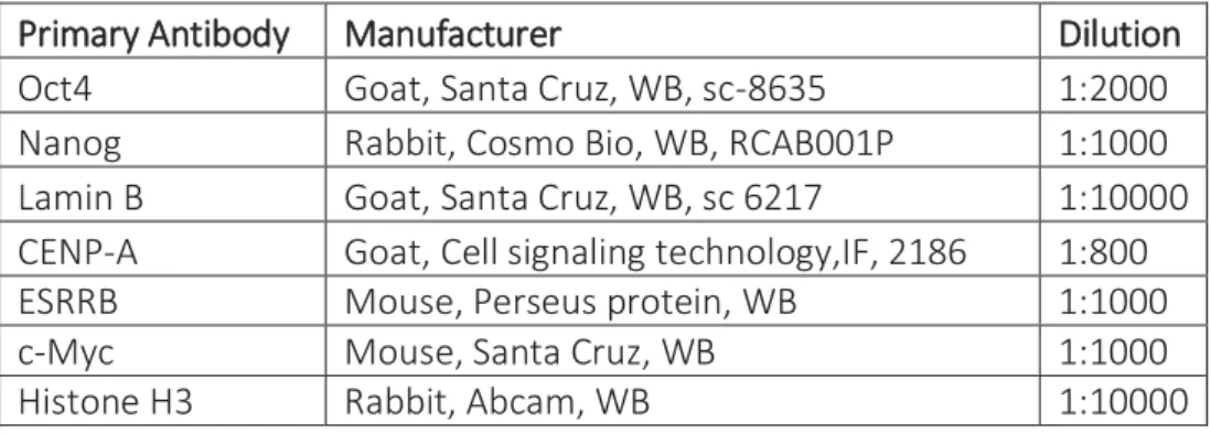
Table 2-3 Mouse gene specific primers Table 2-4 List of antibodies
Abbreviations
2i two inhibitors
5-AzaC 5 -deoxyazacytidine
AP Alkaline Phosphatase
AR Aspect Ratio
BSA Bovine Serum Albumin
Cast Mus musculus castaneous CENPA Centromere Protein A
chIP chromatin ImmunoPrecipitation CLEM Correlative light Electron Microscopy
CRISPR Clustered Regulatory Interspaced Short Palindromic Repeat CryoEM Cryo electron microscopy
DAPI 4', 6'-diamidino-2-Phenylindol DMEM Dubelcco's modified Eagle's medium DMSO Dimethyl sulfoxide
DNA Desoxyribonucleic acid
DNA FISH DNA Fluorescence in Situ Hybridization Dom Mus musculus domesticus
EBV Epstein Barr Virus
EDTA Ethylene Diamine Tetraacetic Acid
EM Electron Microscopy
ES cell Embryonic Stem cell
Esrrb Estrogen-Related Receptor Beta FACS Fluorescence-Activated Cell Sorter
FCS Foetal Calf Serum
gDNA genomic DNA
GFP Green Fluorescent Protein HAT Histone Acetyl Tranferase HDAC Histone deacetylase Hi-C High chromosome contact
ICM Inner Cell Mass
iPS Induced Pluripotent Stem cell IRES Internal Ribosome Entry Site LIF Leukemia Inhibitory Factor MEF Mouse Embryonic Fibroblast
miRNA micro RNA
OSKM Oct4, Sox2, Klf4, C-myc
PAB Polyamide buffer
PEG Poly Ethylene Glycol
PFA Paraformaldehyde
PI Propidium Iodine
piRNA Piwi Interacting RNA
RNA Ribonucleic acid
RT-PCR Reverse Transcriptase PCR RT-PCR Reverse Transcriptase PCR
RT-qPCR quantitative Reverse Transcription-PCR SCNT Somatic Cell Nuclear Transfer
SDS SodiumDodecyl Sulphate
SIM Structured Illumination Microscopy siRNA Small Interfering RNA
SNP Single Nucleotide Polymorphism TEV protease Tobacco Etch Virus protease TAD Topologically Associated Domain
UTR Untranslated region
WT Wild Type
Xa Active X chromosome
XCI X Chromosome Inactivation
Xp paternal X
Abbreviation of Units
Å Ångström kDa KiloDalton bp base pair Kp Kilopair L Litre M Molar nm NanometerRPM Rotation per Minute μm Micrometer
Chapter 1
:
I
NTRODUCTION
1.1 Epigenetic mechanisms and inheritance
A multicellular organism is composed of a myriad of differentiated cells with specific roles, transcriptomes and proteomes, which ultimately the vast majority of them possess the same genome. Therefore, differences in gene expression between various cell types are caused by modifications on chromosomes without altering the DNA sequence. This phenomenon of epigenetics encompasses DNA methylation, histone modifications and variants, as well as non-coding RNA (Figure 1). In the following section I will attempt to give an account of these mechanisms.
FFigure 1-1: Epigenetic mechanisms involved in gene expression.
Methylation of DNA on specific promoters inhibit gene transcription. Histone modifications can either repress or activate gene expression depending on the residues present on the histone tails. Non-coding RNA such as Xist can trigger gene silencing, or in this particular case, the silencing of an entire genome. (Zaidi et al., 2010)
1.1.1
DNA
METHYLATIONOf the three different types of epigenetic mechanisms known so far, DNA methylation has been the most extensively studied. DNA methylation was first described in the 1950 by Hotchkiss (Hotchkiss, 1948) but it wasn’t until the 1970s that DNA methylation was associated with gene regulation (Holliday and Pugh, 1975). DNA methylation consists of the addition of a methyl group (-CH3) on DNA, preferentially at the 5-position of cytosine residues (5mC) at CpG dinucleotides (Robertson, 2005).
DNA methylation is a mechanism important for cell differentiation and gene expression. DNA methylation prevents the binding of transcription factors or the recruitment of proteins (Methyl-CpG-binding proteins) implicated in gene repression. Examples of gene silencing associated with DNA methylation include imprinting (Elhamamsy, 2017) and X chromosome inactivation (Kaslow and Migeon, 1987), the details of which I shall elaborate upon later on. DNA methylation is catalyzed by the DNA methyltransferase (DNMTs) enzymatic family which can be separated into two distinct groups depending on the DNA substrate they use (Bestor, 2000; Bestor et al., 1988; Cheng and Blumenthal, 2008). The de novo methyltransferases, DNMT3a and DNMT3b, establish methylation on DNA soon after the embryo’s implantation, whereas DNMT1 is associated with the maintenance of DNA methylation throughout cell mitosis. Indeed, DNMT1 binds on hemi-methylated DNA and methylates the newly synthesized DNA strand using the parental strand as a template (Figure 2) (Hermann et al., 2004). DNMT3L, a DNMT-related protein, associates with DNMT3a and DNMT3b to regulate their catalytic activity, though DNMT3L itself does not possess DNA-methyltransferase activity (Ooi et al., 2007). Previous work demonstrated that Dnmt1 knock-out mice are embryonically lethal, (Lei et al., 1996). Dnmt3a knockout mice die soon after birth, whereas Dnmt3b KO mice are not
viable (Okano et al., 1999). These studies illustrate the importance of DNA methylation in mammals.
Finally, the last member of the DMNTs family is DNMT2. Its biological role is not quite clear yet. One study however showed that DNMT2 methylates tRNA at cytosine 38 preventing tRNA segmentation (Goll et al., 2006).
FFigure 1-2: Mechanisms of DNA methylation.
Unmethylated DNA is methylated by de novo methyltransferases during embryogenesis post implantation. Other epigenetic mechanisms, such as histone modifications, are involved to successfully silence a gene. During DNA replication the newly synthesized strand is unmethylated leading to a DNA hemi-methylated. DNMT1 faithfully establishes the methylation mark on the unmethylated DNA strand using the methylated parental strand as a template. Methylation is erased during reprogramming towards pluripotency. (Issa,2004)
1.1.2
H
ISTONE MODIFICATIONSBesides DNA methylation, another epigenetic modification important for gene regulation is histone modification. Gene expression is regulated through this mechanism by either altering chromatin structure or by recruiting histone modifiers. DNA methylation together with histone modifications have an intrinsic relation regarding the chromatin state (Cedar and Bergman, 2009).
Histones are proteins which are the building block of the first order of compaction of chromatin. Two copies of each core histone (H2A, H2B, H3 and H4) form an octamer, around which 147bp of DNA is wrapped to create nucleosome (Kornberg, 1974; Kornberg and Thonmas, 1974). The core histone possesses an N-terminal protrusion (histone tail) which can be modified according to the type of residue they have. Histone modifications are post translational modifications that happen on core histones on several residues (lysine, arginine serine and threonine). The long list of histone modifications encompasses, among others, methylation, acetylation, phosphorylation, ubiquitylation, and sumonylation (recapitulated in Table 1). Depending on the type of histone modifications and DNA methylation pattern, chromatin can either adopt an open or closed configuration. Open chromatin is associated with transcriptionally active regions named euchromatin, whereas closed chromatin is correlated with transcriptionally inactive regions that are referred to as heterochromatin (Figure 3).
TTable 1-1: Different classes of modification in histones
Chromatin Modifications Residues Modified Functions Regulated
Acetylation K-ac Transcription, Repair,
Replication, Condensation Methylation (lysines) K-me1 K-me2
K-me3
Transcription, Repair
Methylation (arginines) R-me1 R-me2a R-me2s
Transcription
Phosphorylation S-ph T-ph Transcription, Repair, Condensation
Ubiquitylation K-ub Transcription, Repair
Sumoylation K-su Transcription
ADP ribosylation E-ar Transcription
Deimination R > Cit Transcription
Proline isometization P-cis > P-trans Transcription
The most well studied histone modifications are methylation and acetylation. Generally speaking, acetylation is associated with transcriptionally active chromatin whereas methylation is a feature of transcriptionally silenced chromatin. However, there are few exceptions where histones methylation is a transcriptionally active mark, such as H3K4me3,
Histone acetylation is catalyzed by histone acetyltransferase (HAT). Acetylation of histones neutralizes the positive histone charges, thus decreasing the interaction between DNA (negatively charged) and acetylated histones. This allows the unravelling of DNA making it accessible to proteins such as transcription factors to bind on the DNA and promote transcription of genes (Grunstein, 1997). Acetylation can be removed from histones by the Histone DeAcetylase (HDAC) enzymatic family.
Histone methylation consists of the addition of one, two or three methyl groups on the histone tail which occurs in the presence of histone methyltransferase (HMT) and will either repress or activate chromatin. Heterochromatin can either be facultative or constitutive. Facultative heterochromatin is enriched in H3K27me3, a histone modification strongly present on the inactive X chromosome (further details below). Constitutive heterochromatin is characterized by the presence of H3K9me3 and is localized at the centromeric and telomeric regions (Saksouk et al., 2015).
FFigure 1-3: Histone modification and chromatin compaction.
(A) Schematic representation of open and closed chromatin according to histone modification. (B) Example of histone modification on different histone subunits and histone residues. Adapted from (Marks et al., 2001)
Little is known about the role of histone phosphorylation in gene expression. However, several studies have suggested that phosphorylation of histone is important during mitosis. Indeed, Fischle et al.,2005 have shown that phosphorylation of Serine 10 on histone H3 (H3S10) by Aurora B kinase during mitosis is important in chromatin condensation (Fischle et al.,2005). In this study, the authors indicate that H3S10 displace HP1 from H3K9me.The same year, Dai et al., demonstrated that phosphorylation of Threonine 3 on histone H3 is essential for alignment of metaphase chromosome.
Another histone, histone H1 (known as linker histone), plays also an important role in chromatin compaction and higher order structure (Thoma et al., 1979). H1 binds to the nucleosome and interacts with linker DNA, maintaining the DNA wrapped around the nucleosome (Thomas, 1999). H1 is the histone protein that possesses the most variants, however little is known about H1 modifications (Wiśniewski et al., 2007). An elegant study has demonstrated, using Fluorescent Recovery After Photobleaching (FRAP), the “stop and go” mechanism of H1 binding to nucleosomes highlighting the dynamics of chromatin structure (Catez et al., 2006). The “stop phase” occurs when H1 binds to the nucleosome, and its duration varies according to the modifications of the core histones. The duration of the stop phase is significantly longer than the “go phase”, which occurs when H1 binds to another nucleosome.
1.1.3
N
ON-
CODINGRNA
Another element affecting chromatin states are long non coding RNAs (lnc-RNA), which are RNA transcribed from non-protein coding regions of the genome (Kapranov et al., 2007; Amaral and Mattick, 2008). Nc-RNA encompass several types of RNA which can be divided into two categories based on their size: Small nc-RNA and long nc-RNA. Small nc-RNA includes among others piRNA, miRNA, siRNA. Nc-RNA uses two mechanisms to regulate gene expression: small nc-RNA bind complementarily to mRNA and either inhibit their translation (miRNA) or degrade the RNA itself (siRNA) (Filipowicz et al., 2005). PiRNA is involved in gene
Lnc-RNA have been shown to be crucial for cell pluripotency and differentiation as well as cell growth (Cesana et al., 2011; Wang et al., 2013). Lnc-RNA may be used as precursor for small nc-RNA, however recently it has been shown that lnc-RNA can also regulate gene silencing by direct interaction with DNA or chromatin modifiers (Marchese and Huarte, 2014). Indeed, the Hox locus (HOTAIR) lnc-RNA interacts with the PRC2 protein complex to silence a chromosomal domain (Rinn et al., 2007). Other lnc-RNA have been shown to recruit chromatin modifiers in the imprinting process to preferentially silence one allele. Another example of gene silencing mediated by lnc-RNA is the inactivation of the X chromosome triggered by the lnc-RNA Xist which acts in cis and adheres to specific loci on the Xi then spreads to inactivate an entire chromosome (see section 3.1 of the introduction for more details). These two examples demonstrate the variety of mechanisms used by Lnc-RNA to silence specific gene loci.
1.1.4
M
ITOTIC CHROMOSOMES AND EPIGENETIC MEMORY 11.1.4.1 Mitotic chromosomesOne of the most striking phenomenon that occurs during mitosis is the formation of mitotic chromosomes, which are a complex entity that arise from the compaction of chromatin. Mitotic chromosomes are condensed chromatin with the addition of proteins important for the compaction, structure and segregation of chromosomes. In this section, I will discuss the proteins important for mitotic chromosome structure.
Scaffolding proteins are important for the higher order structure of mitotic chromosomes. Two major members of this group of proteins are Topoisomerase II (Topo II) and condensin I and II (Earnshaw and Heck, 1985; Gasser et al., 1986; Saitoh and Laemmli, 1994). Topo II has different roles according to the cell cycle stage. During DNA replication, Topo II breaks and rejoins double-strand DNA in order to remove supercoiled or intertwined DNA, while during mitosis it acts as a scaffold. The other protein complex that acts as a scaffold during mitosis is the highly conserved condensin, composed of the five subunits. Two of these subunits are part of the
components called Condensin Associated Proteins (CAP) and are CAP-G/G2, CAP-D2/D3 and CAP-H/H2 (Ono et al., 2003). Maeshima and Laemmli demonstrated in an elegant study that Topo II and a condensin subunit (13S) localize on a vertical line in mitotic chromosomes. From these finding they proposed that Topo II and condensin intertwined to form a ‘Barber Pole’ pattern that acts as a scaffold in mitotic chromosomes (Maeshima and Laemmli, 2003). Condensin depletion generates mitotic chromosomes that appear to be larger and less condensed (Hudson et al., 2003) whereas lack of Topo II alpha diminishes chromatin condensation (Farr et al., 2014) but not the loading of condensin, suggesting two distinct pathways for Topo II alpha and condensin to act as a scaffold in mitotic chromosomes (Cuvier and Hirano, 2003).
Another protein important for mitotic chromosome structure is the cohesin protein complex. Cohesin is a tetrameric protein composed of 4 subunits: SMC1, SMC3, RAD21 and STAG. Its role is to hold the two sister chromatids, established during S phase, together during mitosis until the anaphase step of mitosis. The cohesin complex forms a ring-like structure within both sister chromatids are trapped. Cohesin counteracts the forces of the mitotic spindle, hence allowing the correct alignment and segregation of the sister chromatids (Nasmyth et al., 2000). Several studies using cohesin mutants showed that they were unable to hold sister chromatids together during metaphase (Guacci et al., 1997; Michaelis et al., 1997). At the onset of mitosis (prophase), cohesin is removed from mitotic chromosomes. Only a small percentage remain on mitotic chromosomes throughout the prophase-to-metaphase transition, where cohesin subunit’s Rad21 can only be detected at the centromeric region (Waizenegger et al., 2000). To release the two sister chromatids in anaphase, cohesin needs to be cleaved by separin, leading to the release of cohesin from mitotic chromosomes and the separation of the chromosome arms. Rad21 links together all the other subunits of the cohesin complex. Destruction of Rad 21 results in the dissociation of the other subunits of cohesion from the chromosomes (Xu H et al, 2004).
as well as the segregation of chromosomes (Kunitoku et al., 2003; Zeitlin et al., 2001). Human CENP-B proteins bind to a 17 bp region named the CENP-B box and may be involved in chromatin structure(Tanaka et al., 2001). CENP-C is a member of CENPA-nucleosomes associated complex that is a key factor in mitotic progression, chromosome segregation and kinetochore assembly (Gascoigne et al., 2011). DNMT3B is recruited by CENP-C to methylate centromeric and pericentromeric regions (Gopalakrishnan et al., 2009).
Another component of mitotic chromosomes which are also present in interphase chromosomes are the telomeres located at either end of each chromosome arm. Telomeres are made of kilo bases of nucleotides repeats and possess specific telomere proteins known as sheltering complex proteins. At each cell division, the nucleotides repeats are shortened until it reaches a critical point. When cells possess critically short telomeres they become senescent and stop proliferating. The shelterin complex is composed of five proteins TRF1, TRF2, Rap1, Pot1, Tin2 and TPP1. Shelterin proteins prevent shortening of the telomere and chromosomes end fusion by forming a T-loop DNA structure that avoids the end of a chromosomes from being identified as DNA damage (de Lange, 2005).
11.1.4.2 Epigenetic memory
Epigenetic mechanisms are maintained throughout cell division allowing, during symmetrical division, both daughter cells to possess the same phenotype as the parental cell. During mitosis, cells undergo drastic nuclear and cellular changes. One of the most evident changes is the compaction of chromatin into mitotic chromosomes and loss of the nuclear envelope. Another modification that happens during mitosis is the cessation of gene transcription and the removal of most proteins, including RNA polymerase and most transcription factors, from mitotic chromosomes (Spencer et al., 2000).
To maintain the epigenetic memory throughout the cell cycle, several mechanisms takes place. For instance, during S phase to retain methylation newly synthesized DNA is methylated by the DNMT1 enzyme. Eed protein, a component of PRC2 protein complex ensure the propagation of H3K27me3 during DNA replication (Margueron et al., 2009). These process ensure the stability of the epigenetic marks during cell cycle.
Following the transition between mitosis and G1 phase of the cell cycle, transcription is resumed in daughter cells in order to obtain a gene expression pattern similar to the parental cell in the case of symmetrical cell division. To allow the maintenance of the cell fate and proliferation throughout several cell cycles, a mechanism, named mitotic bookmarking, has been proposed. Mitotic bookmarking is a relatively recent concept and its details still remain to be fully elucidated. Mitotic bookmarking is the retention of specific transcription factors on the mitotic chromosomes to ensure an accurate and rapid transition from mitosis to G1 phase of the cell cycle. These bookmarking transcription factors are involved in cell fate or cell proliferation and even in oncogenesis (Zaidi et al., 2014). A recent study has shown using high-resolution microscopy techniques that in ES cells, the pluripotency associated factor Sox2 remains attached on mitotic chromosomes (Deluz et al., 2016) allowing the daughter cells to maintain their ES cell like state throughout rapid cell cycling. Another study demonstrated that Esrrb, another pluripotency-associated factors, remains bound to mitotic chromosomes (Festuccia et al., 2016). These findings corroborate the fact that mitotic bookmarking is important for the maintenance of cell fate.
1.2 Pluripotent Stem Cells and reprogramming towards pluripotency
1.2.1
P
LURIPOTENTS
TEM CELLSPluripotency is the ability of a cell to differentiate into the three primary germ cell type of the early embryo, therefore leading to the genesis of all the cells present in an adult body. Pluripotent Stem Cells are present in vivo specifically during embryogenesis. Pluripotent Stem Cells are defined by their ability to differentiate into the three primary germ layers (endoderm, ectoderm and mesoderm) and to self-renew with cell division. Other characteristics of pluripotent cells include the formation of teratomas (tumors that have the ability to form three different germ cell lines) in vivo and the formation of chimeras. In embryogenesis, pluripotent stem cells are found at the blastocyst developmental stage. Embryonic stem cells (ES cells) are
Two different types of culture conditions are used to maintain pluripotency in vitro: by the addition of Leukemia Inhibitory Factor (LIF) to the ES cell media, or alternatively by using two small molecules (Chir99021 and PD0325901) known as 2 inhibitors (2i) media. ES cells cultured in 2i media maintain their pluripotent state by inhibiting mitogen-activated protein kinase (MEK) and glycogen synthase kinase-3 (GSK3) (Ying et al., 2008). The gene expression profile of ES cells maintained in 2i media condition is different from ES cells maintained in LIF (Marks et al., 2012). ES cells cultured in 2i media express a stable level of Nanog contrary to ES cells in serum with LIF media that shows a periodicity in Nanog expression from Nanog-high to Nanog-low state. The Nanog low population is more prone to respond to differentiation (Singh et al., 2007).
Pluripotency is regulated by three main group of transcription factors, Oct4 (Pou5f1), Sox2 (SRY box2) and Nanog (Kim et al., 2008; Orkin et al., 2008). These three transcription factors represent the core factors of pluripotency. Oct4 is expressed in pluripotent ES cells both in vivo and in vitro (Nichols et al., 1998). Sox2 have been shown to be crucial for the generation of pluripotent epiblast stem cells and tightly regulates Oct4 expression in ES cells (Avilion et al., 2003). Nanog overexpression have been shown to promote ES cell self-renewal in the absence of Leukemia Inhibitory Factor (LIF) (Mitsui et al., 2003). The concentration of these three transcription factors is stringently controlled as their overexpression or loss leads to differentiation of ES cells. ES cells harboring a low level of Nanog are more prone to differentiate compared to ES cells with a high level of Nanog (Filipczyk et al., 2015). Small increases in Sox2 expression in ES cells leads to the differentiation of ES cells towards several lineages including neuroectoderm or mesoderm (Kopp et al., 2008). Slight overexpression of Oct4 in ES cells triggers differentiation towards mesoderm whereas knock-down of Oct4 enhances the differentiation towards trophectoderm (Hay et al., 2004).
ES cells have unique chromatin states that differ from differentiated cells. In ES cells histones can simultaneously possess both active (H3K4ac) and repressive histone modifications (H3K27me3). This status is called bivalency (Bernstein et al., 2006). Upon differentiation, one of the two histone modifications is lost while the other one remains. Furthermore, ES cells possess a global low level of DNA methylation and an enrichment of acetylation (Azuara et al., 2006).
1.2.2
R
EPROGRAMMINGReprogramming is the conversion of one cell type to another that can either be induced chemically or by ectopic expression of specific proteins. Reprogramming towards pluripotency is the process by which somatic cells are converted to ES cells. Reprogramming towards pluripotency can be achieved in vitro by three different ways: nuclear somatic cell transfer, cell fusion, and transcription-factor transduction induced pluripotent stem cells (iPS). The discovery of iPS cells has tremendous clinical applications, and more studies need to be conducted in order to fully understand the mechanisms regulating the reprogramming towards pluripotency process. Understanding the molecular mechanism can be done using the three in vitro reprogramming techniques. In this section I will briefly describe the three reprogramming methods.
Figure 1-4: Cell fusion mediated nuclear reprogramming towards pluripotency.
Cell fusion mediated reprogramming is where a pluripotent cell (e.g ES cells) is fused with a somatic cell to form a single entity called a heterokaryon that share one cytoplasm and several nuclei. Heterokaryons can then give rise to hybrid cells that only
11.2.2.1 Nuclear Transfer
Nuclear transfer occurs when a nucleus from a somatic cell is transplanted inside an enucleated oocyte, followed by the nuclear reprogramming of the somatic cell nucleus leading to the creation of an entire individual possessing the exact same genome of the somatic cell nucleus. Briggs and Kings 1952 were the first to demonstrate the potential of nuclear reprogramming mediated by nuclear transfer. This experiment was carried out by inserting an early blastocyst cell nucleus into an oocyte from amphibians. This first study demonstrated the plasticity of somatic cells and their epigenetic mechanisms.
Later on, Gurdon performed the same experiment using a nucleus from fully differentiated tadpoles and successfully obtained normal adult frogs (Gurdon, 1962). These findings lead to the creation of Dolly the sheep (Wilmut et al., 1997) decades after. A year later, cloned mice were generated by nuclear transfer (Wakayama et al., 1998). In addition to the low efficiency of the somatic cell nuclear transfer (SCNT) process, cloned mice can have several abnormalities ranging from abnormal gene expression in embryos to shorter life span, suggesting that the erasure of the epigenetic memory, such as DNA methylation or histone modification, is not complete in cloned mice obtained by SCNT. Indeed, addition of a histone deacetylase inhibitor increases the efficiency of reprogramming by SCNT. Recently, a study was performed with enucleated human oocytes and human fibroblasts (Tachibana et al., 2013). This could be useful for future clinical application.
1.2.2.2 Transcription factor mediated reprogramming
In 1987, Gehring and colleagues showed in D.melanogaster that ectopic overexpression of a homeotic gene, antennapedia, led to a change in body plan by adding a pair of legs where the antenna should have been (Hiromi and Gehring, 1987). The same year, Weintraub and colleagues discovered that ectopic expression of myoD in fibroblasts in mice lead to their conversion to myogenic lineage (Davis et al., 1987). Another finding demonstrated that the
et al., 1999). In 2006 the generation of pluripotent stem cells from mouse fibroblasts has been achieved by overexpressing Oct4, Sox2, Klf4 and c-Myc (OSKM) using retro-viral vectors and a DNA library of genes expressed in ES cells (Takahashi and Yamanaka, 2006).
The pluripotency status of those derived cells was assessed by their capacity to generate teratomas. Those cells were named iPS (induced Pluripotent Stem cells). iPS cell generation is a lengthy (up to three weeks) and low efficiency process (Hochedlinger and Plath, 2009). Its mechanism can be dissected into three phases: initiation, maturation and stabilization. In the initiation phase the cell cycle of somatic cells changes towards a more ‘ES cell like’ cell cycle profile, and a change in cellular morphology can be seen (e.g cells become smaller and round). A few days after transfection, Mesenchymal to epithelial transition (MET) occurs, indicated by the formation of small, round shape colonies. MET is the exact opposite of epithelial to mesenchymal transition (EMT) that happens during embryonic development. During the maturation/intermediate phase, cells start expressing pluripotency associated markers such as Alkaline Phosphatase, the cell surface marker SSEA1 and are self-renewable (Brambrink et al., 2008). Most of the cells transfected with OSKM remain stuck at the maturation stage indicating that there is an epigenetic barrier preventing them to move towards the fully pluripotent status. It has been shown that the choice of somatic cells is important, less differentiated somatic cells have a higher efficiency in reprogramming towards pluripotency (Raab et al., 2014). For example, neural stem cells already express Sox2 and a low level of Klf4, thus their reprogramming efficiency is higher compared to fibroblasts. Recently people have tried to increase the efficiency of iPS by various methods by either controlling the microenvironment of by changing the stoichiometry of the ectopic factors (Luni et al., 2016; Tiemann et al., 2011). Overall these studies indicate that ectopic expression of specific proteins as well as deletion of proteins can change the cell fate of differentiated cells demonstrating the plasticity of somatic cells and their epigenetic memory.
11.2.2.3 Cell fusion mediated reprogramming
Experimental cell fusion was described by Henry Harris and Nils Ringertz in the 1960s (HARRIS et al., 1969). A few years later, production of monoclonal antibody was achieved with hybridomas generation by cell fusion between B cells and plasmacytomas NS1 (Köhler and Milstein, 1975). More recently cell fusion has been used to study the molecular mechanism of trans-acting elements from one genome to another one. Cell fusion studies showed that the fate of a somatic cell can be reversible and requires regulators such as proteins. Specifically the cell fusion technique demonstrated that when fusion occurs between ES cells and somatic cells, pluripotency associated factors from ES cells reprogram the nucleus of the somatic cells leading to chromatin remodeling and reactivation of previously silenced genes (Pereira et al., 2008). Cell fusion consists of the fusion of two or more cell types to generate one single entity which are heterokaryons or hybrid cells (Figure 1-4). The main difference between those two cells is that hybrid cells can undergo mitosis and possess one nucleus contrary to heterokaryons which have a short life-span and possess multiple nuclei. Upon cell fusion, RNA and protein are synthesized suggesting that cell fusion does not change fundamental cellular metabolism.
Cell fusion is useful to study events of reprograming such as DNA demethylation or X chromosome reactivation (Cantone et al., 2016; Piccolo et al., 2013). Surani and colleagues showed that when fusing embryonic germ cells with somatic thymocytes from mice, hybrid cells were generated (Tada et al., 1997). They demonstrated that the generated hybrid cells possessed pluripotent properties such as formation of chimera and self-renewal. Later on Tada et al took advantage of the hybrid system to show that hybrid cells created from the fusion between ES and somatic cells can acquire a pluripotent state (Tada et al., 2001). In this study they fused male ES cells with female somatic cells that contained a GFP reporter transgene driven by the promoter of Oct4. They found that some X-linked genes from the previously inactive X were reactivated and that GFP was expressed indicating the re-activation of Oct4. Subsequent studies with human ES cells showed the generation of tetraploid hybrid cells when fusing with human fibroblasts (Cowan et al., 2005). Those hybrid cells successfully express Oct4
useful to study early reprogramming events (Pereira et al., 2008). Changes in the chromatin environment are also detected, high level of acetylation and presence of histone modifications such as H3k4me3 can be detected (Kimura et al., 2004).
Cell fusion mediated reprogramming is highly dependent on the somatic cell partner. It has been shown that neural progenitors have a higher reprogramming efficiency compared to fibroblast (Silva et al., 2006). In this study, they also showed that overexpression of Nanog
increases the efficiency of reprogramming towards pluripotency mediated by cell fusion (Silva et al., 2006). Increasing the reprogramming efficiency mediated by cell fusion is also achieved by treatment with the demethylating agent 5-AzaC, showing once more the importance of the chromatin environment to fully reprogram somatic cells towards pluripotency (De Carvalho et al., 2010).
1.3 Epigenetic mechanisms behind X chromosome inactivation and
reactivation
1.3.1X CHROMOSOME INACTIVATION
In placental female mammals one of the two X chromosomes is randomly inactivated (Lyon, 1962). This biological process ensures that both males and females express the same dosage of X-linked genes. In mammalian cells the inactive X chromosome (Xi) forms a Barr body that is characterized by an intense signal when cells are stained with DAPI (Barr, M.L. & Bertram, 1949). X chromosome inactivation happens only when there are more than one X chromosomes. As an example, individuals possessing only one X chromosomes and no other sex chromosomes (i.e X 0) have a female phenotype. This phenomenon is known as counting. Random Xi leads to mosaicism where some cells express genes from one X chromosome whereas other cells express X-linked genes from the other allele (Figure 1.2 A).
In mice, the first X chromosome inactivation occurs at the 4-cell stage, where the paternal X chromosome (Xp) is preferentially inactivated (Mak et al., 2004; Okamoto et al., 2004).This process is an example of imprinting as the maternal X chromosomes is prevented to not express Xist. Extraembryonic lineage such as trophectoderm and primitive endoderm will then maintain the inactive paternal X chromosome (Takagi and Sasaki, 1975). It is not fully understood why the Xp chromosome is preferentially inactivated, but it is hypothesized that the maternal X chromosome (Xm) possesses an epigenetic mark preventing it from being inactivated at the 4-cell stage. A Recent study suggests that H3K9me3 located at the Xist promoter on the Xm plays a role in preventing Xist expression from Xm (Fukuda et al., 2014). Xp inactivation is reversed at the blastocyst stage. At this stage random Xi occurs.
Random silencing of X-linked genes is initiated in epiblast cells of the blastocyst and is maintained thereafter throughout subsequent cell divisions (Rastan, 1982; Takagi et al., 1982). X chromosome inactivation (XCI) is an exemplar of epigenetic silencing that requires the presence of a specific region called the X Inactivation Center (Xic) located on both X
region of XIC contains genes coding only for Ln-RNA such as Xist, RepA, Tsix, Xite and Jpx. A Recent study demonstrated that silencing of an autosome can be achieved by insertion of the core region of XIC on this autosome (Tang et al., 2010). Other element important for XCI are Ln-RNA Ftx, Tsx and the protein RNF12. XCI happens in a specific order that can be separated into an initiation phase and maintenance phase. These phases involving several long non-coding RNAs (Tian et al., 2010), histone modifications (Heard et al., 2001; Plath et al., 2003) and chromatin remodeling. Specifically, XCI is initiated by the transcription of Xist, a 17 kb long non-coding RNA localized on the Xic locus that is expressed and coats the future inactive X chromosome (Xi) in cis (Panning et al., 1997). Expression of Xist is tightly controlled by several lnc-RNA such as, Jpx, ftx, Tsix and proteins such as RNF12 (Sun et al., 2013). The most well-known lnc-RNA regulating Xist is Tsix, which when expressed prevents Xist expression (Lee et al.,2004). Another study revealed that RNF12 is essential of X inactivation by degrading Rex1 a pluripotency-associated factors that inhibits Xist transcription (Barakat et al., 2011; Jonkers et al., 2009).
FFigure 1-5: Random X inactivation mechanism.
(A) Upon differentiation somatic cells retain one X active and one inactive to ensure the same level of X-linked genes in male and female cells. This process occurs randomly. (B) Schematic representation of Xi. First Xist is expressed from the future inactive X closely followed by the exclusion of the transcription machinery such as RNA Pol II. PRC1 and PRC2 are recruited and catalyzed the histone marks H2AK119ub1 and H3K27me3, respectively. This lead to a late replication of the inactive X in S phase and a global hypo-H4 acetylation of the inactive X.
Xist expression triggers nuclear delocalization of Xi, closely followed by the exclusion of RNA polymerase II (Chaumeil, 2006) creating a repressive compartment on the Xi characterized by the lack of active histone modifications such as histone acetylation. Then chromatin modifiers such as polycomb repressive complex 1 and 2 (PRC1 and PRC2) are recruited to catalyze ubiquitination of H2A at lysine 119 (Fang et al., 2004) and histone H3 lysine27 tri-methylation (H3K27me3) (Csankovszki et al., 2001; Silva et al., 2003) respectively. Although it has been shown that Xist is necessary for the initiation of inactivation, it is not essential for maintaining the inactive state (Csankovszki et al., 1999). The maintenance phase involves DNA methylation and recruitment of heterochromatin proteins to the Xi. DNA (cytosine-5)-methyltransferase 1 (Dnmt1) has been shown to play a crucial role in the stabilization and maintenance of the Xi (Sado et al., 2000) by hypermethylating gene rich region on the Xi. Another key protein involved in the maintenance of the Xi is the structural-maintenance-of-chromosomes hinge domain containing 1 (Smchd1) protein. Its role is to hypermethylate CpG islands on the inactive X chromosome (Blewitt et al., 2008). Another factor affecting the stability of the Xi is macroH2A, a histone variant that is preferentially bound to Xist (Costanzi and Pehrson, 1998; Mermoud et al., 1999). The combination of different epigenetic factors is thought to make the Xi process highly stable.
1.3.2X CHROMOSOME REACTIVATION
11.3.2.1 Xi reactivation in vivo
In mice, as well as in humans, reactivation of the Xi has been used as an indicator of the naïve pluripotent state (Plath and Lowry, 2011). Reactivation of the Xi chromosome happens both in vivo and in vitro. During mouse development, Xi reactivation takes place at three different stages of embryonic development. Xi reactivation first occurs at the two cell stage of the embryo, where the paternal X chromosome (Xp) is reactivated (green arrow figure 1-6). The second Xi reactivation occurs at the blastocyst stage, where cells from the Inner Cell Mass (ICM) reactivate the previously inactive paternal X chromosome (Lee et al., 2013). Xp reactivation is characterized by the loss of Xist expression, followed by loss of the repressive mark H3K27m3 and loss of PRC2. Surprisingly, the expression of some X-linked genes have been detected prior Xist and H3K27me3 loss indicating that Xpi reactivation occurs while Xist and H3K27me3 are present on the Xi (Williams et al., 2011).
The third Xi reactivation happens during the formation of primordial germ cells (PGCs), where the pluripotency gene network is re-expressed (de Napoles et al., 2007; Sugimoto and Abe, 2007). Embryonic stem cell lines can be derived from these Primordial Germ Cells (PGCs) (i.e. embryonic germ cells, EGCs). PGCs are derived from epiblasts where the Xi silencing has already occurred. Xi reactivation is initiated when PGC cells begins to migrate towards the genital ridge. The erasure of the epigenetic memory in PGCs occurs during this migration, and includes reactivation of Xi. Xi reactivation is a process that includes several steps. Loss of Xist expression and H3K27m3 are the first event to occur followed by reactivation of some X-linked genes. DNA demethylation and chromatin remodeling are required for the epigenetic reprogramming that happens during the formation of PGCs and this coincides with X-linked reactivation.
FFigure 1-6: X inactivation and reactivation during mouse embryonic development.
After fertilization of the oocyte by the sperm and the first mitosis, both X chromosomes become active (First reactivation indicated by the green arrow). Then, the paternal X chromosome (Xp) become inactive and will remain inactive in the extraembryonic tissues. In contrast at the blastocyst stage Xp (blue) is reactivated (Second reactivation). Soon after this reactivation, random X inactivation happens (blue arrow). The last reactivation takes place in PGCs before oogenesis. Adapted from Ohhata and Wutz,2013.
1.3.2.2 Xi reactivation in vitro
In vitro the Xi re-acquires an active state during pluripotent reprogramming of a somatic cell, achieved by various approaches including exogenous factors (Maherali et al., 2007; Pasque et al., 2014), nuclear transfer (Eggan et al., 2000) or cell fusion (Tada et al., 2001; Takagi et al., 1983; Ying et al., 2002) (Recapitulated in Figure 1-7). Furthermore, some X-linked genes can be reactivated by chemicals such as 5-AzaC (Csankovszki et al., 2001). Xi reactivation is one of the last events during reprogramming and results in loss of expression of Xist RNA, displacement of polycomb-group repressor proteins and loss of chromatin-associated silencing marks such as H3K27me3.
FFigure 1-7: In vitro system for Xi reactivation.
(A) Using SNCT the female somatic cell nucleus is inserted inside and enucleated oocyte. This gives rise to an embryo with two active X chromosomes. (B) In iPS system, female somatic cells are transfected with four transcription factors upon which the somatic cells will become pluripotent, thus having two active X chromosomes. (C) Finally in cell fusion, the female reprogrammed cells will also possess two active X chromosomes. Adapted from Ohhata and Wutz 2013
Generation of cloned mice by SNCT, has shown that the Xi from the somatic donor cell is preferentially chosen for the inactivation in extraembryonic lineage (Figure 1-7.A). This process is similar to the inactivation of the paternal X chromosomes in extraembryonic tissues from fertilized eggs. If the donor cell is female random Xi occurs in extraembryonic tissues. However random inactivation is observed in cloned mice suggesting that the process of reactivation and inactivation of the X chromosomes is normal (Eggan et al., 2000). The SNCT technique has a lot of caveats, its efficiency is low and individuals generated from it carry genetic abnormalities due to failed erasure of epigenetic mark. In female embryos derived from nuclear transfer down-regulation of X-linked genes is observed due to improper XCI activation and Xist expression. Ectopic Xist expression has been shown to increase the efficiency of nuclear cloning (Inoue et al., 2010).
In iPS generation, Xi reactivation appears to be one of the last events in the epigenetic remodeling (Figure 1-7.B) (Stadtfeld et al., 2008). In this system, studies demonstrated that Xist repression is not sufficient to induce Xi reactivation they also found that DNA demethylation happens at a late stage during Xi reactivation and that DNA demethylation itself is also not sufficient enough to induce Xi reactivation (Pasque et al., 2014).
Cell fusion between mouse ES cells or Embryonic Carcinoma (EC) cells and female somatic cells showed Xi reactivation, demonstrating that mouse ES cells possess the chromatin environment necessary for Xi reactivation (Takagi et al., 1983). Xi reactivation is also achieved when fusing EG cells with female somatic cells. In this particular fusion system global DNA demethylation was detected (Tada et al., 1997). Cell fusion studies have been able to shed some light regarding the mechanism of reprogramming. Indeed, it has been shown that PRC2 components are required for cell fusion mediated reprogramming (Pereira et al., 2010). In contrast, DMNTs are not necessary for reprogramming mediated by cell fusion. This highlights the potential of cell fusion to understand the mechanism of Xi reactivation (Figure 1-7.C). Recent studies demonstrated that some X-linked genes are reactivated upon reprogramming towards pluripotency (Cantone et al., 2016).
X chromosome inactivation and reactivation has been associated with diseases. Despite recent advances in understanding molecular mechanism implicated in Xi reactivation, a lot still need to be discovered for potential clinical applications regarding the treatment of X-linked gene diseases. Females are less affected by X-linked genes diseases compared to male due to the silencing of one of the two X chromosomes. Rhett syndrome is an X-linked genes diseases that affects female and is due to a mutation on the MeCP2 gene (Lyst and Bird, 2015). Other studies have demonstrated that X chromosome could be involved in cancer. It has been shown that in breast cancer the Barr body which represents the inactive X chromosome disappear (Barr and Moore, 1957). This lead to the assumption that in cancer cell the Xi is reactivated.
1.4 Chromosomes visualization
It was in the 19th century that mitotic chromosomes were observed for the first time. The process of how chromatin compacts into mitotic chromosomes still remains elusive. The vast majority of the discovery made on mitotic chromosome structures were done using microscopy techniques. Currently, four mitotic chromosome models exist. The first model consists of DNA wrapping around nucleosome which then will compact gradually into fibers (30 nm) to form chromatid (Belmont et al., 1987). The second well-known model hypothesizes that DNA form loops around a core unit of scaffold proteins (Earnshaw and Laemmli, 1983). In this elegant study, Earnshaw and Laemmli depleted purified human mitotic chromosomes of histones and imaged them by EM. They found that histone depleted human mitotic chromosome still harbor a metaphase chromosome shape (X shape) surrounded by a halo of DNA. The third proposed model suggests that DNA is stacked onto a 6nm layers perpendicular to chromosome axis (Daban et al, 2015). Finally, the last model propose that mitotic chromosomes are not based on scaffolding proteins but instead consist of chromatin cluster separated by proteins (Poirier and Marko, 2002). All of these models even though they have been scientifically demonstrated still remain debatable. These models were developed based on data obtained from microscopy. In this section I will review the recent advance of chromatin visualization by both high resolution light and electron microscopy.
1.4.1LIGHT MICROSCOPY CHROMOSOMES VISUALIZATION
Several light microscopy methods have been used to study the chromatin compaction during mitosis. Light microscopy allow us to visualize mitotic chromosomes during the different stages of mitosis. However, little information could be extracted from it due to the resolution limits of standard light microscopy techniques (250nm). To obtain a better resolution, in the last couple of years, several new microscopy techniques were developed named high resolution microscopy techniques. The most performing technique, PhotoActivated Localization Microscopy (PALM) allowed us to achieve a resolution up to 10nm. Some of these techniques were used to decipher the substructures of mitotic chromosomes.
In 2010 Matsuda et al were able to image mitotic chromosomes from D.melanogaster at a resolution up to 20nm using PALM technique and H2A tagged with GFP. This study suggested that mitotic chromosomes from D.melanogaster are formed by an assembly od 70nm blocks (Matsuda et al., 2010). Two years later, 3D- Structured Illimunation Microscopy (SIM) technique was used to study the role of condensin in metaphase chromosomes (Green et al, 2012). They showed the importance of condensin in maintaining the structure and physical properties of mitotic chromosomes. In 2015, another group studied the role of condensin and topo II with super resolution microscopy and electron microscopy. This study suggests that both condensin and Topo II act as a scaffold for mitotic chromosomes by forming a helix. Condensin topo II and cohesion role in mitotic chromosome structure was further assessed a year later with high resolution microscopy. They discovered that blocking cohesin has little effect on mitotic chromosomes whereas inhibition of topo II prevent sister chromatid resolution (Nagasaka et al., 2016). Assessing the role of proteins involved in mitotic chromosomes structure is easier than assessing the compaction of DNA itself. Indeed, DNA has to be stained to be able to visualize it. The staining may generate artefacts regarding the structure of DNA. Dong et al, 2016 developed a new technique to image DNA. They used the autofluorescence property of the DNA and Stochastic Optical Reconstruction Microscopy (STORM) technique to image chromatin without staining DNA. In their study they were able to see the typical X shape of human mitotic chromosomes (Dong et al., 2016). The data collected on mitotic chromosomes suggest area were the DNA in more compacted than other areas.
1.4.2VISUALIZING CHROMOSOMES AND DNA BY ELECTRON MICROSCOPY
Electron microscopy was first use as a tool to assess chromosomes structure in mitotic cells. In 1977 Paulson and Laemmli depleted chromosomes from histone H1, revealing a loop structure with scaffold using EM (Paulson and Laemmli, 1977). Later on, in 1983, Earnshaw and Laemmli developed a method to visualize intact mitotic chromosomes using EM and their findings corroborate with the results obtained from 1977. The same year X-ray scattering and EM was used to postulate the theory of a 30nm fiber as a substructure of interphase chromosomes (Langmore et al., 1983). More recently, reconstructed nucleosomes were analyzed by EM and showed also a redundant pattern identified as a 30nm fiber (Huynh et al., 2005). A new EM techniques visualized for the first time frozen intact mitotic chromosomes within the cells (Eltsov et al, 2008). Their data revealed that compacted DNA could be imaged using cryo-sectioning and EM. Another EM called FIB (Focused ion beam) combined with high resolution scanning electron microscopy technique was used to image mitotic chromosomes of plants (Schroeder-Reiter et al., 2009).
Another breakthrough regarding DNA imaging happened a couple of years later. In 2012, lambda DNA was imaged by Transmitted electron microscopy (TEM) showing for the first time an image of the double stranded DNA (Gentile et al., 2012). More recently, 3D-Clem was used to characterize chromosome volume of distinct chromosomes (Booth et al., 2016). They also were able to image Ki-67 a protein that surrounds mitotic chromosomes. 3D-CLEM is a hybrid technique that can image both fluorescence and electron microscopy. A year later, Ou et al used ChromEMT (ChromEM tomography), to study the structure of mitotic chromosomes and showed that the chromatin was not as ordered as one thought (Ou et al., 2017).
Aim of the study
Since iPS generation, the reprogramming of somatic cells towards pluripotency have been intensively studied in order to fully understand the molecular mechanisms behind the generation of pluripotent cells from differentiated cells. Despite great advances in the field many questions remain, such as the contribution of different transcription factors or the role of ploidy in reprogramming. Cell fusion is a rapid and efficient way to reprogram somatic cells towards pluripotency. It allows us to study early reprogramming events.
Here I am using cell fusion between pluripotent stem cells and somatic cells to generate heterokaryons and hybrid cells, which can be analyzed to understand the molecular mechanisms important for reprogramming somatic cells towards pluripotency and the reactivation of genes following successful reprogramming. Specifically, first I am first using hybrid cells originating from fusion between ES cells and fibroblasts to investigate the kinetics of Xi reactivation. I am then investigating the role of ploidy in reprogramming towards pluripotency. To do so I will use both heterokaryons and hybrid cells using human B lymphocytes and mouse B lymphocytes respectively. Both of these studies have potential clinical applications for X-linked diseases such as Rett syndrome or Duchennes muscular dystrophy as well as helping iPS to be safer for potential therapeutics.
I then focus my interest in using previously established techniques and adapting them for my purpose. In this section I used flow karyotyping to isolate a specific set of chromosomes to them image them by high-resolution microscopy techniques including cryo-EM. This newly established pipeline of techniques might then be used for biological applications to provide a better understanding of mitotic chromosomes structures and the role of specific proteins in maintaining chromosome structure.
Chapter 2
:
M
ATERIALS AND
M
ETHODS
2.1 Cell culture:
Mouse ES cells were grown on 0.1% gelatin (Sigma) -coated surfaces. E14Tg2a cells were cultured in KO-DMEM medium (Gibco) plus 10% FCS, non-essential amino acids (NEAA) (Gibco), 2mM L-glutamine, 50 μM 2-mercaptoethanol, 10 μg/ml Penicillin and Streptomycin, and 1000 U/ml of homemade leukaemia inhibitory factor (LIF).
Immortalized Mouse embryonic fibroblasts (MEFs) were cultured in KO-DMEM medium (Gibco) plus 10% FCS, non-essential amino acids (NEAA) (Gibco), 2mM L-glutamine, 50 μM 2-mercaptoethanol, 10 μg/ml Penicillin. For the experiments the cells were used between passage P8-P12. The cells were obtained from the laboratory of Professor Brockdorff.
Immortalized Human embryonic fibroblasts were cultured in KO-DMEM medium (Gibco) plus 10% FCS, non-essential amino acids (NEAA) (Gibco), 2mM L-glutamine, 50 μM 2-mercaptoethanol, 10 μg/ml Penicillin. The cells were obtained from Lonza and immortalized using TERT.
Lymphoblastoid cells were cultured in RPMI medium (Gibco) supplemented with 10% delta FCS, 2mM L-glutamine, 50 μM 2-mercaptoethanol, 10 μg/ml Penicillin and Streptomycin. Kind gift of Dr Karen Cheung.
Abelson transformed Oct4-GFP (GOF18ΔPE) Puromycin-resistant mouse B cells were cultured in RPMI medium (Gibco) supplemented with 10% delta FCS, 2mM L-glutamine, 10 μg/ml Penicillin and Streptomycin. Those cells were a kind gift of Prof. A. Surani.
EBV-transformed Puromycin-resistant human B cells were cultured in RPMI medium (Gibco) supplemented with 10% delta FCS, 2mM L-glutamine, 50 μM 2-mercaptoethanol, 10 μg/ml Penicillin and Streptomycin.
Mouse haploid ES cells were grown on 0.1% gelatin (Sigma)-coated surfaces. H129 and AN3-12 cells were cultured in 2i + LIF medium containing N2B27 medium (NDiff 227; StemCells Inc), 3uM CHIR99021 (ReagentsDirect), 0.1mM PD0325901 (ReagentsDirect), 5ml of 7.5% BSA fraction V solution (Life technologies), 10 μg/ml Penicillin and Streptomycin and 1000 U/ml of homemade leukaemia inhibitory factor (LIF). The cells were obtained from Prof. A. Wutz.
Pre B cells Rad21TEV/TEV cells were cultured in IMDM medium (Gibco) supplemented with 10% delta FCS, 2mM L-glutamine, 50 μM 2-mercaptoethanol, 10 μg/ml Penicillin and Streptomycin. Those cells were derived and immortalized in my host laboratory.
2.2 Hybrids experiment:
Hybrids were generated by fusing mouse pluripotent cells (mESCs E14tg2a mcherry puromycin resistant) and mouse embryonic fibroblast in 1:1 ratio using 50% polyethylene glycol (PEG 1500) (Roche Diagnostics) as described in Pereira and Fisher, 2009. Briefly, cells were harvested, mixed in an appropriate ratio and washed twice in serum-free DMEM. The supernatant was completely removed and typically 1ml of PEG (37°C) was added to the pellet of cells over 60 seconds and incubated at 37°C for 90 seconds with constant stirring. Then, 1.4 ml of serum-free DMEM were carefully added over a period of 4 minutes, followed by 10 ml of serum-free DMEM and incubation at 37°C for 3 minutes. After centrifugation (1350 rpm, 5 minutes), the pellet was allowed to swell in complete mES medium for 3 minutes. Cell mixtures were then resuspended and cultured under conditions promoting the maintenance of undifferentiated mESC.18h after fusion, puromycin (1.5μg/ml) and HAT were added to the media to remove the unfused cells.
2.3 Taqman assay:
RNA was extracted using the RNeasy mini Kit (Qiagen). Residual DNA was removed with DNA-free Kit (Ambion). 1μg of extracted RNA was used for cDNA synthesis with the Superscript First-Strand Synthesis system (Qiagen).
DNA extraction was done by phenol chlorophorm procedure. Briefly, cells were incubated overnight at 55 degree Celsius with proteinase K (200 μg/ml) and lysis buffer containing Sodium chloride, Tris, EDTA, Sarcosyl. Same volume of Phenol solution (Sigma) was added after the overnight incubation. Samples were spun for 5 min at maximum speed. The aqueous phase was collected. The same procedure was done with phenol chlorophorm solution (Sigma) and chlorophorm alone.
DNA was used to confirm the presence of both alleles within the genome by Taqman assay. RNA was used to check the biallelic expression of X-linked genes. Allele-specific Taqman probes
Taqman Universal Master Mix (Applied Biosystem). The assay was done according to the manufacturer instruction. Data analysis was performed using the following formula (NRFU1)/(NRFU1+NRFU2) where NRFU1 and 2 stands for Normalized Relative Fluorescent Unit. Fluorescence 1 corresponds to the FAM fluorescent dye and the second fluorescent correspond to the VIC fluorochrome. Each fluorescent dye corresponds to a specific allele.
2.4 RT-PCR analysis:
RNA extraction and cDNA synthesis was done as before. RT-PCR was performed using Phusion enzyme (ThermoFisher) according to the manufacturer instruction with different sets of primers (Table 2).
2.5 Purifying the haploid G1 cell population by
Fluorescence-Activated Cell Sorter (FACS):
Prior to staining, the cells were harvested and collected in a tube to perform the Hoechst staining. After centrifugation the cells were resuspended in 2i media containing 15ug/ml of Hoechst 33342. The tube containing the cells was then place inside an incubator for 20 minutes at 37 degre in the dark. Before sorting, cells were filtered and placed on ice. The haploid G1 cell population and the diploid G2 cell population were collected in 15ml falcon tube containing 2i media complemented with Gentamycin to avoid contamination. After sorting cells were, centrifuged and resuspended in 2i media and plated on gelatin-coated plates. The sorts were performed on a BD Fusion containing a 355 nm laser.