Jantima Roongruangchai, D.D.S., Ph.D.*, Yadaridee Viravud, D.D.S., M.Sc.*, Vasana Plakornkul, D.V.M., M.Sc.*, Kesorn Sripaoraya, M.Sc.*, Wanida Boonmark, M.Sc.*, Kosol Roongruangchai, M.Sc., M.D.**
*Department of Anatomy, **Department of Parasitology, Faculty of Medicine Siriraj Hospital, Mahidol University, Bangkok 10700, Thailand.
The Teratogenic Effects of Monosodium Glutamate
(MSG) on the Development of Chick Embryos
Correspondence to Kosol Roongruangchai E-mail: [email protected]
Received 22 June 2017 Revised 26 September 2017 Accepted 4 October 2017 doi:10.14456/smj.2018.83
ABSTRACT
Objective: The present study was conducted to evaluate the toxicity and teratogenic effects of MSG in chick embryo development.
Methods: Various concentrations of MSG solution, 1 mg, 1.5 mg, 2.5 mg and 3 mg of MSG/gm egg weight were injected into the fertilized hen’s egg at 21 hrs of incubation and further incubated until day 3, 6 and 10. The day 3 and day 6 were processed for serial section, while the day 3 was processed for total mount and day 10 were observed totally.
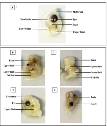
Results: The effects of MSG were growth retardation, embryonic death and congenital malformations of several organs such as brain (rachischisis and anencephaly), eye (microphthalmia and anophthalmia), ectopiacordis, ectopia viscera, opening of posterior neuropore (spinal bifida) and under development of heart and viscerae.
Conclusion: MSG produced congenital deformities and growth retardation in chick embryos which may predict to cause the same results to human embryos. Pregnant women should be advised not to consume MSG to avoid potential harmful effects to the unborn child.
Keywords: Monosodium glutamate (MSG); teratogen; chick embryo (Siriraj Med J 2018;70: 514-522)
INTRODUCTION
MSG is the most commonly used ingredient for cooking process in many countries. In United Kingdom, people consumed MSG about 4 grams (less than one teaspoon) per week while the U.S. showed 0.55 grams per week, while Taiwan reported a higher level of about 3 grams per day.1,2 MSG is a natural neurotransmitter
of the brain and induced symptoms known as “Chinese restaurant syndrome” which can be detected after the consumption of Chinese meals. It is characterized by headache, chest discomfort and facial flushing due to abnormal function of glutamate receptors. MSG was
reported to relate with neurological diseases especially retinal degeneration, brain trauma and endocrine disorder causing obesity and gonad dysfunction.3,4 The
excessive dose and long term usage of MSG in cooking affect tissue organ and embryonic development if it is taken by pregnant women during pregnancy.5 To confirm
the teratogenic effects of MSG, it was easier to use chick embryo because there were plenty of databases compared with human (Hamburger and Hamilton, 1952).6 Chick
In 2012, Fatma and Anan studied the effects of MSG on chick embryo and reported different congenital malformations such as growth retardation, subcutaneous bleeding, abdominal hernia, brain deformation, monophthalmia and beak malformation. Histological study of liver in the treated group showed less cell density and a dilation of venous canals and blood sinusoids, fibrosis, bleeding, hemorrhage and congestion, lipid droplets and necrosis. It was concluded that MSG caused growth retardation, congenital malformation and liver degeneration,1 although they showed no illustration of
such malformations except for the histology of liver. In 2016, Mahaliyana et.al. studied toxicity of MSG on embryonic development of zebrafish (Daniorerio).There was no observable malformation in low dose group. However, at higher concentrations there were abnormal developments such as growth retardation, shrinkage of the chorion, yolk sac edema, lack of pigmentation, tail deformities and scoliosis.7 As in the previous teratogenic
studies the toxicity and teratogenic effects of MSG were still unclear, the present study was conducted to study the toxicity and teratogenic effects of MSG on the 3rd,
6th and 10th day chick embryos which were injected by
different concentrations of MSG into the yolk sac of fertilized egg. The mortality rates were recorded and the teratogenic results were revealed by gross structure anomalies and histological changes of chick embryos by total mount and serial sections of the experimental groups compared to the control group.
MATERIALS AND METHODS Ethics Statement
According to German animal care guidelines, no IACUC approval was necessary to perform the embryo experiments. According to the local guidelines, only experiments with chick embryos E18 and older need IACUC approval. However, the embryos used in this study were all in early stages of embryonic development (between E1 and E11).8
ACUC Guideline
The Use and Euthanasia Procedures of Chicken/ Avian Embryos Avian embryos are not considered live animals under PHS policy. However, there is a consensus in the scientific community that at a certain point in development, avian embryos can experience pain. Because that exact point is not known for chicken embryos, chicken use and euthanasia guidelines differ across institutions. Cal Poly Pomona has chosen to adopt a guideline with the belief that pain occurs on or after gestation day 13, in anticipation of reviewing protocols including them.
However, this study used earlier stages of chick embryos, day 3-day 10.9
Fertilized eggs of white leg horn hen (Gallus gallusdomesticus) were collected fresh from the Department of Animal Science, Faculty of Agriculture, Kasetsart University. They were divided into 2 groups, the control group (n = 6) was injected with 0.15 ml of normal saline and treated groups (n = 60) were injected with 0.15 ml of the MSG solution with the concentrations 1 mg, 1.5 mg, 2.5 mg and 3 mg of MSG/gm egg weight into the yolk sac before incubation. The eggs were cleaned with 70% ethanol and incubated at 37°c. After 21 hours of incubation, all eggs were removed from the incubator and cleaned at the blunt ends of the eggshell with 70% ethanol before injecting into the yolk sac with the equal volume of 0.15 ml of NSS and the MSG solutions. After injection, the eggs were further incubated until day 3, 6 and 10. Embryos were removed from 2 eggs of control group and 5 eggs of each treated group for observing embryonic abnormalities and mortality. The day 3 embryos were processed for total mount and serial section while day 6 were processed for the serial section and day 6 and 10 embryos were processed for removing of visceral organs and alcian blue and alizalin red staining of cartilages and bones. Then the surviving chick embryos were observed for their external morphology indicating any gross malformations and more detail of malformations were confirmed by the serial section.
RESULTS
The mortality rate of the day 3 chick embryos The living embryo was indicated by observing the heartbeat and blood circulation. The percentages of survival and mortality were presented in Table 1.
The results showed the lowest mortality rate was 0% in the control group and the MSG 1 mg/gm egg weight groups. The mortality rates were increasing from 20% to 40% in the MSG concentration of 1.5 to 2.5 and 3 mg/ gm egg weight, respectively. The dead embryos did not exceed 50% in all groups.
The mortality rate of the day 6 chick embryos The living day 6 chick embryos were indicated by observing the heartbeat and blood circulation. The percentages of survival and mortality were presented in
Table 2.
Group (mg/gm egg weight) Survival (%) Mortality (%)
Control (n=5) 100 (5) 0 (0)
1 (n=5) 100 (5) 0 (0)
1.5 (n=5) 80 (4) 20 (1)
2.5 (n=5) 60 (3) 40 (2)
3 (n=5) 60 (3) 40 (2)
TABLE 1. Percentages of the survival and mortality rates of the day3 chick embryos of the experimental groups compared with control group
TABLE 2. Percentages of the survival and mortality of the day 6 chick embryos of the experimental groups compared with the control group
Group (mg/gm egg weight) Survival (%) Mortality (%)
Control (n=5) 100 (5) 0 (0)
1 (n=5) 100 (5) 0 (0)
1.5 (n=5) 80 (4) 20 (1)
2.5 (n=5) 60 (3) 40 (2)
3 (n=5) 60 (3) 40 (2)
The mortality rate of the day 10 chick embryos The percentages of survival and mortality of the day 10 chick embryos were presented in Table 3. The lowest mortality rate was 0% in the control, 20% in 1 and 1.5 mg/gm egg weight groups and 40% in 2.5 and 3 mg/gm egg weight groups, respectively. The dead embryos were not more than 50% in all groups.
Then the surviving chick embryos were observed for the external morphology indicating any gross malformations and more detail malformations were confirmed by serial section of the suspected organs. The cartilage and bone malformations were indicated by the alcian blue /alizarin red staining of the eviscerated embryos.
TABLE 3. Percentages of the survival and mortality of the day 10 chick embryos of the experimental groups compared with control group
Group (mg/gm egg weight) Survival (%) Mortality (%)
Control (n=5) 100 (5) 0 (0)
1 (n=5) 80 (4) 20 (1)
1.5 (n=5) 80 (4) 20 (1)
2.5 (n=5) 60 (3) 40 (2)
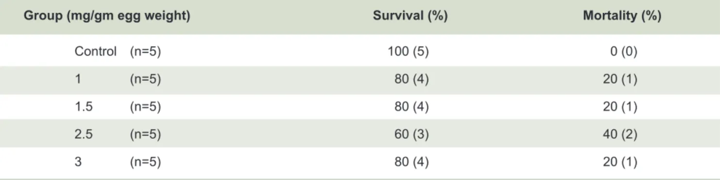
The total mount of the day 3 chick embryos (Fig 1)
The total mount of the day 3 chick embryos showed that the control group had normal development as classified by Hamburger and Hamilton10 as stage 18 (36 somites)
which the brain formation composed of the 5 brain vesicles (telencephalon, diencephalon, mesencephalon, metencephalon and myelencephalon). There were 2 flexures (cervical flexure and cephalic flexure). The body of embryo was rotated to the right side. Normal optic cup at diencephalon was large, horse-shoe shaped and lens located at the middle of the optic cup. The heart was S-shaped. There were 4 branchial arches. Anterior and posterior limb buds were presented. Somites extended to the caudal end and the neural tube appeared as two parallel dense lines.
The experimental group of 1 mg of MSG/gm egg weight (B) showed retardation of development of the brain with the development of only one flexure (cephalic
flexure). The optic cup and lens were smaller than the control. The branchial arch showed only one. The heart was irregular U-shaped with dilated lumen. There was irregular twisting of the body. The somites did not extend to the caudal end and the neural tube appeared as non-parallel dense lines. The limb buds and tail fold were absent. The (C) 2.5 mg of MSG/gm egg weight. (AN = Anterior neuropore, H = Heart) and 3 mg (D) of MSG/gm egg weight showed common malformations, the opening of anterior neuropore, the absence of eye primordia (anophthalmia), the absence of branchial apparatus, the dilated heart loop, retardation of somite formation and the absence of limb buds and tail fold.
The eye of the day 3 (Fig 2)
The eyes of the control comprised optic cup, optic stalk and lens vesicle. The optic cup was large and oval shaped structure connected with the diencephalon by the optic stalk. It was composed of the outer pigment layer and inner nervous layer which lied in closed contact to obliterate the intraretinal space. The lens was composed of cuboidal cell layer, the anterior lens epithelium, and the elongated cell layer, posterior lens fiber. All experiment groups showed more or less growth retardation represented by smaller optic cup with wide opening of the intraretinal space, with separation between the outer pigment and inner nervous layers. The lenses were smaller with the persistence of lens pit in the 1.5 and 2.5 mg groups
(Fig 2C and 2D). All experiment groups showed no
elongation of the posterior lens fiber.
Fig 1. Total mount of day 3 chick embryo of the control group (A), (B) the experimental group of 1 mg of MSG/gm egg weight. (TC=Telencephalon, DC=Diencephalon, MS=Mesencephalon, MT=Metencephalon, MC=Myelencephalon, OC=Optic cup, LV=Lens vesicle, O=Otocyst, H=Heart, PA=Pharyngeal arch, ALB=Anterior limb buds, NT=Neural tube, SM=somite, T=Tail fold, PN=Posterior neuropore), (C) 2.5 mg of MSG/gm egg weight. (AN=Anterior neuropore, H=Heart), (D) 3 mg of MSG/gm egg weight.
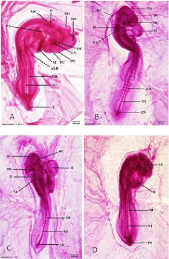
The external morphology of day 6 chick embryos (Fig 3)
The external morphology of day 6 chick embryos (HH stage 29) revealed the diameter of head was larger than the body. Eyes were large round-shaped and symmetrical. Beak appeared prominent but absence of egg’s tooth. The heart and viscerae situated in the thoracic and abdominal cavities was covered by the skin. Upper and lower limbs were paddle-shaped with no sign of the formation of digits.10,11,12
The results showed normal external morphology in the control group while experimental groups showed congenital malformations and growth retardation in all experimental groups. The abnormalities comprised opening of anterior neuropore (anencephaly), absence of eye (anophthalmia), small eye (microphthalmia). The visceral organs situated outside the body cavity (ectopia viscera). The heart situated outside the thoracic cavity (ectopiacordis) and opening of posterior neuropore.
Fig 3. The photographs of the day 6 chick embryos showed normal development of the control group in Fig A and showed congenital malformations in all of the experimental groups in Fig B was 1 mg treated group, showed opening of anterior neuropore called anencephaly, small eyes (microphthalmoa) and ectopia viscera, Fig C was 1.5 mg, showed absent both eyes called anopthalmia and opening of the anterior neuropores, Fig D was 2 mg, showed opening of both anterior and posterior neuropores and Fig E was 3 mg, showed opening of anterior neuropore called anencephaly and ectopia cordis.
The eyes of day 6 embryos indicated in the serial section
(Fig 4)
The eye of the control group showed normal development indicated by Fig A. The optic cup was oval-shaped and comprised outer pigment layer and inner nervous layer which lied close to each other. The intraretinal space was obliterated. The lens comprised anterior lens epithelium which appeared as simple cuboidal epithelium and posterior lens fiber which elongated dramatically and obliterated the lens cavity.
All experimental groups showed more or less abnormalities of the eye development. Some embryos of the experimental group of dose 2.5 mg of MSG/gm egg weight (Fig 4C) showed retardation of eye development indicated by the persistence of the intraretinal space
and the lens cavity. Fig 4B and Fig 4D were the 1 and 3 mg of MSG/gm egg weight which showed abnormal development of brains together with absent formation of both eyes (anophthalmia).
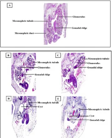
The abnormal development of mesonephros and gonad
(Fig 5)
The control group indicated in Fig 5A, showed a large pair of mesonephros comprised of numerous mesonephric tubules and one duct with glomeruli situated at the medial aspect. The gonad was large mass attached to the medial surface of the mesonephros. All experiment groups showed abnormality of mesonephros and gonadal ridge as indicated in Fig 5 B-D and showed very small mesonephros with fewer tubules and also very small gonadal ridge. Fig 5D showed large cystic cavities and
Fig 5E showed rudimentary of mesonephric tubules with cystic formation.
Fig 5. The micrographs of the development of mesonephros and gonad of the control group in Fig A and the experimental groups (B, C, D and E) showed more or less abnormals development, Fig B and C (1, 1.5 mg) showed small size and underdeveloped of mesonephric tubules and corpuscles with small gonads, Fig D and E (2.5 and 3 mg) showed more abnormality, apart from the small size and lesser number of tubules and absent of mesonephric corpuscle, there were large cystic formation and very small gonad.
Common abnormalities among all treated groups The serial section of day 6 embryo
1. Dosage of 1 mg of MSG/gm egg weight (Fig 6)
group showed widely opened of anterior neuropore and unidentified portion of the forebrain (Fig 6 A), unidentified eye at the level of diencephalon (anophthalmia) and absent otocyst (Fig 6B), and unfusioned spinal cord. At the level of thoracic and abdominal cavity it showed small stomach and dilated liver cord (Fig 6C). The ectopia cordis (Fig 6C) had underdeveloped heart. There was thinned wall atrium and ventricle with unfused endocardial cushion
(Fig 6C). At the level of mesonephros and gonad it
showed under development of the organs with irregular smaller mesonephric tubules without gonad (Fig 6D).
Fig 6. A, B, C, D were serial sections of day 6 chick embryo injected with 1 mg of MSG, section number 5, 15, 45 and 55, respectively to show multiple malformations in the same embryo. Fig 6A and 6B were at the head region to show irregular shaped brain, 6B showed the side opening of anterior neuropore. Fig 6B showed irregularity of diencephalon with rudimentary eye primordium beside. Fig 6C showed small viscerae within the body cavity and the heart situated outside (ectopiacordis). Fig 6D showed small and under developed mesonephros. This embryo showed anencephaly, anophthalmia, ectopiacordis and under developed of all visceral organs.
2. Dose 1.5 mg of MSG/gm egg weight (Fig 7)
mesonephros showed small and abnormal spinal cord, large cystic cavity in mesonephros, very small number of mesonephric tubules and caudal degeneration was indicated in this embryo.
Fig 7. A, B, C, D were serial section of day 6 chick embryo injected with 1.5 mg of MSG, section number 5, 20, 30 and 35, respectively to show multiple malformations in the same embryo. Fig 7A and 7B were at the head region to show irregular shaped of brain. Fig 7B showed the widely opened of the neural tube dorsally with ectopia cordis ventrally. Fig 7C showed small stomach and liver situated outside the body cavity. Fig 7D showed small and under developed mesonephros. The brain and spinal cord were irregularly-shaped, the spinal cord was small and rudimentary. This embryo showed anencephaly, anophthalmia, spina bifida, ectopia cordis and under developed visceral organs and ectopia viscerae.
3. Dose 2.5 mg of MSG/gm egg weight (Fig 8)
The serial section of dose 2.5 mg of MSG/gm egg weight showed opening anterior neuropore and unidentified portion of brain at the level of approximately diencephalon showed no primordial of both eyes called anophthalmia, at the level of approximately myelencephalon showed small otocyst, acousticofacial ganglion and stomodeum, at the level of mesonephros and gonad showed small number of mesonephric tubules, ectopiacordis, underdevelopment with thinned wall atrium and ventricle without endocardial cushion, abnormal spinal cord and ectopia viscera of liver at the relation of ventricle and opened posterior neuropore were also indicated.
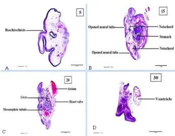
4. Dose 3 mg of MSG/gm egg weight (Fig 9)
The serial section of dose 3 mg of MSG/gm egg weight showed severe abnormality of neural development. The rachischisis of the neural tube was at the level of brain. The spinal cord showed double spinal cords with double notochords. There was underdevelopment of stomach. There was ectopia cordis with heart valve, abnormality
Fig 8. A, B, C, D were serial section of day 6 chick embryo injected with 2.5 mg of MSG, section number 10, 20, 30 and 40, respectively to show multiple malformations in the same embryo. Fig 8A and 8B were at the head region to show irregular shape brain with no eye primordium. Fig 8B showed irregular myelencephalon with small otocyst. Fig 8C showed small spinal cord and viscerae situated inside the body cavity with the heart situated outside, small and under developed mesonephros situated inside the body cavity. Fig 8D showed spinal cord was small and rudimentary. This embryo showed anencephaly, anophthalmia, abnormal spinal cord, ectopia cordis and uner developed visceral organs.
and large cystic cavity of atrium. There was dilated liver cord and liver sinusoid and very small number of mesonephric tubules of mesonephros.
There were common types of malformation which occurred in the treated groups of all concentrations. The abnormalities comprised anencephaly, rachischisis, spina bifida, anophthalmia, microphthalmia, ectopia cordis, ectopia viscerae, underdeveloped heart and all viscerae. All survival day 10 embryos showed normal development except one of the 3 mg injection group showed ectopia viscerae. All parameters measured showed no significant difference when compared with the control. The dead embryos were less than 50% in all groups.
DISCUSSION
Several studies reported in animal models and humans that MSG effected central nervous system, reproductive system, adipose tissue and liver.11 The effect of MSG in
this study showed that the mortality rate increased in higher doses while the abnormalities were identified in all doses, which corresponded with the incidence of death and malformation rate in pregnant rats that received MSG.13
The effects of MSG on the brain were small size, opened anterior neuropore (anencephaly) and the brain vesicle was not completed on day 3 chick embryos in dose 1.5 mg and 3 mg of MSG/gm egg weight. The day 6 chick embryos showed opened of anterior neuropore and unidentified part of brain in dose 1 mg, 1.5 mg and 2.5 mg of MSG/gm egg weight and rachischisis in dose 3 mg of MSG/gm egg weight. The results agreed with the study of Jurand, in 1973 who studied the teratogenic activity of methadone hydrochloride in mouse and chick embryos and showed that the anterior neuropore was opened with rachischisis of brain.12 Moreover, in 2009,
Schmidt et al., reported maternal caffeine consumption showed increased risk of NTDs and anencephaly was also observed.13
The effect of MSG on the eye development revealed both retardation and abnormality. The retardation was indicated by the persistence of intra retinal space and lens pit. The abnormalities included microphthalmos and anophthalmos. This result agreed with Qudsi et al., who studied the effect of monosodium glutamate on chick embryo which also revealed monophthalmia.1 In 2014,
Zheng et al., studied excess caffeine exposure which impaired eye development during chick embryogenesis and reported it caused asymmetrical micropthalmia by increasing ROS production and perturbed Pax6 expression due to the effect of high salt-exposure on the development of retina and lens.
The effect of MSG on the heart development showed elongated heart tube and U-shaped heart looping, thinned wall and dilated lumen on day 3 chick embryos. This study corresponding with the study of Abdelkader et al.,
about teratogenicity and brain aromatase-induction of monosodium glutamate in estrogen-responsive mosaic transgenic zebra fish Daniorerio showed elongated heart and pericardial edema.14 In 2016, Mahaliyana et al., studied
the effects of MSG on embryonic heart development of zebrafish (Daniorerio), a promising animal model, and demonstrated internal anatomical deformities such as elongated heart, cardiac sac edema and yolk-sac edema.15
The effect of MSG on chick embryos to the anterior body wall showed ectopia viscera with ectopia cordis in all doses such as 1 mg, 1.5 mg, 2.5 mg and 3 mg of MSG/ gm egg weight observed by the external morphology and the serial section of day 6, 10 chick embryos. The result corresponded with the study of Qudsi et al., who studied the effect of MSG on chick embryo development which showed different congenital malformations in treated chick embryos such as abdominal hernia, bleeding, monophthalmia and brain deformation.1
CONCLUSION
The striking abnormalities caused by MSG were NTDs, brain showed irregularly shape and unidentified portion, widely opened of anterior neuropore, and severe opening of the neural tube or rachischisis of brain and spinal cord. Some embryos showed opening of the posterior neuropore. Eyes were also the target organ of the MSG as there were several types of abnormality, microphthalmia and anophthalmia, retardation of eye development indicated by persistent of intraretinal space and lens pit. The heart showed ectopiacordis, underdeveloped with thinned wall atrium and ventricle and unfused endocardial cushion. The visceral organs were totally underdeveloped. The mesonephros was smaller than the control and showed abnormal development with lesser number of mesonephric tubules and bilateral large cystic cavities. The gonads were underdeveloped with smaller size of gonadal ridges. This experiment clearly indicated that MSG was a potent teratogen. Therefore, it is essential to make people aware of the effects of MSG, especially for the pregnant women who should avoid consumption altogether.
REFERENCES
1. Qudsi FA, Jahdali AA. Effect of Monosodium Glutamate on chick development. J Am Sci 2012;8(10):499-509.
2. IFIC. Review on Monosodium Glutamate: Examining the
3. Mai A. Effects of Monosodium Glutamate and Acrylamide on the liver tissue of adult Wistar Rats. Life Sci J 2013;10(2s):35- 42.
4. Kumbhare VA, Gajbe UJ, Singh BR, Reddy AN, Shukla SA. Histological & Histochemical changes in liver of adult rats treated with monosodium glutamate: A light microscopic study. World Journal of Pharmacy and Pharmaceutical Sciences 2015;(04):898-911.
5. Prastiwi1 D, Djunaidi1A, Partadiredja G. High dosage of monosodium glutamate Purkinje cells of rats. Human and Experimental Toxicology 2015, Vol. 34(11)1171–1179.
6. Hamburger V, Hamilton HL. A series of normal stages in the development of the chick embryo. Journal of Morphology 1992;88(1): 49–92.
7. Mahaliyana AS, Fasmina MF, Alahakoon AM and Wickrama GM. Toxicity effects ofmonosodium glutamate (MSG) on embryonic development of zebrafish(daniorerio); a promising model to study excitotoxins. International Journal of Scientific and Research Publications 2016; 6(3):229-234.
8. Rashidi H, Sottile V. The chick embryo: hatching a model for contemporarybiomedical researches. Bioassays 2009; 31(4): 459-65.
9. Roongruangchai J, Viravud Y, Pilakasiri K, Pornkunatham A, Chuncharunee A, Plakornkul V, Sangvichien S. Embryology
for medical student. 4th ed. Bangkok: Samcharoenpanit publishing; 2009.
10. Roongruangchai J. Embryology theory and lab. 2nd ed. Bangkok: Supanawit publishing; 2012.
11. Husarova V, Ostatnikova D. Monosodium glutamate toxic effects and theirimplications for human intake: a review. JMED Research 2013;2013:1-12.
12. Jurane A. Teratogenic activity of methadone hydrochloride in mouse and chick embryos.J Embryol.exp. Morph 1973;30(2): 449-458.
13. Schmidt RJ, Romitti PA, Burns TL, Browne ML, Druschel CM, Olney RS. Maternal caffeine consumption and risk of neural tube defects. Birth Defects Research Part A Clinical and Molecular Teratology 2009;85:879-889.
14. Abdelkader TS, Chang SN, Kim TH, Juha S, Dongso K, Park JH. Teratogenicity and brain aromatase-induction of monosodium glutamate in estrogen-responsive mosaic transgenic zebra fish daniorerio. African Journal of Biotechnology 2012;11(48):10816- 10823.