
*Correspondence to Author: Rachuonyo HO
Department of Microbiology,
Kenyatta University
e-mail: [email protected]
How to cite this article:
Rachuonyo HO, Gatheri GW and Nyamache AK. Antimicrobial ac-tivity of crude leaf extracts from medicinal plants against Enterococ-cus faecalis. International Journal of Traditional and Complementary Medicine 2016; 1(2): 0001-0009.
Accepted 27 July 2016; published July 27 2016.
eSciPub LLC, Houston, TX USA. Website: http://escipub.com/
International Journal of Traditional and Complementary Medicine
(IJTCM)
Research Article IJTCM (2016), Vol. 1, Issue 02
Antimicrobial activity of crude leaf extracts from medicinal plants against
Enterococcus faecalis
Enterococcus faecalis
is a Gram negative, commensal enteric
bacterial pathogen usually located in the intestinal tracts of
either animals or human beings. The pathogenic members of
enteric bacteria’s are usually associated to infections that are
characterized by enteric fevers, abdominal pain, and diarrhoea
and vomiting. This study aimed at determining the effects of
the selected medicinal plants extracts against Clinical isolate
of
Enterococcus faecalis
obtained from Kenyatta University
health Centre. Kirby Bauer method was used to determine the
antimicrobial activity of the plants extracts against
Enterococcus
faecalis
. All the plant extracts showed antimicrobial activity
against
Enterococcus faecalis
with Tagetes minuta producing
the largest average zones of inhibition of 18.67±1.03mm when
compared to the other plant extracts. Vernonia lasiopus was
more active at low concentrations (MIC 3.9µg/ml; MBC 5.0µg/ml)
while Aloe secundiflora was less active (MIC 7.0µg/ml; 9.7µg/ml).
Ciprofloxacin (5µg/ml) was used as a positive control producing
an average zone of inhibition of 21.67±2.66mm while Methanol
(33.50±2.56mm) and 4% Dimethyl sulphoxide (0.00±0.00mm)
were used as negative control. Qualitative phytochemical
screening showed the presence of four phytochemicals namely;
saponins, tannins, alkaloids and flavonoids. The study provides
insight about the antimicrobial activity of the plant extracts and
if they can be used in treatment of as an antimicrobial agent
against infections caused by
Enterococcus faecalis
.
Rachuonyo HO
1*, Gatheri GW
2, and Nyamache AK
11
Department of Microbiology, Kenyatta University
2Department of Plant sciences, Kenyatta University
Keywords
Qualitative phytochemical screening;
Antimicro-bial agents;
Enterococcus faecalis
;
Ciprofloxa-cin; Clinical isolate.
INTRODUCTION
Medicinal plants since memorial have been used in many communities as a source of medicine [13]. Medicinal plants play an important role in medical system to counter some of the serious diseases in the world. Pharmacological investigations of herbal plants have been carried out overtime to find nov-el drugs or active components for the devnov-elopment of new therapeutic agents [3]. Aloes are perennial succulent xerophytes which develops water storage tissues in leaves to survive in areas with low or er-ratic rainfall [28]. Aloe secundiflora has been used in treating ailments including; chest problems, polio, malaria and stomach ache by herbalists in the Lake Victoria region [21]. Aloe secundiflora leaf compo-nents have been credited for antibacterial, antifungal and antiviral and antihelmintic medicinal properties [18]. Aloes contain over 75 nutrients and 200 active compounds including enzymes, vitamins, minerals, lignin, sugars, saponins, anthraquinones amino ac-ids and salicylic acid. Extracts of Aloes especially its leaf gel have shown antibacterial activity by inhibiting the growth of both Gram negative bacteria and Gram positive bacteria [11]. Bulbine is a genus of plants in the family xanthorrhoeaceae and sub family aspho-deloideae and its members are well known for their medicinal value [1]. They are succulent plants with most of the species having yellow flowers where as some of them have white, orange or pink flowers. Bulbine frutescens is mostly grown as ornamental plant in flower garden at homes in South Africa [31]. Bulbine plant has been used for medicinal purposes in the early stages of the eighteen century by Dutch and British settlers of South Africa in treating vari-ous ailments [31]. The leaves of the plant have been used in the treatment of wound thought to be infect-ed with bacterial pathogens and it has shown anti-bacterial properties [20]. Some of the species of the plant found in South Africa have been used for blood cleansing, treatment of ringworms and gravel rush by some local communities such as the Xhosa [8]. A decoction of bulbs and roots of some of the species has been used in the treatment of some of the
ve-nereal diseases in women and stomach upsets [30]. Vernonia shrubs is a herbaceous grow in tropical Africa and have a height of about 2-5 metres, ellipti-cal leaves of up to 20 centimetres and a rough bark [15]. The plants in this genus usually have bitter taste and in English they are called bitter leaf [15]. Some of the common African names of plants in this genus are Olusia (Luo), Mululuza (Luganda), Onugu (Igbo), Grawa (Amharic) and Chusar-doki (Hausa) [22]. Ver-nonia lasiopus decoctions from the stems and leaves have been traditionally been used by herbalists in East Africa to treat, malaria, worms and gastrointes-tinal problems [19]. The genus Tagetes belongs to the Asteraceae family which presently comprises of 56 species, 27 biennials and 29 perennials. Tagetes species are grown all over the world as multipurpose plants [27]. Tagetes species and chemo-types from its genus have been largely examined for biological active metabolites that can be used in industry and medicine [12]. Compounds that have antimicrobial activity in the Tagetes minuta plant are said to be accumulated in the organs of the plant and their es-sential oils have not only antimicrobial effect but also insecticidal properties. Plant parts such as flowers and leaves have been known to contain flavonoids that are scavengers for free radicals which enhanc-es the antimicrobial activity of the Tagetes minuta extracts [9]. Phytochemicals from the plant such as carotenoids have also been used in pharmacologi-cal preparations and they have been found to contain anti-aging and anti-cancer effects [4]. The plant ex-tracts have been used in treating intestinal and stom-ach problems [5] [29].
MATERIALS AND METHODS
Plant material collection and sampling
washed in running water to remove debris and dust particles and then rinsed using distilled water and fi-nally air dried.
Preparation of plant extract
The air dried leaves from the plants were grinded into powder and 500 grams were soaked in 750 millilitres of Analar grade (AR) methanol in a conical flask for 72 hours, placed in a Gallenkamp shaker rotating at 65 revolutions per minute. The contents were homog-enized and filtered using whatman filter paper no. 1. The filtrate was poured into a round bottom flask and concentrated using a Buchi Rotavapor R-200 yield-ing 1.8 grams of Bulbine frutescens, 3.1 grams of
Aloe secundiflora, 2.6 grams of Tagetes minuta and 2.1 gram of Vernonia lasiopus. The extracts were then stored in a labelled amber glasses bottle slightly opened where they were further left to dry bottle at room temperature away from light and heat in a lam-inar flow before being used for antimicrobial efficacy test [24].
Preparation of media
The media used were Muller Hinton agar (Sharau®) was prepared according to commercially given in-structions. 38mg of Muller Hinton agar powder was added into one litre of distilled water in a flat bottomed conical flask. The mixture was heat with frequent ag-itation and boiled for one minute to completely dis-solve the media. The flask was then tightly closed us-ing cotton wool and further covered by aluminum foil. The mixture was autoclaved for 15 minutes at 121 degree celsius after which it was left to cool down to room temperature. The media was poured in the petri dishes in a laminar flow to give uniform depth of 3 - 4millimetres. The petri dishes containing the media were then placed in a sterile plastic bags and stored at a temperature of 2 - 8 degree celsius before use. Preparation of discs
Discs of 6 milliliters were prepared from whatman no.1 filter paper. This was done by punching the fil-ter papers using a paper punch. The discs prepared filled four McCartney bottles. The discs were then sterilized by autoclaving at 121 degree celsius for 15 minutes after which the autoclave was left to cool be-fore removing the McCartney bottles containing the discs. The discs were dried on hot air oven at 50 de-grees celsius to remove moisture [2]. The discs used for the antimicrobial activity were impregnated with formulated stock solution of the plant leaf extracts
of 1000µg/ml of Tagetes minuta, Aloe secundiflora, Bulbinefrutescens and Vernonia lasiopus. A forceps used for picking the discs was first sterilized using a spirit lamp and left to cool. The forceps were then used in picking the sterilized discs and placing them in the stock solutions of the plants leaf extracts. The forceps were sterilized after every pick. The disc were then left to stay in the plant leaf extracts stock solu-tion for two hours. The discs were then removed and placed in sterile petridish in a laminar flow and left to dry for 30 minutes. The discs impregnated with leaf extracts from each plant were then picked by ster-ilized forceps and stored in a sterster-ilized McCartney bottle and stored in a refrigerator at a temperature of 4 - 8 degree celsius before being used for the antimi-crobial susceptibility test.
Test bacterial organisms
The test microorganism used in the study was clinical isolate of Enterococcus faecalis. The microorganism was isolated from samples collected from first time patients at Kenyatta University Health Centre Lab-oratory, Nairobi. The samples were collected from patients who showed symptoms associated with en-teric bacterial infections such as fever, abdominal pain, diarrhoea, vomiting and have no known drug resistance. Isolation, morphological identification and biochemical test were carried out in Microbiology lab-oratory, Department of Microbiology. The samples used for isolation of Enterococcus faecalis was from faecal material. The collected samples were isolated and identified by streaking of the samples collected on selective media for characteristic morphological identification based on the type of colonies formed. The morphologically identified microorganisms were then subjected to biochemical test for identification up to biochemical level.
Isolation and Morphological identification
isolat-ed by streaking the dilutisolat-ed faecal sample of 10-6 on Columbia agar with 5% sheep blood. The petridish tightly closed used parafilm and incubated at 37 de-grees celsius for 24 hours. The plate was then later observed for growth. Growth of diplococcus colonies with gamma hemolysis on Columbia agar with 5% sheep blood is a morphological growth characteristic of Enterococcus faecalis.
Biochemical identification
The biochemical test carried out was based on the capability of the isolated and morphologically iden-tified test microorganisms to cause fermentation of sugars and oxidation. The Medias used to carry out the test were prepared according to the manufacturer commercially used procedure and poured into cork screwed centrifuge tubes. Durham tubes were insert-ed to minsert-edia containing broth and later observinsert-ed for gas production. If there was gas production the re-sult was termed as positive and no gas production as negative. The tubes were then closed and auto-claved at 121 degree celsius for 15 minutes and left to cool before being stored in a refrigerator before use. 1 millilitres inoculum of the isolated test mi-crobes from diluted sample of 10-6 were introduced in the media and incubated at 37 degrees celsius and observation done after 24 hours and compared with the standard controls. The tubes containing solid me-dia were observed for colour change. Where colour change occurred the results were termed as positive and no colour change as negative [14].
Antimicrobial susceptibility testing
The discs were impregnated with the extracts from the highest concentration of 1000mg/ml to the lowest concentration of 1mg/ml [17]. The antimicrobial effi-cacy test was carried out using Kirby Bauer method [25]. Muller Hinton agar was used in the spread plate technique where Enterococcus faecalis was spread using sterilized cotton wool swab and exposed to extracts impregnated discs in milligrams per micro-liter from Aloe secundiflora, Tagetes minuta, Verno-nia lasiopus and Bulbine frutescens. The discs were placed with equal distance between them on agar plates inoculated with Enterococcus faecalis. Posi-tive control discs contained ciprofloxacin while neg-ative control discs were impregnated with dimethyl sulphoxide and distilled water. The Petri dishes were incubated at 37ºC for 24 hours. Zones of inhibition formed were measured in millimetres and their aver-age determined. The experiment was carried in dupli-cates and the diameter of zones of inhibition formed
measured.
Minimum inhibitory concentration and Minimum bactericidal concentration
Minimal inhibitory concentration (MIC) was determine using the broth tube method [10]. 100µl of 250mg/ ml of methanol extract was added to 100µl of sterile bacteriological peptone in the first well of the 96 well micro plate and mixed well with a micropipette. 100µl of this dilution was transferred subsequently to wells two folding each dilution of the original extract. This was done to the extracts of Aloe secundiflora, Bulbine frutescens, Vernonia lasiopus, and Tagetes minuta. An inoculum of 100µl (0.5 McFarland standard) of overnight clinical culture of Enterococcus faecalis was added in each of the wells. Triplicate of each micro plate were made and the procedure repeated. The plates were then incubated at 37ºC for 24 hours. After incubation 40µl of 0.2 mg/µl of INT was added in each of the wells and the plates examined after an additional 60 minutes of incubation. Growth was indicated by a red colour (conversion of INT to for-mazan). The lowest concentration at which the colour was apparently invisible as compared to the next dilu-tion was taken as the minimum inhibitory concentra-tion [26]. Minimum bactericidal concentraconcentra-tion (MBC) was determined by taking 100µl of suspension from micro plate wells that demonstrated no growth and inoculated on agar plates. The plates were incubated at 37ºC for 24 hours. In the case where, there was no bacterial growth and value not greater than the min-imum inhibitory concentration the concentration was used as the minimum bacterial concentration.
Qualitative phytochemical analysis
Presence of saponins, tannins, flavonoids and alka-loids in the crude extract were determined [6]. Tannins: Each of the extracts was weighed to 0.5mg and dissolved in 1 ml of distilled water. Filtration was carried out after 2ml of FeCl3 was added. If there was presence of a blue or black precipitate then it indicat-ed the presence of tannins.
Flavonoids: Each of the extracts was weighed to 0.5mg and dissolved in 1 ml of ethanol and filtered. 2ml of 1% HCl and magnesium ribbon was added to the filtrate. If there was formation of a pink or red colour it indicated the presence flavonoids.
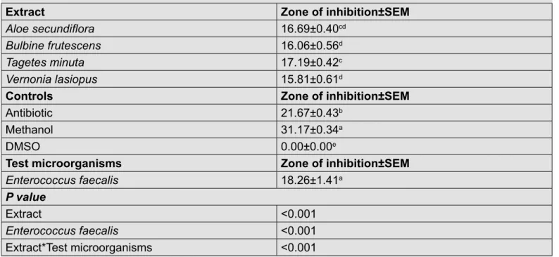
Table 2: Interactions between the plants extract and Enterococcus faecalis
Extract Zone of inhibition±SEM
Aloe secundiflora 16.69±0.40cd
Bulbine frutescens 16.06±0.56d
Tagetes minuta 17.19±0.42c
Vernonia lasiopus 15.81±0.61d
Controls Zone of inhibition±SEM
Antibiotic 21.67±0.43b
Methanol 31.17±0.34a
DMSO 0.00±0.00e
Test microorganisms Zone of inhibition±SEM
Enterococcus faecalis 18.26±1.41a
P value
Extract <0.001
Enterococcus faecalis <0.001
Extract*Test microorganisms <0.001
The value of average zones of inhibition ± standard error of mean (SEM) after two-way ANOVA followed by Tukey`s test.
A value followed by the same superscript within the same column are not significantly different (P˃0.05). Key: Antibiotic (ciprofloxacin), DMSO - dimethyl sulphoxide
Table 1: Average zones of inhibition in millimeters when plant extracts are used against Enterococ-cus faecalis
Plant extracts Zone of Inhibition±SD (mm)
Tagetes Minuta 18.67±1.03c
Aloe secundiflora 17.67±1.63c
Bulbine frutescens 18.50±1.05c
Vernonia lasiopus 18.00±0.89c
Antibiotic 21.67±2.42b
Methanol 28.67±2.34a
4%DMSO 0.00±0.00e
The value of average zones of inhibition ± standard deviation (SD) after one-way ANOVA followed by Tukey`s test. A
value followed by the same superscript within the same column are not significantly different (P˃0.05). Key: 4% Dimethyl sulphoxide (Negative control); Ciprofloxacin (Positive control), Plant extracts concentration (1000µg/ml).
Table 3: Phytochemical tests on the plant extracts
Plant extracts Saponins Tannins Alkaloids Flavonoids
Aloe secundiflora + + + +
Vernonia lasiopus + + + +
Bulbine frutescens + + + +
Tagetes minuta + + + +
1% HCL was added to the filtrate and the solution heated. Mayor`s reagent was added drop wise and if there was formation of any colored precipitate it indi-cated the presence of alkaloids.
Saponins: Each of the extracts was weighed to 0.5mg and dissolved in 1 ml of methanol and filtered. Distilled water was added and shaking done for a few minutes. If there was persistence frothing then it indi-cated the presence of saponins.
STATISTICAL ANALYSIS
The data collected was exported to Microsoft excel spreadsheet where descriptive statistics were car-ried out. The data was analyzed using SAS version 9.1 one-way ANOVA (Analysis of variance) was car-ried out to show statistical difference using the vary-ing zones of inhibition when the plant leaf extracts from Tagetes minuta, Aloe secundiflora, Vernonia lasiopus and Bulbine frutescens were used against Enterococcus faecalis. Two-way ANOVA was also carried to determine if there was any interaction be-tween the plant extracts and Enterococcus faecalis. P ≤ 0.05 considered significant in both test. Further-more, a post hoc test was done to find the difference between the means.
RESULTS
All the plant extracts showed antimicrobial activi-ty against Enterococcus faecalis. The average of the zones of inhibition formed ranged from 17mm - 19mm. Tagetes minuta extractproduced the largest zones of inhibition of 18.67±1.03mm while Vernonia lasiopus extract produced the least zones of inhibition of 18.00±0.89mm (Table 1).The standard antibiotic
used as a positive control (Ciprofloxacin) produced
the largest zone of inhibition when compared to all the plant extracts 21.67±2.66mm. This showed that Tagetes minuta plant leaf extract was more effective against Enterococcus faecalis as compared to the other plant extracts. The average zones of inhibition formed by the extracts when used against Enterococ-cus faecalis were not significantly different; P˃0.05 (Table 1). However, the average zones of inhibition formed by antibiotics (positive control), methanol and DMSO (negative control) were significantly different from those formed by the plant extract P˂0.05 (Table 4.1).
The average zone of inhibition formed by the plants extracts when used against Enterococcus faeca-lis were significantly different to those formed by antibiotic, methanol and DMSO (P<0.05; Table 2). Moreover, zones formed by Tagetes minuta extract against Enterococcus faecalis was significantly dif-ferent to those formed by Vernonia lasiopus and Bul-bine frutescens (P<0.05; Table 2). The interaction between Enterococcus faecalis and the plant extracts was significant (P<0.05; Table 2).
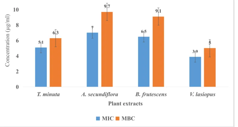
When the plant extracts were used in low concentra-tions against Enterococcus faecalis, Vernonia lasio-pus was the most active with a minimum inhibitory concentration of 3.9µg/ml and maximum bactericidal concentration of 5.0µg/ml. Aloe secundiflora extract was the less active against Enterococcus faecalis with a minimum inhibitory concentration of 7.0µg/ml and a minimum bactericidal concentration of 9.7µg/ ml when compared to all the other plant extracts. The other two extracts, Bulbine frutescens and Tagetes minuta also had a pronounced antimicrobial activity against Enterococcus faecalis with minimum inhibito-ry concentration of 6.5µg/ml and 5.1µg/ml; maximum bactericidal concentration of 9.1µg/ml and 6.3µg/ml respectively (Figure 1.0)
The qualitative phytochemicals test on plant extracts from Tagetes minuta, Aloe secundiflora, Bulbine frutescens and Vernonia lasiopus showed the pres-ence of phytochemicals. The extracts contained sa-ponins, tannins, alkaloids, and flavonoids (Table 3).
DISCUSSION
in the plant extracts. From the study, the extract from Vernonia lasiopus contained pharmacologically ac-tive compounds; flavonoids, saponins, alkaloids and tannins which might be responsible for the antimicro-bial activity. Some of the compounds have been as-sociated with antimicrobial activity in medicinal plant extracts against bacterial microorganisms [3] [19].
Aloe secundiflora extarct showed the less antimi-crobial activity among all the extracts used against Enterococcus faecalis. This showed that, although the extracts from Aloe secundiflora had antimicrobial potential, a lot of it is required to cause antimicrobial effect at low concentrations when compared to other extracts. The extract also had secondary metabolites that might be responsible for its antimicrobial activ-ity against Enterococcus faecalis. Similar studies previously carried out have shown that some of the secondary metabolites are pharmacological active components with antimicrobial activity [2] [24]. The extracts from Bulbine frutescens and Tagetes minuta also showed antimicrobial activity agaisnt Enterococ-cus faecalis. Tagetes minuta extract was more active against Enterococcus faecalis at low concentrations when compared to Bulbine frutescens. Preliminary phytochemical screening of Tagetes minuta extract showed the presence of secondary metabolites; sa-poninns flavonoid, alkaloids and tannins. The antimi-crobial activity might be associated with the presence of the secondary metabolites that may be pharmaco-logically active against both Gram positive and Gram negative bacterial microorganisms [16] [23].The ex-tract from Bulbine frutescens also contained the sec-ondary metabolites that might be responsible for its antimicrobial activity against Enterococcus faecalis. Similar studies carried out have shown that some of the se compounds are phamacologically active and can cause antimicrobial activity [7] [32].
CONCLUSION
Plants of medicinal value have been used by tradi-tional healers to treat a lot of infectious diseases. The plant leaf extracts showed antimicrobial activity against Enterococcus faecalis with extract from Ver-nonia lasiopus showing a higher activity at low con-centrations (MIC) when compared to extracts from the other plants. The present study showed that ex-tracts from the used medicinal plants can be used to treat bacterial infections caused by Enterococcus faecalis. Qualitative phytochemical analysis showed that the plant extracts contained saponins,
flavo-noids, tannins and alkaloids. These secondary me-tabolites found in most medicinal plants are known to be responsible for the antimicrobial activity. Howev-er, further biochemical, chemical and pharmacologi-cal investigation must be carried to identify purified bioactive components that are responsible for the antimicrobial activity and there mechanism of action.
REFERENCES
1. Acock, J.P.H.. Veld types of South Africa. Memoirs of Botanical Survey of South Africa Botanical Research Institute. Pretoria, Department of Agriculture and Water Supply, 1975, p. 57.
2. Arunkumar, S. and Muthuselvam, M. Analysis
of phytochemical constituents and antimicrobial activities of Aloe vera L. against clinical pathogens. World Journal of Agricultural Sciences. 2009, 5(5):572 - 576.
3. Ayoola, G. A., Coker, H. A. B., Adesogun, S. A. and
Adepoju-Bello, A. A.. Phytochemical Screening and Antioxidant activities of some selected Medicinal Plants Used for Malaria therapy in south-western Nigeria. Tropical journal of Pharmaceutical Reseach, 2008 , 7 (3), pp. 1019 1024.
4. Basile, A., Sorbo, S., Giordano S., Ricciardi, L.,
Ferrara, S., Montesano, D.. Antimicrobial resistance of Shigella isolates in Bangladesh, 1983 - 1990: increasing frequency of strains multiply resistant to ampicillin, trimethoprim-sulfamethoxazole,and nalidixic acid. Journal of Clinical Infectious Diseases. 1992, 14:1055 - 1060.
5. Broussalis, A.M., Ferraro, G.E., Martino, V.S., Pinzon, R., Coussio J.D., Calle, A.J. . Argentine plants as potential source of insecticidal compounds. Journal of ethno pharmacology. 1999 , 67:219 - 223.
6. Congesta W.T, C. . Preliminary screening of some
folklore medicinal plants from Preliminary screening of some folklore medicinal plants from Western India for potential antimicrobial activity estern India for potential antimicrobial activity. Indian Journal of Pharmacology, 2005, 37(6), 408-409.
7. Coopoosamy, R.M. . Traditional information and
antibacterial activity of four Bulbine species (Wolf). African Journal of Biotechnology. 2011, 10 (2):220 - 224.
8. Coopoosamy, R.M., Magwa, M.L., Mayekiso, B. .
Proceedings: Science and Society University of Fort Hare, Bhisho, Eastern Cape, South Africa, 2000.
9. De las Rivas, J. . Reversed-phase high performance
liquid chromatographic separation of lutein and lutein
fatty acid esters from marigold flower petal powder.
10. Eloff, J.N. . A sensitivity and quick microplate method
to determine the minimal inhibition concentration of plant extracts for bacterial organisms. Medicinal plants journal. 1998, 64(8):711 - 713.
11. Ferro, V.A., Bradbury, F., Cameron, P., Shakir, E.,
Rahman, S.R., Stimson, W.H. . In vitro susceptibilities
of Shigella flexineri and Streptococcus pyrogenes to
inner gel of Aloe barbadensis Miller. Antimicrobial agents and chemotherapy p. 2003, 1137 - 1139.
12. Green, M.M., Singer, J.M., Sutherland D.J., Hibben,
C.R. . Larvicidal activity of Tagetes minuta (marigold) toward Aedes aegypti. Journal of mosquito control association. 1993, 7:282 - 286.
13. Hoareau, L.,and Dasilva, E.J. . Medicinal plants:
a re-emerging health aid. Electronic Journal of Biotechnology 1999, 2(2): 56-69.
14. http://www.microbiologyinfo.com/biochemical test
(15th June 2014).
15. Ijeh, I.I., Ejike, C.E. . Current perspectives on
the medicinal potential of Vernonia amygdalina Del. Journal of Medicinal Plants Research. 2011, 5(7):1051 - 1061.
16. Irum, S., Amjad, H., Ummara, W.K., Mohammad,
M.S. . Evaluating biological activities of the seed extracts from Tagetes minuta L. found in Northern Pakistan Journal of medicinal plants research. 2010, Vol. 4(20):2108 - 2112.
17. Joshua, M., Ngonidzashe M., Bamusi, S. . An
evaluation of the antimicrobial activities of Aloe barberdensis, A. chabaudii and A. arborescens leaf extracts used in folk fore veterinary medicine in Zimbabwe. Journal of animal and veterinary advances. 2010, 9(23):2918 - 2923.
18. Kaingu, F., Kibor, A., Waihenya, R., Shivairo, R.,
Mungai, L. . Efficacy of Aloe secundiflora Crude
Extracts on Ascaridia galli in Vitro. Sustainable Agriculture Research. 2013, Vol. 2, No. 2.
19. Kareru, P. G., Gachanja, A. N., Keriko, J. M., Kenji, G. M. . Antimicrobial activity of some medicinal plants used by herbalists in Eastern province, Kenya. African Journal of Traditional Complementary And alternative Medicine. 2007, 5(1):51 - 55.
20. Kelmanson, J.E., Jager, A.K., Van Staden, J. . Zulu
medicinal plants with antibacterial activity. Journal of ethno pharmacology. 2000, 69:241 - 246.
21. Kigondu, E.V.M., Rukunga, G.M., Keriko, J.M.,
Tonui, W.K., Gathitwa, J.W., Kirira, P.G., Irungu, B., Ingonga, J.M., Ndiege, I.O. . Anti-parasitic activity and cytotoxicity of selected medicinal plants from Kenya. Journal of ethno pharmacology. 2009, 123(3):504 - 509.
22. Kokwaro, J. . Medicinal Plants of East Africa (3rd Edt). Nairobi, Kenya: University of Nairobi Press 2009.
23. Lubna, T., Naeem, K. . Antibacterial potential of
crude leaf, fruit and flower extracts of Tagetes minuta
l. Journal of Public Health and Biological Sciences. 2012, Vol. 1(3):74 - 78.
24. Mariitah, R.M., Orodho, A.J., Okemo, O.P.,
Kirimuhuzya, C., Otieno, J.N., Magadula, J.J .
Methanolic extracts of Aloe secundiflora Engl. inhibits
in vitro growth of tuberculosis and diarrhea-causing bacteria. Pharmacognosy Research. 2011, 3(2):95 - 99.
25. Newall, C.A., Anderson, L.A., Phillipson, J.D. . Herbal medicines. The pharmaceutical Press London, 1996, p. 25.
26. Rabe T., Mullholland D., Van Staden J. . Isolation and
identification of antibacterial compounds from Vernonia
colorata leaves. Journal of Ethnopharmacology. 2002, 80:91 - 94.
27. Soule, J. A. . Tagetes minuta A Potential New Herb
from South America: New Crops, Proceedings of the New Crops Conference. 1993, p. 649 - 654.
28. Talmadge, J., Chavez, J., Jacobs, L., Munger,
C., Chinnah, T., Chow, J.T., Williamson, D.Y. .
Fractionation of Aloe vera L. inner gel, purification and molecular profiling of activity. International Immuno
pharmacology journal. 2004, 4:1757 - 1773.
29. Tereschuk, M.L., Riera, M.V.Q., Castro, G.R., Abdala, L.R. . Antimicrobial activity of Flavonoids from leaves of Tagetes minuta, Journal of Ethno pharmacology. 1997, 56:227 - 232.
30. Van Wyk, B.E. . A broad review of commercially
important Southern African medicinal plants. Journal of ethno pharmacology. 2008, 119:342 - 355.
31. Van Wyk, B.E. and Gericke, N. . People’s plants:
A guide to useful plants in Southern Africa. Briza Publishers, South Africa 2000.
32. Yakubu, M. T. and Afolayan, A. J. . Effect of aqueous
0
2
4
6
8
10
12
T. minuta
A. secundiflora
B. frutescens
V. lasiopus
Conc
ent
ra
tion (
µg
/ml)
Plant extracts
5.1
7
6.5
3.9 6.3
9.7
9.1
5
![Crystal structure of bis(μ {2 [(5 bromo 2 oxidobenzylidene)amino]ethyl}sulfanido κ3N,O,S){2,2′ [(3,4 dithiahexane 1,6 diyl)bis(nitrilomethanylylidene)]bis(4 bromophenolato) κ4O,N,N′,O′}dicobalt(III) dimethylformamide monosolvate](data:image/gif;base64,R0lGODlhAQABAIAAAP///wAAACH5BAEAAAAALAAAAAABAAEAAAICRAEAOw==)