This article on respiratory system assessment is the first of a four-part series. Future articles will include instructions on focused examinations of the car-diac, gastrointestinal, and neurological systems. A systematic method of collecting both subjective and objective data will guide the healthcare clinician to make accurate clinical judgments and develop interventions appropriate to the home healthcare environment.
T
he health exam is an opportunity toex-plore patients’ subjective symptoms and objective signs, screen for diseases, and identify risk for future medical problems. Al-though technology for disease detection is con-stantly improving, skilled physical assessment may lead to fewer unnecessary diagnostic tests and increased patient satisfaction (Verghese & Horwitz, 2009). In addition, many clinical signs cannot be fully appreciated without a physical as-sessment, which is necessary to recognize subtle individual changes and ultimately improve patient outcomes (Zambas, 2010). This article, the first in a four-part series, focuses on examination of the respiratory system.
Subjective Data
A focused assessment of the respiratory system includes a review for common or concerning symp-toms including: Cough—productive/nonproductive, hoarse, or barking; Sputum characteristics—clear, purulent, bloody (hemoptysis), rust colored, or pink and frothy; Dyspnea (shortness of breath) with or without activity, wheezing, or stridor; Chest pain—on inspiration, expiration, or with coughing and location of pain. Ask about associ-ated symptoms such as cold symptoms, fever, night sweats, and fatigue. For positive responses, ask when symptoms started (duration), location, severity, setting, time of day, alleviating factors (what helps), and aggravating factors (what makes it worse). In addition, ask about smoking history, environmental exposure, past medical and family history, and current medications (Bickley, 2012; Mansen & Gabiola, 2015).
Because older adults are at increased risk for respiratory disease due to loss of elasticity and decreased ventilation of the lower lobes, specifically inquire about fatigue, weight change, dyspnea on exertion, flu and pneumonia vaccine status, and change in number of pillows used at night (Hogstel & Curry, 2005).
Assessment
of the
Respiratory
System
Objective Data
Inspection
Visual inspection begins with observation of facial expression, skin color, moisture, and temperature. Skin should be warm and dry, and skin color should be uniform and consis-tent with ethnicity. Facial expression should be relaxed, without signs of distress or ap-prehension. Any indication that breathing is a
Continuing Education
1.5
conscious effort may be a sign that something is wrong. Observe nail beds, lips, mouth, ears, and conjunctiva for oxygen saturation. A bluish color indicates cyanosis and hypoxia. Clubbing of the fingers may indicate chronic hypoxemia. Observe the neck for contraction of the ster-nomastoid muscles; any use of neck muscles to breathe signals difficult breathing (Bickley, 2012; Mansen & Gabiola, 2015).
Particularly in the winter, be alert to signs of carbon monoxide poisoning, a significant home health problem that occurs in poorly ventilated areas due to faulty furnaces, heaters, clothes dryers, stoves, and fireplaces. Early symptoms are headache, dizziness, confusion, diminished visual acuity, and nausea (McDonald et al., 2013).
With the patient properly draped and sitting upright, observe the respiratory pattern for a
the anterior and posterior chest. It should be free of tenderness, pain, or masses. A cracking sensation on palpation is crepitus, as minute air collections are displaced with fingertip pres-sure. This occurs when air from the lungs is introduced into the subcutaneous space, usually with a pneumothorax (Bickley, 2012; Mansen & Gabiola, 2015).
Vocal fremitus is a vibration felt on the pos-terior chest using the ulnar side of the hand. Instruct the patient to say “baby” to create vibra-tions, each time the hands are moved from one area to another. Solid areas of consolidation such as with pneumonia or tumors will have increased vibration; air-filled areas such as with chronic obstructive pulmonary disease (COPD) or pneu-mothorax will have less vibration. (Bickley, 2012; Mansen & Gabiola, 2015).
Percussion
Percussion is performed by placing the middle finger of the nondominant hand against the chest wall. The tip of the middle finger on the dominant hand is used to strike the distal phalanx of the middle finger between the cuticle and the first joint. Percussion is helpful to determine the den-sity of the underlying lung tissue and identify the position of the diaphragm during inspiration and expiration. Percuss the posterior chest in each intercostal space, avoiding the ribs and scapula, full minute. Ordinarily, control of ventilation is
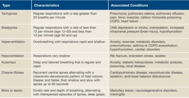
involuntary and is mediated in the brain stem. Respiratory centers in the brain integrate input from neural and chemical receptors and provide neuronal drive to the respiratory muscles, which maintain upper airway patency and drive the tho-racic bellows to determine the level of ventilation. Normal adult respiratory rate is 14 to 20 with a regular rate and frequency and should be quiet. Breathing is considered abnormal if the rate is irregular, too fast, too slow, or shallow (Table 1). Observe the shape of the thorax; it should be symmetrical with equal chest movement. Retrac-tion or bulging of the interspaces may indicate obstructed airways. Following a fall, ribs may be fractured and the injured area may cave inward on inspiration, and outward with expiration (Bickley, 2012). For the patient who cannot sit up, the respiratory assessment can be performed by roll-ing to one side, then the other.
Palpation
Place your index finger in the suprasternal notch at the base of the trachea. The trachea should be midline and slightly moveable. Pulling of the trachea to either side of the neck results from unequal intrathoracic pressure within the chest cavity and may indicate partial to complete pneumothorax, or other serious conditions. Using the palmar surface of the fingers, palpate
Type Characteristics Associated Conditions
Tachypnea Regular respirations with a rate greater than 20 breaths per minute
Pneumonia, pulmonary edema, pulmonary effusion, pain, fever, exercise, carbon monoxide poisoning, COPD, heart failure
Bradypnea Regular respirations with a rate of less than 12 per minute (age 12–50) and less than 13 per minute (age 50 and up)
CNS depression or stroke, oversedation, increased intracranial pressure (brain injury), hypothyroidism Hyperventilation Overbreathing with respirations rapid and shallow Anxiety, exercise, metabolic disorders,
pneumothorax, asthma or COPD exacerbation, hyperthyroidism, cardiac disorders
Hypoventilation Respirations very shallow Rib fracture, brainstem stroke, opiates Kussmaul Deep and labored breathing that is regular and
rapid
Anxiety, diabetic ketoacidosis, metabolic acidosis, poisoning, renal disease
Cheyne-Stokes Recurrent central apnea alternating with a crescendo–decrescendo pattern of tidal volume. Deeper and faster, then shallow and slow with apnea up to 60 seconds
Cardiopulmonary disease, neuromuscular disease, sedation, acid-base balance disturbances Biots or ataxic Erratic rate and depth of breathing, alternating
with interspersed episodes of apnea, deep gasps
Medullary lesion, neurodegenerative disorders, meningitis
pleural linings rubbing together and can be de-scribed as the sound made by treading on fresh snow. This occurs when the pleural layers are inflamed and have lost their lubrication. Fric-tion rub sounds that continue while the patient is holding their breath are most likely cardiac related (Bickley, 2012; Mansen & Gabiola, 2015). comparing one side with the other, using the
side-to-side ladder pattern, striking in each place twice (Figure 1). Percussion sounds should be low-pitched, hollow, and long in duration, or resonant. In contrast, dullness occurs when fluid or solid tissue replaces the normally air filled lung and are thud-like with medium pitch and duration. Dull tones may indicate pneumonia, pleural effusion, or atelectasis. Very loud, lower pitch, and longer percussion sounds, hyperresonance, when unilat-eral may indicate emphysema or pneumothorax (Bickley, 2012; Mansen & Gabiola, 2015).
Auscultation
Ask the patient to breathe slowly and deeply through their open mouth. Using the diaphragm of your stethoscope, listen in the ladder pattern posterior (Figure 1) and anterior (Figure 2), noting the breath sounds (Table 2). Listen in each area for at least one full breath. In the person unable to sit up without help—percuss the upper lung and ascultate the dependent lung on each side. Vesicular breath sounds are soft and generated by airflow of normal lungs. Bronchial breath sounds are normally heard over the larger airways and trachea. Bronchial breath sounds occurring over lateral or posterior chest walls may indicate con-solidation, as in pneumonia. Bronchovesicular breath sounds normally heard between the scap-ula are abnormal if heard over peripheral lung fields and indicate lung tissue is dense, possibly due to consolidation, infection, or compression.
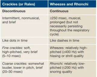
Listen for any adventitious or added sounds (Table 3). Crackles are caused by the small air-ways reopening as the chest wall expands, forc-ing air through passages narrowed by fluid, mu-cous, or pus, and is heard most frequently in the bases due to hypoventilation. The sound of hair being rubbed between one’s fingers simulates this sound. Rhonchi are coarse rattling respi-ratory sounds somewhat like snoring, usually caused by secretions in bronchial airways. A wheeze is a continuous, coarse whistling sound and suggests narrow airways (bronchospasm); and common in asthma, COPD, and bronchitis. If wheezing is heard on one side of the chest only, it may be the result of compression from a tumor or foreign body. Stridor is a medical emergency and is loud, rough, continuous, and high pitched due to upper airway obstruction, heard loudest over the trachea. Pleural friction rub is the squeaking or grating sound of the
Figure 1. Percussion and auscultation pattern for posterior chest. From Bickley, Bates’ Guide to Physical Examination and History-Taking 11E. Reprinted with permission of Wolters Kluwer Health.
Figure 2. Percussion and auscultation pattern for anterior chest. From Bickley, Bates’ Guide to Physical Examination and History-Taking 11E. Reprinted with permission of Wolters Kluwer Health.
1
2
3
4
5
1
2
3
4
5
6 6
7 7
1 1
2 2
3 3
4
4 5
5
brain is less sensitive to hypoxia (low oxygen) and hypercapnea (higher than normal carbon dioxide) and higher residual volume. As a result of these changes, older persons are at increased risk for pneumonia and bronchitis (Minaker, 2011; Sharma & Goodwin, 2006).
A great deal of information may be obtained simply by use of our senses—hearing, vision, touch, and even smell and taste. Knowing what to ask, look and listen for, and feel when you assess the lungs and thorax will allow you to spot early signs of dis-tress and intervene in a timely manner.
Deborah Fritz, PhD, FNP, ANP-BC, is a Family Nurse Practitioner, St. Louis Veterans Administration Medical Center, St. Louis, Missouri. The author and planners have disclosed no potential conflicts of interest, financial or otherwise.
Address for correspondence: Deborah Fritz, MSN, APRN, ANP-BC, 915 North Grand Blvd., St. Louis, MO 63106 ([email protected]). DOI:10.1097/NHH.0000000000000283
REFERENCES
Bickley, L. (2012). Bates’ Guide to Physical Examination and History Taking (11th ed.). Philadelphia, PA: Lippincott Williams & Wilkins. Hogstel, M., & Curry, L. (2005). Health Assessment Through the Life
Span (4th ed.). Philadelphia, PA: F.A. Davis.
Mansen, T., & Gabiola, J. (2015). Patient-Focused Assessment. Bos-ton, MA: Pearson Education, Inc.
McDonald, E. M., Gielen, A. C., Shields, W. C., Stepnitz, R., Parker, E., Ma, X., & Bishai, D. (2013). Residential carbon monoxide (CO) poisoning risks: Correlates of observed CO alarm use in urban households. Journal of Environmental Health, 76(3), 26-32. Minaker, K. L. (2011). Common clinical sequelae of aging. In L.
Gold-man & A. I. Schafer (Eds.), Goldman’s Cecil Medicine (24th ed.). New York: NY: Elsevier Saunders.
Sharma, G., & Goodwin, J. (2006). Effect of aging on respiratory system physiology and immunology. Clinical Interventions in Aging, 1(3), 253-260.
Verghese, A., & Horwitz, R. I. (2009). In praise of the physical exami-nation. British Medical Journal, 339, b5448.
Zambas, S. I. (2010). Purpose of the systematic physical assess-ment in everyday practice: Critique of a “sacred cow”. Journal of Nursing Education, 49(6), 305-310.
Physical Changes Associated With Aging Common significant deviations of the chest for older adults include marked dorsal curvature, kyphosis, increased AP diameter of the chest (barrel chest), and diminished chest expansion (Hogstel & Curry, 2005). Bones become thinner, more rigid, and change shape, and muscles may become weakened. This results in a lower oxygen level with less carbon dioxide removed from the body, and decreased ability to cough. Osteoporo-sis causes decreased thoracic vertebrae height. Aging also causes the alveoli to lose their shape causing shortness of breath. Due to diminished cough reflex with decreased sensitivity, large amounts of particles that are more difficult to ex-pectorate can collect in the lungs. In addition, the
Type Location Heard Characteristics
Tracheal Over trachea and anterior neck Loud, high-pitched, hollow, and harsh; inspiration = expiration
Bronchial Anterior over manubrium and posteriorly between C7-T3 vertebrae (between scapula)
Loud, high-pitched, and hollow; inspiration < expiration Bronchovesicular Anterior over mainstem bronchi and upper two
intercostal spaces and posteriorly left and right of the T3-T5 vertebrae
Softer than bronchial sounds with tubular quality; inspiration = expiration
Vesicular Normal lung sounds all lobes except over scapula Soft, low-pitched, and breezy; inspiration is 2–3 times > expiration
Table 2. Normal Breath Sounds
For more than 170 additional continuing nursing education activities on home healthcare topics, go to nursingcenter.com/ce.
Table 3. Adventitious or Added Breath Sounds
Crackles (or Rales) Wheezes and Rhonchi
Discontinuous Continuous
Intermittent, nonmusical, and brief
≥250 msec, musical, prolonged (but not necessarily persisting throughout the respiratory cycle)
Like dots in time Like dashes in time
Fine crackles: soft, high-pitched, very brief (5–10 msec)
Wheezes: relatively high-pitched (≥400 Hz) with hissing or shrill quality
Coarse crackles: somewhat louder, lower in pitch, brief (20–30 msec)
Rhonchi: relatively low-pitched (≤200 Hz) with snoring quality