Methylomic profiles reveal sex-specific differences in leukocyte composition
associated with post-traumatic stress disorder
Grace S. Kim
a,b, Alicia K. Smith
c,j, Fei Xue
d, Vasiliki Michopoulos
c, Adriana Lori
c,
Don L. Armstrong
e, Allison E. Aiello
f, Karestan C. Koenen
g, Sandro Galea
h, Derek E. Wildman
i,
Monica Uddin
i,⁎aNeuroscience Program, University of Illinois at Urbana-Champaign, Urbana, IL, USA bMedical Scholars Program, University of Illinois College of Medicine, Urbana, IL, USA cDepartment of Psychiatry & Behavioral Sciences, Emory University, Atlanta, GA, USA dDepartment of Statistics, University of Illinois at Urbana-Champaign, Urbana, IL, USA
eCarl R. Woese Institute for Genomic Biology, University of Illinois at Urbana-Champaign, Urbana, IL, USA fGillings School of Global Public Health, University of North Carolina – Chapel Hill, Chapel Hill, NC, USA gDepartment of Epidemiology, Harvard T.H. Chan School of Public Health, Boston, MA, USA
hDepartment of Epidemiology, Boston University School of Public Health, Boston, MA, USA iGenomics Program, College of Public Health, University of South Florida, Tampa, FL, USA jDepartment of Gynecology and Obstetrics, Emory University, Atlanta, GA, USA
A R T I C L E I N F O
Keywords: PTSD Monocytes
Leukocyte composition DNA methylation Sex differences
A B S T R A C T
Post-traumatic stress disorder (PTSD) is a debilitating mental disorder precipitated by trauma exposure. However, only some persons exposed to trauma develop PTSD. There are sex differences in risk; twice as many women as men develop a lifetime diagnosis of PTSD. Methylomic profiles derived from peripheral blood are well-suited for investigating PTSD because DNA methylation (DNAm) encodes individual response to trauma and may play a key role in the immune dysregulation characteristic of PTSD pathophysiology. In the current study, we leveraged recent methodological advances to investigate sex-specific differences in DNAm-based leukocyte composition that are associated with lifetime PTSD. We estimated leukocyte composition on a combined me-thylation array dataset (483 participants, ∼450 k CpG sites) consisting of two civilian cohorts, the Detroit Neighborhood Health Study and Grady Trauma Project. Sex-stratified Mann-Whitney U test and two-way ANCOVA revealed that lifetime PTSD was associated with significantly higher monocyte proportions in males, but not in females (Holm-adjusted p-val < 0.05). No difference in monocyte proportions was observed between current and remitted PTSD cases in males, suggesting that this sex-specific difference may reflect a long-standing trait of lifetime history of PTSD, rather than current state of PTSD. Associations with lifetime PTSD or PTSD status were not observed in any other leukocyte subtype and our finding in monocytes was confirmed using cell estimates based on a different deconvolution algorithm, suggesting that our sex-specific findings are robust across cell estimation approaches. Overall, our main finding of elevated monocyte proportions in males, but not in females with lifetime history of PTSD provides evidence for a sex-specific difference in peripheral blood leukocyte composition that is detectable in methylomic profiles and that may reflect long-standing changes associated with PTSD diagnosis.
1. Introduction
Post-traumatic stress disorder (PTSD) is a debilitating mental dis-order that is precipitated by a traumatic experience involving direct or indirect exposure to actual or threatened death, serious injury, or sexual violence (American Psychiatric Association, 2013). PTSD presents with
intrusive and persistent re-experiencing of the traumatic event, avoid-ance of distressing, trauma-associated stimuli, negative alterations in cognition and mood, and alterations in arousal/reactivity that persist for longer than a month (American Psychiatric Association, 1994; 2013). While most individuals are exposed to a potentially traumatic event at some point in their lives, only some develop PTSD (Breslau,
⁎Corresponding author at: Genomics Program, College of Public Health, University of South Florida, 3720 Spectrum Blvd., Ste. 304, Tampa, FL 33612, USA.
E-mail address:[email protected](M. Uddin). https://doi.org/10.1016/j.bbi.2019.06.025
Received 1 February 2019; Received in revised form 18 June 2019; Accepted 18 June 2019 Available online 19 June 2019
2015); moreover, given the well-known sex difference in PTSD pre-valence and in immune response (Klein and Flanagan, 2016; Osborne et al., 2018), sex-stratified investigation relating to these dual factors is warranted to identify potential differences in variability by sex that may be missed in analyses combining both sexes.
Peripheral blood-based methylomic profiles are comprised of two dynamic components: 1) profiles reflecting proportions of immune cell subtypes (i.e., leukocyte composition) and 2) alterations in DNAm le-vels at CpG sites genome-wide (i.e., differential methylation). Epigenome-wide association studies (EWAS) often seek to identify dy-namic differential methylation marks and treat cellular heterogeneity as a major confound that must be addressed to improve signal detection. However, differences in leukocyte subtypes provide key insights into immunological changes and warrant examination themselves. Recent developments in deconvolution algorithms and cell-type discriminating reference databases have improved estimates (Titus et al., 2017; Teschendorff et al., 2017; Koestler, 2016; Salas, 2018) and enabled utility of DNAm-based leukocyte subtype estimates as proxies for white blood cell differential-based metrics (Wiencke, 2017; Koestler, 2017). Leveraging these recently developed methods, here we use leukocyte-derived methylomic profiles combined from two civilian cohorts to investigate our hypothesis that PTSD is associated with sex-specific differences in leukocyte composition, detectable by DNAm-based esti-mates. To our knowledge, this study is the first to investigate leukocyte composition profiles in PTSD using these new DNAm-based approaches for immune profiling.
2. Materials and methods
2.1. Study participants
Samples from trauma-exposed, adult participants with available Illumina HumanMethylation450 (450 K) BeadChip array data were se-lected from two predominantly African-American, community-based cohorts examining biological and environmental correlates of PTSD, namely the Detroit Neighborhood Health Study (DNHS; n = 192) and Grady Trauma Project (GTP; n = 422). The DNHS, based in Detroit, MI, was approved by the institutional review boards of the University of Michigan and University of North Carolina at Chapel Hill. The GTP, based in Atlanta, GA, was approved by the institutional review boards of Emory University School of Medicine and Grady Memorial Hospital. All participants provided written informed consent prior to data col-lection. Details regarding the DNHS (Uddin, 2010; Goldmann, 2011; Meyers, 2015) and GTP (Gillespie, 2009; Binder, 2008; Zannas, 2015) were published previously. While neither study excluded participants based on illness, women known to be pregnant (in the GTP) were ex-cluded from estimation and analyses, due to well-known/known sig-nificant differences in leukocyte composition during pregnancy (Luppi, 2003). Collected demographic data included self-reported gender, race, age, and current smoking, which was defined as any cigarette smoking in the past 30 days.
2.2. Assessment of PTSD
Study participants were assessed for PTSD, as defined by the Diagnostic and Statistical Manual of Mental Disorders, Fourth Edition (DSM-IV) (American Psychiatric Association, 2013). In the DNHS, study participants were assessed for PTSD using the well-validated self-report PTSD Checklist, Civilian Version (PCL-C) (Blanchard et al., 1996; Grubaugh et al., 2007; Wilkins et al., 2011; Ruggiero et al., 2003) and additional questions about duration, timing, and impairment due to symptoms, via structured telephone interviews (Breslau, 1998; Uddin, 2010; Koenen, 2011). Participants who met all six DSM-IV criteria in reference to their worst traumatic event or to a randomly selected traumatic event (if the participant experienced more than one trauma), were considered affected by lifetime PTSD. Those that met criteria
2009; Kessleretal.,1995;Kilpatrick,2013; Liu,2017;Benjet,2016; Kessler, 2005;KesslerandWang,2008),suggesting thatthedisorder reflectsadistinctinabilitytoreinstatehomeostasisafterpsychological traumainvulnerableindividuals(Yehudaetal.,2011).
Epidemiologicalstudieshaveidentifiedsextobeasignificant vul-nerabilityfactorfordevelopingPTSD,withwomentwiceaslikelyto havelifetimePTSDthanmen,evenwhenriskofexposureandtypesof traumaaretaken intoconsideration(Kessler etal.,1995; Kilpatrick, 2013; Kessler and Wang,2008; Breslau, 1998; Breslau et al., 1997; TolinandFoa,2006).Thissexbiasin diseaseprevalence isalso ob-served in other stress-related mood and anxiety disorders (Kessler, 1994), including depression (Kessler et al., 1993). Preclinical and clinical investigationshaveidentifiedsexualdimorphisminstress re-sponse systemsthat maybe involvedinthe increasedprevalence of stress-related psychopathologies in women (Hodes, 2013; Bangasser andValentino,2014).Furthermore,inadditiontosexualdimorphismin the neurobiological underpinnings of stress/trauma response, recent animalstudiessuggestthatbehavioralresponsetotraumaticstressis fundamentally differentb etweenm alesa ndf emalesa nds houldbe consideredininterpretationof results(Pooley,2018).Inhumans, re-sponse tostress/traumaexposure involvesbothbiologicalandsocial contributorscorrespondingtosexandgender-relatedvariables.While theeffectsofsexandgenderaredifficulttodisentangle,investigations stratifiedbybiologicalsex,understoodtointeractwithgender-related variables, are warranted to improve our currently limited under-standingofthesex-specificbiologicalprocessesdysregulatedinPTSD pathophysiology.
Mounting evidence suggests a key role for stress-induced in-flammationa ndi mmunea lterationi nt hed evelopmenta nd main-tenance of PTSD and other stress-related psychiatric disorders. Althoughfindingsfromhumanliteraturehavebeenmixed,PTSDhas generally beenassociated withincreasedpro-inflammatoryt one, ba-sallyandinresponsetoimmunechallenge,viabothcytokinesignaling and changes in immune cell distribution/function (Passos, 2015; Michopouloset al.,2017;Wang andYoung,2016;Kawamura etal., 2001; Altemus et al., 2003; Gloveret al., 2005; Gillet al., 2009; Gotovac,2010; Lindqvist, 2014; Aiello, 2016; Lindqvist, 2017; Sondergaardetal.,2004;Sommershof,2009;Hoge,2009;Michopoulos etal.,2015;Bersani,2016;Wangetal.,2016).Investigationsinanimal models,primarily inmalerodentstudies,haveprovidedmechanistic insightsintohowperipheralimmunecellresponse/signalingand dis-tributionis linkedwithmicroglialactivationandneuroinflammatory dynamicstotriggerstress-inducedanxietybehaviorandmemory im-pairment(Wohleb,2011;Wohlebetal.,2013;Wohleb,2014;McKim, 2016a,b;WohlebandDelpech,2017;Yin,2019).
times interquartile range (IQR) from the lower quartile or > 3 times IQR from the upper quartile); 4) greater than three standard deviations of the mean bisulfite conversion control probe signal intensity (Salas, 2018). Additionally, samples were checked for gender discordance based on median total intensity of X and Y-chromosome mapped probes (as implemented inminfi) (Aryee, 2014) and removed if predicted sex differed from self-reported gender. Five samples were removed among DNHS samples for gender discordance, and six samples were removed among GTP samples (two for data quality and four for gender dis-cordance). After within-array background correction and dye-bias equalization using out-of-band control probes (ssNoob,minfi) (Fortin et al., 2017; Triche et al., 2013), probes with detection p-value > 0.001 in > 10% of samples (Barfield et al., 2012) and cross-reactive probes (Chen, 2013) (i.e., cross-hybridized between autosomes and sex chromosomes) were removed. Beta-mixture quantile (BMIQ) normal-ization (ChAMP) (Morris, 2014; Tian, 2017) was used to correct for type II probe bias (Teschendorff, 2013).
To control for technical artifacts (e.g., sample processing and ima-ging batch effects), principal components (PCs) based on non-negative control probe signal intensity (Xu et al., 2016) were removed from BMIQ-normalized M-values (i.e., logit-transformed beta-values) sepa-rately for each study. PC correlation heatmaps were used to check for successful removal/reduction of batch effects, especially chip and row effects, while maintaining signal from biological variables. The DNHS and GTP datasets were then combined and an empirical Bayes method (i.e., ComBat (Johnson et al., 2007) in the svapackage (Leek et al., 2012) was used on the combined M-values to account for study effects while controlling for sex and age. Data quality assessment, QC probe filtering, and first step of batch removal were study-specific, while pre-processing steps (ssNoob + BMIQ) implemented within-array ap-proaches unaffected by study. Only probes that passed QC in both studies (n = 455,072 probes) were included in the combined dataset.
2.5. Leukocyte composition estimation
Leukocyte composition was estimated on ComBat-adjusted beta-values using theEpiDISH(Teschendorff et al., 2017) reference database, which is informed by cell-type specific DNase hypersensitive sites (DHS; based on the NIH Epigenomics Roadmap database (Roadmap Epigenomics, 2015)) and is optimized for discriminating granulocytes,
Table 1
Key demographic characteristics, by Sex.
Female(n = 330) Male(n = 153) Total(n = 483)
Study
DNHS 111 (33.6%) 64 (41.8%) 175 (36.2%)
GTP 219 (66.4%) 89 (58.2%) 308 (63.8%)
Race
AA 291 (88.2%) 140 (91.5%) 431 (89.2%)
CA 33 (10.0%) 11 (7.2%) 44 (9.1%)
Other 6 (1.8%) 2 (1.3%) 8 (1.7%)
Median Age
49.0 (38.0–55.0) 48.0 (36.0–56.0) 48.0 (37.5–55.0) Current Smoking
no 210 (63.6%) 72 (47.1%) 282 (58.4%)
yes 109 (33.0%) 78 (51.0%) 187 (38.7%)
Missing 11 (3.3%) 3 (2.0%) 14 (2.9%)
Lifetime PTSD
no 135 (40.9%) 71 (46.4%) 206 (42.7%)
yes 195 (59.1%) 82 (53.6%) 277 (57.3%)
This table describes the subset of participants included in primary analyses investigating sex-specific associations between DNAm-based cell estimates and lifetime PTSD, by sex. For the 2-way ANCOVA, 14 participants were excluded due to missing current smoking data.
DNHS: Detroit Neighborhood Health Study; GTP: Grady Trauma Project
based onsymptomsreportedwithin thepastmonthwereconsidered affectedbycurrentPTSD.Analysisofdatafromtheclinicalinterviews showed that the PTSD instrumentused during structured telephone interviews had excellent internal consistency and highconcordance (Uddin,2010;Uddin,2011).ThePCL-CyieldedaCronbachcoefficient alpha(α)of0.93.UsingclusterscoringbasedonDSM-IVcriteria(i.e.to beacase,theparticipant’ssymptomshadtomeetallsixcriteria),the instrumenthadaspecificity(SP)of0.97,sensitivity(SE)of0.24, po-sitivepredictivevalue(PPV)of0.80,negativepredictivevalue(NPV) 0.72,andanareaundertheROCcurve(AUC)of 0.76,aspreviously reported(Uddin,2010;Uddin,2011).IntheGTP(Jovanovic,2010), studyparticipantswereassessedforlifetimeandcurrentPTSDusingthe Clinician-Administered Post-traumatic Stress Disorder Scale (CAPS, DSM-IV),astructuredinterview administeredbyaclinicianthathas previously been shown to have excellent reliability (i.e., consistent scoresacrossitems,raters,testingoccasions)andexcellentconvergent anddiscriminant validity in large scalepsychometricstudies(Blake, 1995; Weathers et al., 2001). For lifetime analyses, participants meeting criteria for PTSDat any point (including current andpast PTSD)wereconsideredcasesandcomparedtotrauma-exposedcontrols wholackedahistoryofPTSDatanypointintheirlifetime.For com-parisons of current PTSD with remitted PTSD and trauma exposed controls,participantswithlifetimePTSDwerefurtherseparatedinto twogroups:thosewithcurrentPTSDandthosewithlifetime(butnot current)PTSD.
2.3. Sample processing
IntheDNHS, sampleswereobtained viaanin-homeblooddraw performedbyaclinician,processedinthelabwithintwohours,and storedat−20°Cuntiltesting.Detailedmethodsregardingbiospecimen processingintheDNHSareavailableinaseparatepublication(Weckle, 2015).IntheGTP,DNAwasextractedfromwholeblood,aliquoted,and frozen at−80°C withintwo hoursofcollection.GenomicDNA was extractedfromperipheralbloodusingtheDNAMiniKit(Qiagen, Ger-mantown, MD) in the DNHS andthe GentraPuregene Kit (Qiagen, Germantown,MD)intheGTP.Bothstudiesbisulfite-convertedtheDNA samplesusingtheZymoEZ-96DNAmethylationkit(ZymoResearch, Irvine, CA) and used 500ng of DNA per sample for whole-genome amplification,fragmentation,andhybridizationtotheIlluminaHuman Methylation450KBeadChiparray(Illumina,SanDiego,CA),according to the manufacturers’ recommended protocols. Sample processing procedureshavebeenpublishedpreviouslyforboththeDNHSandGTP (Wolf, 2018; Zannas, 2015; Ratanatharathorn, 2017; Uddin, 2018; Mehta,2013).
2.4. Quality control and pre-processing of 450 K data
The raw .idat filesw erei mportedi ntoR ( version3 .5.1)(R FoundationforStatisticalComputing,Vienna,Austria,2018),usingthe
minfi package(Aryee,2014),availableonBioconductor(version3.7) (Huber, 2015; Gentleman,2004),for all subsequentdataprocessing andanalyses.Afterqualitycontrol(QC)(Aryee,2014;Barfieldetal., 2012;Xuetal.,2016),datapre-processing(LiuandSiegmund,2016; Fortinetal.,2017;Chen,2013;Teschendorff,2013;Morris,2014;Tian, 2017) was conducted on all QC’ed samples (DNHS: n=187; GTP: n=416).Thisincludedduplicates(n=12)intheDNHSand partici-pantswithknownpregnancy(n=26)ormissingPTSDphenotypedata (n=82)intheGTP.Analyseswereconductedonuniqueparticipants that passed QC, as described below, and had PTSD data available (Table1).
CD14+monocytes, CD8+T cells, CD4+T cells, CD19+B cells, and
CD56+ natural killer cells. Robust partial correlation (RPC; robust
multivariate linear regression, non-constrained projection) was used as the primary deconvolution algorithm andEpiDISH’simplementation of linear, constrained projection (CP), originally introduced byHouseman (2012), was used to calculate a second set of estimates for comparison.
2.6. Ancestry estimation
DNAm-based ancestry PCs were derived on cleaned beta-values after regressing out sex and age from batch-adjusted M-values. Ancestry PCs were calculated on a subset of 2,317 ancestry informative CpG probes included in two published ancestry informative CpG lists that accounted for confounders (Rahmani, 2017) and that included probes within 10 base pairs (bp) of single nucleotide polymorphisms (SNPs) (Barfield, 2014). The first 2 PCs based on this subset of probes were used as ancestry PCs (ancPCs) after checking for strong association with self-reported race and effective separation of self-reported races.
2.7. Statistical analysis
The Shapiro-Wilk test was used to assess normality and Levene’s test was used to compare equality of variance among groups. Since cell estimates had dissimilar, non-normal distributions, non-parametric tests were used for all initial group comparisons of leukocyte subtypes. The two-sample Kolmogorov-Smirnov (KS) test was used to compare distribution of cell estimates when variances were unequal between groups and Mann-Whitney U test was used to compare mean ranks of cell estimates otherwise. Spearman’s rank correlation was used to assess agreement between estimates based on RPC and CP deconvolution approaches. A threshold of 0.05 was used for p-values and p-values were adjusted for multiple comparisons using Holm’s method (Holm, 1979), unless otherwise specified.
To test our main hypothesis—that PTSD is associated with sex-specific differences in leukocyte composition—initial sex-stratified analyses were conducted on all leukocyte subtypes using the
non-parametric Mann-Whitney U test. For leukocyte subtypes determined to be significantly associated with lifetime PTSD in either sex based on initial Mann-Whitney U tests, a two-way analysis of covariance (ANCOVA; Type III) controlling for age, ancestry (based on DNAm ancestry PCs), and current smoking, was performed with post-hoc comparison of estimated marginal means to examine the effects of sex and lifetime PTSD on transformed cell estimates. Transformation for cell estimate was conducted to meet modeling assumptions for ANCOVA and was informed by Tukey’s Ladder of Powers. Power parameter (λ) was computed to maximize normality based on the Shapiro-Wilks W statistic. Sex-stratified Kruskal-Wallis and post-hoc Dunn tests were conducted as additional follow-up to investigate pos-sible differences in cell proportions by PTSD status (i.e., trauma-ex-posed controls, remitted PTSD, and current PTSD).
3. Results
3.1. Demographic characteristics of sampled study participants from the DNHS and GTP
The demographic characteristics of study participants included in primary analyses investigating sex-specific associations between DNAm-based cell estimates and lifetime PTSD are presented inTable 1. Of the 483 participants from the combined DNHS and GTP sample, 57.3% had a lifetime diagnosis of PTSD, 68.3% were female, and 38.7% were current smokers. The study population was predominantly African-American (89.2%), based on self-reported race, and the median age was 48 years (IQR: 17.5; 37.5–55 years).
3.2. Comparison of leukocyte subtype estimates by deconvolution approach
Good overall agreement was observed between RPC and CP esti-mates, as measured by Spearman’s correlation (i.e., RPC-CP correla-tion), but CD8+ T cells (CD8T) showed much poorer agreement,
ρs(4 8 1) = 0.83, relative to the other leukocyte subtypes,
ρs(4 8 1) > 0.94 (Fig. 1). Since the main objective of this study was to Fig. 1.Correlation (Spearman’s rho) between cell estimates derived using robust partial correlation (RPC) and constrained projection (CP) deconvolution algorithms is high in all leukocyte subtypes, with CD8+T cells (CD8T) showing the worst agreement atR= 0.8 in females andR= 0.87 in males. CD8T cells also showed the
monocyte proportions than trauma-exposed controls, U= 2100,
Z= −2.9,p= 0.004,adj. p= 0.026,r= 0.23. In contrast, no differ-ence in monocyte estimates was found between groups in females,
U= 13000,Z= −0.58,p= 0.6,adj. p= 1,r= 0.03. Lifetime PTSD-associated differences were not observed in any other leukocyte sub-types in either sex.
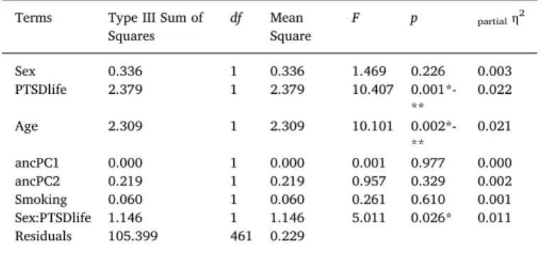
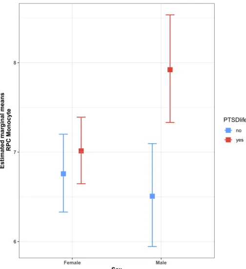
A two-way ANCOVA was conducted to investigate whether sex moderated the effects of lifetime PTSD on transformed monocyte esti-mates, while accounting for age, ancestry, and current smoking (Table 2). Monocyte estimates were square root transformed for the ANCOVA to meet model assumptions (i.e., normality) and were in-formed by Tukey’s Ladder of Powers (RPC: λ = 0.43; CP: λ = 0.5). A significant interaction was found between sex and lifetime PTSD,F(1, 461) = 4.89,p= 0.027,ηp2= 0.011. Post-hoc comparison of estimated
marginal means (EMMs) for lifetime PTSD by sex (Fig. 5; Table 3) showed a significant mean difference between lifetime PTSD cases and controls in males, ΔEMM= 0.26,SE= 0.08,t(4 6 1) = 3.32,p= 0.001, where mean monocyte estimates were higher in lifetime PTSD cases than controls. No significant mean difference was observed between PTSD cases and controls in females, ΔEMM= 0.05, SE= 0.05, t
(4 6 1) = 0.89,p= 0.37, confirming findings from initial sex-stratified analyses. Together, our results suggest that male PTSD cases have sig-nificantly elevated monocyte proportions compared to trauma-exposed controls and that this lifetime PTSD-associated difference is not ob-served in females.
3.5. Association between monocyte proportions and lifetime PTSD in males is independent of current PTSD status
To investigate whether participants with current PTSD exhibited a different monocyte profile from those with remitted PTSD, a sex-stra-tified Kruskal-Wallis test was conducted for PTSD status (i.e., trauma-exposed controls, remitted PTSD, and current PTSD; Fig. 6). A sig-nificant difference in monocyte estimates was observed in males, H
(2) = 8.2,p= 0.017, but not in females, H(2) = 1.1, p= 0.59, con-firming findings from analyses for lifetime PTSD. The post-hoc Dunn test revealed significant differences between PTSD case groups and trauma-exposed controls (current PTSD vs. controls: Z= 2.31,
Fig. 2.Distribution of leukocyte subtypes based on robust partial correlation (RPC) estimates, by sex. Sex differences in CD8+T and CD56+NK cell distributions
were found to be prominent.
investigate sex-specificdifferencesin leukocytecomposition, compar-isonofRPCandCPestimateswasstratifiedbysex.Sex-stratified RPC-CP correlationrevealedthatthelargestdifferenceinRPC-CP correla-tionbetweensexeswasalsofoundinCD8Tcells,|Δρs| =0.07,suchthat
females showed poorer correlation, ρs(328)=0.80, than males,
ρs(151)=0.87. Forthe other leukocyte subtypes, the differencein
RPC-CP correlationbetween sexes(|Δρs|),rangedfrom 0.01to0.03,
withCD56+naturalkiller(NK)cellshavingthesecondlargest
differ-ence in correlation between sexes (female: ρs(328)=0.93; male:
ρs(151)=0.96).Inallleukocytesubtypes,exceptCD19+Bcells,
fe-maleshadlowercorrelationcoefficientsthanmales.Detailedresultsfor RPC-basedcellestimatesarereportedbelowandcorrespondingresults basedonCP-basedestimatesarereportedinsupplementarymaterials, duetostrongagreementbetweenfindingsfrombothsetsofestimates.
3.3. Comparison of leukocyte subtype estimates by sex and lifetime PTSD
Cellestimateswerecomparedbysex,lifetimePTSD,andstudyin each leukocyte subtype to establish overall differences.Significant overallsexdifferenceswereobservedinthedistributionsofNK(KS:
D =0.19,adj. p =0.007)andCD8Tcellproportions(KS:D =0.16;adj. p =0.04)inRPCestimates.Malesshowedgreatervariabilitythan fe-malesforbothNKandCD8Tcells(malevs.female–IQRNK:6.2%vs.
4.15%;IQRCD8T:9.5%vs.6.2%),aswellashighermedianNK(5.5%vs.
4.4%) and lower median CD8T (9.0% vs. 9.7%) cell proportions (Fig.2).Nosignificantoveralldifferences(i.e.,inbothsexescombined) wereobservedbetweenlifetimePTSDcasesandtrauma-exposed con-trols in any leukocyte subtype (Mann-Whitney).Additional analyses comparing leukocyte subtype proportions between participating co-hortsandassessingage effectsineachcelltypearereportedin sup-plementarymaterials.
3.4. Elevated monocyte proportions were associated with lifetime PTSD in males, but not females
p= 0.021, adj. p= 0.042, r= 0.23; remitted PTSD vs. controls:
Z= 2.40,p= 0.016,adj. p= 0.049,r= 0.22), but no significant dif-ference between current and remitted PTSD groups,Z= 0.18,p= 0.86,
adj. p= 0.86, r= 0.02. These findings suggest that the association between monocyte proportions and lifetime PTSD in males is in-dependent of current PTSD state and may reflect long-standing changes associated with lifetime history of PTSD diagnosis.
Comparative analyses based on CP monocyte estimates showed si-milar results to RPC-based results (see Supplementary Materials); however, CP-based results consistently presented smaller effect sizes than RPC-based results across all analyses and follow-up comparisons between PTSD case groups and trauma-exposed controls did not reach significance after p-value adjustment in post-hoc Dunn test using CP monocyte estimates in males.
Fig. 3.Violin plots of RPC estimates for lifetime PTSD cases and controls, stratified by sex. Only monocyte proportions in males showed a statistically significant difference between lifetime PTSD cases and controls, based on Mann-WhitneyUtest (p-value = 0.004; Holm-adjusted p-value = 0.026). For figure labels on x-axis: B = CD19+B cells; NK = CD56+NK cells; CD4T = CD4+T cells; CD8T = CD8+T cells; Gran = Granulocytes; Mono = CD14+monocytes.
Fig. 4.Density plots for RPC monocyte estimates in lifetime PTSD cases and controls, stratified by sex, show distinctly higher monocyte levels in males with lifetime PTSD compared to trauma-exposed controls. This difference in monocyte levels between lifetime PTSD cases and controls is not observed in females.
Table 2
Two-way ANCOVA Table for RPC monocyte estimates (n = 469). Terms Type III Sum of
Squares df MeanSquare F p partialη 2
Sex 0.336 1 0.336 1.469 0.226 0.003
PTSDlife 2.379 1 2.379 10.407
0.001*-** 0.022
Age 2.309 1 2.309 10.101
0.002*-** 0.021
ancPC1 0.000 1 0.000 0.001 0.977 0.000
ancPC2 0.219 1 0.219 0.957 0.329 0.002
Smoking 0.060 1 0.060 0.261 0.610 0.001
Sex:PTSDlife 1.146 1 1.146 5.011 0.026* 0.011 Residuals 105.399 461 0.229
4. Discussion
Methylomic profiles derived from peripheral blood offer a wealth of information and can be harnessed to detect two dynamic measures of immune state: 1) differences in leukocyte composition (i.e., proportions of peripheral immune cell subtypes); and 2) true alterations in
methylation involved in epigenetic regulation of immune processes. They are particularly well-suited for investigating PTSD because DNAm encodes individual response to trauma and may play a key role in PTSD-associated immune dysregulation. Given the prominent sex dif-ferences in both PTSD prevalence (Kessler et al., 1995; Kilpatrick, 2013; Kessler and Wang, 2008; Breslau, 1998; Breslau et al., 1997; Tolin and Foa, 2006) and immune response (Klein and Flanagan, 2016; Osborne et al., 2018), the primary goal of the present study was to investigate whether PTSD is associated with sex-specific differences in leukocyte composition, detectable by DNAm-based estimates. We found that males with lifetime PTSD showed significantly higher monocyte pro-portions than trauma-exposed males without PTSD; this difference was not observed in females. No difference in monocyte proportions was observed between current and remitted PTSD cases in males, suggesting that this sex-specific difference may reflect a long-standing trait of lifetime history of PTSD diagnosis, rather than current state of PTSD. These findings were observed in both the primary RPC and comparison CP-based sets of cell estimates, which were derived using non-con-strained vs. connon-con-strained projection deconvolution algorithms, respec-tively. Overall, our main finding of elevated monocyte proportions in males, but not females with lifetime history of PTSD provides evidence for a sex-specific difference in peripheral blood leukocyte composition that may reflect long-standing changes associated with PTSD diagnosis and is detectable in methylomic profiles, consistently across different deconvolution algorithms.
Fig. 5.Lifetime PTSD by sex interaction plot for estimated marginal means (EMMs) of RPC monocyte estimates. Interaction plot shows a significant EMM difference between lifetime PTSD cases (red) and controls (blue) in males, where mean monocyte estimates are higher in cases than controls. No significant EMM difference was observed between PTSD cases and controls in females. (For interpretation of the references to colour in this figure legend, the reader is referred to the web version of this article.)
Table 3
Summary for RPC monocyte estimates by group.
Sex PTSD n mean SE EMM SEEMM lower.CL upper.CL
Female no 135 7.113 0.2443 6.758 0.2214 6.330 7.200 Female yes 184 7.182 0.1691 7.014 0.1893 6.647 7.391 Male no 70 6.803 0.3228 6.507 0.2926 5.945 7.095 Male yes 80 8.103 0.3147 7.921 0.3063 7.331 8.535
This table describes untransformed RPC monocyte estimates by group (i.e., sex and lifetime PTSD); n = count per group; EMM = estimated marginal means (i.e., least squares means); SE = standard errors for regular means; SEEMM= standard errors for EMM.
In our study, we leveraged recent advances in reference-based de-convolution methods (Teschendorff et al., 2017; Koestler, 2016; Salas, 2018) – specifically theEpiDISHalgorithm (Teschendorff et al., 2017), which (i) employs DNase hypersensitive site (DHS) data of leukocyte subtypes to inform probe selection for their reference database and (ii) introduces RPC, a non-constrained projection approach for reference-based deconvolution. A comparative validation study on in-silico mix-tures of purified cell DNAm profiles previously showed this newer RPC approach to consistently perform better than the widely used CP ap-proach (Houseman, 2012), based on root mean square error (RMSE) and R2, at low noise levels (Teschendorff et al., 2017) typically en-countered in real data (Teschendorff et al., 2017; Salas, 2018). Relevant to our results, the study showed the difference in RMSE and R2 to be the most prominent in CD8T cells (Teschendorff et al., 2017). This is con-sistent with our comparison between RPC and CP estimates, which showed CD8T cells to exhibit poorer correlation between RPC and CP estimates relative to other leukocyte subtypes and the largest difference in RPC-CP correlation between sexes. Similarly, the validation study reported better performance of RPC, compared to CP, in monocytes, with higher RMSE and lower R2 in CP compared to RPC, suggesting RPC-based estimates were more robustly associated with true weights. In light of the validation study, this suggests that our RPC-based esti-mates were better able to resolve male-specific association of monocyte proportions with lifetime PTSD. In all, our results were consistent with the previously published validation study (Teschendorff et al., 2017) and favored use of RPC estimates for modeling leukocyte composition. However, as these methods have been developed recently, further va-lidation and comparative studies are warranted.
Comparison of leukocyte subtype estimates by sex revealed sig-nificant baseline sex differences in the distributions of NK cell and CD8T cell proportions, with males showing greater median NK and lower median CD8T cell proportions, using both RPC and CP based estimates. This finding is consistent with a previous study that reported sex differences in both leukocyte subtypes using estimates based on
minfi’simplementation of the Houseman approach (Inoshita, 2015) and with immunology literature that reported higher NK cell counts and proportions in males compared to females (Abdullah, 2012). A recent study that modeled cell-specific methylation profiles also reported ro-bust sex differences in CD56+NK methylation patterns (White, 2017),
suggesting that this leukocyte subtype may be regulated by DNAm in a sex-specific manner. Additionally, DNAm dynamics have been found to drive effector functions in CD8T cells after stimulation (Scharer et al., 2013; Suarez-Alvarez et al., 2012). Development of reference databases that resolve the six main leukocyte subtypes to consider proportions of subsets with shared lineage but different functionality/phenotype (e.g., naïve vs. memory vs. regulatory subtypes) may allow us to explore this hypothesis and would greatly enrich our understanding of immune activity.
gene expression (Neylan, 2011) of CD14+monocytes isolated from
per-ipheral blood. Given the inherent sex differences in innate immune response (Klein and Flanagan, 2016; Nunn et al., 2009), as understood in the context of infection, injury, and treatment of inflammatory disorders, sexually di-morphic dynamics and effects may also exist in the context of neuroimmune response to stress/trauma exposure (Bekhbat and Neigh, 2018). One re-levant sex difference in monocytes involves the expression of IL-6, which was suggested to be important for stress-induced anxiety-like behavior and social avoidance in the RSD model (Niraula et al., 2019). Independent of reproductive hormones (i.e., estradiol, dehydroepiandrosterone [DHEA], progesterone), women were shown to have greater monocyte expression of IL-6 across a circadian period than men, and sympathovagal balance was negatively associated with monocyte IL-6 expression only in women (O'Connor et al., 2007). On the other hand, a study examining sex differ-ences in regulation of inflammatory cell recruitment and cytokine synthesis found that ovarian hormones regulate phenotype, function, and numbers of macrophages, but not T lymphocytes, in females (Scotland et al., 2011). This fundamental sex difference may underlie more efficient recognition and elimination of infectious stimuli without recruitment of circulating neutrophils or excessive cytokine production in females, compared to males (Scotland et al., 2011), and may also have implications in the context of psychosocial stress exposure. Relatedly, statins, which have anti-in-flammatory activity, modulate monocyte migration in a sex-specific manner, such that both spontaneous and lipopolysaccharide-induced mi-gration of isolated monocytes were found to be inhibited by statins in women, but not men (Ruggieri, 2014).
Our observation of male-specific increase in monocyte proportions as-sociated with lifetime PTSD may reflect fundamental sex differences in leukocyte trafficking, tissue distribution, and thus composition in blood, with implications for stress/trauma-induced neuroimmune alterations and behavior. Of note, while the effect size detected in males translates to an absolute difference of only 1.3% in monocyte proportions between parti-cipants with vs. without PTSD (∼8.1% vs. 6.8%;Table 3), it also corre-sponds to an increase of approximately 19% in overall monocytes among men with lifetime PTSD. Furthermore, the lack of difference between re-mitted and current PTSD observed in males may have a number of im-plications for PTSD pathophysiology, including adverse health con-sequences associated with PTSD across the life course in men, which may be distinct from PTSD-associated health trajectories in women (Dedert et al., 2010; Mitchell, 2013; McLean et al., 2011).
Although the current dataset combined two cohorts and known pregnancies were excluded from our study, sample size and unavailable phenotype data on pregnancy, timing of the menstrual cycle, hormonal birth control use, well-harmonized measures of depression, and health status, as well as gender-related variables, such as coping mechanisms, are all limitations of this study. Future studies that account for hormone levels and other fundamental physiological sex differences may help identify female-specific associations between PTSD and leukocyte composition and clarify if hormone-dependent processes influence leukocyte composition dynamics. Additionally, both cohorts included in this study are civilian, urban, and predominantly African-American, so generalizability of our findings may be limited to this population.
Overall, our study implements current state-of-the-art methods to illus-trate feasibility of using DNAm-based leukocyte composition estimates to probe immune profiles. Our literature-supported finding of higher DNAm-based monocyte proportions in males may be an informative metric to in-clude as part of a diagnostic biomarker panel for PTSD in males, and future study in females, with consideration for hormonal status, may elucidate a female-specific panel as well. Differential methylation markers discovered in sex-stratified EWAS, which account for these cell estimates as covariates, are other prime candidates to be included in such sex-specific biomarker panels. Furthermore, in addition to being able to infer leukocyte composi-tion when complete blood count data is not available, these DNAm-based estimates of leukocyte composition can be used to determine cell-specific differential methylation profiles. In fact, methods and validation for cell-specific differential methylation analyses have been published very recently
1999;deKloet,2007;Vidovic,2011,2007;Jergovic,2014)andthose based on both sexes did not conduct sex-stratifieda nalyses(Aiello, 2016;Sommershof,2009;Rohlederetal.,2004).Toourknowledge,no authoritativestudyofsexdifferencesincompletebloodcountsinPTSD hasyet beenpublished,andstudiesof sexdifferencesi nPTSDhave generallybeenlacking,withanumberoflarge-scalestudiesconducted inpredominantlymalemilitarycohorts(oronfemale-onlycohorts,e.g. Nurses’HealthStudyII)(Koenen,2009;Sumner,2017).
WhilenotforPTSD,astudyofdepressionthatexaminedwhiteblood celldifferentialcountnotedasignificantincreaseinmonocytecountand proportionsamongmaleswithmajordepressivedisorder(MDD),butnot females, and asignificants exb yd iagnosisi nteraction(Maes,1992). Likewise,a separatelongitudinalstudyfollowingMDDinpatientsalso reportedelevatedmonocytecountsinpatientscomparedtocontrols,and thiswasdrivenbymen(Seidel,1996).Additionally,adecreasein de-pression severitywasassociated witha decreasein monocytecounts (Seidel, 1996),suggesting thatmonocytes may be related to clinical improvement. Similarly, thepresence and severityof atherosclerosis, anotherconditionlinkedtoPTSDviasystemiclow-gradeinflammatory state (Brouwers etal.,2015),arealsoassociated withanincreasein monocytecountinmales,butnotfemales(Huang,2001).
Furtherprospective investigation of PTSD isneeded to determine whetherthehighermonocyteproportionobservedinmalesreflectsan increased susceptibility for developing PTSD or if it reflectsa n im-munologicalshiftinresponsetotheprecipitatingtraumaassociatedwith PTSDpsychopathology.However,studiesinamalerodentmodel pro-videstrongevidenceforthelatterandhavebeenimportantfor estab-lishingtherelationshipbetweenperipheralimmunecellsandthebrainin the context of psychosocial stress and associated behavior. Repeated social defeat(RSD)wasfound toinducemyelopoiesisand release in-flammatory( Ly6Chi)m onocytesi ntoc irculationv ias ympathetic
sig-naling,andthisincreasedlevelofcirculatingperipheralmonocyteswas associated with recruitment of pro-inflammatory monocytes/macro-phagestothebrainandneuroinflammation(Wohleb,2011;Engleretal., 2004; Weber et al., 2017). Increased proportion of these peripheral monocytesandmacrophagerecruitmenttothebrainwerealso demon-stratedtocorrespondwithdevelopment,maintenance,and re-establish-mentofRSD-inducedanxiety-likebehavior;blockadeofthisrecruitment (via splenectomy or β-adrenergicreceptor blockage) beforeRSD was foundtopreventdevelopmentofanxiety-likebehavior(Wohleb,2014; McKim,2016).Additionally,arecentpaperdiscernedthatstress-induced anxiety-likebehaviorandsocialavoidancearedependentonanincrease in IL-6 after stress exposure,which induces a primed transcriptional profileinmonocytesrecruitedtothebrainandpropagatesIL-1β medi-atedinflammationassociatedwithanxiety-likebehavior(Niraulaetal., 2019).Thesestudiesimplicateperipheralmonocytesindirectlyaffecting relevant PTSD-like behaviorafter stress exposure in males(Bierhaus, 2003;Grisanti, 2010).Very recently,thefirsts tudyu singamodified versionoftheRSDparadigmwasconductedinfemalemiceandreported asimilaronsetofanxiety-likeandsocialavoidancebehavior,increasein myelopoiesis,increaseinperipheralmonocyteproportions,and recruit-mentofperipheralmyeloidcellsto thebrain,14hafterthelastRSD cycle(Yin,2019).Continuedworkbasedonthisparadigmatmultiple timepointsmaybefruitfulforinvestigatingiftherearesexdifferencesin thekineticsof leukocytetraffickingan dti ssuedi stribution,especially sincerecentinvestigationsinotherPTSD-relevantrodentmodelssuggest fundamentalsexdifferencesinneurobiologicalresponsetotrauma ex-posure(Pooley,2018)andinregulationofstress/trauma-induced neu-roinflammatoryp riming/neuroimmunea lterations( Fonken,2018; Bekhbat and Neigh, 2018). Furthermore, a social stress paradigm in pregnantratsreportedlowernumbersofmonocytesinstressedfemales than control femalerats (Stefanski etal., 2005),illustrating the im-portanceofconsideringdifferentparadigmsandbreeds/species.
5. Conclusion
By combining DNA methylation datasets from two civilian cohorts, the current study found significantly higher monocyte proportions in males with lifetime PTSD compared to trauma-exposed controls, a dif-ference that was not observed in females. This sex-specific difdif-ference in peripheral blood leukocyte composition may reflect a long-standing trait of PTSD diagnosis, rather than current state of PTSD. While this finding was confirmed using two different cell estimation approaches (i.e., deconvolution algorithms), the recently developed non-con-strained projection approach (RPC) appears better suited for modeling leukocyte composition. Methylome-based characterization of immune profiles holds special promise for the study of PTSD and continued development of reference databases and validation of methods will build on these recent improvements to enrich our understanding of sex-specific immune dysregulation associated with PTSD.
Funding
NIH grants: R01MD011728; 3R01MD011728-02S1; R01 DA022720; DA022720-S1; RC1MH088283; MH096764; MH071537; University of Illinois: CompGen Fellowship.
Declaration of Competing Interest
None.
Acknowledgments
We appreciate the time and effort of study participants, staff and volunteers of the Detroit Neighborhood Health Study and Grady Trauma Project.
Appendix A. Supplementary data
Supplementary data associated with this article can be found, in the online version, athttp://dx.doi.org/10.1016/j.bbi.2019.06.025.
References
Abdullah, M., et al., 2012. Gender effect on in vitro lymphocyte subset levels of healthy individuals. Cell Immunol 272, 214–219.https://doi.org/10.1016/j.cellimm.2011. 10.009.
Aiello, A.E., et al., 2016. PTSD is associated with an increase in aged T cell phenotypes in adults living in Detroit. Psychoneuroendocrinology 67, 133–141.https://doi.org/10. 1016/j.psyneuen.2016.01.024.
Altemus, M., Cloitre, M., Dhabhar, F.S., 2003. Enhanced cellular immune response in women with PTSD related to childhood abuse. Am. J. Psychiat. 160, 1705–1707. https://doi.org/10.1176/appi.ajp.160.9.1705.
Altemus, M., Dhabhar, F.S., Yang, R., 2006. Immune function in PTSD. Ann. N Y Acad. Sci. 1071, 167–183.https://doi.org/10.1196/annals.1364.013.
Alvarez-Errico, D., Vento-Tormo, R., Sieweke, M., Ballestar, E., 2015. Epigenetic control of myeloid cell differentiation, identity and function. Nat. Rev. Immunol. 15, 7–17. https://doi.org/10.1038/nri3777.
Aryee, M.J., et al., 2014. Minfi: a flexible and comprehensive Bioconductor package for the analysis of Infinium DNA methylation microarrays. Bioinformatics 30, 1363–1369.https://doi.org/10.1093/bioinformatics/btu049.
American Psychiatric Association, 1994. Diagnostic and statistical manual of mental disorders. Fourth ed. (American Psychiatric Association, Washington, DC). American Psychiatric Association, 2013. Diagnostic and statistical manual of mental
disorders. 5 ed.
Bam, M., et al., 2016. Dysregulated immune system networks in war veterans with PTSD
interleukin-12 and interferon gamma, in peripheral blood mononuclear cells from PTSD patients. J. Neuroimmune. Pharmacol. 11, 168–181.https://doi.org/10.1007/ s11481-015-9643-8.
Bangasser, D.A., Valentino, R.J., 2014. Sex differences in stress-related psychiatric dis-orders: neurobiological perspectives. Front. Neuroendocrinol. 35, 303–319.https:// doi.org/10.1016/j.yfrne.2014.03.008.
Barfield, R.T., et al., 2014. Accounting for population stratification in DNA methylation studies. Genet. Epidemiol. 38, 231–241.https://doi.org/10.1002/gepi.21789. Barfield, R.T., Kilaru, V., Smith, A.K., Conneely, K.N., 2012. CpGassoc: an R function for
analysis of DNA methylation microarray data. Bioinformatics 28, 1280–1281. https://doi.org/10.1093/bioinformatics/bts124.
Bekhbat, M., Neigh, G.N., 2018. Sex differences in the neuro-immune consequences of stress: Focus on depression and anxiety. Brain Behav. Immun. 67, 1–12.https://doi. org/10.1016/j.bbi.2017.02.006.
Benjet, C., et al., 2016. The epidemiology of traumatic event exposure worldwide: results from the World Mental Health Survey Consortium. Psychol. Med. 46, 327–343. https://doi.org/10.1017/S0033291715001981.
Bersani, F.S., et al., 2016. A population of atypical CD56(-)CD16(+) natural killer cells is expanded in PTSD and is associated with symptom severity. Brain Behav. Immun. 56, 264–270.https://doi.org/10.1016/j.bbi.2016.03.021.
Bierhaus, A., et al., 2003. A mechanism converting psychosocial stress into mononuclear cell activation. Proc. Natl. Acad. Sci. U.S.A. 100, 1920–1925.https://doi.org/10. 1073/pnas.0438019100.
Binder, E.B., et al., 2008. Association of FKBP5 polymorphisms and childhood abuse with risk of posttraumatic stress disorder symptoms in adults. JAMA 299, 1291–1305. https://doi.org/10.1001/jama.299.11.1291.
Blake, D.D., et al., 1995. The development of a Clinician-Administered PTSD Scale. J. Trauma Stress 8, 75–90.
Blanchard, E.B., Jones-Alexander, J., Buckley, T.C., Forneris, C.A., 1996. Psychometric properties of the PTSD Checklist (PCL). Behav. Res. Ther. 34, 669–673.
Boscarino, J.A., Chang, J., 1999. Higher abnormal leukocyte and lymphocyte counts 20 years after exposure to severe stress: research and clinical implications. Psychosom. Med. 61, 378–386.
Breslau, N., et al., 1998. Trauma and posttraumatic stress disorder in the community: the 1996 Detroit Area Survey of Trauma. Arch. Gen. Psychiat. 55, 626–632. Breslau, N., 2009. The epidemiology of trauma, PTSD, and other posttrauma disorders.
Trauma Viol. Abuse 10, 198–210.https://doi.org/10.1177/1524838009334448. Breslau, N., Davis, G.C., Andreski, P., Peterson, E.L., Schultz, L.R., 1997. Sex differences in
posttraumatic stress disorder. Arch. Gen. Psychiat. 54, 1044–1048.
Brouwers, C. J., Wolf, J. M. von Känel, R. 2015. Comprehensive Guide to Post-Traumatic Stress Disorder Ch. Chapter 54–1, 1–13.
Chen, Y.A., et al., 2013. Discovery of cross-reactive probes and polymorphic CpGs in the Illumina Infinium HumanMethylation450 microarray. Epigenetics 8, 203–209. https://doi.org/10.4161/epi.23470.
Chen, L., et al., 2016. Genetic drivers of epigenetic and transcriptional variation in human immune cells. Cell 167, 1398–1414.https://doi.org/10.1016/j.cell.2016.10.026. de Kloet, C.S., et al., 2007. Leukocyte glucocorticoid receptor expression and
im-munoregulation in veterans with and without post-traumatic stress disorder. Mol. Psychiat. 12, 443–453.https://doi.org/10.1038/sj.mp.4001934.
Dedert, E.A., Calhoun, P.S., Watkins, L.L., Sherwood, A., Beckham, J.C., 2010. Posttraumatic stress disorder, cardiovascular, and metabolic disease: a review of the evidence. Ann. Behav. Med. 39, 61–78. https://doi.org/10.1007/s12160-010-9165-9.
Engler, H., Bailey, M.T., Engler, A., Sheridan, J.F., 2004. Effects of repeated social stress on leukocyte distribution in bone marrow, peripheral blood and spleen. J. Neuroimmunol. 148, 106–115.https://doi.org/10.1016/j.jneuroim.2003.11.011. Fonken, L.K., et al., 2018. Neuroinflammatory priming to stress is differentially regulated
in male and female rats. Brain Behav. Immun. 70, 257–267.https://doi.org/10.1016/ j.bbi.2018.03.005.
Fortin, J.P., Triche Jr., T.J., Hansen, K.D., 2017. Preprocessing, normalization and in-tegration of the Illumina HumanMethylationEPIC array with minfi. Bioinformatics 33, 558–560.https://doi.org/10.1093/bioinformatics/btw691.
Gentleman, R.C., et al., 2004. Bioconductor: open software development for computa-tional biology and bioinformatics. Genome Biol. 5, R80. https://doi.org/10.1186/gb-2004-5-10-r80.
Gill, J.M., Saligan, L., Woods, S., Page, G., 2009. PTSD is associated with an excess of inflammatory immune activities. Perspect. Psychiat. Care 45, 262–277.https://doi. org/10.1111/j.1744-6163.2009.00229.x.
Gillespie, C.F., et al., 2009. Trauma exposure and stress-related disorders in inner city primary care patients. Gen. Hosp. Psychiat. 31, 505–514.https://doi.org/10.1016/j. genhosppsych.2009.05.003.
Glover, D.A., Steele, A.C., Stuber, M.L., Fahey, J.L., 2005. Preliminary evidence for lymphocyte distribution differences at rest and after acute psychological stress in PTSD-symptomatic women. Brain Behav. Immun. 19, 243–251.https://doi.org/10. 1016/j.bbi.2004.08.002.
Gola, H., et al., 2013. Posttraumatic stress disorder is associated with an enhanced spontaneous production of pro-inflammatory cytokines by peripheral blood mono-nuclear cells. BMC Psychiat. 13.https://doi.org/10.1186/1471-244x-13-40. Goldmann, E., et al., 2011. Pervasive exposure to violence and posttraumatic stress
dis-order in a predominantly African American Urban Community: the Detroit Neighborhood Health Study. J. Trauma Stress 24, 747–751.https://doi.org/10. 1002/jts.20705.
Gotovac, K., et al., 2010. Natural killer cell cytotoxicity and lymphocyte perforin
(Zhengetal.,2018;Lietal.,2019)andmayenablethenextsignificant advanceinextractinginsightsfrommethylomicprofilesbycontextualizing howdifferentialmethylationinspecificleukocytesubtypesalterregulatory dynamicsintheimmunesystem.Ultimately,thisworkmayhelptoshape futurestudiesdesignedtodeterminewhethersex-specificmethylomic me-tricsofperipheralimmunestatuscaninformusaboutsexdifferencesin neuroinflammationandcorrespondingbehaviorinresponsetotrauma ex-posure.
isanoutcomeofalteredmiRNAexpressionandDNAmethylation.Sci.Rep.6,31209. https://doi.org/10.1038/srep31209.
expression in veterans with posttraumatic stress disorder. Prog.
Neuropsychopharmacol. Biol. Psychiat. 34, 597–604.https://doi.org/10.1016/j. pnpbp.2010.02.018.
Grisanti, L.A., et al., 2010. Pro-inflammatory responses in human monocytes are beta1-adrenergic receptor subtype dependent. Mol. Immunol. 47, 1244–1254.https://doi. org/10.1016/j.molimm.2009.12.013.
Grubaugh, A.L., Elhai, J.D., Cusack, K.J., Wells, C., Frueh, B.C., 2007. Screening for PTSD in public-sector mental health settings: the diagnostic utility of the PTSD checklist. Depress Anxiet. 24, 124–129.https://doi.org/10.1002/da.20226.
Hodes, G.E., 2013. Sex, stress, and epigenetics: regulation of behavior in animal models of mood disorders. Biol. Sex Differ. 4, 1.https://doi.org/10.1186/2042-6410-4-1. Hoge, E.A., et al., 2009. Broad spectrum of cytokine abnormalities in panic disorder and
posttraumatic stress disorder. Depress Anxiety 26, 447–455.https://doi.org/10. 1002/da.20564.
Holm, S., 1979. A simple sequentially rejective multiple test procedure. Scand. J. Statist. 6, 65–70.
Houseman, E.A., et al., 2012. DNA methylation arrays as surrogate measures of cell mixture distribution. BMC Bioinformat. 13, 1–16. https://doi.org/10.1186/1471-2105-13-86.
Huang, Z.S., et al., 2001. Correlations between peripheral differential leukocyte counts and carotid atherosclerosis in non-smokers. Atherosclerosis 158, 431–436.https:// doi.org/10.1016/S0021-9150(01)00445-2.
Huber, W., et al., 2015. Orchestrating high-throughput genomic analysis with bio-conductor. Nat. Methods 12, 115–121.https://doi.org/10.1038/nmeth.3252. Inoshita, M., et al., 2015. Sex differences of leukocytes DNA methylation adjusted for
estimated cellular proportions. Biol. Sex Differ. 6, 11.https://doi.org/10.1186/ s13293-015-0029-7.
Ironson, G., Cruess, D., Kumar, M., 2007. inPsychoneuroimmunology (Fourth Edition)(ed Robert Ader) 531–547. Academic Press.
Irwin, M.R., Cole, S.W., 2011. Reciprocal regulation of the neural and innate immune systems. Nat. Rev. Immunol. 11, 625–632.https://doi.org/10.1038/nri3042. Jergovic, M., et al., 2014. Patients with posttraumatic stress disorder exhibit an altered
phenotype of regulatory T cells. Allergy Asthma Clin. Immunol. 10, 43.https://doi. org/10.1186/1710-1492-10-43.
Johnson, W.E., Li, C., Rabinovic, A., 2007. Adjusting batch effects in microarray ex-pression data using empirical Bayes methods. Biostatistics 8, 118–127.https://doi. org/10.1093/biostatistics/kxj037.
Johnson, G.J., Slater, B.C., Leis, L.A., Rector, T.S., Bach, R.R., 2016. Blood biomarkers of chronic inflammation in Gulf War Illness. e0157855. PLoS One 11.https://doi.org/ 10.1371/journal.pone.0157855.
Jovanovic, T., et al., 2010. Impaired fear inhibition is a biomarker of PTSD but not de-pression. Depress Anxiety 27, 244–251.https://doi.org/10.1002/da.20663. Kawamura, N., Kim, Y., Asukai, N., 2001. Suppression of cellular immunity in men with a
past history of posttraumatic stress disorder. Am. J. Psychiat. 158, 484–486.https:// doi.org/10.1176/appi.ajp.158.3.484.
Kessler, R.C., et al., 1994. Lifetime and 12-month prevalence of DSM-III-R psychiatric disorders in the United States. Results from the National Comorbidity Survey. Arch. Gen. Psychiat. 51, 8–19.
Kessler, R.C., et al., 2005. Lifetime prevalence and age-of-onset distributions of DSM-IV disorders in the National Comorbidity Survey Replication. Arch. Gen. Psychiat. 62, 593–602.https://doi.org/10.1001/archpsyc.62.6.593.
Kessler, R.C., McGonagle, K.A., Swartz, M., Blazer, D.G., Nelson, C.B., 1993. Sex and depression in the National Comorbidity Survey. I: Lifetime prevalence, chronicity and recurrence. J. Affect. Disord. 29, 85–96.
Kessler, R.C., Sonnega, A., Bromet, E., Hughes, M., Nelson, C.B., 1995. Posttraumatic stress disorder in the National Comorbidity Survey. Arch Gen Psychiat. 52, 1048–1060.
Kessler, R.C., Wang, P.S., 2008. The descriptive epidemiology of commonly occurring mental disorders in the United States. Annu. Rev. Public Health 29, 115–129.https:// doi.org/10.1146/annurev.publhealth.29.020907.090847.
Kilpatrick, D.G., et al., 2013. National estimates of exposure to traumatic events and PTSD prevalence using DSM-IV and DSM-5 criteria. J. Trauma Stress 26, 537–547.https:// doi.org/10.1002/jts.21848.
Klein, S.L., Flanagan, K.L., 2016. Sex differences in immune responses. Nat. Rev. Immunol. 16, 626–638.https://doi.org/10.1038/nri.2016.90.
Koenen, K.C., et al., 2009. Protocol for investigating genetic determinants of posttrau-matic stress disorder in women from the Nurses' Health Study II. BMC Psychiat. 9, 29. https://doi.org/10.1186/1471-244X-9-29.
Koenen, K.C., et al., 2011. SLC6A4 methylation modifies the effect of the number of traumatic events on risk for posttraumatic stress disorder. Depress Anx. 28, 639–647. https://doi.org/10.1002/da.20825.
Koestler, D.C., et al., 2016. Improving cell mixture deconvolution by identifying optimal DNA methylation libraries (IDOL). BMC Bioinformat. 17, 120.https://doi.org/10. 1186/s12859-016-0943-7.
Koestler, D.C., et al., 2017. DNA methylation-derived neutrophil-to-lymphocyte ratio: an epigenetic tool to explore cancer inflammation and outcomes. Cancer Epidemiol. Biomarkers Prev. 26, 328–338.https://doi.org/10.1158/1055-9965.EPI-16-0461. Kwapis, J.L., Wood, M.A., 2014. Epigenetic mechanisms in fear conditioning: implications
for treating post-traumatic stress disorder. Trend. Neurosci. 37, 706–720.https://doi. org/10.1016/j.tins.2014.08.005.
Leek, J.T., Johnson, W.E., Parker, H.S., Jaffe, A.E., Storey, J.D., 2012. The sva package for removing batch effects and other unwanted variation in high-throughput experi-ments. Bioinformatics 28, 882–883.https://doi.org/10.1093/bioinformatics/bts034. Li, Z., Wu, Z., Jin, P., Wu, H., 2019. Dissecting differential signals in high-throughput data
from complex tissues. Bioinformatics.https://doi.org/10.1093/bioinformatics/ btz196.
Lindqvist, D., et al., 2014. Proinflammatory milieu in combat-related PTSD is in-dependent of depression and early life stress. Brain Behav. Immun. 42, 81–88. https://doi.org/10.1016/j.bbi.2014.06.003.
Lindqvist, D., et al., 2017. Increased pro-inflammatory milieu in combat related PTSD – a new cohort replication study. Brain Behav. Immun. 59, 260–264.https://doi.org/10. 1016/j.bbi.2016.09.012.
Liu, H., et al., 2017. Association of DSM-IV posttraumatic stress disorder with traumatic experience type and history in the world health organization world mental health surveys. JAMA Psychiat. 74, 270–281.https://doi.org/10.1001/jamapsychiatry. 2016.3783.
Liu, J., Siegmund, K.D., 2016. An evaluation of processing methods for
HumanMethylation450 BeadChip data. BMC Genom. 17, 469.https://doi.org/10. 1186/s12864-016-2819-7.
Luppi, P., 2003. How immune mechanisms are affected by pregnancy. Vaccine 21, 3352–3357.https://doi.org/10.1016/S0264-410x(03)00331-1.
Maddox, S.A., Schafe, G.E., Ressler, K.J., 2013. Exploring epigenetic regulation of fear memory and biomarkers associated with post-traumatic stress disorder. Front. Psychiat. 4, 62.https://doi.org/10.3389/fpsyt.2013.00062.
Maes, M., et al., 1992. Leukocytosis, monocytosis and neutrophilia: hallmarks of severe depression. J. Psychiatr. Res. 26, 125–134.https://doi.org/10.1016/0022-3956(92) 90004-8.
Malan-Muller, S., Seedat, S., Hemmings, S.M., 2014. Understanding posttraumatic stress disorder: insights from the methylome. Genes Brain Behav. 13, 52–68.https://doi. org/10.1111/gbb.12102.
McGowan, P.O., et al., 2009. Epigenetic regulation of the glucocorticoid receptor in human brain associates with childhood abuse. Nat. Neurosci. 12, 342–348.https:// doi.org/10.1038/nn.2270.
McKim, D.B., et al., 2016a. Sympathetic release of splenic monocytes promotes recurring anxiety following repeated social defeat. Biol. Psychiat. 79, 803–813.https://doi. org/10.1016/j.biopsych.2015.07.010.
McKim, D.B., et al., 2016b. Neuroinflammatory dynamics underlie memory impairments after repeated social defeat. J. Neurosci. 36, 2590–2604.https://doi.org/10.1523/ JNEUROSCI.2394-15.2016.
McLean, C.P., Asnaani, A., Litz, B.T., Hofmann, S.G., 2011. Gender differences in anxiety disorders: prevalence, course of illness, comorbidity and burden of illness. J. Psychiatr. Res. 45, 1027–1035.https://doi.org/10.1016/j.jpsychires.2011.03.006. Mehta, D., et al., 2013. Childhood maltreatment is associated with distinct genomic and
epigenetic profiles in posttraumatic stress disorder. Proc. Natl. Acad. Sci. U.S.A. 110, 8302–8307.https://doi.org/10.1073/pnas.1217750110.
Meyers, J.L., et al., 2015. Frequency of alcohol consumption in humans; the role of metabotropic glutamate receptors and downstream signaling pathways. e586. Transl. Psychiat. 5.https://doi.org/10.1038/tp.2015.70.
Michopoulos, V., Norrholm, S.D., Jovanovic, T., 2015. Diagnostic biomarkers for post-traumatic stress disorder: promising horizons from translational neuroscience re-search. Biol. Psychiat. 78, 344–353.https://doi.org/10.1016/j.biopsych.2015.01. 005.
Michopoulos, V., Powers, A., Gillespie, C.F., Ressler, K.J., Jovanovic, T., 2017. Inflammation in fear- and anxiety-based disorders: PTSD, GAD, and beyond. Neuropsychopharmacology 42, 254–270.https://doi.org/10.1038/npp.2016.146. Miller, C.A., Campbell, S.L., Sweatt, J.D., 2008. DNA methylation and histone acetylation
work in concert to regulate memory formation and synaptic plasticity. Neurobiol. Learn Mem. 89, 599–603.https://doi.org/10.1016/j.nlm.2007.07.016.
Mitchell, K.S., et al., 2013. PTSD and obesity in the Detroit neighborhood health study. Gen. Hosp. Psychiat. 35, 671–673.https://doi.org/10.1016/j.genhosppsych.2013.07. 015.
Morris, T.J., et al., 2014. ChAMP: 450k chip analysis methylation pipeline. Bioinformatics 30, 428–430.https://doi.org/10.1093/bioinformatics/btt684.
Neylan, T.C., et al., 2011. Suppressed monocyte gene expression profile in men versus women with PTSD. Brain Behav. Immun. 25, 524–531.https://doi.org/10.1016/j. bbi.2010.12.001.
Niraula, A., Witcher, K.G., Sheridan, J.F., Godbout, J.P., 2019. Interleukin-6 Induced by social stress promotes a unique transcriptional signature in the monocytes that fa-cilitate anxiety. Biol. Psychiat. 85, 679–689.https://doi.org/10.1016/j.biopsych. 2018.09.030.
Nunn, C.L., Lindenfors, P., Pursall, E.R., Rolff, J., 2009. On sexual dimorphism in immune function. Philos. Trans. R Soc. Lond. B Biol. Sci. 364, 61–69.https://doi.org/10. 1098/rstb.2008.0148.
O'Connor, M.F., Motivala, S.J., Valladares, E.M., Olmstead, R., Irwin, M.R., 2007. Sex differences in monocyte expression of IL-6: role of autonomic mechanisms. Am. J. Physiol. Regul. Integr. Comp. Physiol. 293, R145–151.https://doi.org/10.1152/ ajpregu.00752.2006.
O'Donovan, A., et al., 2011. Transcriptional control of monocyte gene expression in post-traumatic stress disorder. Dis. Markers 30, 123–132. https://doi.org/10.3233/DMA-2011-0768.
Osborne, B.F., Turano, A., Schwarz, J.M., 2018. Sex differences in the neuroimmune system. Curr. Opin. Behav. Sci. 23, 118–123.https://doi.org/10.1016/j.cobeha. 2018.05.007.
Pace, T.W., Heim, C.M., 2011. A short review on the psychoneuroimmunology of post-traumatic stress disorder: from risk factors to medical comorbidities. Brain Behav. Immun. 25, 6–13.https://doi.org/10.1016/j.bbi.2010.10.003.
Passos, I.C., et al., 2015. Inflammatory markers in post-traumatic stress disorder: a sys-tematic review, meta-analysis, and meta-regression. Lancet Psychiat. 2, 1002–1012. https://doi.org/10.1016/S2215-0366(15)00309-0.