MONOCLONAL ANTIBODY-MEDIATED TUMOR TARGETING AND DRUG DELIVERY TO SOLID TUMORS
Dylan Michael Glatt
A dissertation submitted to the faculty at the University of North Carolina at Chapel Hill in partial fulfillment of the requirements for the degree of Doctor of Philosophy in the Department
of Pharmaceutical Sciences (Molecular Pharmaceutics) in the UNC Eshelman School of Pharmacy.
Chapel Hill 2016
ABSTRACT
Dylan Michael Glatt: Monoclonal antibody-mediated tumor targeting and drug delivery to solid tumors
(Under the direction of Russell J. Mumper)
Antibody-drug conjugates (ADCs) are an emerging class of targeted anticancer
therapeutics. Premised on Paul Ehrlich’s magic bullet hypothesis and clinical need to improve the tumor selectivity of anticancer drugs, ADCs marry the tumor homing properties of
monoclonal antibodies (mAbs) with the cell-killing properties of cytotoxic agents like chemotherapy. Conceptually, this is achieved by simultaneously improving the therapeutic effects and diminishing the toxic effects of the cytotoxic agent, thereby increasing the therapeutic index compared to standard chemotherapy. Recent FDA-approvals of ADCETRIS® (2011) and KADCYLA® (2013), as well as the FDA breakthrough designation of inotuzumab ozogamicin (2015), inspire confidence in ADCs to continue advancing state-of-the-art cancer treatment. New research in cancer biology influences trends in ADC design. Advances in antibody engineering, linker design, and bioconjugation continue to improve the chemistry,
bodies from the daily barrage of pathogens but also shield diseases like cancer, HIV/AIDS, and Alzheimer’s from the drugs we use for treatment.
To my grandparents, Benjamin and Evelyn Glatt:
ACKNOWLEDGEMENTS
Successful defense and completion of my PhD dissertation research would not have been possible without significant contributions from others, many of whom I acknowledge below.
First and foremost, I acknowledge my research advisor, Dr. Russell Mumper, whose management, mentorship, clear and articulate communication, and attention to detail improved the science and scientific approach of this dissertation research. Aside from the scientific training, I gleaned much from Dr. Mumper professionally by watching him interact and manage others, myself included; for example, Dr. Mumper taught me to always under promise and over deliver, use time methodically and deliberately, and a that phone call can serve as the best and most efficient line of communication. I am grateful for these lessons and the countless others, and for his steady hand guiding me through my PhD training. I acknowledge my committee chair, Dr. Mike Jay, whose calm helped even the keel during the occasional turbulent waters, particularly after Dr. Mumper transitioned to his new role at the University of Georgia. From Dr. Jay I learned to bring your boss solutions rather than problems. I acknowledge my committee members: Dr. Mike Jarstfer for, besides an infectious scientific curiosity and gravitational hipness, affirming the “daily existential debate” as normal and okay; Dr. Matt Parrott for demanding clarity in science and communication, and also hip-checking my ego, all while warmly welcoming me to his lab; and Dr. Chris Luft, for offering his space, time,
office door open, and reminding me that in life, like PV=nRT, you can hope for the ideal but never depend on it, and Dr. Rahima Benhabbour, for her mentorship, friendship, and unfettering support, personally and professionally. To these mentors, advisors, and committee members, thank you for at times picking me up and at others knocking me down.
I acknowledge Dr. Denis Beckford Vera, whose technical contributions are incalculable, and who approaches science with a sword in one hand and shield in the other – a posture I hope to carry forward. I also acknowledge Mumper lab members, particularly Shamit Prabhu, Shelby Hudson, Amanda Lucier, Amy Webster, Shalini Minocha, Lai, Lan, Clint, and Ping, and Jay lab members, particularly Dr. James Huckle, Dr. Matthew Sadgrove, Jay Kim, and Carla Coste Sanchez, as well as past and present friends and colleagues within MOPH and UNC ESOP community, including Dr. Dan Crona, Kristian Becker, Dr. Brian Ferslew, Dr. Nathan Pfeifer, Dr. Julie Lauffenburger, Dr. Chintan Kapadia, Onyi Okolie, Matt Haynes, Dongfen Yuan, Shaye Hagler, Holly Schroeder, Jarod Waybright, Matt Gallovic, Mike Collier, and Dr. Kevin Peine for scientific contributions, entertaining conversations, cups of coffee, Merritt’s BLTs, occasional teasing, unnecessary equipment labels, colored tape art, and generally enjoyable workplace antics. For your contributions, friendship, and camaraderie, I thank you.
Carrboro at 6AM on the coldest, darkest days of the year (Ken Nowell), and introduce me to the world of crossfit-style high intensity interval work and strength training (Crossfit Carrboro). I cherish these experiences and hold fondly every moment of sweat and smiles we shared together.
I acknowledge the Orange High School soccer community, an experience that taught me as much about myself as any other, and in particular acknowledge Palmer Bowman and Jenna Hartley for their mentorship and friendship. I’ve never met two finer human beings in my life.
I acknowledge those involved with the UNC Graduate and Professional Student
Federation, the caring and compassionate souls of UNC student body who offer large swaths for their own time and resources to the betterment of the graduate and professional student
community and quality of life at UNC. My time in a leadership role with GPSF was exhilarating and fulfilling, not as much for what we accomplished but for the outstanding people I had the opportunity to work with. Kiran Bhardwaj, JoEllen McBride, Autumn McClellan, Xin Liu, Julie Lauffenburger, Dean Steve Matson, Dean Leslie Lerea, and the many others – I thank you.
I acknowledge my friends, near and far, those people who continue to share their “one wild and precious life” with me, and particularly acknowledge my roommates, Lera Yavich and Libby McClure – thank you for the memories and the feels.
Finally, I acknowledge my family, who continually support my pursuits and yet who also help clarify what is most important in life. In particular, I acknowledge my mother, Sandy Glatt, for showing me what it means to “strive valiantly” not despite but rather in honor of the “dust and sweat and blood” life can sometimes demand. I will always strive to live life “in the arena.”
TABLE OF CONTENTS
LIST OF TABLES ... XI LIST OF FIGURES ... XII LIST OF ABBREVIATIONS ... XVII CHAPTER 1: MONOCLONAL ANTIBODY AND ANTIBODY-DRUG
CONJUGATES IN TUMOR TARGETING AND CANCER THERAPY ... 1
Cancer, treatment, and drug delivery ... 5
Immunoglobulin and the monoclonal antibody ... 15
Receptor-ligand interactions ... 34
Barriers to mAb tumor targeting and drug delivery ... 38
MAb-mediated drug delivery and the antibody-drug conjugate (ADC) ... 56
Antibody-drug conjugates (ADCs): synthesis and design ... 60
Structure-activity relationships of ADCs ... 79
State-of-the-art ADCs and development frontiers ... 87
CHAPTER 2: SUMMARY OF DISSERTATION RESEARCH, AIMS, STATEMENT OF PROBLEM, AND RESEARCH HYPOTHESES ... 100
Summary of dissertation research ... 100
Aim 1 ... 100
Aim 2 ... 101
CHAPTER 3: THE INTERPLAY OF ANTIGEN AFFINITY, INTERNALIZATION AND PHARMACOKINETICS ON CD44-POSITIVE TUMOR TARGETING OF
MONOCLONAL ANTIBODY ... 103
Introduction ... 103
Materials and methods ... 106
Results ... 111
Discussion ... 141
CHAPTER 4: ON TUMOR TARGETING AND DEVELOPMENT OF A BROAD ACTING PRECLINICAL IMMUNOPET IMAGING AGENT FOR EGFR-OVEREXPRESSING SOLID TUMORS ... 148
Introduction ... 148
Materials and methods ... 152
Results ... 157
Discussion ... 171
CHAPTER 5: SYNTHESIS AND CHARACTERIZATION OF DISULFIDE-LINKED ANTIBODY-DOCETAXEL CONJUGTAES THAT IMPROVE SELECTIVITY AND ACTIVITY OF DOCETAXEL TO EGFR-OVEREXPRESSING CANCERS ... 179
Introduction ... 179
Materials and methods ... 182
Results ... 190
Discussion ... 226
CHAPTER 6: SUMMARY OF CONCLUSIONS AND FUTURE DIRECTION OF DISSERTATION RESERACH ... 234
LIST OF TABLES
Table 1-1. MAbs currently indicated for treatment of metastatic colorectal cancer ... 24
Table 1-2. Properties of common radionuclides used in bioD and PET imaging studies ... 42
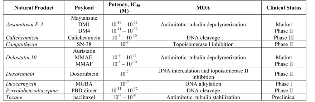
Table 1-3. Potency, linker compatibility, and clinical status of clinical ADC payloads ... 76
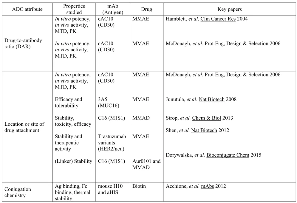
Table 1-4. Critical attributes and key papers linking ADC design to in vivo activity, safety, efficacy, and mode of action ... 80
Table 1-5. Quality attributes and analytical characterization of ADCs ... 85
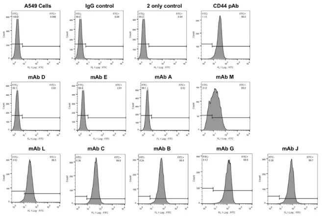
Table 3-1. CD44 mAbs screened for A549 NSCLC tumor targeting ... 113
Table 3-2. Radiochemical yield and specific activity of radioiodinated CD44 mAb ... 125
Table 3-3. Tumor-to-non-tumor tissue ratios of 125I-CD44 mAb in A549 subcutaneous xenograft tumor-bearing mice ... 136
Table 3-4. Associations between A549 tumor exposure and critical property attributes of CD44 mAb in A549 tumor-bearing mice ... 141
Table 4-1. Cell uptake behavior of 89Zr-labeled EGFR mAbs by A431 cells ... 160
Table 4-2. Compiled affinity (Kd) and specificity (IC50) of intact, DFO-conjugated, and radiolabeled EGFR mAbs and fragments EGFR-overexpressing A431 cells ... 173
Table 4-3. Affinity (Kd) of anti-EGFR mAbs, fragments, and radioimmunoconjugates with EGFR expressing human cancer cells ... 175
Table 4-4. Cellular retention and uptake of 89Zr-DFO-N by EGFR-expressing cancer cells ... 176
Table 5-1. Reaction yield of-anti-EGFR ADCs ... 209
Table 5-2. Affinity (Kd) of anti-EGFR-ADCs to EGFR-expressin g A431 cells ... 214
Table 5-3. Cytotoxicity of anti-EGFR ADCs on A431 cells, 4 h treatment ... 215
Table 5-4. Drug-loading and 14-day drug stability of anti-EGFR ADCs ... 216
Table 5-5. Cytotoxicity and the EGFR specificity ratio of anti-EGFR ADCs ... 220
LIST OF FIGURES
Figure 1-1. Schematic of drugs and drug carriers used in cancer therapy ... 14
Figure 1-2. Structure of immunoglobulin ... 16
Figure 1-3. Clinical indications of therapeutic mAbs, 2010-2015 ... 22
Figure 1-4. Humanization of therapetuic mAbs in the clinical pipeline, 2010-2015 ... 27
Figure 1-5. ADC mechanisms of antitumor activity. ... 58
Figure 3-1. CD44 expression by 4T1, MDA-MB-231 and A549 cells ... 112
Figure 3-2. FACS histograms of A549 cell-surface binding of CD44 mAb ... 114
Figure 3-3. A549 cell-surface CD44 mAb binding screen ... 115
Figure 3-4. Saturation binding of CD44 mAb B, C, G and J to CD44-positive A549 cells ... 116
Figure 3-5. Saturation and competitive equilibrium binding of CD44 mAb to A549 cells ... 117
Figure 3-6. CD44 mAb internalization by A549 cells ... 119
Figure 3-7. Cell uptake, endosomal accumulation and retention of mAb G by A549 cells ... 121
Figure 3-8. CD44 mAb 48 h-spatiotemporal endosome tracking in A549 cells ... 122
Figure 3-9. CD44 mAb 48 h-spatiotemporal lysosome tracking in A549 cells ... 123
Figure 3-10. Colocalization of CD44 mAb within the endosome and lysosome of A549 cells ... 124
Figure 3-11. PD-10 column purification of 125I-CD44 mAb B, C, G, J and X ... 125
Figure 3-12. Cell-surface binding and internalization of 125I-CD44 mAb by A549 cells ... 127
A549 tumor-bearing mice ... 129
Figure 3-15. Impact of tumor size and Lugol's iodine preconditioning on tumor uptake and biodistribution of 125I-CD44 mAb in A549 tumor bearing mice ... 130
Figure 3-16. Biodistribution of 125I-CD44 mAb following i.v. tail injection in A549 subcutaneous xenograft tumor-bearing mice ... 131
Figure 3-17. Time-course tumor exposure of 125I-CD44 mAb in A549 tumor- bearing mice ... 132
Figure 3-18. 24, 48, 72 and 120 h tumor uptake of 125I-CD44 mAb in A549 tumor-bearing mice ... 133
Figure 3-19. Tumor uptake of CD44 mAb in A549 tumor-bearing mice by IHC ... 134
Figure 3-20. Tumor-to-blood and tumor-to-muscle ratios of 125I-CD44 mAb ... 137
Figure 3-21. CD44 mediates tumor uptake in A549-tumor bearing mice ... 138
Figure 3-22. Quantification of percent free iodine-125 by TCA precipitation following i.v. tail vein injection of 125I-labeled CD44 mAb to A549 tumor-bearing mice. ... 139
Figure 3-23. PK and selected tissue uptake of 125I-CD44 mAb in A549 tumor- bearing mice ... 140
Figure 4-1. Saturation binding of EGFR mAbs and DFO-conjugates to A431 cells ... 157
Figure 4-2. Saturation binding of 89Zr-labeled EGFR mAbs to A431 cells ... 158
Figure 4-3. Competitive equilibrium binding of 89Zr-labeled EGFR mAbs to A431 cells ... 159
Figure 4-4. Fraction unbound, surface-bound, and internalized of 89Zr-labeled EGFR mAbs following 8 h exposure to A431 cells ... 160
Figure 4-5. Saturation binding of 89Zr-DFO-N by EGFR expressing human cancer cells ... 161
Figure 4-6. Competitive equilibrium binding of 89Zr-DFO-N with native N to EGFR- expressing human cancer cells ... 162
Figure 4-7. Cell uptake behavior of 89Zr-DFO-N in EGFR-expressing human cancer lines ... 163
Figure 4-9. Saturation binding of 64Cu-NOTA-C Fab to EGFR expressing
human cancers. ... 166
Figure 4-10. Competitive equilibrium binding of 64Cu-NOTA-C Fab with native C to EGFR-expressing human cancer cells ... 167
Figure 4-11. Saturation binding of panitumumab Fab’ to EGFR expressing human cancers ... 168
Figure 4-12. Saturation binding of 64Cu-NOTA-P Fab’ to EGFR expressing human cancers ... 169
Figure 4-13. Competitive equilibrium binding of 64Cu-NOTA-P Fab’ with native P to EGFR-expressing human cancer cells ... 170
Figure 5-1. Synthesis of LC-DX prodrug from the LC-SPDP heterobifunctional linker ... 190
Figure 5-2. Chromatograms of DX, LC-SPDP linker, and LC-DX conjugate by RP-HPLC ... 191
Figure 5-3. Cytotoxicity of the LC-DX prodrug, DX,and the LC-SPDP with A431 cells. ... 192
Figure 5-4. pH-sensitive hydrolysis of LC-DX. ... 193
Figure 5-5. Degradation of DX-prodrugs ... 195
Figure 5-6. Synthesis of SC-DX prodrug from the SPDP heterobifunctional linker ... 196
Figure 5-7. Reaction schemes covalently modifty anti-EGFR mAbs PAN and CET with a dipyridyl disulfide containing docetaxel prodrug ... 197
Figure 5-8. Time- and concentration-dependent thiolation of panitumumab ... 198
Figure 5-9. Thiolation of cetuximAb with NHS-PEG2000-SH ... 199
Figure 5-10. Common agents used to reduce mAb disulfide bonds ... 199
Figure 5-11. Quantification of the number of free thiols on panitumumab and cetuximab following reduction with DTT, TCEP, 2-MEA, or 2-ME ... 200
Figure 5-12. Time- and concentration-dependence on the disulfide reduction of cetuximab and panitumumab with TCEP ... 201
Figure 5-14. PEGylated mAb generated by reduction-alkylation scheme ... 202
Figure 5-15. PEGylation of cetuximab and panitumumab by molecular weight. ... 203
Figure 5-16. PEGylation of cetuximab and panitumumab analyzed by thiol content ... 203
Figure 5-17. Number of free thiols formed after reduction (black bars) of CET and PAN incubated with DTT, TCEP, or 2-MEA ... 204
Figure 5-18. Characterization of LC-DX alkylation to reduced CET and PAN ... 205
Figure 5-19. Enhancing solubility of LC-DX and SC-DX prodrugs in aqueous-organic co-solvent systems ... 206
Figure 5-20. Binding affinity of anti-EGFR mAbs following DMSO exposure ... 208
Figure 5-21. Preparation and characterization of anti-EGFR ADCs ... 210
Figure 5-22. MALDI-TOF of CET and anti-EGFR ADC C-DX ... 211
Figure 5-23. MALDI-TOF of CET and anti-EGFR ADC C-LC-DX ... 211
Figure 5-24. MALDI-TOF of PAN and anti-EGFR ADC P-DX. ... 212
Figure 5-25. MALDI-TOF of PAN and anti-EGFR ADC P-LC-DX. ... 212
Figure 5-26. Saturation binding of cetuximab DX-ADCs and pantitumumab DX-ADCs with EGFR expressing human cancer cells ... 213
Figure 5-27. Cytotoxicity of anti-EGFR antibody-docetaxel conjugates. ... 215
Figure 5-28. SE-HPLC chromatograms of P-DX, P-LC-DX, and the high DAR conjugates of C-DX and C-LC-DX ... 217
Figure 5-29. 14-day pharmacokinetic profile of anti-EGFR mAb-DX ADCs in healthy mice ... 218
Figure 5-30. Cytotoxic activity of anti-EGFR antibody-docetaxel conjugates is EGFR-dependent, whereas cytotoxic activity of docetaxel alone is not ... 219
Figure 5-31. PAN and CET ADC cytotoxicity on EGFR+++ A431 cells. ... 221
Figure 5-32. Saturation binding of anti-EGFR ADCs to EGFR-expressing human cancer cells by ELISA. ... 222
Figure 5-34. Multi-dose efficacy of P-DX and C-DX ADCs in A431 tumor-bearing mice. ... 224 Figure 5-35. 14-day pharmacokinetic profile of anti-EGFR P-DX and C-DX in
A431 tumor-bearing mice. ... 225 Figure 5-36. P-DX and C-DX efficacy in A431 tumor-bearing mice ... 232 Figure 6-1 Critical molecular properties of CD44 mAb that impact tumor targeting to
LIST OF ABBREVIATIONS
Ab Antibody
ACN Acetonitrile
ADC Antibody-drug conjugate
ADCC Antibody-dependent cellular cytotoxicity
AE Auristatin E
AML Acute myeloid leukemia ANOVA Analysis of variance
ATCC American type culture collection AUC Area under the curve
BACE1 Beta-site APP cleaving enzyme 1 biAb Bispecific antibody
bioD Biodistribution
BSB Binding site barrier
C1q Complement component 1q
Cmax Maximum concentration CAIX Carbonic anhydrase IX
CD44 Cluster of differentiation 44 (aka HCAM or Pgp-1) CDC Complement dependent cytotoxicity
CDR Complementarity determining region CEA Carcinoembryonic antigen
CET (or C) Cetuximab (aka Erbitux®)
CI Colocalization index
CH1 (IgG) constant heavy 1 domain CH2 (IgG) constant heavy 2 domain CH3 (IgG) constant heavy 3 domain CL (IgG) constant light domain cGLP Current good laboratory practice cGMP Current good manufacturing practice CMC Chemistry, manufacturing, and control
CPM Counts per minute
CRS Cytokine release syndrome
CSC Cancer stem cell
CTLA4 Cytotoxic T-lymphocyte antigen 4
Da Dalton
DAR Drug-to-antibody ratio
DCM Dichloromethane
DFO Desferoxiamine
DLL3 Delta-like 3 protein (receptor) DMAP 4-dimethylaminopyridine
DMEM Dulbecco’s modified eagle medium
DMSO Dimethylsulfoxide
DNA Deoxyribonucleic acid
DOX Doxorubicin
DX Docetaxel
EDTA Ethylenediaminetetraacetic acid EEA1 Early endosome-associated protein 1 EGF Epidermal growth factor
EGFR Epidermal growth factor receptor ELISA Enzyme-linked immunosorbent assay EpCAM Epithelial cell adhesion molecule
EPR Enhanced permeability (permeation) and retention effect
f Fractional occupancy
FACS Fluorescence-activated cell sorting Fab Fragment, antigen binding, monovalent
Fab’ Fragment, antigen binding, monovalent, with hinge region F(ab’)2 Fragment, antibody binding, bivalent
Fc Fragment, constant (aka fragment, crystallizable) FcRn Neonatal Fc receptor (aka Brambell receptor)
Fc!R Fc-gamma receptor
FBS Fetal bovine serum
FDA Food and Drug Administration FGF Fibroblast growth factor
FGFR4 Fibroblast growth factor receptor 4 FITC Fluorescin isothiocyanate
GSH Glutathione
HA Hyaluronic acid
HC (IgG) heavy chain
HER1 Human epidermal growth factor receptor 1 (see EGFR) HER2 Human epidermal growth factor receptor 2
HER2/neu Human epidermal growth factor receptor 2 (murine species) HNSCC Head and neck squamous cell carcinoma
HPLC High pressure liquid chromatography IC50 Inhibitory concentration, 50%
Ig Immunoglobulin
IgG Immunoglobulin gamma
IHC Immunohistochemistry
IF Immunofluorescence
immunoPET Antibody-based positron emission tomography
IP Immunoprecipitation
IT Immunotoxins
ITAM Immunoreceptor tyrosine-based activation motif ITIM Immunoreceptor tyrosine-based inhibitor motif kint Kinetic cell internalization rate
Kd Equilibrium dissociation constant
kDa Kilodalton
L Ligand
LAMP-1 lysosome-associated membrane protein 1
LC-DX Long-chain docetaxel prodrug
LC-SPDP Long-chain SPDP heterobifunctional linker
LeY Lewis Y antigen
m/z Mass per charge
mAb Monoclonal antibody
MAC Membrane attack complex
MALDI-TOF Matrix-assisted laser desorption/ionization-time of flight (MS) MBC Metastatic breast cancer
mCRC Metastatic colorectal cancer mTG Microbial transglutaminase
MED Minimum effector dose
MMAE Monomethyl auristatin D
MMAE Monomethyl auristatin E
MMAF Monomethyl auristatin F MPS Mononuclear phagocyte system MRP1 Multidrug resistance protein 1
MS Mass spectroscopy
MTD Maximum tolerated dose
NAC N-acetyl cysteine
NEM N-ethyl maleimide
NHS N-hydroxysuccinimide
NIMO (or N) Nimotuzumab
NOTA 1,4,7-triazacyclononane-triacetic acid
NP Nanoparticle
NSCLC Non-small cell lung cancer
O/N Overnight
pAb Polyclonal antibody
pAcPhe Para-acetylphenylalanine
PABC p-aminobenzyloxycarbonyl (spacer) PAN (or P) Panitumumab (aka Vectibix®)
PAN-DX (or P-DX) Panitumumab-docetaxel antibody-drug conjugate PBD Pyrrolobenzodiazepine (dimer)
PBS Phosphate buffered saline
PD Pharmacodynamics
PDX Patient-derived xenograft PD-1 Programmed death receptor 1 PEG Poly(ethylene)glycol
PET Positron emission tomography
PET/CT Positron emission tomography-computed tomography
PFA Paraformaldehyde
PgP P-glycoprotein (aka MDR1)
PK Pharmacokinetics
PK/PD Pharmacokinetic/pharmacodynamic
PS Penicillin-streptomycin
PS80 Polysorbate 80
R Receptor
RANKL Receptor activator of nuclear factor kappa-B ligand RES Reticuloendothelial system
RIC Radioimmunoconjugate
RL Receptor-ligand complex
RME Receptor-mediated endocytosis
RNA Ribonucleic acid
RP-HPLC Reverse-phase HPLC
RT Room temperature
RTK Receptor tyrosine kinase scFv Short-chain variable fragment SC-DX Short-chain docetaxel prodrug
SDS-PAGE Sodiumdodecylsulfide-poylacrylamide gel electrophoresis
Sec Selenocysteine
SE-HPLC Size exclusion HPLC
SMCC Succinimidyl 4-(N-maleimidomethyl)cyclohexane-1-carboxylate
SOC Standard of care
SPDP Succinimidyl 3-(2-pyridyldithio)propionate) t1/2 (Time to one) Half-life
T-DM1 ado-trastuzumab emtansine TAA Tumor-associated antigen
TCA Trichloroacetic acid (precipitation) TCEP tris(2-carboxyethyl)phosphine
TI Therapeutic index
TIC Tumor-initiating cell
TIL Tumor-infiltrating lymphocyte TKI Tyrosine kinase inhibitor TLC Thin-layer chromatography TMDD Target-mediated drug disposition TNF Tumor necrosis factor
TNF" Tumor necrosis factor-alpha TNF-R2 Tumor necrosis factor receptor 2 TRAIL TNF-related apoptosis inducing ligand VEGF Vascular endothelial growth factor
VEGFR Vascular endothelial growth factor receptor
VH (IgG) variable heavy domain
VL (IgG) variable light domain
WB Western blot
2-ME 2-mercaptoethanol
2-MEA 2-mercaptoethylamine
CHAPTER 1: MONOCLONAL ANTIBODY AND ANTIBODY-DRUG CONJUGATES IN TUMOR TARGETING AND CANCER THERAPY
“We have to learn how to aim chemically,” Paul Ehrlich once said [1]. He was speaking about the need to find or design molecules capable of targeting specific cells, cellular receptors, or pathogens infiltrating the human body. Ehrlich came to this conclusion after years of studying how colorful dyes interact and localize within specific histological and cellular structures. Among many preeminent discoveries, Ehrlich demonstrated for the very first time that the biological effect of a chemical compound depended on its chemical composition and the cell on which it acts. Thus, Ehrlich elucidated an inextricable connection between chemistry and biology that is the basis for modern drug discovery, design, and delivery.
After shifting his focus to plant toxins, the development of high titer anti-diphtheria sera, and standardizing sera for determining antibody concentration, Ehrlich began musing about specific interactions that occur between toxins and anti-toxins. He postulated the existence of receptors, first termed “side chains,” capable of binding toxins. Ehrlich thought receptors associated with the cell or distributed broadly in the blood stream in response to an antigen. He also believed various types of receptors exist, each compositionally and structurally unique with different binding groups that contribute to the nature, strength, and specificity of the interaction. Over time, Ehrlich’s receptor theory shifted from binding of toxins to binding – and later,
Combining his studies visualizing the differential affinity of chemical dyes with specific biological structures and developing of a high titer anti-diphtheria serum, Ehrlich postulated that molecules exist or could be generated to bind specifically to a pathogen. In a theory presented first in 1908, he called these molecules “magic bullets” and described their ability to home to a specific cell structural target. Ehrlich believed “magic bullets” existing innately or synthesized chemically could efficaciously attack pathogens yet remain harmless to the healthy tissues of the host. To test his “magic bullet” postulated, Ehrlich chemical altered and screened a library of compounds he hypothesized could treat syphilis spirochete. In 1908, the same year he received the Nobel Prize for Physiology or Medicine, Ehrlich’s laboratory detected the anti-syphilitic activity of arsphenamine. His methodological approach to optimize the biological activity of a chemical compound through systematic chemical modification was the first of its kind in drug discovery.
Ehrlich also imagined his molecular “magic bullet” theory might apply to tumor cells and provided some of the earliest hypotheses on the treatment of cancer. He was the first to suggest, boldly at the time, that small molecules might be used in the management of cancer. The
suggestion that the immune system can prevent tumors was perhaps the most controversial and yet least scientifically established of the theories Ehrlich promoted. Naturally, this too turned out to be true. With many of his controversial postulates resoundingly well established in current research, it should be no surprise that Paul Ehrlich is affectionately known as the founder of chemotherapy.
However, it was those who followed that succeeded in identifying receptors overexpressed preferentially on the surface of tumor cells. In 1975, nearly seven decades after Ehrlich won the Nobel Prize, Kohler and Milstein developed hybridoma technology for the production of
monoclonal antibody (mAb) [2], and Ehrlich’s 1908 theory of molecular “magic bullets” came to fruition. The advent of hybridoma technology enabled the generation of immune-like molecules that bind with high affinity and exquisite specificity to any pharmacological target of interest, including pathogens, cell receptors, tumor cells, small molecule toxins, and more.
Kohler and Milstein’s hybridoma technology and generation of a monoclonal antibody is perhaps the greatest discovery in the world of pharmaceutical research ever. It has provided a set of research grade reagents that make up an estimated $1.6B worldwide market annually, $700M accounted for in the US alone [3], and nearly $30B market of therapeutic medicines in the US alone. In fact, mAbs have become the leading biotechnology pursued for medical applications spanning therapy, drug delivery, and prophylactic vaccines. To date, more than 30 mAbs have been approved by the FDA for therapeutic indications in immunology, cancer, cardiovascular disease, inflammation, and allergy.
Cancer, treatment, and drug delivery
Cancer is characterized by a rapid, uninhibited growth of malignant cells capable of infiltrating tissues and starving essential organs of the necessary nutrients to survive.
Oncologists today have a number of tools to help a patient overcome the onslaught of cancer. The three most common approaches are surgery, radiation, and chemotherapy.
According the American Cancer Society, surgery is used to prevent, diagnose, stage, and treat cancer. Curative surgery aims to completely remove the tumor mass from the body and results in the greatest effect when the tumor is localized (or isolated) to only one part of the body. Chemotherapy and/or radiation complement curative surgery in a standard treatment plan. Other surgical approaches include palliative surgery, which focuses on treating cancer-associated pain; debulking or supportive surgery, which removes some of the tumor tissue or alters the tissue to be more susceptible to other forms of treatment; and preventative (or prophylactic) surgery, an elected procedure to remove a tissue with high likelihood to become cancer. Surgery accounts for the vast majority of first line cancer treatment.
Radiation therapy utilizes high energy particles or waves to destroy or damage cancer. Radiation therapy comes in a variety of different forms and has been used in cancer treatment for many decades with success, as it offers access to precious or hard-to-reach tissues often
long periods of time. Brachytherapy has proven an effective treatment for cervical, prostate, breast, and skin cancer. Unsealed source radiotherapy, or the administration of a radioactive isotope by injection or ingestion, can result in tumor growth inhibition or regression following exposure of the tumor to the radioactive agent. Iodine-131 accumulates in the thyroid and works by beta radiation to destroy thyroid tissue and any thyroid cancer that takes up the radioactive iodine. Other common unsealed source radiotherapy agents include yttrium-90, radium-223, strontium-89, and sarmarium-153. Like other treatment options, radiation is often used in
combination with surgery and/or chemotherapy to elicit complete response and eradication of the tumor.
Advances in cancer treatment
Dr. Siddartha Mukherjee, author of New York Times best-selling book “The Emperor of all Maladies: A Biography of Cancer,” elegantly elucidates the impossible characterization of the archetypal cancer patient and archetypal cancer treatment. Instead, he lauds the deeper
molecular understanding of cancer and its inherent diversity – there is no single “cancer” but many diseases that are diverse, complex, and intermingled – and reveals a genuine anticipation that such understanding will improve cancer treatment and prevention. Needless to say, cancer treatment has not significantly changed in the past century. While surgery, radiation, and chemotherapy effectively eradicate cancer in some individuals, each approach has shortcomings that limit application, utility, and, ultimately, success in treating the cancer. It is estimated on average that more than 99.9% of cells in the tumor must be killed to achieve remission in the patient.
As the biological understanding of cancer pathogenesis and cancer treatment evasion matures, two emerging approaches represent the cutting-edge in cancer therapy today. The first harnesses the power of the innate immune system to fight cancer at the cellular level, while the second targets specific phenotypes, pharmacological pathways, and metabolic pathways found upregulated and otherwise aberrant in cancer to fight the disease at the gene or protein level.
advanced, unresectable, or metastatic diseases for which no other therapy has proven effective. For example, recent FDA approvals of programmed death receptor-1 (PD-1) immune checkpoint inhibitors nivolumab (Opdivo®) and pembrolizumab (Keytruda®) join the cytotoxic
T-lymphocyte associated protein 4 (CTLA-4) inhibitor ipilimumab (Yervoy®) as effective immune modulating molecules used in anticancer regimens. Moreover, a host of advanced adoptive T-cell therapies designed to train the innate immune T-cells to become potent antitumor assassins are under current investigation in small clinical trials. However, the application of adoptive T-cell therapy has proven challenging. For example, on July 7, 2016 Juno Therapeutics halted the ROCKET phase 2 clinical trial of JCAR015 (NCT01044069) following two patient deaths. JCAR015 is a molecularly engineered chimeric antigen receptor (CAR) T-cell and arguably the most advanced IO technology in the clinic today.
The second major advance in cancer treatment relies on phenotypically or
Drug delivery: exploiting tumor pathophysiology and phenotypic changes on cancer cells Chemotherapy can provide enormous benefits to patients with both solid and hematological cancers in early stage, late stage, and those with substantial, unlocalized
metastases. However, significant drawbacks in safety and toxicity limit the efficacy and utility of chemotherapy in many patients across a spectrum of cancer indications. Specifically, limited accessibility of chemotherapy to the tumor tissues, the requirement of high dose, intolerable cytotoxicity, multiple drug resistance, and non-target tissue uptake impair the clinical use of chemotherapeutics in cancer therapy [4]. Addressing these shortcomings of standard chemotherapy treatment is the major charge of modern drug delivery.
The ability to efficiently deliver a drug to a tumor site is dependent on a wide range of physiologically imposed design constraints, as well as constraints dictated by the nature, size, location, stage, and phenotype of the tumor. Drug delivery attempts to overcome, and in many instances exploit, these constraints. The two main approaches to cancer drug delivery are summarized as passive targeting, using carrier systems to deliver drugs to cancerous tissue, or active targeting, exploiting genetic or phenotypic features of the cancer cell that differentiate it from other healthy cells of the body. Generally, passive targeting relies on the pathophysiology of the tumor and the tumor microenvironment to improve tumor-specific drug delivery, whereas active targeting takes advantage high affinity, high specificity interactions unique to the targeting ligand and the tumor cell or cancerous tissue.
systems that exploit the changes – the anatomical, metabolic, and phenotypic differences between normal tissues and tumor tissue.
Tumor vasculature in particular differs from healthy, normal tissue vasculature. Tumors exhibit high vascular density, and the vessels are larger in size, more permeable, and more leaky than the tight endothelium of normal blood vessels. This vascular architecture results from elevated levels of vascular mediators, like the vascular endothelial growth factor (VEGF), fibroblast growth factor (FGF), and prostaglandins, as well as the physical stress and consumptive energy needs of the rapidly proliferating cells within the tumor.
The enhanced permeability and retention (EPR) effect is a phenomenon characterized by the impact of rapidly growing cancer cells on the junctions between epithelial cells of the local capillaries in concert with the impaired lymphatic drainage of macromolecules in solid tumors. In effect, when a solid tumor grows, it puts undue pressure on the nearby blood vessels. That pressure stretches the local vessel, thinning out the epithelial cell wall and creating gaps between the epithelial cells. These gaps result in a “leaky vessel.” Depending on the size of tumor and architecture of the nearby vessel, “leaky vessels” allow entities as large as 1 µm to extravasate from the blood vessel and enter, and subsequently interact with, the extra-vascular tissue. Solid tumors also exhibit poor lymphatic vascular density and slow draining rates, which can result in prolonged retention of high molecular weight drugs and drug carriers that accumulate in solid tumors. The EPR effect has been extensively characterized in preclinical tumor models and is a primary driver of tumor uptake and retention of passive targeting drug carrier systems.
Tumor tissue also greatly differs from healthy, normal tissues. Solid tumors are
including the growth rate, metastatic ability, and sensitivity to anticancer therapy, and together form a complex and dynamic environment of tumor vasculature, stroma, and parenchyma, each with unique features, growth needs, and contributions to the tumor microenvironment.
Due to this dynamic and highly complex microenvironment within the tumor, tumors contain specific antigens coined tumor-associated antigens (TAAs) that are often found expressed on the surface of cancer cells. Most function to support the rapid growth, highly proliferative cells that constitute the tumor mass. TAAs vary in expression and function, but most are either selectively expressed or preferentially overexpressed at the tumor compared to normal, healthy tissues throughout the body. While most TAAs function endogenously as growth-promoting cell surface receptors on the membrane of cancer cells, more recently TAAs have been identified on tumor-associated stroma and progenitor cancer cells called cancer stem cells (CSCs, also known as tumor initiating cells, or TICs). Expression of these unique antigens within the tumor microenvironment and receptors on the surface of cancer cells provide the rationale for active tumor targeting, which takes advantage of the difference in the expression level of a TAA on the cancer cell (or within the tumor tissue) to preferentially accumulate at the tumor compared to healthy, normal tissues.
Passive targeting: nanotechnology and the EPR effect
Using nanotechnology to improve tumor targeting and therapeutic drug delivery is the most pursued approach in preclinical cancer treatment today. Nanotechnology in cancer therapy refers to small particles, polymers, nanotubes, or other carriers that can encapsulate, bind, or complex a drug or drugs. While the nanocarrier is primarily designed to increase drug
metabolism and degradation [5]. Advances in material science and basic studies of the colloidal properties of various materials and mixtures of materials have produced in an enormous diversity and versatility of nanocarrier systems for use in drug delivery, each designed and suited for a specific application.
Nanocarriers improve tumor uptake by taking advantage of the pathophysiology of the tumor. The EPR effect, in particular, results in high accumulation of molecules small enough to permeate the “leaky” tumor vessel, yet large enough to avoid rapid clearance by the draining lymphatic system. Despite these advantages, nanocarriers must overcome serious drawbacks that limit its utility in tumor targeting and drug delivery. Among the most commonly faced hurdles – which are generally described here, but many more hinder specific drug carrier systems – low drug encapsulation, poor control over drug release, limited tumor uptake, unacceptably high liver uptake, and rapid clearance by the mononuclear phagocyte system (MPS, a revised nomenclature for the reticuloendothelial system, RES) significantly impair the wide-spread pursuit of clinical grade nanotechnology-based medicines for cancer treatment.
A number of nanoparticle-based drugs have reached clinical development, though few have been FDA approved. Doxil® was the first FDA-approved nanocarrier (1995), which is a liposomal formulation of doxorubicin. Abraxane®, a formulation of containing albumin-bound paclitaxel, was approved in its first indication 2005. Maqibo®, DaunoXome®, and Myocet® are other well-known FDA-approved medicines indicated in cancer.
Active targeting: tumor-associated antigens and tumor-targeting ligands
accumulation of the medicine at the tumor. Importantly, this approach spares normal cells and healthy tissues from significant exposure to the medicine, thereby reducing the toxicity of anti-cancer drugs to a patient. The combined effect of increasing target tissue uptake while lowering exposure to healthy tissue results in a significantly improved therapeutic window.
In general, phenotypic differences between cancer cells and the cancer microenvironment offer a host of targets to increase the accumulation of anti-cancer drugs to the tumor, as well as target drugs to the tumor stroma and pro-growth tumor factors at high concentration in the tumor microenvironment. Active tumor targeting relies on high-affinity, high-specificity interactions between the TAA and the tumor targeting ligand. TAAs exist primarily as cell-surface receptors found selectively expressed or overexpressed on the tumor cell surface. Increasingly, soluble growth factors and receptors found on the tumor extracellular matrix and neovasculature of the tumor serve as effective TAAs. Commonly pursued tumor targeting ligands include endogenous receptor ligands, recombinant proteins, RNA constructs, DNA aptamers, mAbs and various engineered mAb fragments. Selection of the ligand for tumor targeting should take into account a variety of parameters, including amenability to chemical modification, drug loading,
Figure 1-1. Schematic of drugs and drug carriers used in cancer therapy. A tumor cell (blue filled circle) can be treated by a toxic chemotherapeutic drug (red star) or a pharmacological agent (green arrow). Pharmacological agents bind specific receptors on the cell surface (purple V) to induce antitumor activity. Nanocarriers (black circle) can improve safety and efficacy and antitumor chemotherapeutics, particularly when targeted with an antigen-specific ligand (yellow arrow) to a tumor-associated antigen (black V).
Immunoglobulin and the monoclonal antibody
Immunoglobulin (Ig, or antibody, Ab) are large, complex glycoproteins secreted by B-cells in response to a foreign antigen. Ig survey nearly every organ and tissue of the human body, including blood and mucus secretions, and serve to protect the body from invasion and propagation of foreign pathogens. Antibodies recognize foreign antigens by binding to the antigenic epitope, a short amino acid structural sequence, and marking the foreign body for removal. Once bound, the antibody recruits other immune cells to act on the foreign body. Thus, antibodies act specifically and efficiently to facilitate the removal of a foreign pathogen invading healthy tissues of the body. Antibodies, particularly immunoglobulin gamma (IgG) and secretory immunoglobulin alpha (sIgA), also prevent pathogens from gaining access to healthy tissues or inhibit viruses from cell- or tissue-specific infiltration and access to intracellular machinery for replication and propagation within the host organism.
Specific functional characteristics of Ig facilitate its ability to fight foreign viruses,
bacteria, other pathogens. For example, vaccines work on the principle of priming the body for a rapid and robust antibody response when the body is challenged by a foreign pathogen. It is these unique properties – and the ability to manipulate, modulate, and otherwise engineer improved properties – that make Ig attractive molecules for applications in cancer therapy and drug delivery.
Elements of immunoglobulin G structure and function
LC-HC-HC-LC homodimer. The Y-shaped four subunit protein of approximately 150 kDa is the characteristic base structure of Igs.
Figure 1-2. Immunoglobulin, a homodimer of heterodimers, is composed of two identical heavy chains (grey), two identical light chains (orange), and an extensive network of inter- and intrachain covalent yet reducible disulfide bonds (black).
In nature, the basic Y-shaped Ig structure can exist in various orientations and
stoichiometry greater than one [8]. Igs of different classes serve specific functions and localize to specific tissues and fluids. For example, sIgA antibody is an Ig dimer primarily found in mucosal secretions, whereas IgM antibody is an Ig pentamer found in the blood and works as the first antibody defense to infection. Five Ig classes – IgG, IgA, IgM, IgD, and IgE, distinguished by their constant regions – exist in humans, each with unique structural and functional attributes.
This dissertation will focus exclusively on the immunoglobulin G (IgG) class,
IgG quaternary, tertiary, secondary, and primary structure
The macroscopic structure of IgG antibody is a dimer of dimers. Heavy chain (HC) and light chain (LC) form a heterodimer known as “half IgG” or “reduced IgG” of approximately 75,000 Da in molecular weight. A homodimer of two HC-LC heterodimers forms full-length, intact IgG of approximately 150,000 Da in molecular weight. The full IgG molecule contains, on average, approximately 25,000 atoms – four polypeptides totaling 1400-1600 amino acid residues (another word for amino acid). Each HC-LC heterodimer is held together through a variety of non-covalent intermolecular forces, such as salt bridges, hydrogen bonding, and hydrophobic interactions, as well as an extensive network of covalent yet reducible interchain disulfide bonds. For example, the two heavy chains of each HC-LC heterodimer are linked to each other by disulfide bonds, and each HC is linked to a LC by a single disulfide bond. Intrachain disulfide bonds further aid the structural integrity within each heavy and light chain domain. Moreover, each domain within the HC (4 domains per HC) and LC (2 domains per LC) contains one intramolecular disulfide bond and two stacked antiparallel beta sheets. The beta sheet found in each domain of an IgG antibody typically contains 4 antiparallel sheets stacked with 3 antiparallel sheets. On the whole, IgG antibody are 40 – 70% beta sheet and 5 – 10% alpha helix.
The HC of an IgG antibody is approximately 50 kDa, or 450 amino acid residues, and consists of four distinguishable regions, listed from the carboxy (COO-) terminus to the amino (NH3+) terminus: the constant heavy 3 domain (CH3), constant heavy 2 domain (CH2), constant heavy 1 domain (CH1), and variable heavy domain (VH). The LC of an IgG antibody is
domain (VL) located at the amino terminus (N-term). As the names suggest, the variable (V) domains differ them from the constant (C) domains of the IgG heavy and light polypeptide chains by distinguishable differences in amino acid composition.
The HC and LC polypeptides differ in both structure and function. For example, the class and effector function of an antibody is defined by the structure of the heavy chain. The majority of interchain disulfide bonds reside in the IgG hinge region, a neck-like portion of the IgG molecule between the CH2 and CH1 domains of the HC. Five types of HC exist, which define the various isotypes of Ig (IgA, IgG, IgD, IgE, and IgM), and two types of LC exist, lambda and kappa. IgG antibody HCs exhibit various glycosylation patterns, which are mostly restricted to the CH2 domain. Glycosylation is thought to improve aqueous solubility, enhance effector functions, especially the interaction and activation of the complement system, and improve serum half-life, but on the whole their function remains poorly understood.
IgG Fab and Fc fragments
IgG antibody can be further broken down and characterized by two main functional elements: the Fc fragment, “c” for constant or crystallizable, and the Fab fragment, “ab” for antigen-binding. Each fragment (“F”) is characterized by a function, which together contribute to the protective activity of endogenous IgG in the body as well as the utility of IgG in cancer treatment and drug delivery. IgG Fab and Fc fragments are commonly produced in cell culture using recombinant DNA technology, but can also be produced by post-translational digestion with a variety of peptidases, including pepsin, papain, ficin, trypsin, or IdeS, and/or full or partial reduction using common reducing agents like dithiothreitol (DTT),
Pepsin and papain are the two most common IgG digestive enzymes. Pepsin
preferentially cleaves the N-term side of hydrophobic amino acids, especially aromatic amino acids like phenylalanine, tryptophan, and tyrosine. These residues commonly reside in the lower hinge region of the IgG HC. Therefore, treatment of IgG antibody with pepsin results in
production of a divalent Fab dimer, connected via retained disulfide bonds within the HC hinge region. This divalent molecule is coined (Fab’)2 and can be further reduced to monovalent Fab’ fragments by treatment with a mild reducing agent. Papain, on the other hand, is a cysteine protease that generally cleaves IgG antibody above the hinge region. Thus, treatment of IgG antibody with papain results in the production of two monovalent Fab fragments along with the intact, unmodified Fc fragment, which retains its hinge disulfide structure.
The Fab fragment, which contains both domains of the LC along with the VH and CH1 domain of the HC, is the portion of IgG responsible for binding to the antigen. Critical to the Fab fragment are six short loops making-up the complementarity determining region (CDR). In total, the 6 CDR loops, of which 3 reside within the VL chain and 3 reside with the VH chain, form the binding pocket that characterizes the affinity and specificity between IgG antibody and antigen. The CDR loops – often described as 3 fingers from two separate hands – are also known as the IgG hypervariable regions, because these six short peptide segments demonstrate the greatest diversity in amino acid sequence, length, and flexibility of any other portion of the IgG molecule.
(Fc!R) and initiate signaling through immunoreceptor tyrosine-based activation motifs (ITAMs) or immunoreceptor tyrosine-based inhibitory motifs (ITIMs) during an antibody-dependent cellular cytotoxicity (ADCC) response. The Fc can also recruit complement component 1q (C1q) to initiate the complement cascade, resulting in tumor cell lysis by the membrane attack complex (MAC).
In addition to engaging the receptors on immune cells, the Fc also interacts with the Fc neonatal receptor (FcRn) found on the surface of epithelial cells in tissues throughout the body. Interaction between the Fc and the FcRn maintains high serum levels of IgG via endothelial cell recycling or, in select organs, mediates the transcytosis of IgG across epithelial barriers [9]. FcRn-mediated recycling occurs due to the pH-dependence of the IgG Fc-FcRn interaction, which is high affinity at acidic pH but low affinity at neutral pH. This phenomenon enables IgG to be bound to FcRn within the early endosome (pH < 6) yet dissociate at the cell surface when exposed to more alkaline conditions (pH 7) [10]. Moreover, high-affinity binding to the FcRn protects the mAb from intracellular catabolism [11]. FcRn expression also significantly impacts mAb tissue distribution [12]. Like with endogenous IgG, the Fc-FcRn interaction enable mAb for cancer therapy and drug delivery to circulate in the blood stream up to 3 weeks or longer, which offers a huge advantage to other platforms evaluated for applications in drug delivery.
Monoclonal antibody
Monoclonal antibody (mAb) is a special type of IgG. Most IgG in humans is polyclonal (pAb), meaning it recognizes multiple epitopes on any one antigen. mAbs, conversely,
Antigen binding affinity and specificity of mAb separate it from most all other biochemical interactions observed in nature. Moreover, the production of hybridomas and application of recombinant DNA technology allow for the precise engineering of mAb that not only bind a specific epitope on an antigen, but whose pharmaceutical, physicochemical, and biological properties can be precisely tuned. The range of engineering, modification, and manipulation afforded by mAbs –from the modulation of FcRn affinity to the removal of
hydrophobic residues in place of hydrophilic ones or incorporation of non-canonical amino acids that allow bioorthogonal chemistry within living cells – have established mAbs as leading molecules in medicine, drug delivery, and assay reagents. Protein engineering for improved properties of mAb in drug delivery will be discussed in a future section.
mAbs as therapeutics
mAbs have and continue to be one of the fastest growing classes of pharmaceutical products today. In 1986, Orthoclone OKT3® became the first FDA-approved mAb for sale in the US. In 2007, a total of 26 FDA approved therapeutic mAbs were valued at more than $13.B [13] and spanned indications in cancers and immunological disease. By 2015, there were 47 FDA-approved therapeutic mAbs with a total market value of nearly $40B [14] and increased disease targets of cardiovascular disease, pulmonary disease, neurodegeneration, and pain treatment. It is estimated that mAb therapeutics will enter the market at a rate of four per year, reaching a total of 70 therapeutic mAbs by 2020 with an estimated market value above $125B [15].
FDA-approval [16]. Regardless of these considerable resource investments, mAbs were 5 of the top 10 drugs sold in 2014 and the pipeline continues to diversify and expand.
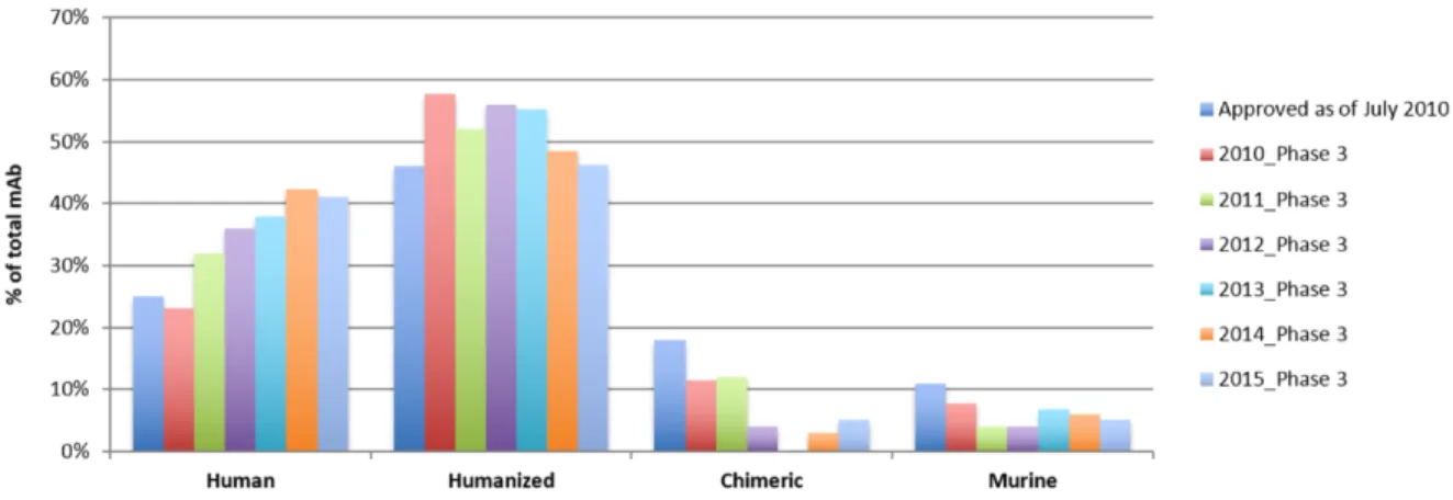
Data compiled from Janice M. Reichert’s annual “Antibodies to watch” series shows mAbs reaching phase 3 clinical trials between 2010 and 2015 (Figure 1-3) [17-23]. As of November 2015, 53 novel antibody therapeutics were in Phase 3 clinical development (data not included). This number represents a 36% and 104% increase compared to the number of
antibody therapeutics in Phase 3 in 2014 and 2009, respectively [24]. Non-traditional indications and immunological indications are the fastest growing molecules as of late, but the 17 of 53 antibody therapeutics in Phase 3 clinical study represent a significant uptick in anticancer antibody therapeutics from 2015.
Figure 1-3. Clinical indications of the 28 FDA-approved mAb-based therapeutics in July 2010, and mAbs reaching Phase 3 between 2010 and 2015 in neoplastic, immunomodulatory, anti-infective and non-traditional indications between 2010 and 2015. [17-23, 25]
present differently. mAbs offer a unique pharmaceutical platform that can feature specific yet diverse pharmacological properties.
Mechanism of action of mAbs in cancer therapy
Anticancer mAbs operate through four main mechanisms to abate tumors and quell the pro-growth, anti-regulatory environment of cancer in the human body [26]. Some mAbs function by (1) binding cancer-associated receptors and ligands expressed at high concentration on tumors and within the tumor microenvironment to antagonize pro-growth or pro-angiogenic pathways. Other mAbs (2) agonize receptors on cancer cells to activate pro-apoptotic pathways within the cancer cell, or disrupt signaling between extracellular environmental stimuli and intracellular anti-apoptotic pathways. Like endogenous mAbs, many therapeutic mAbs (3) are capable of binding to a cancer-associated antigenic epitope found selectively expressed or overexpressed on the cancer cell surface, coat that cell surface, and induce immune-mediated responses of
antibody-directed cellular cytotoxicity (ADCC) or (4) complement-dependent cytotoxicity (CDC). Known as effector functions, or immune-mediated mAb response to cancer, ADCC and CDC occur primarily for the IgG1 isotype mAb. Inducing these effector functions is the only direct cell death pathway of therapeutic mAbs.
Kd) of 390 pM. Panitumumab (Vectibix®) is an IgG2 istoype anti-EGFR human mAb. It was approved in 2006 and antagonizes EGFR with Kd of 50 pM. Bevacizumab (Avastin®) is an IgG1 isotype anti-VEGF humanized mAb approved in 2004. Unlike panitumumab and
cetuximab, which antagonize a cell surface receptor, bevacizumab antagonizes the ligand of the cell surface vascular epithelial growth factor receptor (VEGFR). VEGF is a secreted protein and therefore found extracellular, soluble and freely circulating in the bloodstream and tumor
microenvironment. Bevacizumab binds VEGF with a Kd of 1.1 nM, thereby inhibiting agonism and subsequent activation of VEGFR.
Table 1-1. MAbs currently indicated for treatment of metastatic colorectal cancer
Bevacizumab
Avastin® Cetuximab Erbitux® Panitumumab Vectibix® Dose
5-10 mg/kg every 2 weeks; 7.5-15 mg/kg
every 3 weeks
400 mg/m2 loading dose, 250 mg/m2 weekly thereafter
6 mg/kg every biweekly
Cost $814.42 / 100 mg $629.88 / 100 mg $1144 / 100 mg
FDA approval date 2004 2004 2006
Target VEGF EGFR EGFR
Kd (pM) 1100 390 50
mAb class Humanized Chimeric Fully human
Isotype IgG1 IgG1 IgG2
cancer cells. These immune-mediated tumor cell killing mechanisms might improve anti-tumor activity of cetuximab compared to its higher binding anti-EGFR competitor panitumumab.
Bevacizumab is also an effective agent against metastatic CRC. Rather than antagonizing the receptor, which is expressed on the surface of a cancer cell, bevacizumab antagonizes the VEG factor ligand. Bevacizumab works not directly on the cancer cell itself but rather on the supporting vascular architecture that feeds the tumor blood, oxygen, and other nutrients required to rapidly proliferate. Ready access to its soluble and extracellular target antigen enables the unique activity observed with bevacizumab compared to the higher binding, direct action of cetuximab and panitumumab in mCRC. Other differences between the three mAbs will be discussed later, including mAb humanization, role of antigen affinity on target tissue uptake, and the impact of target antigen expression level and distribution on the safety and efficacy profile of mAbs in clinical study.
It is finally important to note that therapeutic mAbs are rarely indicated as stand-alone agents. The most notable exception is ipilimumab, which activates the immune system by binding to CTLA-4 and exhibits sufficient activity to be used as a single agent in the treatment of solid tumors. Only mAbs indicated for hematological cancers are administered as stand-alone single agents. All remaining FDA-approved mAbs for solid tumors display only modest
mAb development trends
A number of technology improvements continue to drive innovation and improve clinical success of mAb-based therapeutics. Recombinant DNA technology has revolutionized the selection, humanization, and production of antibodies, superseding hybridoma technology and allowing the design of antibody-based reagents of any specificity and for very diverse purposes [29]. These and other mAb development trends are captured and embedded in Reichert’s “Antibodies to watch” series but also discernible in the preclinical pipeline studies found in the peer-reviewed literature of mAbs. The four major trends in mAb development are (1)
humanization, (2) fragmentation, PEGylation, or other post-translational modification, (3) amino acid-level engineering, and (4) organ- and cell-specific drug targeting and delivery.
Humanization
Early mAb-based therapeutics were generated in mice following administration of tumor-associated antigen immunogen. While mAbs are generally well tolerated in humans,
hypersensitivity and immunogenicity reactions plague murine-derived mAb therapeutics, which significantly limit their safety and pharmacological activity, and remain a notable challenge for all mAb-based therapeutics. For example, muromonab-CD3 (Orthoclone OKT3®) is a mouse murine mAb against human CD3 indicated for renal allograft rejection. Upon administration, it can cause cytokine release syndrome (CRS), an acute infusion reaction, as well as cause severe influenza-like symptoms due to an immunogenic response with human anti-mouse IgG
antibodies. The development of human anti-mouse antibodies, also known as anti-drug
mAbs have been replaced with human sequences in an iterative process termed humanization. The first step generated chimeric mAbs, which were part human part mouse. The next step generated humanized mAbs, which contain fully human sequences with only retained murine sequences in the CDR regions of the VL and VH regions. Substituting murine-based amino acid sequences for human ones is a technologically challenging process, yet generally yields
molecules with improved pharmaceutical properties. Like with other mAb development trends, wider access to the technology and the realization of clinical benefits has prompted companies to focus on humanization prior to commercialization of mAb-based therapeutics. In fact, 48 of the 53 (91%) mAb-based therapeutics currently in Phase 2/3 clinical development are humanized or fully human [24], compared to only 56 of the 86 (65%) in development between 1990 and 1999. The species of the mAb is inferred from its name, for example: antibody ending in “-momab” is of murine origin, “-ximab” is chimeric (human mAb with murine variable domains), “-zumab” is humanized (human mAb with mouse CDR sequences), and “–mumab” is human (100% human IgG sequence homology).
Studies of engineered mAbs have shown that immunogenicity is not simply a matter of sequence homology with fully human antibody. In fact, alterations in particular amino acids at certain positions can also influence immunogenicity of mAb-based therapeutics.
Fragmentation, PEGylation, and other post-translational modifications
Monoclonal antibody fragments confer unique properties to molecules of interest in pharmaceutical development [30]. Not only are they smaller in size than intact IgG, fragments offer tunable molecular properties and decreased sensitivity to physical or chemical perturbation [31, 32]. Etanercept (Enbrel®) and certolizumab pegol (Cimzia®) showcase the utility of the and IgG Fc fragment and Fab fragment, respectively, in the development of mAb-based therapeutics, though many others have reached clinical trials and gained FDA approval. Certolizumab pegol also highlights a particular modification, coined PEGylation, which can endow recombinant proteins, mAbs, and mAb fragments improved pharmaceutical features like higher aqueous solubility, lower opsonization, and an extended circulating half-life [33-38]. Though IgG fragments confer many improved properties, their clinical success remains stymied by the lack of proof of concept studies and continued successes of their fully intact IgG
counterparts [39].
Etanercept is an Fc-fusion protein indicated for treatment of autoimmune diseases like rheumatoid arthritis, psoriatic arthritis, psoriasis, and ankylosing spondylitis. It works by inhibiting activity of tumor necrosis factor alpha (TNF"), a master regulator of inflammatory responses in many organ systems. Etanercept was produced using recombinant DNA
serves as decoy receptor for TNF" and showed phenotypic effects in vivo comparable to deletion of the TNF receptor [40].
Etanercept displays exceptionally improved pharmaceutical properties compared to recombinant TNF-R2. The fusion, originally developed as human TNF-R2 fused murine IgG Fc, was shown to be highly active and unusually stable for blockade of TNF in vivo [40, 41]. It also circulated much longer in the blood stream, owing to its increase in molecular weight and FcRn-mediated endothelial cell recycling. Many other Fc-fusion proteins have advanced through clinical trials since the first clinic trials and subsequent FDA approval of Etanercept. Aflibercept is an Fc-fusion protein comprised of the extracellular domains of human VEGF receptors 1 and 2 fused to the Fc portion of human IgG1. It is indicated for wet macular degeneration and more recently as a combination therapy for mCRC. It is therefore a direct competitor, in both indication and mechanism of action, of the mAb-based VEGF antagonist bevacizumab.
Another approach to TNF" therapy employs an anti-TNF" Fab fragment covalently modified with a 40-kDa branched polymer chain of poly(ethylene)glycol. Certolizumab pegol is a PEGylated anti- TNF" Fab fragment that binds with high affinity and specificity to TNF". Unlike etanercept, which serves as a TNF" receptor decoy, certolizumab pegol antagonizes soluble TNF" and prevents its binding to endogenous TNF receptors. Though different in approach, etanercept and certolizumab pegol achieve the same pharmacological effect – lowering the concentration of free, active TNF" in the bloodstream.
targeting studies, Fab fragments exhibit deep tumor penetration and often exhibit significantly improved distribution throughout a solid tumor than mAb. However, their smaller size and lack of Fc fragment result in significantly faster clearance rates than mAb due to renal filtration and abolished FcRn recycling.
efficacy of PEGylated drugs and nanoparticles, mAbs, and engineered fragments like certolizumab pegol.
Engineering for improved physical, chemical, and biological properties
Deletion or substitution of amino acids along the polypeptide chain enable modification of the mAb at the most fundamental level. Such changes are used to engineer mAbs with improved physical stability, added chemical handles for functionalization or biorthogonal conjugation, and enhancing biological properties.
One of the most common applications of protein engineering to mAbs is the removal or substitution of amino acids for improved physical or chemical stability in vivo [43, 44]. mAbs contain 2 to 8 aggregation-prone motifs per heavy and light chain pair, usually comprised of a string of solvent-exposed hydrophobic amino acids [45]. “Hot spots,” as their known, can induce aggregation during storage, especially when formulated at high concentration, or result in opsonization, degradation, and rapid clearance upon administration in vivo [46, 47]. Regions prone to physical or chemical instability also occur at pockets of charge-charge repulsion within the mAb secondary or tertiary structure. Removing “hot spots” from the amino acid sequence can improve the structural stability of the molecule [44]. Often, single point mutations within a hot spots can improve solubility, lower opsonization, and improve the overall PK profile of the mAb [43]. In other examples, reducing the disulfide bond heterogeneity of an IgG4 molecule increased the Fab thermal stability [48], and single oligosaccharide modifications to the Fc – in this case, removing core N-glycan residues – significantly enhanced Fc!R binding and mediated higher antibody-dependent cellular cytotoxicity against human tumor cells [49].
without significant impact on native amino acid of the mAb. This is a critical technology advancement, as the integrity of the basic amino acid sequence is often fundamental to the chemical, physical, and biological activity of the mAb. Site-specific antibody-drug conjugates (ADCs) provide the best example of amino acid substitution or incorporation for the purpose of bioconjugation. For example, Schultz and colleagues were one of the earliest groups to
demonstrate the incorporation of a non-canonical oxime-functionalized phenylalanine into the amino acid backbone of a tumor-targeting human IgG [50, 51]. By incorporating the
non-canonical amino acid into the primary structure of the mAb, the team was able to site-specifically ligate the mAb with a drug at a specific location on the mAb using the biorthogonal chemistry of an azide [50]. In this way, both the location and number of drugs could be controlled with exquisite precision and conjugation performed in the cell without concern for modification to non-target protein – an enormous feat in the world of protein chemistry in general, and the generation of homogenous antibody-drug conjugates in particular [51-60]. The site-specifically-modified conjugate outperformed a non-specific conjugate carrying the same number of drugs [50, 61, 62]. Other more common methods to include the addition of a non-native cysteine or selenocysteine, methods spearheaded by Genentech and the National Institute of Health, respectively.
Modest changes to the amino acid sequence that yield a change in biological function are perhaps the most alluring of protein engineering feats applied to mAbs. Many recent studies modulate the affinity of the Fc to the FcRn neonatal receptor in an attempt to alter residence time or mAb bioD [63, 64]. Therapeutic mAbs that rely on long residence time in the blood to
cleaved during FcRn recycling might desire completely abated affinity from the FcRn without change in activation of effector function. Point mutations within the CDR loops of Fab can increase or decrease in binding affinity between the mAb and the target antigen. Reducing the affinity of a bispecific antibody for the transferrin receptor resulted in improved the brain uptake and subsequent access to therapeutics targets within the brain parenchyma [65].
Given the state of technology in protein engineering, mammalian expression, and purification, the diversity of mAb-based molecules with improved physical, chemical, and biological activity will only continue to grow in the decades to come.
MAb used in tumor targeting and cell-specific intracellular drug delivery
Of the massively wide array of technologies aimed to improve the delivery of drugs and other pharmaceutical agents at high concentration and pharmacologically active form to tumor cells within the body, mAbs, mAb fragments, and engineered derivatives together represent the most clinically advanced and proven vectors to date. The reason is because mAbs are so versatile, having successfully carried drugs, toxins, and radionuclides to tumors and efficiently facilitating internalization by the target cell and subsequent intracellular release of the payload. Moreover, mAbs have permeated the blood brain barrier, one of the most impenetrable barriers in the human body, and homed immune cells to cancer cells (blinotumomab), enhancing immune-mediated anticancer cytotoxicity. The number and diversity of mAb-based constructs under investigation for tumor targeting and drug delivery is unprecedented today. These