Abstract
Endothelial cells (EC), the cells that line blood vessels, can be activated by
pro-angiogenic cues and rearrange their cell-cell junctions to allow for sprouting angiogenesis. Bone morphogenetic protein (BMP) signaling plays a fundamental role in regulating angiogenesis and is pro-angiogenic through ligands BMP2 and BMP6. The BMP pathway is negatively regulated by SMAD6, an intracellular inhibitory SMAD. SMAD6 is anti-angiogenic and modulates EC adherens junction turnover both in vivo and in vitro. SMAD6 is comprised of two MAD
homology domains, MH1 and MH2, which correlate to the N-terminal and C-terminal domains of the protein, respectively. The MH2 domain of SMAD6 is responsible for protein interactions important for its function, but the role of the MH1 domain is not understood. Here, I explore the function of the poorly characterized MH1 domain of SMAD6 in vitro by analyzing
overexpression effects on BMP signaling outputs. I found that the MH1 domain is necessary for proper SMAD6 cellular localization, as its deletion led to SMAD6 mis-localization in the trans-Golgi network. In the presence of BMP6 ligand, overexpression of either the MH1 or MH2 domain alone did not inhibit BMP signaling nor maintain stable endothelial cell junctions to the degree of full-length SMAD6 overexpression. Collectively, my results demonstrate that the MH1 domain of SMAD6 is important for proper localization of the SMAD6 protein, and localization is important for SMAD6 function. Numerous pathologies are associated with dysfunctional BMP signaling, and thus it is critical to fully understand the regulation of this pathway to inform future treatment. Therefore, understanding how the structure of SMAD6, a key inhibitor of BMP
Introduction
Angiogenesis, the growth of new blood vessels from pre-existing vasculature, forms the largest network in our bodies and is essential in many processes including embryogenesis and wound healing.1,2 Proper angiogenesis relies on the coordinated responses of pro-angiogenic signals and the interactions of endothelial cells (EC) through the establishment of cell-cell junctions such as tight and adherens junctions.3,4 However, under diseased conditions, angiogenesis can contribute to tumor growth by facilitating nutrient exchange through blood vessels.5 Therefore, due to its importance in development and contribution to the progression of cancer, understanding the regulation of angiogenesis is important.
Bone morphogenetic protein (BMP) signaling, a member of the TGF-! superfamily, is known to regulate angiogenesis and is essential for the proper function of blood vessels.6,7,8 Our lab recently showed that BMP2 and BMP6 ligands are pro-angiogenic and increase blood vessel network complexity through regulation of lateral branching.9 BMP ligands bind to a hetero-tetrameric receptor complex comprised of two type I and two type II serine/threonine kinase receptors.10,11 Ligand binding causes the type II receptor to phosphorylate and activate the type I receptor, which then phosphorylates intracellular receptor-associated SMAD proteins
(R-SMADs), SMADs 1/5/8, at the C-terminal tail sequence. This complex then binds to the common SMAD, SMAD4, and translocates into the nucleus to regulate transcription.12
discovered a novel role for SMAD6 in stabilization of EC junctions.13 Junctional rearrangement is one of the first things that must occur in order for sprouting angiogenesis to be induced. We showed that loss of SMAD6 leads to disorganized localization of VE-cadherin, the main EC adherens junction protein, and increased VE-cadherin internalization.13 VE-cadherin localization is used as a readout for activated versus quiescent EC, with activated junctions having serrated, punctate VE-cadherin localization between cells, whereas straight or linear VE-cadherin staining between cells is indicative of quiescent, stable EC.15 Increased VE-cadherin internalization also suggests that EC adherens junctions are less stable. BMP6 induces hyper-permeability and disruption of adherens junction by promoting VE-cadherin internalization.15 However, the mechanism whereby SMAD6 affects junctional stability is unknown, and it is unclear how distinct protein domains within SMAD6 contribute to its function.
SMAD proteins are comprised of two evolutionarily conserved MAD homology
domains, MH1 and MH2 (MAD homology 1 and 2), separated by a non-conserved and variable proline-rich linker region that provides sites for non-TGF- ! receptor-driven
phosphorylation.17,18,19 The N-terminal MH1 domain contains a DNA-binding motif, while the C-terminal MH2 domain is responsible for protein interactions, and it mediates the
phosphorylation-triggered binding of SMAD4 and R-SMADs.20,21,22 The MH2 domain is conserved in all SMADs and across species. However, the MH1 domain is poorly conserved across species and among the inhibitory SMADs.22 It is therefore possible that the MH1 domain (amino acids 148-275) of SMAD6 has cell-type specific functions, and thus, we aim to
understand its function in EC.
from the type I receptor, allowing BMP signaling to occur. This suggests that the MH1 domain may play an undiscovered role determined by post-translational modifications.23 Additionally, the full SMAD6 protein and not either domains alone is necessary for normal dorsal/ventral patterning of Xenopus embryos, which is controlled by BMP signaling early in development.24 The relationship of the contribution of the MH1 domain to the primary known function of SMAD6 is thus in question, and is important to understanding the mechanism by which SMAD6 functions in angiogenesis.
While full-length SMAD6 suppresses BMP signaling, the importance of the MH1 domain in this inhibition is not well characterized. Thus, in the present study, I have sought to understand the poorly conserved MH1 domain of SMAD6 and its effect on BMP signaling with and without BMP6 ligand. I hypothesized that although the MH2 is traditionally considered the functional domain of SMAD6, the MH1 domain is necessary for BMP inhibition and contributes to
Materials and Methods:
Cell Culture & Reagents:
Human umbilical vein endothelial cells (HUVEC, Lonza C2519A) were maintained according to the manufacturer’s recommendations and were used in experiments from passages 2 to 4.
HUVEC were grown in endothelial growth medium (EGM-2, Lonza CC-2535) with EC growth kit-VEGF (ATCC, PCS-100-041). All experiments were performed at ~85% confluency.
Plasmids:
To generate a plasmid expressing human SMAD6-tdTomato fusion protein with a doxycycline-inducible promoter (pN1-hSMAD6-tdTomato),9 the full-length human SMAD6 (hSMAD6) coding sequence was amplified from pOTB7 hSMAD6 (GE Dharmacon MHS6278-202829590) using PrimeSTAR MAX polymerase (Clontech R045A) and the following primers: Forward: 5’ATTCACAGATCTGCCACCATGTTCAGGTCCAAA-CGCTCG-3’, containing 6 nt overhang, BglII restriction site and Kozak consensus sequence, Reverse: 5’-ATTCACGGTA-CCCTTCTGGGTTGTTGAGGAGGATCTC-3’, containing 6 nt overhang, KpnI restriction site and 1 nt deletion from stop codon for in-frame read-through to carboxy-terminal tdTomato tag. PCR product was purified and subcloned as described.9
5’-ATTCACGGATCCAGATTCGGGCCCG-CAGAG-3’, containing 6 nt overhang and BamHI restriction site. PCR product was run on a 1% TAE gel with ethidium bromide, purified using NucleoSpin kit (Clontech 740609), and double-digested with HindIII (Life Technologies FD0504) and BamH1 (Life Technologies FD0054) restriction enzymes for 10 min at 37 ºC. pGFP-N1 empty vector (Clontech 632370) was digested with the same restriction enzymes, purified using NucleoSpin kit and de-phosphorylated for 5 min at 37 ºC using FastAP Thermosensitive Alkaline Phosphatase (Life Technologies EF0654). Products were then ligated at a 3:1 (insert:vector) molar ratio using T4 Rapid Ligation Kit (Life Technologies K1422) for 10 min at 37 ºC. Products were then transformed in One Shot Top10 competent cells (Invitrogen C4040-03), purified using Nucleospin Plasmid Kit (Clonetech 740588.25), and sequenced.
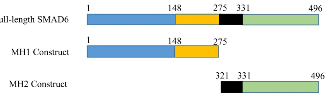
Figure 1. Domain Constructs. Plasmid constructs of just the MH1 (orange) or the MH2 (green) domain compared to full-length SMAD6. The blue and black regions represent the variable and linker region, respectively.
Plasmid DNA Transfections:
HUVEC were transfected with each of the plasmid constructs (described above) using the standard Amaxa optimized protocol (Lonza VPB-1002). HUVEC (5 x 10⁵ cells) were combined with 100 #L of Nucleotransfector solution and 2.5 #g of DNA and placed into a cuvette. The nucleofector program A-034 for HUVECs (Lonza VPB-1002) was applied and transfected cells were immediately transferred into EGM-2 media. HUVEC were then incubated at 37 ºC
overnight before beginning next experiment.
Immunofluorescence:
HUVEC were seeded onto 0.1% gelatin coated coverslips overnight at 37 ºC, pretreated for 4 hours in 0.1 % normal bovine calf serum (NBCS) in OptiMEM (Life Technologies 31985-070), and then treated for 90 min with either vehicle (4mM HCl + 0.5 % bovine serum albumin (BSA)) or recombinant human BMP6 (R&D Systems 507-BP-020) at 200 ng/mL in 0.1% NBCS + OptiMEM. Cells were then fixed for 10 min in 4% paraformaldehyde (PFA), washed 4 x 5 min with phosphate-buffered saline (PBS), and permabilized for 15 min with 0.5% Triton X-100 in
Full-length SMAD6
MH1 Construct
MH2 Construct 1
1
148
148 275
275 331
331
321 496
PBS. Cells were blocked for 1 hour at room temperature with the background suppression agent CAS-block (Life Technologies 00-8120) and then incubated in Rabbit anti-pSMAD1/5 primary antibody (1:1000, Cell Signaling 9516S) for phosphorylated SMAD1 and 5 (pSMAD1/5)
immunofluorescence in CAS-block overnight at 4 ºC. For VE-cadherin, trans-Golgi, or cis-Golgi immunofluorescence, Rabbit VE-cadherin (1:1000, Cell Signaling 2500S), Rabbit anti-TGN46 (1:1000, Sigma SAB4200355), or Rabbit anti-GM130 (1:1000, Abcam ab52649) primary antibody were used, respectively. The following day, coverslips were washed 4 x 5 min with PBS, incubated with either goat anti-Rabbit Alexa Fluor 488 or 555 (1:500) secondary antibodies in CAS-block for 2 hours, and then incubated with DRAQ7 (1:250, Abcam, ab109202) in PBS for 10 min at room temperature. Coverslips were mounted on slides using Prolong Diamond Antifade Mountant (Life Technologies P36961) and sealed with clear nail polish.
Micropattern Plating:
Imaging and Quantification:
All HUVEC fluorescent images were acquired on either an Olympus FV1200 Laser Scanning Confocal Microscope or Olympus FV3000 and FluoView software. FIJI (ImageJ) software was used for all analysis and quantification. pSMAD1/5 nuclear fluorescence intensities were determined on a single-cell basis using FIJI by measuring the mean gray values of pSMAD per nucleus. The brightest slice in the DRAQ7 (nuclear) channel from confocal z-stack images was threshold adjusted into a binary image (black nuclei, white background). Analysis was
re-directed to the pSMAD1/5 channel and the mean gray values per nucleus with a 50-500 #m2 was obtained.9 Similarly, quantification of the mean junctional VE-cadherin signal intensity was measured using the mean gray values of VE-cadherin. The VE-cadherin channel was adjusted into a binary image and mean gray values were measured within a 10 #m x 10 #m box over the middle of the junction.
Analysis:
Results
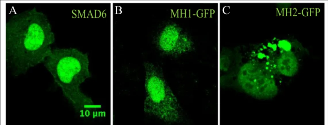
The MH1 domain is necessary for proper localization of SMAD6.
I first examined the effects of overexpression of MH1 or MH2 on SMAD6 localization. Although the MH2 domain inhibits BMP signaling, it is unclear if certain parts of SMAD6 protein control its localization in the cell. I therefore hypothesized that the MH1 domain could be required for proper SMAD6 localization to the cell membrane. HUVEC were transfected with full-length SMAD6, MH1, or MH2 constructs and the subcellular localization of SMAD6 was examined by immunofluorescence. Endogenous SMAD6 is known to be punctate throughout the cell and extends to the cell membrane.9 However, examination of transfected ECs with the domain constructs
showed that the MH2 mutant protein, which is lacking MH1 domain, is mis-localized
compared to full-length SMAD6 (Fig. 2A-C). The MH2 construct mainly demonstrated perinuclear localization (Fig. 2C). In contrast, the MH1 construct localized similarly to full-length SMAD6, residing predominantly in the cytoplasm. These results suggest a novel role for the MH1 domain of SMAD6 in the proper cellular localization of SMAD6.
Figure 2. MH1 domain is needed for localization of SMAD6. Transfection of
HUVEC cells with either full-length SMAD6 (A), MH1 (B), or MH2 (C).
A
B
C
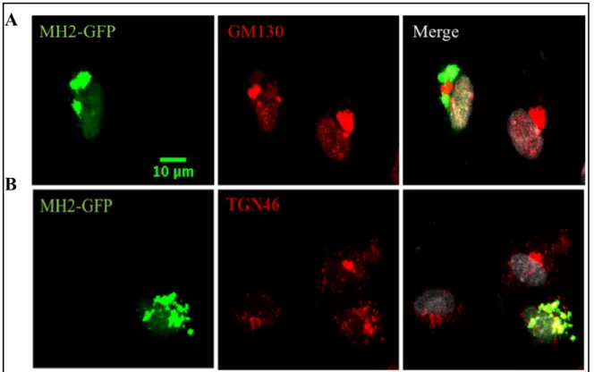
Closer examination of the perinuclear localization of the MH2 construct showed localization to the Golgi network. Specifically, the MH2 construct signal overlapped with the trans-Golgi network (Fig. 3A) more completely than with the cis-Golgi network (Fig. 3B). These findings suggest that the MH2 domain of SMAD6 is not sufficient to properly localize in EC without the MH1 domain and is instead stuck in the trans-Golgi apparatus.
A
B
Figure 3. MH2 mutant protein is localized to the trans-Golgi network (TGN). HUVEC
The MH1 and MH2 domains of
SMAD6 are required for BMP
signaling inhibition.
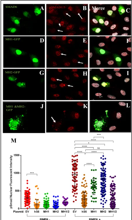
I then examined levels of nuclear phosphorylated SMADs 1 and 5 (pSMAD1/5) in transfected cells with and without the addition of BMP6 ligand, as pSMAD1/5 signal in the nucleus can be used as a readout for BMP signaling. Treatment with BMP ligand induces BMP signaling and thus nuclear pSMAD1/5 localization. I reasoned that the MH2 domain alone should be able to inhibit BMP
signaling, as it is thought to be the functional domain of the protein. However, because I saw that the MH1 domain is required for SMAD6 localization, I hypothesized that it is also required for SMAD6 full function in inhibition of BMP signaling. To test this, HUVEC were transfected with MH1, MH2, or MH1+MH2 constructs,
A
B
C
D
E
F
G
H
I
Merge
Figure 4. Both MH1 and MH2 domains of SMAD6 are required for BMP6 signaling suppression via pSMAD 1/5
nuclear localization. HUVEC cells transfected with full-length
SMAD6 (A-C), MH1 (D-F), MH2 (G-I), and both MH1 and MH2 (J-L) mutant proteins with exogenous BMP6 and stained with pSMAD 1/5 antibody (red) and DRAQ7 (gray). Arrows point to transfected HUVEC cells. (M) Scatterplot of pSMAD 1/5 nuclear fluorescent intensity of the different SMAD proteins. Graph represents 5 experiments. N=100 cells.
L
K
J
treated with either a vehicle or BMP6, and stained with pSMAD1/5 antibody. As expected, BMP6 treatment significantly increased overall nuclear pSMAD1/5 levels in transfected cells compared to controls (Fig. 4M). In the presence of BMP6 ligand, overexpression of full-length SMAD6 was found to decrease the expression of pSMAD1/5 significantly compared to the EV, indicating that full SMAD6 significantly inhibits BMP signaling. 30 However, the MH1 and MH2 domain constructs only partially inhibited BMP signaling (Fig. 4M, compare dark blue and dark green to dark orange). Neither domain construct was able to inhibit pSMAD1/5 nuclear
translocation to the degree of full-length SMAD6, suggesting that individual domains of SMAD6 are not sufficient to inhibit BMP signaling. Thus, both the MH1 and MH2 domains of SMAD6 are necessary for full BMP inhibition. In comparing pSMAD inhibition between the two domain constructs, the MH1 domain suppressed BMP signaling slightly better than the MH2 domain, (Fig. 4M, compare dark green to dark blue). Surprisingly, the overexpression of both the MH1 and MH2 domain constructs together was able to suppress BMP signaling just as well as full-length SMAD6 (Fig. 4M, compare dark orange to dark purple). This further suggests that both domains of SMAD6 are necessary for complete BMP inhibition, although it is unclear how the independent domains co-operate to restore SMAD6 function in EC.
both domains, and that neither MH1 or MH2 domains will stabilize junctions alone. EC that are quiescent have linear junctions whereas EC that are active exhibit serrated and/or punctate VE-cadherin localization.15 To determine the effect of EV, SMAD6 and the different domain constructs on VE-cadherin localization, I transfected HUVEC with full-length SMAD6, MH1, and MH2 constructs and then plated them on cover slips or on H-pattern micropatterns to rigorously quantify the junctions between two EC. I stained with a VE-cadherin antibody to mark the adherens junction and then quantified the mean gray area of VE-cadherin at a cell-cell junction. In vehicle treated HUVEC, overexpression of full-length SMAD6 and the MH1 domain resulted in lower VE-cadherin area (Fig. 5A, C-F). Addition of BMP6 ligand resulted in a
A
B
Figure 5. Both the MH1 and MH2 domains of SMAD6 are required for stable cell-cell junctions. Representative images of HUVEC cells transfected with EV, full-length SMAD6, MH1, and MH2
constructs, stained for VE-cadherin (red) and nucleus (DRAQ7, gray) without (A) and with (B) exogenous BMP6. White boxes indicate areas of higher magnification. H-micropatterns of 2 cells transfected with EV (C), full-length SMAD6 (D), MH1 (E), or MH2 (F) with and without BMP6. (G) Quantification of mean junctional VE-cadherin signal intensity in endothelial cells between transfected cells using H-pattern micropatterns. Graph represents 2 experiments. N=20 cells.
C
D
E
F
G
E V h S 6 M H 1 M H 2 E V h S 6 M H 1 M H 2 0
2 0 4 0 6 0
B M P 6 - B M P 6 +
M e a n J u n c ti o n a l V E -c a d h e ri n S ig n a l In te n s it y * * * * * * * * * n s n s
Discussion
SMAD6 is a major intracellular inhibitory SMAD that limits BMP signaling, yet the precise structure-function relationships of this protein remain elusive.14,26 Collectively, my data indicate that the MH1 domain is necessary for the localization of SMAD6 in EC, and also for the inhibition of BMP signaling and EC adherens junctional stability. My studies show that the MH1 domain of SMAD6 is particularly necessary for proper cellular localization, as overexpression of just the MH2 domain resulted in different localization than full-length SMAD6. This suggests that SMAD6 may be unable to be trafficked out of the trans-Golgi when it lacks the MH1 domain. It is likely that, although the MH1 domain of SMAD6 is not conserved among species, there are certain amino acids and/or regions that are essential for SMAD6 trafficking and localization. However, additional functional studies are needed to determine which specific regions within the MH1 domain of SMAD6 that are required for proper protein localization.
Interestingly, when MH1 or MH2 domains are overexpressed in the presence of BMP6, they were unable to suppress pSMAD1/5 signaling to the degree of full-length SMAD6. The MH2 domain of I-SMADs, such as SMAD6, participates in blocking downstream BMP signaling by competitively binding with type I receptors or SMAD1/4. Several important basic amino acid residues such as Lys-401 and Arg-409 in the MH2 domain of I-SMADs, particularly SMAD7, play an important role in receptor interactions.14,23,27 However, my results show that
though the MH2 domain is thought to be the functional domain for inhibiting BMP signaling, the localization and therefore inhibitory function of SMAD6 is in part, dependent on the MH1 domain. Furthermore, HUVEC transfected with both MH1 and MH2 domain constructs
significantly suppressed nuclear localization of pSMAD1/5 compared to EV. This suggests that the two separate domains may additively interact with each other to inhibit BMP signaling more sufficiently than either domain on its own.24 Though my results show that co-expression of the two domains can block downstream signaling, it is not clear how the two domains interact to accomplish this. Additional studies addressing this can help further characterize the role of the MH1 and MH2 domains of SMAD6 in BMP signaling.
BMP signaling through BMP6 ligand increases internalization of VE-cadherin, and thus controls vascular permeability.16 Decreased barrier function is linked to hemorrhage of blood vessels.28 My results show that overexpression of full-length SMAD6, but not SMAD6 MH1 nor MH2, in the presence of BMP6 ligand led to decreased VE-cadherin area at the EC junction. This suggests that full-length SMAD6 is required to maintain junctional stability, and neither the MH1 nor the MH2 domain is sufficient to maintain this stability. Our lab recently showed that SMAD6 knockdown in HUVEC results in increased levels of phosphor-SRC, which is upstream of VE-cadherin internalization and EC activation.13 This is the first evidence that SMAD6 influences function of the adherens junction protein VE-cadherin and Src.
and junctional proteins will provide insight into the mechanism whereby SMAD6 stabilizes EC junctions. Understanding the structure/function of SMAD6 protein is critical to fully understand how it inhibits BMP signaling and how it regulates other cellular processes, such as adherens junction stability and inform future blood vessel therapies.
Acknowledgements
I would like to express my sincere gratitude to Lyndsay A. Wylie and Victoria L. Bautch for support and guidance throughout this project. I would also like to thank the Bautch Lab members for productive input.
References
1. Adair, T. H.; Montani, J. P. Angiogenesis; Morgan and Claypool life Sciences: San Rafael, 2003.
2. Tonnesen, M. G.; Feng, X.; Clark, R. A. Angiogenesis in wound healing. J. Investig. Dermatol. Symp. Proc. 2000, 5, 40-46.
3. Wallez, Y.; Huber, P. Endothelial adherens and tight junctions in vascular homeostasis, inflammation and angiogenesis. Biochim. Biophys. Acta. 2008, 1778, 794-809.
4. Lampugnani, M. G.; Dejana, E. Adherens junctions in endothelial cells regulate vessel maintenance and angiogenesis. Thromb. Res. 2007, 120, S1-S6.
5. Weis, S. M.; Cheresh, D. A. Tumor angiogenesis: Molecular pathways and therapeutic targets. Nat. Med. 2011, 17, 1359-1370.
6. Benn, A.; Hiepen, C.; Osterland, M.; Schütte, C.; Zwijsen, A.; Knaus, P. Role of bone morphogenetic proteins in sprouting angiogenesis: differential BMP receptor-dependent signaling pathways balance stalk vs. tip cell competence. FASEB J. 2017, 31, 470-4733. 7. Dyer, L. A.; Pi, X.; Patterson, C. The role of BMPs in endothelial cell function and
8. Wiley, D. M.; Kim, J. D.; Hao, J. Hong, C. C.; Bautch, V. L., Jin, S. W. Distinct signaling pathways regulate sprouting angiogenesis from the dorsal aorta and the axial vein. Nat. Cell Biol. 2011, 13, 686-692.
9. Mouillesseaux, K. P.; Wiley, D. S.; Saunders, L. M.; Wylie, L. A.; Kushner, E. J. Chong, D. C.; Citrin, K. M.; Barber, A. T.; Park, Y.; Kim, J-D.; Samsa, L. A.; Kim, J.; Liu, J.; Jin, S-W.; Bautch, V. L. Notch regulates BMP responsiveness and lateral branching in vessel networks via SMAD6. Nat. Comm. 2016, 7, 13247.
10. Heldin, C. H.; Miyazono, K.; ten Dijke, P. TGF-beta signaling from cell membrane to nucleus through SMAD proteins. Nature, 1997, 390, 465-471.
11. Wang, R.; Green, J.; Wang, Z.; Deng, Y.; Qiao, M.; Peabody, M.; Zhang, Q.; Ye, J.; Yan, Z.; Denduluri, S.; Idowu, O.; Li, M.; Shen, C.; Hu, A.; Haydon, R.; Kang, H.; Mok, J.; Lee, M.; Luu, H.; Shi, L. Bone Morphogenetic Protein (BMP) signaling in development and human diseases. Genes and Diseases. 2014, 1, 87-105.
12. Horbelt, D.; Denkis, A.; Knaus, P. A portrait of transforming growth factor b superfamily signaling: background matters. Int. J. Biochem. Cell Biol. 2012, 44, 469-474.
13. Wylie, L. A.; Mouillesseaux, K. M; Chong, D. C.; Bautch, V. L. SMAD6 stabilizes endothelial cell junctions and inhibits developmental angiogenesis. In preparation. 14. Imamura, T.; Takase, M.; Nishiharam, A.; Oeda, E.; Hanai, J.; Kawabata, M.; Miyazono,
K. Smad6 inhibits signaling by the TGF-beta superfamily. 1997, 389, 622-626.
15. Bentley, K.; Franco, C. A.; Philippides, A.; Blanco, R.; Dierkes, M.; Gebala, V.; Stanchi, F.; Jones, M.; Aspalter, I. M.; Cagna, G.; Weström, S.; Claesson-Welsh, L.; Vestweber, D.; Gerhardt, H. The role of differential VE-cadherin dynamics in cell rearrangement during angiogenesis. Nat. Cell Biol. 2014, 16, 309-321.
16. Benn, A.; Bredow, C.; Casanova, I.; Vukičević, S.; Knaus, P. VE-cadherin facilitates BMP-induced endothelial cell permeability and signaling. J. Cell Sci. 2016, 129, 2016-218.
17. Yingling, J. M.; Datto, M. B.; Wong, C.; Frederick, J. P.; Liberati, N. T.; Wang, X. F. Tumor suppressor Smad4 is a transforming growth factor beta-inducible DNA binding protein. Mol. Cell Biol. 1997, 17, 7019-7028.
18. Kim, J.; Johnson, K.; Chen, H. J.; Carrol, S.; Laughon, A. Drosophila Mad binds to DNA and directly mediates activation of vestigial by Decapentagplegic. Nature. 1997, 388, 304-308.
20. Liu, F.; Hata, A.; Baker, J. C.; Doody, J.; Cárcamo, J.; Harland, R. M.; Massagué, J. A human Mad protein acting as a BMP-regulated transcriptional activator. Nature. 1996, 381, 620-623.
21. Massagué, J.; Wotton, D. Transcriptional control by the TGF-!/Smad signaling system. EMBO J. 2000, 19, 1745-1754.
22. Hao, R.; Chen, L.; Wu, J-W.; Wang, Z-X. Structure of Drosophila Mad MH2 domain. Acta Crystallogr. Sect. F Struct. Biol. Cryst. Commun. 2008, 64, 986-990.
23. Xu, J.; Wang, A. H.; Oses-Prieto, J.; Makhijani, K.; Katsuno, Y.; Pei, M.; Yan, L.; Zheng, G.; Burlingame, A.; Bruckner, K.; Derynck, R. Arginine methylation initiates BMP-induced Smad signaling. Mol. Cell. 2013, 51, 5-19.
24. Nakayama, T.; Berg, L. K.; Christian, J. L. Dissection of inhibitory Smad proteins: both N- and C-terminal domains are necessary for full activities of Xenopus Smad6 and Smad7. Mech. Of Dev. 2001, 100, 251-262.
25. Gavard, J.; Gutkind, J. S. EGF controls endothelial-cell permeability by promoting the beta-arrestin-dependent endocytosis of VE-cadherin. Nat. Cell Biol. 2006, 8, 1223-1234. 26. Hata, A.; Lagna, G.; Massagué, J.; Hemmati-Brivanlou, A. Smad6 inhibits BMP/Smad1
signaling by specifically competing with the Smad4 tumor suppressor. Genes and Development. 1998, 12, 186-197.
27. Hanyu, A.; Ishidou, Y.; Ebisawa, T.; Shimanuki, T.; Imamura, T.; Miyazono, K. The N domain of Smad7 is essential for specific inhibition of transforming growth factor-signaling. J. Cell Biol. 2001, 155, 1017-1027.