RICKY L. LANGLEY. Fungi as a Cause of Indoor Air Pollution : A Literature Review and Case Report. (Under
The Direction of Dr. David Fraser)
Adverse health problems resulting from indoor air pollution have become more apparent over the last two decades. Because of the Legionella outbreaks in the 1970's, biologic agents have been recognized as one of the important causes of indoor air pollution. Biologic agents can cause either infections or allergies, and techniques for collection and handling of biologic samples are
frequently unfamiliar to the industrial hygenist.
This paper is a review of indoor air pollution due to
fungi and describes infections and allergies which may occur in building occupants. An approach to investigating sick building syndrome is also described. Also, an example of sick building syndrome at a local university
ill
TABLE OF CONTENTS
I Introduction...1
II Exposure to Indoor Air Pollutants...2
III Sources of Pollutants...4
IV Tight (Sick) Building Syndrome...5
V Frequency of Sick Building Syndrome...6
VI Biological Aerosols...8
VII Factors Involved in the Development of Disease from Microorganisms...10
VIII Taxonomy of Fungi...12
IX Characteristics of Fungi...13
X Indoor Contamination by Fungi...19
XI Adverse Effects of Fungi on Man...22
XII An Overview of Fungal Infections...23
XIII Indoor Infections due to Fungi...25
XIV Diagnosis of Fungal Infections...32
XV Treatment of Fungal Infections...35
XVI An Overview of Allergic Diseases...37
XVII Mechanisms of Allergic Reactions...40
XVIII Fungi as a Cause of Allergic Disease...41
XIX Diagnosis of Allergic Diseases due to Fungi .... .48
XX Treatment of Allergic Diseases...54
XXI Fungal Sampling Equipment...59
XXII Sampling Airborne Fungi: Important Considerations...63
XXIV Investigation of Sick Building Syndrome...67
XXV Prevention and Control of Fungi in the Indoor Environment...74
XXVI Case Report...79
XXVII Appendices...101
XXVIII Figures...105
XXIX Tables...108
INTRODUCTION
Ever since man learned to control fire, he has been
faced with the potential health hazards associated with indoor air pollution. Soot has been found on the ceilings of prehistoric caves. It has been known for several years that burning of fossil fuels has been associated with outdoor air pollution and adverse health effects, but research on the health effects of indoor air pollution
earnestly began in the late 1960's (1).
Indoor air quality, unlike outdoor air quality, is not directly regulated. Under the authority of the Clean Air Act, the Environmental Protection Agency regulates outdoor air quality. The EPA has constructed a framework for implementing ambient air quality standards (1) . It also has a set of regulations for controlling air
pollution sources.
Six criteria pollutants have been established which
include total suspended particulates, sulfur dioxide,
carbon monoxide, lead, nitrous oxides, and ozone. Recentimprovements in the outdoor concentrations of these
pollutants, except NO?., have been noted (1) .
However, as previously stated, no single agency has
buildings, has been increasing (2). From 1980 to mid 1981, 139s of requests to the National Institute for Occupational Safety and Health (NIOSH) for health hazard
evaluations were from workers in nonindustrial settings who felt their offices to be hazardous (3).
As the hazards from outdoor air pollution are being decreased from better air pollution control devices and
the threat of legal suits to companies that exceed standards set by the government, more attention is being paid to the problems of indoor air pollution. Research has shown that individuals in more developed countries are spending less of their time outdoors. Thus indoor
concentrations of air pollutants may be the main
determinant of exposure for many of these pollutants (4). Table 1 shows the average hours spent per day in various
locations by adults in 44 U.S. cities (1).
EXPOSURE TO INDOOR AIR POLLUTANTS
As can be seen from table 1, we spend a great deal of
our time indoors. Although we consider a house or building a safe environment from the outside elements,
potential health risks also exist in many of these
dwellings. When evaluating health risks from exposure to
air pollution, one must consider the dose that reaches the
the indoor or outdoor air. One can usually safely assume,
however, that the higher the concentration of pollutant,
the more likely it is to cause an adverse effect.
Personal exposures to pollutants represent time
weighted average exposures from different locations (1).
The determinants of indoor concentration depends upon many
factors, including the following: outdoor level of the pollutant, indoor sources, the rate of exchange between
indoor and outdoor air, and other characteristics of the
structure and its furnishings (4).
Outdoor pollutants may enter a building through the ventilation system or through open windows, doors, and cracks in the structures. The indoor concentration of a
pollutant from an outside source depends upon the
concentration of the pollutant, the rate of air
infiltration, the reactivity of the contaminant, the
filter efficiency of the ventilation system, and upon
particle size and shape (1).
Because of the oil embargo and increasing energy prices during the 1970's, individuals and institutions have been seeking ways to become more energy efficient. More than one third of the energy used in the U.S. is consumed by buildings (5). Common approaches to decrease
the use of energy has led to adding insulation, reducing
air exchange rates, and fuel switching. This increased
expense of the comfort and health of individuals (5,6).
, SOURCES OF POLLUTANTS
The focus of this paper will be on microorganisms,
fungi in particular, as a cause of indoor air pollution.
However, many other agents may cause similar symptoms and will be briefly discussed. Since remedial action tends to be nonspecific for the majority of cases of buildings related illnesses, principles applicable in dealing with fungal related problems are also applicable in dealing
with other viable and nonviable agents. ,
The 83 million housing units and hundreds of thousands of office buildings in the U.S. contain numerous
sources of pollutants (1,4,5,6). Due to the energy
crisis, the construction of homes and buildings has
decreased the average air exchange rate into these
facilities in order to save money. These "tight" and "super tight" homes have air exchange rates as low as 0.1 to 0.5 per hour, while more conventional homes have
exchange rates between indoor and outdoor air around 1.0 per hour (1). Also, the American Society of Heating,
Refrigeration, and Air Conditioning Engineers has lowered
its recommendations for the amount of fresh air per person
An often unrecognized source of indoor pollution is from transportation. It is estimated that about 5% of our
time is spent in transit, while the remainder of our day is spent at home or in the office (1). In mass transit systems, the occupant to air volume ratios are much higher than in most indoor environments. Thus, substantial
exposure to pollutants may occur (1).
Table 2 is a listing of many of the pollutants that have been found in indoor environments. Over 30 types of organic chemicals have been detected in buildings (6) .
These contaminants can arise from sources located outside
the building, the building materials, building maintenance and cleaning materials, or the building inhabitant and the
products they use (8).
TIGHT (SICK) BUILDING SYNDROME
(4,6,9) .
The following is a partial list of symptoms reported
in investigations of sick building syndrome; aching
joints, muscle twitchings, back pain, hearing disturbance, dizziness, dry skin, discolored skin, skin irritation or itching, heartburn, nausea, detectable odors, sinuscongestion, sneezing, chest tightness, wheezing, eye
irritation, problems wearing contact lens, headache, fatigue, drowsiness, sensation of too hot or cold (2,5). The most common complaints include eye, nose, and throat irritation, headache, fatigue, sneezing, and difficultywearing contacts.
Most individuals say that the severity of their
symptoms increases during the day, and often over the
course of a week, but symptoms improve when they leave the building or take a vacation (6). Even short breaks while at work to different areas inside or outside the building are often associated with noticeable improvement insymptoms.
FREQUENCY OF SICK BUILDING SYNDROME
general, about 15-20% of workers in any office building
will complain of nonspecific symptoms (9, 10). Hence, the syndrome is defined in the epidemiologic sense as the occurrence of symptoms above the background level.Currently, more than one half of the workplaces in
the United States are offices. It has been estimated that
30 percent of newly constructed and remodeled offices have
signs of the tight building syndrome, and in those buildings, between 10-30% of the occupants are affected(11) . These statistics do not take into account the new homes and mobile homes that are being built for energy efficiency (1).
No one knows for sure how common sick building
syndrome is, but based on a telephone survey of 600 office workers in the United States, it is possible that 20% of office workers are exposed to environmental conditions described as sick building syndrome (9). A recent New England study demonstrates how serious the problem can be (9). State government workers in Maine and New Hampshire took a survey to identify the extent of sick building syndrome. Fifty-one percent of the workers were bothered by stuffy air most of the time, 16% at specific times and
27% occasionally. Thirty percent of the workers said that
attention for health problems related to poor air quality.
If this survey is any indication of the problem in the total U.S., then millions of days of work and potentially
billions of dollars are being unnecessarily wasted.
BIOLOGICAL AEROSOLS
There are numerous biological agents that can be
transmitted by the aerosol route and that can cause discomfort or illness in man. For example, molds, dust, bacteria, viruses, fungi, danders, pollens, and insect
parts can all be transmitted by the air.
Viruses and bacteria cause about 69,000 deaths per year due to respiratory illness in the U.S. They are also
the most important cause of acute disabling illness with an average of 1.22 disabling colds per person per year in
the United States (12).
Airborne allergenic agents are felt to be responsible
for millions of disabling episodes of asthma and allergic
rhinitis that occur each year (12). The aerosol route is
also felt to be responsible for many of the hospital
acquired infections that occur each year.
for 809s of lab acquired infections is unknown, it is felt that a majority of these are due to aerosol spread (13).
The most important single parameter useful in
explaining the behavior of an aerosol is its size (14).
Particle sizes may vary from 10"'' cm to 10 ^-'' cm, and
particles less than 5-10 microns are usually considered
respirable (14).
Aerosols may be conveniently classified into two
groups: Those >5 microns and those <5 microns. Particles
greater than 5 microns tend to be removed in the nasopharynx, while particles less than 5 microns can reach the alveoli. The size of fungal species varies considerably, so they may be filtered out in the nose or
they may reach the alveoli.
At least three mechanisms of deposition are known to
occur (15). Impaction refers to a particle's failure to turn corners and thus impinge on the mucus surfaces of the nose and pharynx. This mechanism is effective for removal of most particles >3 microns in diameter. Sedimentation
is the settling of particles due to gravity and is
important for particles between 1-5 microns. Sedimentation occurs extensively in the small airways.
Deposition by diffusion takes place usually in the alveoli.
Many particles that are introduced do not settle at all but are exhaled with the next breath. These particles
are often too small to impact or sediment to a major
extent but are too large for significant diffusion to
occur.
Some factors involved in determining the respiratory risk from aerosol exposure include; concentration of viable organisms, the influences of air volume and
ventilation rate, environmental factors such as
temperature and humidity, breathing rate, settling rate of particles, particle size, virulence of the organism, and
susceptibility of the host .
FACTORS INVOLVED IN THE DEVELOPMENT OF DISEASE FROM
MICROORGANISMS
It was known for a while that certain bacteria, viruses and fungi could be transmitted by the aerosol route. However, quantitative data on indoor air pollution and transmission of microbiologic agents is limited for most organisms (29). With the Legionella outbreak that occurred during a convention in Philadelphia in 1975 and the subsequent detection of the organism in the water of HVAC units, the importance of microorganisms as a cause of
After investigating many episodes of Sick Building Syndrome, it has been shown that fungi are one of the microorganisms responsible for illness in building
occupants.
Characteristics of the host, agent, and environment
all determine whether an individual will develop allergies or infection from microorganisms. Host factors that are important include age, sex, ethnic group, genetics, physiologic and immune status of the individual,
nutritional status, human behavior, and preexisting disease. An individual has both specific and nonspecific
resistance factors that protect him from disease. Included among the nonspecific resistance factors are the
normal indigenous microflora, genetic factors, morphologic
integrity of the skin and mucus membranes, nutrition, acute phase reactants, and hormones. Among the specific host resistance factors are immunoglobulins, complement,
and cell mediated immunity.
Important aspects of the infectious agent that must be considered are its phylogenetic class, reservoir, life
cycle, geographic distribution, latency, transmissibility,
and pathogenicity. Pathogenicity or the ability to
establish an infective process depends on the organism's
invasiveness, evasiveness, and virulence.
Environmental factors that must be considered can be
biological, Eind socioeconomic. Physical factors such as climate and geology are important in determining whether an organism is likely to occur in an area. Biologic factors include local flora, fauna, and human population. Occupation, urbanization, and sanitation are important
socioeconomic factors that must be considered.
TAXONOMY OF FUNGI
Taxonomists initially divided the living world into two kingdoms; the nonmotile, photosynthetic plants and the motile, nonphotosynthetic animals (16). Traditionally, fungi were classified as primitive plants. However, there are many characteristics of fungi that are not shared by
plants. Because of the lack of agreement among taxonomists on how to classify fungi and certain bacteria, a third kingdom was proposed in 1866 by Haeckel. This
kingdom, the Protista, is distinguished from the plant and animal kingdoms by their relatively simple organization (16). Controversy still exists in the classification of
life forms, and Whittaker has proposed five kingdoms (17): Monera (bacteria, actinomyces and blue green algae),
Protista (protozoa and other unicellular organisms), Fungi, Plantae, and Animalia. This last classification scheme will be used during this paper.
The classification of fungi is largely based on the
13
present during the sexual stage of their life cycle (18,
19) . However, the sexual spores and fruiting bodies (pei'fect life cycle) of many fungi are unknown. These imperfect fungi are classified on characteristics other than their sexual stage, such as the morphology of their asexual spores and thalli (the whole fungus including
nonsexual portions and specialized structures (18).
The Fungal (myceteae) kingdom has three divisions:
Gymnomycota, Mastigomycota, and Amastigomycota. The Amastigomycota is the division of primary concern to human health. Most forms included in the Amastigomycota class produce extensive, well developed mycelia, consisting
either of septate or aseptate hyphae, although some single-celled organisms are placed here (19). Five classes. Zygomycetes, Trichomycetes, Ascomycetes, Basidiomycetes and Deuteromycetes, are included in this division.
CHARACTERISTICS OF FUNGI (16, 17, 18, 19,)
bener_al...fiBst:ri_£)tj.jjn. ,
--Fungus is a general term encompassing yeast, molds,
and mushrooms. Yeast are oval, spherical (3-5 microns in diameter) or elongated cells which reproduce by budding.
Molds are characterized by tubular branching cells which
microns in diameter. As a thallus grows, its hyphae form
a mass of intertwining strands called a mycelium. The mycelium forms the visible, usually dry colony of mold
observed on natural substrate or on culture media.
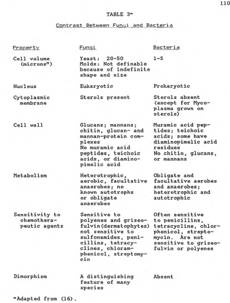
The fungi have several distinguishing features from
bacteria. Fungi possess rigid cell walls composed of
certain polysacchai'ides such as chitin and mannan . The
cytoplasmic membrane of fungi contain sterols. All fungi
reproduce asexually and most can reproduce sexually.
Fungi may be unicellular or multicellular. All fungi are
heterotrophic, requiring organic foodstuffs, and most are
obligate aerobes. See Table 3.
a r"j d H1 r b o r n b 1J :i. s> c
ͣͣ
-. i"- i ͣ-_ͣͣ I ourxon
Water and soil are two principal reservoirs for fungal populations. High humidity is required for fungal
growth and spore germination.
Climactic conditions are important in the
distribution of airborne fungal spores throughout the air, as well as in the return of the spores to soil, water, or
ground. All atmospheric air contains certain varieties
and amounts of fungal spores. The concentration of the
spores differs according to location, altitude, time of day, season of year, condition of surrounding area,
temperature, humidity, rain, snow, sunshine, windspeed,
15
Most airborne fungi are found as spores or hyphal
fragments. All fungi that cause allergies are airborne
fungi. The most common airborne fungi belong to the
genera Cladosporium, Alternaria, Aspergillus, Penicillium,
Helminthosporium, Auerobasidium, Phoma, Nigrospora,
Rhizopus, Mucor, Epicoccum, Stamphylium, Curvularia,
Fusarium, Scopulariopsis, Cephalasporium, Chaetomium,
Trichoderma, Streptomyces, Candida, Cryptococcus, andRhodotorula (20) .
F? e n I'" o rj 1 ..1 r t i Q n
In addition to growth by apical extension and
branching, all fungi reproduce by asexual and most by
sexual processes.
1 - Asexual reproduction
Three mechanisms of asexual (vegetative) reproduction
are known: 1) sporulation, followed by germination of the
spores; 2) budding; and 3) fragmentation of hyphae (15).
Asexual reproduction involves the formation of a new clonewithout involvement of gametes and without nuclear fusion.
Asexual spores are sometimes referred to as conidia
that form at the tips and sides of hyphae. Others develop
within hyphae. The asexual spores aid in the
identification of some species. These spores may be small,
single cell microconidia or large single or multicelled
are characteristic for a given spore (16, 21). There are
several types of asexual spores (Table)
Budding is the primary asexual reproduction process
in yeast, although some divide by fission. As the bud
bulges out from the mother cell, the nucleus from the
mother cell divides and passes into the bud. Cell wall
material then is laid down between the mother and daughter
cell, and the daughter cell eventually breaks away (16) .
The third mechanism of vegetative reproduction is by
fragmentation of hyphae . New colonies are capable of
being formed from these fragments.
2 - Sexual Reproduction
For most species of fungi, except mushrooms,
sexual spores are produced less freguently and less
abundantly than asexual spores. They may be produced only
under special conditions and not detected on usual culture
(22,18) .
Sexual reproduction can be accomplished in several
different ways. The basic steps involved are the
following: 1) The haploid nucleus of a male penetrates
the cytoplasm of the female. 2) The nuclei of both fuse
to form a diploid zygote nucleus, and 3) by meiosis - the
diploid nucleus gives rise to four haploid nuclei (16).
One type of sexual spore is formed by fertilization
of the contents of a female structure (oosphere) by male
17
Another type of sexual reproduction occurs when the tips
of two hyphae come together and their contents fuse. A
zygospore is formed from this process. When the sexual
spores are formed in a sac called an ascus, the spores are
called ascospores, When the sexual spores are formed in a
structure known as a basidium, the spores are known as
basidospores (18).
Charac:terij51 i cs __ and__lo^uXJ- £A.CLi:tij3.D___h}'f__f''.ir;.Q.3.i___9J'___u^SiS'sl..
ImDortance ͣͣ-:
The identification of fungi depends on several
factors. Among these are the shape, size, texture, color,
number of cells, and thickness of the cell walls of the
spores. Other features often examined are the fungal
colony characteristics on growth media, the temperature,
pH, light, nutritional requirements, presence of septae,
and mycelial structure. More specialized antigenic tests
can also be used to identify the fungi (22).
The major classes of fungi that are pathogenic to man
are the Zygomycetes, Ascomycetes, Basidomycetes, and the
Deuteromyctes (13, 17, 18). All except the Deuteromycetes
have sexual and asexual spores. The Deuteromycetes, or
Fungi Imperfecti, lack a sexual stage of reproduction.
The Zygomycetes are often referred to as bread molds.
Most produce well developed hyphae that lack septae (17,
sexual reproduction is by fusion of hyphal tips forming zygospores (18, 22). Examples of human pathogens include
Rhizopus, Absidia, Mucor, and Basidiobolus (22),
The Ascomycetes are characterized by well developed
mycelia that are septate (IV, 18, 19). Sexual reproduction is by formation of a diploid ascus and after meiosis, eight haploid ascospores are formed. Asexual reproduction is by externally born spores (conidia) formed on normal hyphae (18, 22). Yeast forms reproduce by
either budding or fission (18). Examples of human diseases caused by this class include histoplasmosis,
blastomycosis, candidiasis, dermatophytosis, and some
mycetomas (17).
The Basidomycetes include those fungi with which the
layman is most familiar such as mushrooms, puffballs, stinkhorns, bracket and jelly fungi, rusts, and smuts.
They are characterized by septate hyphae and the presence
of sexual spores, basidiospores, formed on the surface ofa specialized structure called the basidium (17, 18, 19,
22). Asexual reproduction may occur by various means,
including the production of conidia (18, 22). An example
of a human disease produced by these fungi is
cryptococcosis.
19
reproduction is by conidia. A few yeast belong in this category, but most fungi have well developed septate hyphae (18, 22). If upon further study, a fungus in this group is found to have a sexual stage, then it is
reclassified and placed in another class. Most of the
human pathogens occur in this class. For example,
Alternaria, Penicillium, Aspergillus, Cladosporium, Candida, Helminthosporium, Fusarium, Gliocladium, Stemphylium, Phoma, Scopulariopsis, Epicoccum, Trichoderma, Nigrospora, Trichospora, Pityrosporum,
Microsporum, Trichophyton, and Coccidioides are included
in this class (17, 18, 20).
INDOOR CONTAMINATION BY FUNGI
Fungi are ubiquitous organisms and can be found most anywhere they are sought. Given a carbon, nitrogen, and water source, fungi will grow under very extreme conditions and in very unusual places. The air in any "clean house" contains hundreds of types of biological and
nonbiological particles (23).
Most indoor contamination results from outdoor sources and may build up to concentrations high enough to
adversely affect the health of man (2, 23). Indoor
buildup of bioaerosols results from material being shed
and accumulating indoors and actual growth on interior
The penetration of bioaerosols into a building seems
to depend most on the extent of mass flow via windows and
doors (2, 24). Additional ventilatory factors that are important include the incident wind speed and direction, negative pressurization either by fans and exhaust stacks, and airleak between structural cracks (2, 24). Windows and doors also contribute to bioaerosol entry into a
building.
Fungi will grow indoors if given a carbon and water source, A relative humidity level greater than 70% is optimal for fungal growth. Numerous sources in a building will support the growth of fungi by producing standing
water reservoirs. Humidifiers, evaporative coolers,
self-defrosting refrigerators, and flush toilets all have a potential for becoming contaminated. Water disasters such
as leaks in the roof or ruptured water pipes, often will allow abundant fungal
growth in carpet, furniture, or wood (2). Many of the appliances mentioned above are in contact with a moving stream of air which can pick up small particulates and spray them into a room or throughout a building depending on the ventilation system.
Another common source of molds in the indoor environment is house dust (24, 25, 26). House dust is a
complex mixture of organic and inorganic compounds,
21
Gravesen at. al., rooms with carpets had higher dust
levels than rooms without carpets (25). It has been
demonstrated that schools with carpets have an increased frequency of students with allergic symptoms than schools without carpets (27) . Indoor surface contamination by biological organisms is dangerous usually only when the
particles become airborne (24) . However, practically any
human or animal activity inside a building can elevate the
background level.
Volumetric fungal spore studies in domestic interiors give counts that range from 1-6000 per cubic meter with maximum levels usually below 1600 per cubic meter (2). Indoor levels of fungi usually average about 40-503o of outdoor levels (2). Cladosporium species are the taxa most frequently recovered both outdoors and indoors in the United States during the summer. Levels are always more
abundant outdoors (2). On the other hand, Penicillium
isolates usually dominate in the winter and are often more frequent indoors than outdoors . Outdoor factors that appear to affect the indoor mold level include marked shade, marked levels of organic debris near the home, and
natural or unkept property.
There are no set guidelines or levels that one can use to state whether a building has an excess amount of biological material in the air. As noted earlier, a wide
If the interior is used specifically for handling or processing biological material, then substantially higher counts can be found. For example, barns with moldy hay may have levels of millions of spores/m-"' (2) .
A comraittee on bioaerosols has presumed a normal
concentration of culturable spores to be 100-200 colony forming units (CFU) per m~^ and 200 CFU/m-' of nonviable spores (28). They suggest that if levels greater than 1000 CFU/m--'^ in affected areas, and if less than
800-1000 CFU/m-' in nonaffected locations are found, then
consider cleaning the office, inspecting the HVAC system
and continue looking for the source. If elevated levels of nonviable spores (above outdoor levels) are found or elevated levels of any single spore type are found indoors but not outdoors, then attempt to identify the source
(28) .
ADVERSE EFFECTS OF FUNGI ON MAN
Fungi can adversely affect the well being of man in
at least six different ways: (22)
1 - Infection - invasion of the body
a - superficial - on the skin, hair or nails
b - superficial - on the mucus membranes or genitalia
c - localized, deep invasion of tissue
d - widespread systemic invasion of the body
23
3 - Poisoning by eating toxic fungi
4 - Poisoning by mycotoxins produced by fungi growing on
stored food
5 - Starvation due to spoilage of stored food by fungi 6 - Starvation from crop failure due to fungal disease
This paper will first look at fungi as a cause of
infection, then as a cause of allergic disease.
AN OVERVIEW OF FUNGAL INFECTIONS
Infections due to fungi can be divided into four groups, differing in the level of tissue invasion. These
four groups include the following: (16)
1) Systemic or deep mycoses which involve the internal organs. They often disseminate widely and invade
different tissues.
2) The subcutaneous mycoses involve the skin, subcutaneous tissue, fascia and bone.
Z) The cutaneous mycoses involve hair, nails, and the epidermis. The responsible fungi are known as
dermatophytes.
4) The superficial mycoses involve only the hair and the
most supei'ficial layers of the skin.
expose individuals to pathogenic fungi which possibly may
lead to a serious infection.
Most microorganisms studied in the laboratory have
caused accidental lab acquired infections. Dr. Pike has analyzed over 3900 cases of lab acquired infections (31) . Of these cases 353 were due to fungi. Coccidioidomycosis, Histoplasmosis and Blastomycosis, fungal infections which
can often be life threatening , accounted for almost 50S6
of these cases,
Because the deep mycoses have caused lab acquired infection and are potentially fatal diseases, these will be examined in some detail. In general, the deep mycoses
are caused by saprophytic fungi (an organism that normally
exists on dead organic material) in the soil, and
inhalation of spores is usually the main route of infection. The earliest lesions are usually pulmonary, and the initial acute pneumonitis is often self limited. In their subsequent chronic stage, the diseases often
begin insidiously and progress slowly and are
characterized by granulomatous lesions (16). Fungi may
spread via the blood stream and produce metastatic lesions
in other parts of the body.
The subcutaneous mycoses are caused by saprophytic
fungi in soil and on plants. Infection often occurs by
direct implantation of spores or mycelial fragments into a
ͣ
' ͣ 25
progresses and is characterized by localized subcutaneous
abscesses and granulomas (15). The infection may extend
directly into adjacent tissue or may be spread by
lymphatic channels. Rarely, it may spread systemically.
The cutaneous mycoses are obligate parasites of man
and animals and only rarely soil saprophytes. The
dermatophytes tend to produce inflammatory lesions of the
skin. The hair and nails are also frequently involved.
Part of the inflammatory lesion may be allergic in nature
(16, 21) .
The superficial mycoses are localized along hair
shafts and in the nonviable layers of the skin. The
infections are rarely of clinical significance (16).
INDOOR INFECTIONS DUE TO FUNGI
Fungi can cause infections by many routes, but this
paper is mainly concerned with airborne spread of the
agents and will focus on certain fungi that have caused
"indoor infections" by this route. Even though the deep
mycoses are rarely associated with indoor infections, they
have been responsible for infections in diagnostic labs.
Infectious fungi, with the exception of the
dermatophytes are usually saprophytic. Those fungi that
cause infection, do so by becoming adapted to the host so
Often, these infections occur in a host who has an
immunologic abnormality.Of the infectious mycoses that can be spread by the
aerosol route, the ones of main concern are the following:
Cryptococcus, Histoplasma, Blastomyces, Coccidioides,
Aspergillus, and Sporothrix . Other fungi may cause
infections, but primarily via contact or inoculation,
therefore, they will not be included in this paper.Cryptococcus is a member of the class Basidomycetes
and is a spherical to oval budding yeast-like organism
(4-20 microns in diameter) with a polysaccharide capsule (16, 21, 39). High concentrations of this organism are foundin pigeon droppings and other avian excreta and in soil
contaminated by this material. Inhalation of yeast cellsis assumed to cause pulmonary infection with subsequent
spread via the hematogenous system. Cryptococcus has a
predilection for the central nervous system. It is
assumed that many infections are subclinical and that most
infections occur in immunocompromised hosts (16, 39) .
However, healthy individuals can become infected, and
often these cases are associated with heavy exposure to
dust (39) .
Cryptococcus has occurred in certain occupations
within fertilizer, flour or textile mills. Also,
individuals that have worked in dusty barns, corn cribs
' ' 27
Coccidiodes is a dimorphic member of the class
Deuteromycetes (40). In infected tissue, it appears
primarily as spherules that range from 5-50 microns in
size. These spherules are filled with many endospores
ranging in size from 2-5 microns (15). Coccidioidomycosis
occurs usually in the South Western United States and is
often called San Joaquin or Valley Fever.
Infection develops when arthrospores (2x5 microns)
are inhaled and reach the lungs. Entry into the host
leads to conversion of these spores into spherules (38).
In 50Ss of infected persons, the disease remains
asymptomatic (16, 21). About 40% of individuals develop
an acute pneumonitis, often with pleurisy, and 3-5% of
these may develop skin eruptions. The lesion is primarily
granulomatous in nature and 1-5% of people develop a
chronic pulmonary cavitary disease resembling tuberculosis
(15, 21). Rarely, systemic spread may also occur.
Outbreaks have occurred in archaeologists and among
farm workers exposed to the soil. However, laboratory
personnel have often become infected from the arthrospores
and many deaths have occurred (31).
Histoplasma is a dimorphic fungus occurring in vivo
as a typical oval yeast (2-4 microns in length) within
macrophages (21). Histoplasma is a member of the class
Because of epidemiologic studies on student nurses in
the 1940's for TB, it was noted that individuals often had
pulmonary calcifications on chest x-ray but negative TB
skin tests. Subsequent skin testing with other antigens
and soil studies concluded that Histoplasma capsalstum was
responsible for many cases of pulmonary calcification
(16) .
Hi stop J asms capsulatum is a common soil organism, and
inhalation of spores is responsible for infection. It is
felt that bats may also serve as a carrier of this fungus
in their intestinal tract. The infection may be
inapparent, may appear as a primary acute pulmonary
disease, may appear in a chronic cavitary form , or it may
rarely dissiminate throughout the body (15, 21, 41), The
inoculum or dose inhaled and the immunologic status of the
host are factors that determine the severity of the
disease.
Farmers, gardeners, nursery men, military personnel,
and spelunkers are individuals likely to contract this
disease. Many lab infections have been due to inhalation
of cultured spores (31). Epidemics have been associated
with bird roost, caves, cellars, chicken houses, cleaning
or demolition of old buildings, farms, soil, and trees
(41) .
<..y , ^ 29
Ascomycetes and grows as a budding yeast in human tissues.
Infections occur by inhalation of spores 3-5 microns in size or by direct inoculation into the tissue.
The natural reservoir remains unresolved (42). The
substrate for growth appears to be organic debris close to
the soil. Infection apparently begins in the lung and spreads hematogenously. Destructive lesions are often noted in many organs. Skin lesions may result either from direct inoculation or hematogenous spread. The
asymptomatic or mild self-limited form of pulmonary disease rarely occurs as with the other deep mycoses.
The disease occurs frequently in individuals that
work outdoors, but no well defined association with occupation is known (42) . There have been at least five reported outbreaks of Blastomycosis. All individuals
involved had contact with the soil or organic debris.
Also, several lab acquired infections have occurred (31).
Although not generally considered deep-seated
mycoses, both Aspergillus and Sporothrix may rarely cause
severe infections, especially in immunocompromised
patients. Pulmonary sporotrichosis is a rare disease, while Aspergillus usually causes an allergic disease
instead of an infection.
to the class Deuteromycetes. The organism's natural habitat is the soil, sphagnum moss and other plant
mat erial . • ,
Most cases of sporotrichosis are due to direct
inoculation, but cases of pulmonary sporotrichosis have occurred (38, 4 3). Pulmonary sporotrichosis usually presents as an upper lobe infiltrate and may cavitate. Inhalation of spores occurs in areas that allow large amounts of fungal growth or in areas where the fungus has grown, sporulated and then dried, allowing dispersal of
the spores.
Persons at risk are primarily foresters, gardeners,
construction workers, farmers, and children (43). Children often play with plant material and soil infected with Sporothrix.
Aspergillus is usually associated with allergic
diseases but may cause infections, especially in immunocompromised hosts. Aspergillus either belongs to the class Ascomycetes or Deuteromycetes, depending on the species. Most aspergilli are not dimorphic, growing only in mycelial form (16) . W. furffigatus accounts for over 90% of all infections due to Aspergillus. Spores are 2-5
microns in diameter, and the fungus is capable of growing
over a wide temperature range (44). Inhalation of spores
, . ͣͣͣ ^ - 31
minimal tissue invasion, 2) active invasive granuloma
giving rise to necrotizing pneumonitis and occasional dissemination (this form occurs primarily in
immunocompromised patients), and 3) "fungus ball" growth
in a preexisting cavity without invasion of lung parenchyma (32).
Agricultural workers, cane sugar processors, or
cleaners exposed to spores in rye flour are often exposed
to high concentrations of this organism (44).
Immunosuppressed hospitalized patients often can develop infections from this organism, and often the source is
external to the hospital (23, 38, 45).
There are a few other fungi that rarely can be
transmitted by the aerosol route and cause serious infections, however the host usually has some type of underlying metabolic (diabetes mellitus) or immunologic disorder. With the AIDS epidemic, many microorganisms that were previously considered innocuous have been found to produce disease. All of the organisms previously
mentioned can cause an infection in healthy individuals, thus they are covered in more detail.
contact with dressing room floors that are contaminated by fungi (athlete's foot).
DIAGNOSIS OF FUNGAL INFECTIONS
The diagnosis of infections from fungi can be roughly broken down into three broad categories: (1) history,
physical exam, and chest x-ray, (2) culture and
histopathology, and (3) serological and immunologic
testing.
The patient's history often gives clues to the
diagnosis. For example, in a patient with a cough, fever, chest pain and recent travel to the San Joaquin Valley, the possibility of coccidioidomycosis should be considered. Also the occupational history may give clues to potential exposures. If several employees working in the same area become ill and are found to have the same disease, then the physician should suspect the same agent in a coworker with similar symptoms. A good history will
often point to the right diagnosis.
The physical exam is rarely diagnostic. However, certain lesions, in conjunction with the patient's history, may point to a fungal infection. For example, erythema nodosum or erythema multiforme, along with hepatosplenomegaly may be noted in histoplasmosis (46). Lesions that follow the lymphatic channels are often seen
33
chronic meningitis with signs and symptoms of fever,
headache, disorientation, and papilledema (21, 39).
Lesions may occur throughout the body if the fungus
disseminates, and these may be noted on physical exam or
by x-ray examination. As mentioned previously,
histoplasmosis can cause calcifications on chest x—ray
that may appear similar to tuberculosis.
Histopathologic examination of sputum, pus, or tissue may identify a fungus as the cause of a patient's illness.
Special stains may be needed to detect certain fungi. For example, sporotrichosis is difficult to detect by
conventional histologic stains (43). Once stained, the
shape of the hyphae, spores, or yeast-forms is used to identify the organism (48). The demonstration by
histologic examination of fungi invading tissue is
indicative of an infectious process.
The standard method of diagnosing an infection is by culturing urine, pus, blood, other body fluids, or tissue samples on different types of media. Sabouraud's glucose agar is frequently used to culture fungi (16, 21). Fungi tend to grow slower than bacteria, and by adding drugs, maintaining a low pH and low temperature, bacterial contaminants are prevented from growing (16). Fungal
cultures are often incubated at different temperatures to
depending on the growth media (48). Once grown, microscopic evaluation of the colony is done to identify
shape and size of the spores and hyphae. In certain
conditions, the recovery of fungi by culture may not
indicate an infection, but rather only fungal colonization
or possibly contamination of the media.
If unable to classify the genus and species by
morphology alone, then biochemical tests can be done to
determine the identification of the organism (46). These
biochemical tests often involve substrate utilization.
More recently serologic and immunologic testing has
been developed to evaluate exposure to fungi (46, 48, 49,
50). Three serologic tests are frequently used to
diagnose fungal infections. These include
immunodiffusion, complement fixation, and agglutination
tests (48) .
Immunodiffusion techniques detect the reaction of
antigen and antibody by the precipitation reaction (51).
Immunodiffusion is one of the simplest and most direct
means of demonstrating antigen-antibody reactions in the
clinical laboratory. Reactions may be classified as single or double immunodiffusion depending upon the
movement of the reactants. Basically, two circular wells
ͣ
35
that fungal antigen, the immune complexes will form and
precipitate, and a line will be visible in the agar.
However, this test is not positive in all cases of
infection (41, 48, 49, 50).
In a complement fixation test, the fixation of
complement occurs during the interaction of antigen and
antibody (51, 52). The antigen-antibody complex uses up the complement. When antibody coated red cells are then added to the solution, these will be lysed by any remaining complement. The amount of remaining complement or red cell lysis is inversely proportional to the
concentration of antigen-antibody complex. Rising titers
indicate progressive infection, while decreasing titers indicate regression. As noted with immunodiffusion, this
test may not be positive in all patients with infection.
Agglutination reactions involve clumping of antigenic
particles by antibodies (51, 52). The reaction may be
classified as direct or indirect. In the simple direct
technique, a cell or insoluble particulate antigen is
agglutinated directly by antibody. Indirect agglutination
refers to agglutination of antigen-coated cells or inert
particles which are passive carriers of soluble antigens.
The agglutination technique detects the presence of
specific antibody. The titer of agglutinating antibody is
solutions of antibody. The final dilution at which clumping occurs is referred to as the specific antibody
titer. ͣ)
Skin testing, using antigens derived from certain fungi has been used in the diagnosis of infection. Skin testing has been developed for histoplasmosis, coccidiodomycosis, and aspergillosis (41, 44, 46, 50). However, negative skin tests may occur in individuals with
overwhelming infections, and cross reactivity between
antigens may give false positive skin tests (16, 50).
TREATMENT OF FUNGAL INFECTIONS
Dissemination of fungi within the host is often fatal if untreated. Localized infections may occasionally
reoccur without treatment. Whether or not to treat may be
a difficult decision. Therapy is probably indicated if
symptoms persist, if evidence suggests local progression
of disease, if the patient is an infant, has a concurrent
illness, an immune impairment, or belongs to a racial
group predisposed to handling the infection poorly (45, 5 0) .
Surgery as well as chemotherapy may be indicated in
the patient. Surgical excision may be useful in some
cases with residual pulmonary, cutaneous, or bony lesions.
Chemotherapy is usually considered part of the therapy
37
It has only been since 1959 that effective antifungal agents were developed (15). Today, there are still only a few antifungal drugs available for treatment of infections. For the treatment of deep mycotic infections, Amphotericin B has been the mainstay. It is effective
against all of the deep mycotic agents and most other fungi that may become invasive in the host. However it is
not effective against the superficial mycoses (16). Amphotericin B, like some other antifungal agents, is derived from various species of Streptomyces. It works by binding to ergosterol in the plasma membrane, thus disrupting the fungal cell (16, 21) .
Newer antifungal drugs such as Flucytosine, Ketoconazole, and Miconozole have been used alone or in conjunction with Amphotericin B to treat deep seated fungal infections (53). As with other drugs, adverse effects from these are multiple and potentially serious, so one must monitor the hematologic and metabolic systems
carefully .
AN OVERVIEW OF ALLERGIC DISEASES
Numerous agents in the indoor environment can cause
allergies. Included in this group are pollens, fungi, algae, actinomycetes, arthropod fragments, dust, and pumices (24). A large number of indoor particles reflects
Numerous factors are important in the development of allergies, among which a history of atopy is very important. Atopy is a term that refers to the familial occurrence of common allergic disease and immediate allergies acquired by natural sensitization to common allergens. Some factors in the development of atopy include: season of the year, family history of allergies, geographic and racial factors, mucosal permeability, and immunologic disorders (54). Subjects are classified according to the degree of reaction to a battery of common allergens (55) . Atopic siibjects are more predisposed to occupational sensitization and allergic reactions to numerous agents than subjects that are not atopic.
Allergic reactions can occur in many organ systems such as the eyes, skin, nose, airways, and alveoli. Allergies affect 175S (35 million) of the population in the
United States (56). The overall prevalence of asthma is 39s, but it is felt to be increasing in frequency. Atopic
dermatitis occurs in 6.9 persons per 1000 (55). Twenty
percent of the population experience some form of
urticaria or angioedema at some time in their life, and animal allergy prevalence ranges from 11-32% (56). Allergic rhinitis occurs in 10-12% of the population (57). Other allergies to foods, insects, and drugs affect tens
; ; ; - .: ͣ " 39
Asthma and allergic rhinitis are the third leading cause of limited activity in persons less than 45 years of age , and annual work loss due to these problems is reported at 5 million days (55). In the United States, it is felt that 1% of all cases of asthma may be occupationally related, while up to 20% of workers in various occupations in the United Kingdom have asthma (56,
58, ) .
Allergic reactions of the airways may be broadly
classified according to their site of involvement and
nature of reaction into four groups: allergic rhinitis involving the nosej the airways (allergic asthma); the
airways and adjacent alveolar spaces (allergic asthma with
pulmonary eosinophilia); and alveoli or peripheral bronchioles (hypersensitivity pneumonitis (24) . A description of these as they relate to fungi will be discussed later.
Organic as well as inorganic agents can cause
irritation or allergic sensitization of the skin.
Allergic skin reactions may result from direct
sensitization by an agent or from a secondary response to
an infection within the body. Examples of the latter
include the id reaction, erythema nodosum, erythema
multiforme, urticaria, and annular erythema (22).
Other organ systems may be involved with allergic
involve the lungs, skin, the eyes, and possibly the
gastrointestinal tract (22).
MECHANISMS OF ALLERGIC REACTIONS
The amount of tissue damage has conventionally
distinguished reactions due to normal protective immunity from allergic reactions. Four types of allergic reactions
were classified by Gell et. al. in 1953 (51, 59). These reactions are the following: Type I (anaphylactic), type
II (cytotoxic), type III (arthus), and type IV (delayed hypersensitivity)
Type I reactions are immediate and depend on IgE bound to mast cells and basophils. After antigen binds to
the IgE antibodies, these cells release numerous pharmacologic substances. Smooth muscle contraction,
increased vascular permeability, and bronchial spasms are
examples of reactions that frequently occur. Hay fever,
allergic rhinitis, asthma, and occasionally anaphylaxis are examples of clinical diseases due to type I reactions
(51, 59) .
Type II reactions occur when antigens present on cell
membranes react to IgG or IgM antibodies. Cell damage
often occurs from this type of reaction, and it is
41
and thrombocytopenia may result from a Type II drug
reaction due to quinidine. •
Type III reactions, often known as arthus reactions, are due to the formation of complement binding antigen-lgG antibody complexes. Release of lysosomal products and activation of complement components leads to tissue damage. Allergic bronchopulmonary aspergillosis and hypersensitivity pneumonitis are two diseases whose mechanism of damage is mediated by type III reactions.
Type IV or cell mediated immune reactions result from the interactions between actively sensitized lymphocytes and specific antigens. These lymphocytes release
lymphokines which are biologically active and mediate a local inflammatory reaction. Antibody and complement are not involved in these reactions. Examples of these reactions include tuberculosis and also hypersensitivity
pneumonit is .
FUNGI AS A CAUSE OF ALLERGIC DISEASE
Fungi may cause allergies that, although rarely
serious, are often debilitating. Most fungal allergies
involve the respiratory tract, and these will be
considered in more detail. Initial evidence establishing fungi as a cause of respiratory allergy was obtained by
time have contributed to our current understanding on
f IIri'-j 1 ,ino .i 1 1 i:r gic disease .
In the natural environment, man is potentially
exposed to more than 100 different species of fungi, many
of which are present in the indoor environment (51).
There is no doubt that factors other than the mere
existence and amount of fungi play an important role in fungal allergy. For example, the size of the spore is highly important. It is generally thought particles greater than five microns are filtered out in the upper respiratory tract and particles less than five microns can reach the alveoli. This is an oversimplification, because if the person is a mouth breather, then particles greater
then twenty microns may penetrate into the bronchioles
(62) .
From air sampling of mold parts, Lowenstein states that spores comprise greater than 90% of the particles of relevant size (26). It has also been shown that spores and mycelia contain several distinct antigens. Attempts to define fungal allergens, however, are still
preliminary, and standards for testing and reagent preparation are not fully developed.
Prevalence of type I allergy to molds ranges from
ͣ
r 43
-' ^ ͣ . " ͣ ͣ '^ ' ͣ »'
the ignorance of molds as a possible factor in respiratory
allergy. (26) .
The Deuteromycetes is the class of fungi most
important as a cause of respiratory disease in man (20).
The four genera Cladosporium, Alternaria, Penicillium, and
Aspergillus represent the most common allergenic molds based on skin reactivity (20, 37). These genera are also associated with the highest percentage of fungal spore counts obtained by air sampling (23). About 85% of people with mold allergies will react with one of these
allergens.
The respiratory allergic conditions which may be due
to fungi include allergic rhinitis, asthma, allergic
bronchopulmonary aspergillosis, hypersensitivity
pneumonitis, and humidifier fever. Each will be briefly described. The main clinical problem is the extent to
which a mold sensitive person's symptoms can be attributed
to molds (37). This is because exposure to molds is a
continuum without definite seasonal end points. Indoor spores are present throughout the year, and levels increase when human activity is present.
Allergic rhinitis consists of paroxysms of sneezing, nasal itching and congestion, clear rhinorrhea, palatal
itching, and if severe, conjunctival irritation, redness and tearing, ear fullness, and pressure sensation in the
perennial depending on the presence of environmental allergens. IgE mediated rhinitis may be caused by a wide variety of allergens including pollens, molds, dander, mites, excreta, etc. About 10-12% of the United States population suffer from allergic rhinitis, two thirds of which occurs before age 30 (57). Nasal symptoms from
fungi are usually less intense than in pollen hayfever,
persist for longer periods of time, and are more or less intermittent in nature, but showing acute exacerbationafter heavy exposure (53).
As an example of rhinitis problems associated with
molds, a study of nasal hypersensitivity in wood furniture workers by Vilhelmsson, et al, found that 3% of the
workers had allergy to molds and 2% to wood dust (64), However, up to 16% of workers in the furniture factory had nasal perennial hypersensitivity, so for the majority of cases, the etiology of their symptoms was not determined.
Asthma is often defined as reversible obstruction of the airways. This airway narrowing may be due to contraction of airway smooth muscle, edema of the bronchial mucosa, or accumulation of bronchial secretions
or any combination of these. An acute asthmatic reaction
is believed to be an IgE mediated reaction, but a delayed
45
emergency room by Dr. Platt Mills, showed that 90Ss had IgE antibody to one or more major indoor allergens (65).
For more than 60 years, molds have been considered a
cause of asthma (56). It has been shown that fungal
spores of Alternaria and Penicillium both can cause asthma, even though there is a large difference in thesize of the spores (57). The shape of spores may be very
different, their aerodynamic behavior is important in
determining whether they can reach the lower respiratory
tract.
Even though we know fungi can cause asthma, we don't
know how often it happens. There have been reports of
fungi growing within humidifiers and air conditionersleading to exacerbations of asthma (10, 24).
Identification of mold aeroallergens is the main problem
to progress. Detailed information on the kind and amount
of antigen in the air and at different times and places,
and reliable standard allergen extracts for diagnostictesting are required before we can fully correlate signs
of asthma with fungal exposure (55).
Hypersensitivity pneumonitis is an allergic lung
disease that results from sensitization and recurrent
eight hours after allergen exposure. Infiltrates may be seen on chest x-ray, and physiologic changes can be found in pulmonary function that suggests a restrictive disorder. Acute attacks usually clear in one to four
days. Repeat acute attacks or chronic exposure may result
in pulmonary fibrosis.
Numerous microorganisms, including fungi, can cause
hypersensitivity pneumonitis (58, 59, 70). Farmers are the occupational group most involved with up to 8-9% of
farmers affected in some countries (70). Reports of
fungal contaminated hot tubs or HVAC systems leading to hypersensitivity pneumonitis in office workers are
becoming more frequent (32, 34, 71, 72).
Immunologically, this disease has been considered a
classic Type 3 reaction, but recently, evidence of Type 4 involvement has been noted (73). Serum precipitating IgG antibodies to inhaled organic antigens can be found in over 908s of individuals with clinical disease.
Allergic bronchopulmonary aspergillosis (ABPA) is an allergic pulmonary reaction to inhaled HSpsrg i11 as
47
um and/or weight loss, productive cough with typical sput plugs and/or hyphae and/or hemoptysis, high peripheral blood eosinophil counts Q 600 per m"') , positive precipitins against Aspergillus and/or positive specific anti-Aspergillus IgE (74). Disease is certain if criteria one through four and three of the last four are present.
Disease is probable if criteria one through three and four
of the last five are present, and disease is possible if
criteria one through two and three of the last six arepresent.
The immunopathogenesis of ABPA is thought to involve
type I and Type III reactions. During the acute illness, airway obstruction is predominant but over time, signs of interstitial fibrosis occur (73).Although flsperg 111 as fami gatas is widely distributed, ABPA rarely occurs in the United States. It occurs
usually in atopic people with a history of asthma.
Individuals that work at sewage composting run a risk ofheavy exposure to Aspergillus (44).
Humidifier fever is a recently described entity,
probably allergic in nature, that has been reported in
office workers exposed to contaminants in humidification
systems (10, 24, 45). The disease presents with flu-like
symptoms of headache, lethargy, myalgia, arthralgia,
fever, shortness of breath, and occurring four to eight
hours, regardless of continuing exposure. Symptoms recur on reexposure after an absence of several days from work.
Lung function studies during an acute attack show a
restrictive pattern that improves during the working week. The diffusion coefficient is reduced during acute illness and arterial desaturation may occur. The chest x-ray is normal in the acute stage of disease and even after several years of symptoms, thus differentiating this
disorder from hypersensitivity pneumonitis.
The cause is felt to be an antigen or antigens found
in the humidifier (24). Precipitins to different
microorganisms can be found in the serum of affected
individuals, but antibodies may be seen in asymptomatic exposed individuals also. However, serologic testing is
not felt to be a reliable method of determining the cause of the condition because inhalation challenge testing with extracts of organisms to which individuals have
precipitins fail to produce systemic symptoms
DIAGNOSIS OF ALLERGIC DISEASES DUE TO FUNGI
As with the diagnosis of infectious diseases due to
fungi, the diagnosis of allergic disease is multi-faceted.
The requirements for good diagnostic tests are that they
be standardized, accurate, reproducible, inexpensive, easy4 9
Unfortunately, no test presently fulfills all these
requirements. '
»-The initial evaluation of any disease is taking a good history. Symptoms such as cough, wheezing, runny nose, nasal congestion, paroxysms of sneezing, itchy nose and palate, tightness of the chest, shortness of breath, onset of symptoms in relationship to work or environmental exposure, and rash are typical of allergic reactions.
Next, a physical examination should be performed. Typical signs often include pale, swollen nasal mucosa with watery secretions, eyelid edema, and red conjunctiva in allergic rhinitis. With asthma, tachypnea, wheezing, rapid pulse, pulsus paradoxus, hyperresonant lung fields, diminished breath sounds, wheezes and rhonchi may be noted. In hypersensitivity pneumonitis fever, chills, dry cough, tachypnea, cyanosis, and rales are often noted four to eight hours after exposure to environmental fungi that
the patient has become sensitized to. In ABPA fever, bronchospasm, cough and expectoration of rusty brown mucus
plugs are often noted.
Confirmation of clinical suspicions frequently relies
upon laboratory testing. Tests frequently obtained
include a complete blood count with differential, chest
x-ray, pulmonary function testing, nasal swabs for
Infiltrates are often seen on chest x-ray, especially
with hypersensitivity pneumonitis and ABPA. Nasal swabs often show eosinophilia with allergic rhinitis. Complete blood counts frequently show elevation in the eosinophil count and often elevated serum IgE levels are present in
the allergic patient. •
Pulmonary function testing is frequently used to evaluate the respiratory status of the individual. In asthma and the early course of ABPA, evidence of
obstruction is frequently noted. In hypersensitivity
pneumonitis and late in ABPA, evidence of restriction is frequently noted.
More sensitive and specific "immunologic" tests have
been developed to aid in the diagnosis of allergic
disease.. Among these, both in vivo and in vitro
procedures exist. The in vivo tests include skin tests
and provocative challenge studies. Among the in vitro procedures, radioallergosorbent test (RAST), enzyme linked immunosorbent assay (ELISA), histamine release from
basophils, and immunodiffusion tests are frequently used
to diagnose fungal allergies (51, 75). A brief review of each follows.
Skin testing involves placing a suspected allergen
onto the skin after a scratch into the epidermis is made
51
the drop, and the skin is pricked (prick test).
Occasionally, an allergen is injected intradermally under
the skin similar to tuberculin testing. An interaction between the allergen and IgE fixed to mast cells in the
skin with liberation of chemical mediators occurs (75,
78) . Histamine released from the mast cells causes
vasodilation (erythema or flare). Localized edema results
from increased vascular permeability (wheal). Skin
testing is usually positive in most individuals with type
I allergy. A saline control, and a histamine control are usually included in the battery of allergens which aretested. The skin tests are graded on a scale from 0-4+ depending on the presence and size of the wheal and flare. When done correctly, the saline control is 0 and results greater than or equal to 2+ are usually considered
positive. See Table
Both false negative and false positive reactions may
occur, therefore, the skin tests must be correlated with
the history. When discordance between the history and
skin tests is noted, inhalation challenge or in vitrotesting may be used to evaluate the importance of skin
test reactivity.
Two types of challenge test are in use. The first
involves nasal provocation testing, and the second
Nasal provocation tests are considered research tools at the present. Uses include evaluating the effectiveness
of immunotherapy, correlating results of other in vivo and in vitro tests, identifying new allergens, and
substituting for bronchial challenge tests (75).
Bronchial provocation testing is used to evaluate the role of fungal allergens as the cause of an individual's respiratory symptoms. The results of post challenge pulmonary function testing are used to determine whether a patient has a positive reaction. A drop of .>.20% from baseline in the FEVI, FEF 25-75, or flow rate is
considered a positive reaction. Also, an increase in
airway resistance of >50So or a fall in specific airway
conductance of >.35SS is considered a positive reaction. Many problems must be considered when evaluating provocation testing. Included are false positive reactions, difficulty standardizing the techniques, deciding what constitutes a positive reaction, time and
expense involved, inability of patients to cooperate fully, and the frequent adverse reactions that may occur,
some potentially life threatening. However, as technical aspects improve, provocation testing will be increasingly
used to confirm suspected allergens as the cause of
allergic symptomatology.
Numerous in vitro laboratory techniques to diagnose
discussion on Radioallergosorbent test (RAST) and enzyme linked immunosorbent assay (ELISA) will follow. Immunodiffusion methods were discussed in the section on infections. Funderberg et. al. and Newman et. al. provide excellent reviews on the field of in vitro tests (51, 75).
Because immunodiffusion measures IgG, IgM, and IgA, other methods were developed to measure IgD and IgE, which are present in much lower concentrations in the blood. Of
the newer methods, RAST and ELISA are frequently used to detect the presence of specific IgE to fungal allergens. However, as with most tests, each has its advantages and
disadvantages.
For RAST testing, allergens are coupled to a matrix. The patient's serum is then added to the matrix-allergen complex and then incubated. After washing, radio-labeled
iodine-125 anti-human IgE is added. After washing, the remaining bound 1-125 is measured in a gamma counter and
is a measure of the amount of specific serum IgE antibody
to the allergen. This method can detect one nanogram of specific IgE antibody.
Problems with RAST testing for fungal allergies include: difficulty coupling the mold allergens to the matrix; competitive inhibition from other classes of
immunoglobulins; and the failure of RAST to discriminate between monovalent and polyvalent allergens. Monovalent