Bevacizumab and Wound-Healing
Complications: A Systematic Review
By
Ketan Sharma
A Master’s Paper submitted to the faculty of the University of North Carolina at Chapel Hill
In partial fulfillment of the requirements for the degree of Master of Public Health in
the Public Health Leadership program.
Chapel Hill
2011
Advisor _____________________________
Second Reader _____________________________
2 Purpose
With current indications in breast, renal cell, brain, colorectal, and lung cancer, the
anti-VEGF antibody bevacizumab (Avastin©) is increasingly utilized to treat various advanced-stage
malignancies, reflecting the growing understanding of VEGF-mediated angiogenesis for tumor
survival and growth. However, as VEGF also mediates normal physiological processes
including the vasodilation, increased vascular permeability, and angiogenesis crucial for proper
wound-healing, bevacizumab use in the perioperative setting can also be expected to lead to
both undesirable wound-healing complications (WHCs) and their potentially-serious sequelae.
Current recommendations aim to minimize bevacizumab-related WHCs by employing a very
conservative interval between bevacizumab cessation and surgical intervention, but these are
based primarily on provider judgment and not clinical evidence. Therefore, this paper
systematically reviews the current clinical evidence concerning the probability, nature, and
timing of bevacizumab-related WHCs, in hopes to modify and/or refine current management
recommendations.
Introduction
Vascular endothelial growth factor (VEGF) is a family of glycoproteins that activate
receptor tyrosine kinases on endothelial cells and circulating endothelial progenitor cells. While
VEGF has been shown to recruit and differentiate progenitor cells from bone marrow, enhance
vascular permeability, promote monocyte chemotaxis, and regulate immune response, its key
clinically-relevant role is stimulation of angiogenesis.[1] Angiogenesis, the proliferation of blood
vessels from pre-existing vasculature, is crucial for tumor survival and growth.[2] VEGF has
been shown to stabilize and enhance abnormal tumor vasculature[3], while normal human
vasculature remains largely independent of VEGF for survival.[4] Such selectivity renders
3 One such approach is bevacizumab (Avastin), an anti-VEGF monoclonal antibody that
prevents downstream VEGF receptor activation in endothelial cells, causing anti-angiogenic
inhibition of new tumor vasculature with normalization of existing tumor vasculature.[5, 6] These
molecular effects have translated to improvements in clinical outcomes, as bevacizumab with
chemotherapy improves response rates, progression-free survival, and overall survival in
patients with various advanced-stage cancers over chemotherapy alone.[7] Currently,
bevacizumab enjoys FDA indications for first or second-line treatment of metastatic colorectal
cancer (mCRC) in combination with intravenous 5-fluorouracil (5-FU)-based chemotherapy, for
first-line treatment of unresectable, locally advanced, recurrent or metastatic non-squamous
non-small cell lung cancer (NSCLC) in combination with paclitaxel and carboplatin
chemotherapy, for second-line treatment of progressive disease glioblastoma multiforme (GBM)
following prior single-agent therapy, and for metastatic renal cell carcinoma (RCC) in
combination with interferon-alpha.[8]
On the basis that “the modest benefit observed in breast cancer trials to-date with the
substantial adverse reactions observed in breast cancer trials fails to provide a favorable
risk-benefit profile to support continued marketing of Avastin in a first-line metastatic breast cancer
indication”[9], the FDA recently recommended withdrawing bevacizumab’s indication for
metastatic HER2-negative breast cancer in combination with paclitaxel chemotherapy in
patients who have not received prior chemotherapy. However, final decisions will be made
pending a hearing with the manufacturer Genentech, so bevacizumab remains indicated for
breast cancer at this time. Additional clinical trials are currently evaluating any therapeutic
potential for treatment of endometrial, cervical, ovarian, gastric, prostate, and pancreatic
cancers.[10-14]
The mammalian VEGF family consists of five glycoproteins: VEGFA, VEGFB, VEGFC,
VEGFD, and PGF (placental growth factor). Of these, a specific 165-amino acid isoform of
4 receptors on target cells and initiate specific downstream signaling pathways.[16] VEGFR2,
found exclusively on vasculature, mediates VEGF-induced angiogenesis. VEGFR1 is also found
on vasculature and other cells but its function remains less elucidated. VEGFR3 preferentially
binds VEGFC and VEGFD, is expressed on lymphatic endothelial cells, and mediates
cardiovascular development and post-natal lymphangiogenesis.[15]
VEGF promotes tumor angiogenesis through several overlapping mechanisms:
increased proliferation and survival of endothelial cells, increased migration and invasion of
endothelial cells, increased permeability of pre-existing vasculature, and enhanced chemotaxis
of bone marrow-derived endothelial progenitor cells.[15] Other non-vascular, pro-tumor VEGF
effects include autocrine promotion of survival, migration, and invasion, suppression of host
immune response, and aiding metastasis by targeting progenitor cells to destination organs.[17]
Bevacizumab is a humanized monoclonal antibody with a circulating half-life of ~20 days that
neutralizes the activity of VEGF by selectively binding VEGF-A and inhibiting downstream
activation of the VEGF2R receptor on endothelial cells.[5] Inhibition of VEGF activity may block
tumor growth through several parallel and/or overlapping mechanisms. In addition to simply
stopping further outgrowth of pre-existing vessels (so-called classical “sprouting angiogenesis”),
VEGF inhibition may also vasoconstrict and normalize existing tumor vasculature, induce
endothelial-cell apoptosis, sensitize tumor cells to concurrent chemotherapy, prevent
recruitment of hematopoietic and endothelial progenitor cells, and directly impair tumor cell
growth and metastasis.[15]
However, as VEGF also mediates many normal physiological processes, VEGF-targeted
therapy can lead to multiple adverse reactions. For example, VEGF inhibition leads to
decreased nitric oxide (NO) production in arteriolar walls resulting in vasoconstriction and
consequently increased blood pressure[18], while proteinuria results from inhibition of
VEGF-dependent interactions between podocytes and glomerular endothelial cells that disrupt the
5 specific adverse reaction profile relating to inhibition of VEGF-mediated physiological
processes. These toxicities include hypertension, proteinuria, gastrointestinal perforation,
hemorrhage and other bleeding events, arterial thromboembolism, cardiac toxicity,
leukoencephalopathy, rash, infusion-related hypersensitivity reactions, congestive heart failure,
and hypothyroidism.[20-22] Literature regarding the pathophysiology, preclinical and clinical
evidence, and optimal management recommendations for other bevacizumab-associated
toxicities has been well-characterized elsewhere.[20-24]
Notably, angiogenesis is also crucial for proper wound repair[25], so bevacizumab also
poses an increased risk of impaired wound healing[20], an important consideration in the
perioperative care of patients receiving such therapy. Proper wound healing requires a highly
structured and sequential series of events including phagocytosis, coagulation, chemotaxis,
mitogenesis, and synthesis of collagen and other matrix components, and involves recruitment
of various cell lineages including platelets, neutrophils, macrophages, lymphocytes, and
fibroblasts, in that order.[26]
VEGF plays a role in many of these steps. First, after tissue injury, activated platelets
release VEGF which helps recruit macrophages, fibroblasts, and endothelial cells. Second,
monocytes also release VEGF which stimulates other monocytes to migrate into and remodel
clots. Third, VEGF increases microvascular permeability which enhances both the early
inflammatory response where recruited granulocytes clear bacteria and other wound debris and
the late inflammatory response where recruited macrophages phagocytose debris and produce
growth factors necessary for extracellular matrix production. Fourth, fibroblasts that deposit
types I and III collagen to form new extracellular matrix also release VEGF.[26] Increased VEGF
expression correlates with wound hypoxia and occurs as early as 3 days after wound induction
before returning to normal levels by 3 weeks.[27]
Ultimately, VEGF mediates three effects for wound healing: vasodilation, increased
6 and significantly aids oxygen and glucose delivery and waste removal.[26] Increased vascular
permeability permits extravasation of fibrinogen, plasminogen, and other plasma proteins to
help produce a pro-angiogenic extracellular matrix rich in fibrin and fibronectin, a substrate for
further tissue regrowth.[26, 28] Angiogenesis occurs throughout these phases and remains
crucial for proper wound healing.[25]
The above pathophysiological evidence suggests that bevacizumab therapy may
increase the risk of undesirable wound-healing complications (WHCs). In fact, many of the
clinical trials conducted to test the efficacy and effectiveness of bevacizumab in its various
oncological indications have specifically reported wound-healing complications as part of
bevacizumab’s toxicity profile. However, there remains no high-quality systematic review
concerning the probability, timing, and nature of wound-healing complications resulting from
bevacizumab use.
As bevacizumab therapy expands in clinical use in the oncological setting, and as many
cancer patients undergo post-oncological aesthetic reconstruction, knowledge of its specific
toxicity profile will become increasingly important, especially for the plastic surgeon who will
increasingly be entrusted with proper wound care and elective reconstructions in these patients.
As such, knowledge of the incidence, timing, and nature of bevacizumab-induced WHCs is
increasingly critical to guide therapy and outline optimal evidence-based management
recommendations. Therefore, this paper will systematically review the current clinical evidence
to estimate the risk, timing, and nature of WHCs in patients who receive perioperative
7 Methods
The purpose of this paper is to systematically review the current clinical evidence to
assess the probability, timing, and nature of wound-healing complications while receiving
bevacizumab therapy in the oncological setting. Literature regarding the pathophysiology,
preclinical and clinical evidence, and optimal management recommendations for other
bevacizumab-associated toxicities has been well-characterized elsewhere[20-24]. The final
work plans for this review were developed by the authors.
Data Sources and Searches
To identify relevant studies, we conducted a MEDLINE (1980 to June 11, 2011) search
using the MESH headings “bevacizumab”, “avastin”, “mechanism”, “complication”, “wound”,
“surgery”, “colorectal”, “breast”, “renal cell”, “brain”, “lung”, and “cancer.” Each search was
limited to studies and trials performed in humans, as preclinical and pathophysiological
evidence was excluded. Furthermore, studies were limited to those published in the English
language. We also hand searched the bibliographies of included articles, searched our own
files, and queried content experts at our institution to identify additional possibly-relevant studies
as well. In addition, we also performed related articles searches of all included MEDLINE
articles.
Study Selection
We included studies of any design that were conducted in academic institutions and
have been necessarily approved by the Institutional Review Board (IRB). Studies had to specify
an intervention of FDA-approved bevacizumab therapy given to a patient with an oncological
diagnosis. Neoadjuvant bevacizumab had to be given at most 60 days (~8 weeks) before
surgical intervention, while adjuvant bevacizumab had to be given at most 60 days (~8 weeks)
8 nature and timing of WHCs, although this did not have to be the study’s primary outcome of
interest. Furthermore, studies had to report a follow-up period of at least one year, with
wound-healing complications occurring one year after cessation of bevacizumab therapy not
considered a bevacizumab-related WHC. The primary outcome of interest for this study was the
incidence of WHC, with timing and nature being secondary outcomes of interest.
Table 1. PICOTTS Eligibility Criteria for Studies Included In Review.
Inclusion Exclusion
Population Patients with oncological
diagnoses receiving bevacizumab for an FDA-approved indication
Patients with oncological diagnoses receiving bevacizumab as off-label use not currently approved by the FDA, or patients without oncological diagnoses
Intervention Bevacizumab therapy occurring around or during surgical intervention, with or without concurrent chemotherapy
Bevacizumab therapy with or without concurrent chemotherapy occurring without any reported surgical intervention
Control Chemotherapy with surgical intervention, but control group not necessary
Chemotherapy without surgical intervention
Outcome Clinical outcome where the incidence, timing, and nature of predefined wound-healing complications specifically reported; did not have to be primary outcome of study
Wound-healing complications not specifically defined, timing and nature not provided, any intermediate outcomes
Time (intervention) Neoadjuvant bevacizumab given at most 60 days (~8 weeks) before surgery and/or adjuvant
bevacizumab given at most 60 days (~8 weeks) after surgery
Neoadjuvant bevacizumab given more than 60 days (~8 weeks) before surgery and/or adjuvant bevacizumab given more than 60 days (~8 weeks) after surgery Time (follow-up) Patient follow-up of at least one
year after cessation of
bevacizumab therapy, with any reported WHCs occurring one year after bevacizumab cessation discounted
Patient follow-up less than one year after cessation of bevacizumab therapy
Study Design Randomized-controlled trials, cohort studies, case control, or case series of at least 40 patients
9 We excluded studies that did not clearly define what was considered a wound-healing
complication, contained less than 40 patients, received neoadjuvant bevacizumab more than 60
days before surgery and/or adjuvant bevacizumab more than 28 days after surgery, did not
report the timing of wound-healing complications, did not have a quantitative study design, did
not include some type of surgical intervention, applied bevacizumab therapy as off-label use
and/or in patients without oncological diagnoses, did not have at least a one-year median
follow-up period, or were perceived to be very poor quality follow-upon initial examination.
Study Type
The following study types were considered acceptable for inclusion: case series,
case-control analyses, cohort studies, randomized-case-controlled trials. The following study types were
not considered acceptable and excluded: single case report, expert opinion.
Data Extraction
Both authors independently reviewed titles, abstracts, and full articles (if necessary) to
determine inclusion eligibility, with disagreements resolved by discussion. The first author
abstracted study information into tables, with both checking information for accuracy. Both
authors independently assessed study quality and then compared results, with disagreements
resolved by discussion as well.
Quality Assessment
Quality was graded using criteria adapted from the United States Preventive Services
Task Force (USPSTF) which had been used in numerous previous reviews. For each study,
reviewers graded pre-specified criterion as good (G), fair (F), or poor (P). Ratings for each time
were converted into numerical values as follows: good = 2, fair = 1, and poor = 0. Then, a
10 study, with each item weighted equally. The final quality of the study was determined by the
composite score, with 1.5 or higher considered good (G), 1.0 to 1.49 considered fair (F), and
less than 1.0 considered poor (P).
Measurement Assessment
To be included in this review, studies had to specifically define what criteria was used to
define all wound-healing complications. Such criteria had to be constructed before
implementation of the study, had to be consistently applied to all study patients, and had to
consist of clinically-documented events determined to be WHCs by providers. WHC criteria did
not necessarily have to include severity of each complication. The bevacizumab dose had to be
specifically reported and had to be consistent with FDA-approved dosing by indication.
Data Synthesis and Analysis
After data abstraction and quality assessment, this study’s authors met for discussion
about result, comparing and contrasting relevant features of studies, prioritizing certain study
sizes and designs, and continuing iterative review until reaching consensus about key message
and conclusions. No meta-analyses could be performed to the heterogeneity of study
populations, oncological diagnoses, timing and duration of bevacizumab therapy, and
11 Results
Search Results
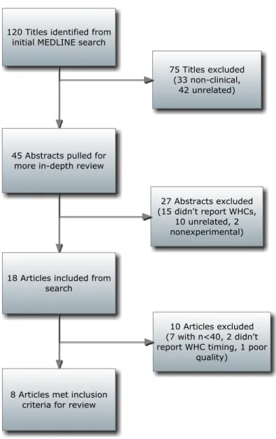
Overall, we identified eight articles that were reviewed in final analysis and included in
our review; all eight were identified through primary literature search and none through hand
review or expert inquiry (Figure 1).
12 Study Design
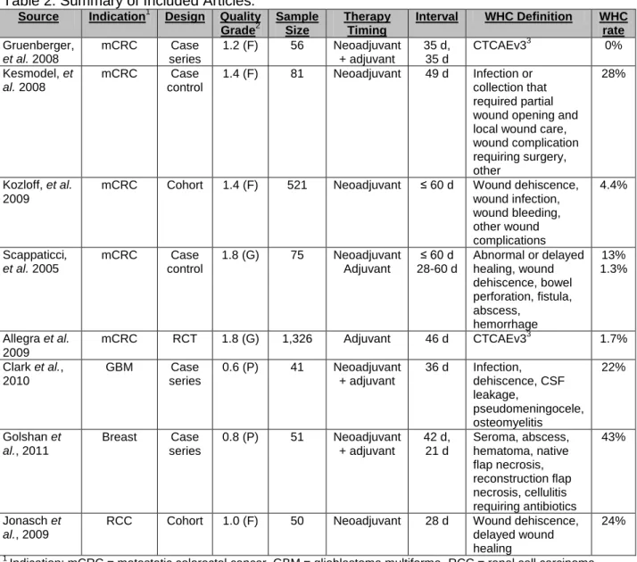
A summary of included studies is provided in Table 2. One (Allegra et al. [29]) was a
high-powered prospective randomized controlled trial. Two (Kozloff et al. [30], Jonasch et al.
[31]) were prospective cohort analyses, one (Gruenberger et al. [32]) was a prospective case
series, one (Scappaticci et al. [33]) was a retrospective case-control study, and another three
(Kesmodel et al. [34], Clark et al. [35], Golshan et al. [36]) were retrospective case series.
Table 2. Summary of Included Articles. Source Indication1 Design Quality
Grade2
Sample Size
Therapy Timing
Interval WHC Definition WHC rate
Gruenberger, et al. 2008
mCRC Case
series
1.2 (F) 56 Neoadjuvant
+ adjuvant
35 d, 35 d
CTCAEv33 0%
Kesmodel, et al. 2008
mCRC Case
control
1.4 (F) 81 Neoadjuvant 49 d Infection or
collection that required partial wound opening and local wound care, wound complication requiring surgery, other
28%
Kozloff, et al. 2009
mCRC Cohort 1.4 (F) 521 Neoadjuvant ≤ 60 d Wound dehiscence,
wound infection, wound bleeding, other wound complications 4.4% Scappaticci, et al. 2005
mCRC Case
control
1.8 (G) 75 Neoadjuvant
Adjuvant
≤ 60 d 28-60 d
Abnormal or delayed healing, wound dehiscence, bowel perforation, fistula, abscess, hemorrhage 13% 1.3%
Allegra et al. 2009
mCRC RCT 1.8 (G) 1,326 Adjuvant 46 d CTCAEv33 1.7%
Clark et al., 2010
GBM Case
series
0.6 (P) 41 Neoadjuvant
+ adjuvant
36 d Infection, dehiscence, CSF leakage, pseudomeningocele, osteomyelitis 22% Golshan et al., 2011
Breast Case
series
0.8 (P) 51 Neoadjuvant
+ adjuvant 42 d, 21 d Seroma, abscess, hematoma, native flap necrosis, reconstruction flap necrosis, cellulitis requiring antibiotics 43% Jonasch et al., 2009
RCC Cohort 1.0 (F) 50 Neoadjuvant 28 d Wound dehiscence,
delayed wound healing
24%
1
Indication: mCRC = metastatic colorectal cancer, GBM = glioblastoma multiforme, RCC = renal cell carcinoma
2 As described in the “Data Extraction and Quality Assessment” subsection of “Methods”, each study’s quality was
graded according to a composite score that ranked each component as good (G), fair (F), or poor (P)
13 Study and Source Populations and Measurement Outcomes
Included studies spanned four of bevacizumab’s five current oncological indications:
metastatic colorectal cancer (Gruenberger et al., Kozloff et al., Kesmodel et al., Scappaticci et
al., Allegra et al.), glioblastoma multiforme (Clark et al.), renal cell carcinoma (Jonasch et al.), and breast cancer (Golshan et al.). Notably, no studies met the inclusion and exclusion criteria
for the remaining indication of non-small cell lung cancer. In addition, the timing of bevacizumab
therapy ranged from exclusively neoadjuvant (Kozloff et al., Kesmodel et al., Scappaticci et al.,
Jonasch et al.) to exclusively adjuvant (Allegra et al.) to combined neoadjuvant and adjuvant
regimens (Gruenberger et al., Clark et al., Golshan et al.). The duration of the
bevacizumab-surgery interval ranged from 28-60 days in the neoadjuvant setting to 21-36 days in the adjuvant
setting.
Three included studies (Gruenberger et al., Allegra et al., Jonasch et al.) were trials
evaluating the efficacy of novel bevacizumab-containing chemotherapy regimens and therefore
had primary efficacy outcomes of all-cause mortality or progression-free survival, though
specific wound-healing complications were reported as part of an overall safety and toxicity
profile. The remaining five studies (Kesmodel et al., Kozloff et al., Scappaticci et al., Clark et al.,
Golshan et al.) were constructed to specifically examine the incidence of bevacizumab-related
healing complications and thus had primary safety outcomes of postoperative
wound-healing complication incidence.
Critical Appraisal of Included Studies
Tables 3a and 3b provide a summary of critical appraisal of the eight included studies;
14 Table 3a. Critical Appraisal of Included Studies.
Gruenberger et al. Kesmodel et al. Kozloff et al. Allegra et al.
Design Prospective, single-center, nonrandomized phase II clinical trial (case series) evaluating efficacy and toxicity of bevacizumab with capecitabine and oxaliplatin as neoadjuvant therapy for metastatic colorectal cancer Retrospective case-control analysis evaluating postoperative complication rate after neoadjuvant bevacizumab vs. chemotherapy and mCRC hepatectomy Prospective, multi-center cohort study to elucidate safety and effectiveness of bevacizumab-containing chemotherapy for mCRC
Prospective, phase III RCT to evaluate the safety and effectiveness of adjuvant
bevacizumab with FOLFOX
chemotherapy for stage II and III CRC. Source
population
Patients from Europe and United States with surgically resectable stage IV colorectal cancer metastasized to liver
Patients from the United States with isolated hepatic metastases from colorectal cancer United States patients with previously untreated mCRC United States patients with stage II and III colorectal cancer
Study population
56 patients with histologically-confirmed resectable mCRC hepatic metastases at high risk for early recurrence, with ECOG performance status 0-1 and adequate bone marrow reserve and renal and hepatic function. Exclusion criteria: prior chemotherapy, prior coagulopathy, CNS metastases, significant CV disease
125 patients who received neoadjuvant chemotherapy (44 chemo only, 81 with bevacizumab) at a median of 58 days prior to hepatic mCRC resection. Study did not report specific exclusion criteria, other than assumed surgical resectability
1,953 patients with locally advanced or metastatic CRC previously untreated receiving a bevacizumab-containing chemotherapy regimen, of which 521 subsequently underwent surgical resection within 60 days. No exclusion criteria specified
2,710 patients with stage II or III CRC randomized to receive adjuvant FOLFOX (1,356) or adjuvant FOLFOX + bevacizumab (1,354) after undergoing surgical resection. Exclusion criteria: history of stroke, TIA, vascular disease, arterial thrombosis (MI). Initial comparability of groups
Not applicable, as no control group
Comparable, as did not differ by age, stage, comorbidity, BMI, tumor size, or procedural variables. Bevacizumab group did have more metastatic liver lesions (p = 0.04).
Not applicable, as no control group
Comparable, as did not differ by age, gender, stage, and race. Study did not report patient comorbidity and procedural variables, but randomized so should be similar too Drop-outs,
adherence, cross-overs
Study did not report any drop-outs or adherence issues; cross-overs not applicable
Study did not report drop-outs, cross-overs, or adherence issues
Of the initial 1,953, 83 (4%) dropped out, 150 (8%) were lost to follow-up, and 88 (4%) withdrew
Of the 1,354 assigned to bevacizumab, 22 (1.6%) dropped out. No cross-overs Selection
bias
Intermediate potential (fair), as only 56 subjects were enrolled and assessed as “surgically resectable” and “high risk of recurrence” according to clinical judgment
Intermediate potential (fair), as subjects were initially enrolled into groups according before study by varying criteria over time
Low potential (good). To reduce selection bias, sites were instructed to recruit all eligible patients, but no non-enrolled patient log was kept
Low potential (good), as both arms were randomized after stratifying by number of positive lymph nodes
Outcome measurement
WHCs graded via CTCAEv3.0
Wound infection or collection requiring surgery, other
Wound dehiscence, infection, bleeding, and other wound complications
WHCs graded via CTCAEv3.0
Measurement bias
Low potential (good), as CTCAEv3.0 provides a systematic and consistent independent guideline to evaluate specific toxicities in oncological patients, thus
Intermediate potential (fair), as WHCs not clearly or systematically defined by
independent system,
Low potential (good). Though WHCs not independently set, high sample size would neutralize random biases from
Low potential (good), as CTCAEv3.0 provides a systematic, independent
15
equal, valid, and reliable and can vary heavily by clinician
clinical judgment, and centers agreed on same WHCs
toxicities, thus equal, valid, and reliable
Confounders Age, tumor stage, patient comorbidity that could independently affect wound-healing (obesity, diabetes, nutrition status)
Age, tumor stage, comorbidity (diabetes, CV disease), BMI, procedural variables (duration, EBL, type)
Age, tumor stage, tumor size and site, comorbidity (diabetes, PAD, BMI), procedural variables
Age, stage, tumor size and site, comorbidity (BMI, diabetes, nutrition status), procedural variables Confounding potential
High potential (poor), as study was single-arm of 56 patients and patient comorbid conditions were not reported or controlled
Low potential (good), as two groups were similar and most known confounders reported and controlled for
Low potential (good), as most known confounders were reported and controlled for in multivariate analysis
Low potential (good), as study was large and randomized, so confounders controlled for even if not reported Analysis Efficacy (tumor response)
and safety (rate of grade 3/4 adverse events)
Incidence of post-operative wound-healing complication
WHC incidence in subgroup of patients undergoing surgery
Effectiveness (PFS) and safety (rate of grade 3/4 toxicities)
Results 0% rate of grade ¾
wound-healing complications
25% chemo vs. 28% bevacizumab, p = 0.68
23/521 (4.4%, 95% CI 2.7-6.2%) WHC rate
1.7% bevacizumab vs. 0.3% FOLFOX only, p < 0.01 Clinical
importance
Significant, as
bevacizumab expands in use as perioperative therapy for oncological patients who undergo surgical resection
Significant, as bevacizumab is increasingly used as neoadjuvant therapy for hepatic mCRC
Significant, as bevacizumab is increasingly used as neoadjuvant therapy for hepatic mCRC
Significant, as bevacizumab is increasingly used in adjuvant setting for CRC, even if not metastatic Internal
validity
Good. Strict inclusion and exclusion criteria.
Fair. Moderate inclusion criteria, no exclusion criteria Fair. Inclusion criteria specified first-time treatment Good. Strict inclusion and exclusion criteria. External validity
Fair. Strict inclusion and exclusion criteria
decreased external validity, as mCRC patients who are not surgically resectable or considered to be “high risk of recurrence” were excluded, and results are less applicable to broader source population
Good. Inclusion criteria broadly-defined and no exclusion criteria, so results more
applicable to broader source population of mCRC patients with hepatic metastases
Good. Broad inclusion criteria roughly applicable to source population of mCRC patients not previously treated, with no exclusion criteria
Fair. Applicable only to stage II and III colorectal cancer patients and not stage IV metastatic, which is the population in which bevacizumab is most applied in the oncological setting Comments Although limited sample
size and high potential for confounding, study did indicate that neoadjuvant bevacizumab for hepatic metastectomy can be well-tolerated, indicating need for further research
Although
retrospective, study best controlled for known confounders and did not find increased post-operative WHC risk in bevacizumab-containing regimen, though may be underpowered due to overall small n
16 Table 3b. Critical Appraisal of Included Studies (continued).
Scappaticci et al. Clark et al. Golshan et al. Jonasch et al.
Design Retrospective, case-control analysis to assess
postoperative WHC rate after neoadjuvant bevacizumab from two randomized studies in mCRC patients
Retrospective single-center case-series to assess wound healing complication risk of second or third craniotomies for recurrent GBM
Prospective single-center trial to assess surgical morbidity of neoadjuvant
bevacizumab for triple-negative breast cancer
Prospective non-comparative phase II (cohort) trial
evaluating efficacy and safety of neoadjuvant
bevacizumab in RCC Source
population
United States patients with previously untreated mCRC considered surgically resectable United States patients with surgically-resectable recurrent GBM United States patients with triple-negative resectable breast cancer United States patients with surgically-resectable RCC Study population 1,132 previously-untreated mCRC patients: 516 got chemotherapy alone, 616 bevacizumab of which 305 received resection (230 neoadjuvant, 75 adjuvant). Exclusion criteria: major surgery up to 28 days before, vascular disease and/or coagulopathy
209 patients who underwent repeat craniotomy for recurrent GBM and had received adjuvant chemo-radiation after first resection. Exclusion: patients with implantable agents, emergent surgeries, prior scalp infection
51 patients with triple-negative breast cancer who got neoadjuvant
bevacizumab before lumpectomy (n = 29) or mastectomy (n = 22) versus 28 patients who got neoadjuvant cisplatin alone. No exclusion criteria specified.
52 patients with resectable RCC got neoadjuvant bevacizumab before cytoreductive nephrectomy 4 weeks later. Inclusion: adequate bone marrow and liver function. Exclusion: brain metastases, prior systemic therapy Initial comparability of groups
Comparable, as the two groups (chemotherapy alone, chemotherapy + bevacizumab) were pooled from two other randomized studies
Not applicable, as no control group
Groups did not differ by age, mean tumor size, or resection type (BCT vs. mastectomy), but differed by stage
Not applicable, as single-arm and no control group
Drop-outs, adherence, cross-overs
Study did not report drop-outs, cross-overs, or adherence issues
N/a. Retrospective, so no dropouts after initial exclusion
No dropouts, cross-overs, or adherence issues reported
Of 52, 2 (4%) dropped out and 8 (16%) lost to f/u Selection
bias
Low potential (good), as study pooled data from two other randomized studies using computers, which minimizes selection bias.
High potential (poor), as no consistent criteria defined or reported for why study patients got bevacizumab in addition to standard chemoradiation before or after recurrent GBM resection
Intermediate potential (fair). Both arms had similar inclusion criteria that was clearly
specified, though selection into bevacizumab arm or not was offered by clinician without oversight Intermediate potential (fair). Prespecified and clear inclusion criteria, but whether or not bevacizumab was offered
depended on clinical judgment
Outcome measurement
WHCs graded via CTCAEv3.0 Surgical site infection, wound dehiscence, CSF leakage, pseudo-meningocele, bone flap osteomyelitis Seroma, abscess, hematoma, native flap necrosis, reconstruction flap necrosis, cellulitis requiring antibiotics Wound dehiscence, delayed wound healing Measurement bias
Low potential (good), as CTCAEv3.0 provides a consistent, systematic and independent guideline, thus equal, valid, and reliable
Low potential (good), as single reviewer used same specifically-defined WHC criteria for all cases, thus equal, valid, and reliable
Intermediate potential (fair), as authors did use prespecified criteria, but criteria were broad, vague, and not graded
High potential (poor). WHC criteria were vague, especially “delayed wound healing”, so not very equal, valid, and/or reliable
Confounders Age, tumor stage, tumor size and site, comorbidity
Age, tumor stage, tumor size and
Age, tumor stage, tumor location,
17
(BMI, diabetes, CV disease), procedural variables
location, patient comorbidity (BMI, diabetes, nutrition status, CV disease), prior treatment (chemoradiation), procedural variables
patient comorbidity (BMI, diabetes, nutrition status, CV disease), prior radiation therapy, procedural variables size, patient comorbidity (BMI, diabetes, nutrition status, CV disease), prior radiation therapy, procedural data (length, etc…) Confounding
potential
Low potential (good), as data was pooled from two other randomized studies which minimizes
confounding.
High potential (poor), as possible
confounders not reported or controlled for in analysis
High potential (poor). Small study with no methodology that reported and controlled for many likely confounders
High potential (poor). Study did not report or control for most likely confounders in analysis, simply reporting crude rate
Analysis Postoperative WHC
incidence Postoperative WHC incidence Postoperative WHC incidence Postoperative WHC incidence
Results 3/230 (1.3%, 95% CI
0.3-3.8%) adjuvant; 10/75 (13%, 95% CI 7-23%) neoadjuvant
Overall 9/41 (21.9%) Neoadjuvant 8/23 34.8%), adjuvant 1/18 (5.6%)
11/28 (39%) cisplatin vs. 22/41 (43%) in bevacizumab, p = 0.82
12/50 (24%) WHC rate after
neoadjuvant bevacizumab Clinical
importance
Significant, as bevacizumab is used as adjuvant and neoadjuvant therapy for hepatic mCRC
Significant, as bevacizumab is indicated for recurrent GBM now
Significant, as bevacizumab is used for neoadjuvant therapy, and many breast cancer patients undergo immediate reconstruction Significant, as bevacizumab is increasingly used before cytoreductive nephrectomy for RCC Internal validity
Good. Strict inclusion and exclusion criteria
Fair. Results applicable to study population
Fair. Inclusion criteria defined, no exclusion criteria Good. Strict inclusion and exclusion criteria. External validity
Fair. Strict inclusion and exclusion criteria from randomized studies but no selection bias means study population approximates source population fairly
Poor. Study did not specify inclusion or exclusion criteria or indicate why patients got bevacizumab for recurrent GBM over just chemoradiation, so less applicable to overall source population of recurrent GBM patients
Fair. Clear inclusion criteria but no exclusion criteria, though should apply to source population of triple-negative breast cancer
Good. Study population
approximates source population well, as exclusion criteria did not leave out most patients in the U.S. with RCC
Comments Although retrospective, this study pooled data from two randomized and high-powered studies, with little measurement bias, and thus well evaluates WHC incidence in the
neoadjuvant and adjuvant setting
Retrospective and small study which didn’t report and control for numerous confounders, high selection bias potential, and limited external validity, these findings are uncertain
Although prospective and comparative, this study was small with no method to control for many possible
confounders and/or other biases, and as such results are very uncertain
Retrospective and small, this study didn’t control for most variables that could confound impaired wound-healing, making results very uncertain
18 Measurement Bias Potential
All studies explicitly defined what they considered a wound-healing complication, but this
definition was not necessarily consistent from one study to the next, as evidenced in Table 2.
Inconsistency and inherent subjectivity in WHC definition rendered each of the studies to be at
risk for measurement bias.
Several considerations were consistent across all studies and could have led to possible
measurement bias. First, none of the eight studies included any type of blinding when
measurements were being recorded, as providers were always aware of which patients were on
bevacizumab therapy. This introduces some potential measurement bias in all eight studies.
However, blinding may not be feasible, as wound-healing complications are recorded by the
same surgical oncologists who provide appropriate follow-up after surgical intervention, and
therefore must be aware if the patient received neoadjuvant or adjuvant bevacizumab.
Nonetheless, this consideration should be taken into account when assessing measurement
bias.
Second, the dose of bevacizumab did vary between studies but was consistent with
FDA-approved dosing. For GBM, Clark et al. used 10 mg/kg. For breast cancer, Golshan et al.
used 15 mg/kg. For mCRC, Gruenberger et al., Kozloff et al., Kesmodel et al., Allegra et al., and
Scappaticci et al. all used 5 mg/kg. Finally, for RCC, Jonasch et al. provided 10 mg/kg. The
variations in dosing introduce possible measurement bias as higher bevacizumab doses may
lead to higher reported WHCs.
Five of the eight studies had little potential for measurement bias. Gruenberger et al.,
Scappaticci et al., and Allegra et al. utilized the increasingly-favored CTCAEv3.0[37], which
provides systematic, consistent, and independently-determined guidelines to evaluate and
grade adverse reactions in oncological settings, including wound-healing. This makes
measuring WHCs equal, valid, and reliable, and thereby minimizes measurement bias. On the
19 formation, and osteomyelitis, as the location of the craniotomy incision in the scalp for GBM
warrants these specific considerations as possible sequelae secondary to impaired
wound-healing. Since a single author reviewed all cases retrospectively in this study with a
predetermined and specific set of criteria, measurement bias potential was minimized here too.
Finally, although Kozloff et al. used a broader WHC definition that included dehiscence,
infection, bleeding, and “other”, the large sample size (n = 521) means random biases from
clinical judgment are more likely to be neutralized.
Two of the eight studies had intermediate potential for measurement bias. Kesmodel et
al. defined WHCs as any “infection or collection requiring wound opening and local wound care or wound complication requiring surgery.” The stipulation that the complication would require
subsequent surgical intervention represents an objective measure that somewhat offsets the
broader and vaguer WHC criteria here. Golshan et al. defined a WHC to include seroma,
abscess, hematoma, native or reconstruction flap necrosis, and cellulitis requiring antibiotic
therapy. Including flap necrosis here is justified as many breast cancer patients undergo
reconstruction following mastectomy. However, these umbrella criteria are broader than what is
typically considered a wound-healing issue, such as abscess or cellulitis which may be
unrelated.
The remaining study, Jonasch et al., had a high potential for measurement bias as
WHCs were defined as either wound dehiscence or “delayed wound healing”; the latter
requirement is vague, qualitative, and easily variable depending on clinical judgment.
Three of the studies (Gruenberger et al., Scappaticci et al., Allegra et al.) included a
grading system with also ranked the severity of the reported WHC. Another two (Kesmodel et
al., Golshan et al.) included stipulations in their WHC criteria such that only severe
complications would be reported: Kesmodel et al. required surgical re-intervention, and Golshan
less-20 severe ones. In the lattermost group, the lack of differentiation by WHC-severity introduces bias
as less-severe WHCs (such as healing delayed by one week) are weighted as equally as
more-severe WHCs (such as meningitis or osteomyelitis).
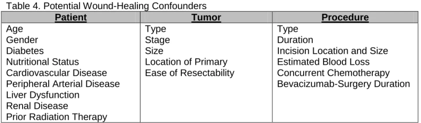
Confounding Potential
Many patient and procedural factors could independently and differentially affect the
postoperative wound-healing status in these studies, other than the presence of bevacizumab
therapy. Known confounders can be classified into patient variables, tumor variables, and
procedural variables (Table 4). Potential patient confounders include age, gender, diabetes,
hypertension, nutritional status, cardiovascular disease, peripheral arterial disease, liver
disease, acute or chronic renal disease, and prior radiation therapy. Potential tumor
confounders include type, stage, size and location of primary, and ease of resectability.
Potential procedural confounders include type, duration of surgery, estimated-blood loss,
location and size of incision, nature of concurrent chemotherapy, and duration between
bevacizumab cessation and surgical intervention.
Table 4. Potential Wound-Healing Confounders
Patient Tumor Procedure
Age Gender Diabetes
Nutritional Status Cardiovascular Disease Peripheral Arterial Disease Liver Dysfunction
Renal Disease
Prior Radiation Therapy
Type Stage Size
Location of Primary Ease of Resectability
Type Duration
Incision Location and Size Estimated Blood Loss Concurrent Chemotherapy Bevacizumab-Surgery Duration
The ability of each study to address potential confounders primarily determined the
certainty of its results. Four of the eight studies were considered to have minimal potential for
21 bevacizumab with chemotherapy against controls of neoadjuvant chemotherapy only before
hepatic metastectomy. The two groups did not differ by age, stage, numerous comorbidities
(cardiovascular disease, hypertension, pulmonary disease, renal disease, hepatobiliary disease,
diabetes, BMI), and numerous procedural variables (procedure type, number and nature of
neoadjuvant chemotherapy regimens, bevacizumab-surgery duration), although the
bevacizumab cases did have a higher number of metastatic liver lesions (p = 0.04). Since the
two groups did not differ significantly by numerous confounders that were properly reported, the
final results are more trustworthy. Kozloff et al.’s cohort study also reported numerous potential
confounders: age, race, gender, primary tumor site, mean preoperative albumin, proportion and
type of prior adjuvant therapy, site of metastatic disease, diabetes, hypertension, arterial
disease, anticoagulation therapy use, and bevacizumab-surgery duration. Many of these were
controlled for via multivariate Poisson regression analysis, and thus adequate reporting with
statistical normalization minimizes confounding here too. The other two studies (Allegra et al.
and Scappaticci et al.) were randomized and high-powered, such that even though not all
known specific confounders were reported, confounding was minimized if not eliminated.
The remaining four studies were all rated as having high potential for confounding.
Gruenberger et al. reported only basic patient information (age, gender, primary tumor site,
hepatic metastases distribution) and ignored other potential confounders including patient
comorbidity and procedural variables Similarly, Golshan et al. reported only basic patient
information, ignored most tumor and procedure confounders, and did not compare the two
groups in a formal, statistical manner. In addition, Clark et al.’s also did not report most patient,
tumor, and procedural confounders, and neither did Jonasch et al. In all four studies, the final
bevacizumab-related WHC incidence is very uncertain, as it remains difficult to attribute the
recorded WHC incidence to bevacizumab therapy as opposed to the numerous potential
22 Overall Study Quality
Overall study quality broke down as follows. Two studies were ultimately categorized as
“Good” quality: Scappaticci et al.’s case-control analysis of neoadjuvant bevacizumab before
hepatic resection of metastatic colorectal cancer and Allegra et al.’s randomized-controlled trial
of adjuvant bevacizumab after primary resection of stage II and III colorectal cancer. Four
studies were ultimately categorized as “Fair” quality: Gruenberger et al.’s case series of
neoadjuvant and adjuvant bevacizumab before and after hepatic resection of metastatic
colorectal cancer, Kesmodel et al.’s case-control analysis of neoadjuvant bevacizumab before
hepatic resection of metastatic colorectal cancer, Kozloff et al.’s cohort study of neoadjuvant
bevacizumab before hepatic resection of metastatic colorectal cancer, and Jonasch et al.’s
cohort study of neoadjuvant bevacizumab before cytoreductive nephrectomy for renal cell
carcinoma. Finally, the remaining two studies were classified as “Poor” quality”: Clark et al.’s
case series of neoadjuvant and adjuvant bevacizumab before and after repeat craniotomy for
recurrent glioblastoma multiforme, and Golshan et al.’s case series of neoadjuvant and adjuvant
bevacizumab before and after surgical resection for triple-negative breast cancer.
The Effect of Bevacizumab Therapy on Wound-Healing
The results of the “Good” studies can be concluded with the most certainty. Scappaticci
et al. observed a 1.3% (95% CI: 0.3-3.8%) WHC rate in the adjuvant setting and a 13% (95% CI: 7-23%) in the neoadjuvant setting. Allegra et al. observed a 1.7% WHC rate with adjuvant
bevacizumab, as opposed to 0.3% with adjuvant chemotherapy alone (p = 0.01). Importantly,
both studies applied the same CTCAEv3.0 criteria to define wound-healing complications. Both
yielded a WHC rate in the adjuvant bevacizumab setting that was similar and low, at ~2%. In
addition, Scappaticci et al. supports the notion that neoadjuvant bevacizumab raises the WHC
23 adjuvant bevacizumab raises the WHC risk more than adjuvant chemotherapy alone (1.7%
versus 0.3%).
The results from the “Fair” studies should be interpreted with more caution, though not
discounted altogether. Gruenberger et al. found a 0% WHC rate amongst 56 mCRC patients,
though only severe grade 3 and 4 complications were recorded, so less-severe WHCs may
have occurred but not been considered. Kesmodel et al. reported a 28% WHC rate following
neoadjuvant bevacizumab that did not differ significantly (p = 0.68) from the 25% WHC rate
following neoadjuvant chemotherapy alone. This finding disagrees with Allegra et al. which
implied a higher WHC risk than chemotherapy alone, though Kesmodel et al. occurred in the
neoadjuvant setting and Allegra et al. in the adjuvant. Kozloff et al. reported a 4.4% (95% CI:
2.7-6.2%) WHC rate after neoadjuvant bevacizumab. This is significantly smaller than
Scappaticci et al.’s 13% rate, though the difference here can be attributed (at least partially) to
differing WHC criteria. Jonasch et al. observed a 24% WHC rate after neoadjuvant
bevacizumab, significantly higher than both Scappaticci et al.’s 13% and Kozloff et al.’s 4.4%
rates, although these differences can be explained in part by both differences in WHC definition
and tumor type (RCC vs. mCRC) and procedure (cytoreductive nephrectomy vs. hepatic
metastectomy).
The results from the two “Poor” studies are highly uncertain and should be interpreted
with utmost caution if used at all. Clark et al. observed an overall WHC incidence that was
higher in the neoadjuvant setting (34.8%) than the adjuvant (5.6%), in agreement with
Scappaticci et al. Golshan et al. reported a 43% WHC incidence after neoadjuvant bevacizumab
24 Discussion
VEGF-targeted therapies, including the anti-VEGF antibody bevacizumab, are
increasingly being investigated and utilized in the treatment of various advanced-stage
malignancies, reflecting the growing understanding of VEGF-mediated angiogenesis in tumor
survival and growth. However, as VEGF also mediates many normal physiological processes,
including the vasodilation, increased vascular permeability, and angiogenesis necessary for
proper wound healing, such bevacizumab use can be expected to lead to impaired wound
healing and other wound-healing complications. The WHC risk confers special clinical
significance as bevacizumab is employed in both neoadjuvant and adjuvant settings before
and/or after surgical resection, and its anti-VEGF activity may impair wound healing of both
primary and surrounding surgical incisions. This delay in wound healing may impair patient
quality of life and in turn lead to serious sequelae including infection, hemorrhage, fasciitis,
osteomyelitis, gross necrosis, and flap and/or reconstruction loss. In addition, it necessarily
delays clinical benefit from resumption of bevacizumab therapy. Therefore, this systematic
review is an attempt to survey the current clinical evidence and assess the incidence, timing,
and nature of bevacizumab-related wound-healing complications. Such considerations should
guide clinical judgment of timing of bevacizumab therapy and management decisions for
bevacizumab-related WHCs.
Of the eight articles included in final analysis, two were judged to be “Good” quality and
another three to be “Fair”. The emerging consensus from the “Good” articles (Scappaticci et al.,
Allegra et al.) stipulates that bevacizumab does appear to raise the WHC risk more than
chemotherapy alone, and that neoadjuvant bevacizumab appears to raise the WHC risk more
than adjuvant bevacizumab. However, two considerations should be taken into account about
these conclusions.
First, the magnitude of these increases is less certain. Both the “Good” articles
25 use (1.3% vs. 1.7%), using the same WHC definition (CTCAEv3.0) in the same clinical setting
(hepatic resection for metastatic colorectal cancer). Moreover, Allegra et al. observed a 1.7%
WHC rate after adjuvant bevacizumab that was statistically-significantly different than the 0.3%
rate after adjuvant chemotherapy alone; however, the absolute increase in risk here is small,
such that the clinical significance of increased WHC risk may not be as prominent. In addition,
Scappaticci et al. observed a significant increase in WHC risk from neoadjuvant over adjuvant
bevacizumab use (13% vs. 1.3%). Clark et al. agreed with this conclusion, but with a far greater
increase (35% vs. 6%).
Second, the applicability of these conclusions to other oncological indications with other
WHC definitions is far less certain. Both “Good” articles employed the CTCAEv3.0 WHC criteria
in patients with colorectal cancer. These conclusions cannot necessarily be generalized to
breast cancer, renal cell carcinoma, glioblastoma multiforme, or non-small cell lung cancer. In
fact, the other included studies performed in these indications sometimes found differing and
opposing results. For example, both Golshan et al. (breast cancer) and Kesmodel et al. (stage
IV colorectal cancer) did not find a significant difference in WHC incidence between
bevacizumab versus chemotherapy alone, in disagreement with Allegra et al. (stage II and III
colorectal cancer). Also, Jonasch et al. (cytoreductive nephrectomy for RCC) observed a higher
WHC incidence after neoadjuvant bevacizumab than Scappaticci et al. (hepatic metastectomy
for mCRC). While these studies were judged to be less quality and their results should
consequently be interpreted with more uncertainty, it is important not to over-generalize results
from the “Good” studies.
Some of the difference in observed WHC risk between studies is attributable to varying
definitions of what constitutes a wound-healing complication. While the CTCAEv3.0 provides a
systematic and consistent guideline that was independently derived and therefore reduces
measurement bias, it is not necessarily the best applicable in all settings. WHC criteria should
26 the incision for hepatic resection occurs in the abdomen, it is justifiable to consider an incisional
hernia as a WHC here, though certainly not for a craniotomy incision occurring in the scalp.
Conversely, a poorly-healing craniotomy wound may produce osteomyelitis, CSF leakage, and
meningoencephalitis, rendering it justifiable to include these as pertinent WHCs in this setting
but not in others. Thus, the specific criteria for wound-healing complications cannot be
standardized across oncological indications, as each indication results in a different procedure
for surgical resection, with an incision that differs by location and size, and a different profile of
potentially-harmful sequelae secondary to impaired wound-healing. As such, meta-analysis of
WHC risk across indication remains not possible.
WHC criteria should be defined for future research purposes. Ideally, these criteria
should be standardized within each oncological indication, independently-constructed by groups
or committees other than the researching investigator, explicitly defined before initiation of the
study, applied consistently to cases and controls, and as objective as possible, to minimize
measurement bias. Furthermore, there should be some grading of WHCs to also report the
severe complications, as is currently done in the CTCAEv3.0. Wound-healing complications can
range from the somewhat mild (such as delayed wound-healing) to the very severe (such as
skull flap necrosis producing meningoencephalitis), and it would be very helpful for providers to
know both the rate of overall WHC and the rate of severe WHC when considering the toxicity
profile of bevacizumab therapy.
Many variables can confound the relationship between treatment and wound-healing.
This review tried to systematically organize them into three categories: patient variables, tumor
variables, and procedural variables. Several of the included studies attempted to methodically
report and control for these potential confounders, thus strengthening their final analysis, while
other studies simply ignored them, making their reported WHC risk far less attributable to the
27 trying to assess the bevacizumab-related WHC risk, especially in the setting of oncological
patients, many of whom suffer from high and complex comorbidity.
General recommendations have already been provided regarding timing of bevacizumab
therapy and onset of surgical intervention: bevacizumab therapy should occur at least 60 days
before or 28 days after surgery[7, 33], should not be initiated until all surgical wounds are fully
healed[7], and should be permanently discontinued if wound dehiscence occurs.[20]
Bevacizumab treatment should also be withheld prior to elective surgery, though the interval
here has not yet been optimized, with some recommending 4 weeks[7] and others 6-8
weeks.[38] These recommendations rely chiefly on preclinical pathophysiological evidence, as
bevacizumab’s long circulating half-life of ~20 days[5] necessitates an appropriate interval
between treatment cessation and surgery to adequately prevent toxicities resulting from
inhibition of VEGF-mediated physiological processes, including wound-healing complications.
Thus, surgical oncologists apply conservative estimates of the bevacizumab-surgery
interval, though these recommendations are not based on current clinical evidence. Importantly,
it should be noted that any clinical benefit obtained by extending this interval and delaying
bevacizumab therapy to minimize toxicity is necessarily offset by loss of bevacizumab’s
therapeutic anti-tumor potential. Therefore, optimizing the duration of the bevacizumab-surgery
interval remains an important goal for current and future research. None of the included studies
specifically examined how the duration of this interval correlated with WHC risk.
The current clinical evidence is unfortunately not of high enough quality to change or
refine these management guidelines. Future research is required to assess the WHC risk of
neoadjuvant and adjuvant bevacizumab therapy and to compare these risks to that of
chemotherapy alone within each oncological indication. Future research is also required to
evaluate how the duration of the bevacizumab-surgery interval correlates with increased WHC
risk, if at all. Ideally, such studies would consist of prospective and high-powered trials within
28 cases (patients who received bevacizumab) and controls (patients who didn’t). Furthermore,
such studies should control for confounding (Table 4), either by multivariate analysis or ideally
through randomization. In addition, such studies should employ clearly-defined,
independently-constructed WHC criteria that include some grading scheme to specify severe WHCs as well.
Until then, the decision to proceed with surgical wound repair a certain time after bevacizumab
cessation should depend on the clinical judgment and expertise of the managing
multi-disciplinary team (including both plastic surgeons and oncologists) that weighs the potential
costs of preventable bevacizumab-associated toxicities against the potential anti-tumor benefits
of bevacizumab use, along with knowledge of the patient’s underlying characteristics and
29 Works Cited
1. Ferrara, N., H.P. Gerber, and J. LeCouter, The biology of VEGF and its receptors. Nat Med, 2003.
9(6): p. 669-76.
2. Folkman, J., Tumor angiogenesis: therapeutic implications. N Engl J Med, 1971. 285(21): p. 1182-6.
3. Inai, T., et al., Inhibition of vascular endothelial growth factor (VEGF) signaling in cancer causes loss of endothelial fenestrations, regression of tumor vessels, and appearance of basement membrane ghosts. Am J Pathol, 2004. 165(1): p. 35-52.
4. Longo, R., et al., Anti-angiogenic therapy: rationale, challenges and clinical studies.
Angiogenesis, 2002. 5(4): p. 237-56.
5. Ignoffo, R.J., Overview of bevacizumab: a new cancer therapeutic strategy targeting vascular endothelial growth factor. Am J Health Syst Pharm, 2004. 61(21 Suppl 5): p. S21-6.
6. Zondor, S.D. and P.J. Medina, Bevacizumab: an angiogenesis inhibitor with efficacy in colorectal and other malignancies. Ann Pharmacother, 2004. 38(7-8): p. 1258-64.
7. Avastin (bevacizumab) package insert. 2008, Genentech, Inc.: San Francisco.
8. Jenab-Wolcott, J. and B.J. Giantonio, Bevacizumab: current indications and future development for management of solid tumors. Expert Opin Biol Ther, 2009. 9(4): p. 507-17.
9. Pazdur, R., Regulatory Decision to Withdraw Avastin (bevacizumab) First-line Metastatic Breast Cancer Indication, FDA, Editor. 2010.
10. Klastersky, J., Adverse effects of the humanized antibodies used as cancer therapeutics. Curr Opin Oncol, 2006. 18(4): p. 316-20.
11. Moore, D.H., Chemotherapy for advanced, recurrent, and metastatic cervical cancer. J Natl Compr Canc Netw, 2008. 6(1): p. 53-7.
12. Aragon-Ching, J.B. and W.L. Dahut, The role of angiogenesis inhibitors in prostate cancer. Cancer J, 2008. 14(1): p. 20-5.
13. Burger, R.A., et al., Phase II trial of bevacizumab in persistent or recurrent epithelial ovarian cancer or primary peritoneal cancer: a Gynecologic Oncology Group Study. J Clin Oncol, 2007.
25(33): p. 5165-71.
14. Cannistra, S.A., et al., Phase II study of bevacizumab in patients with platinum-resistant ovarian cancer or peritoneal serous cancer. J Clin Oncol, 2007. 25(33): p. 5180-6.
15. Ellis, L.M. and D.J. Hicklin, VEGF-targeted therapy: mechanisms of anti-tumour activity. Nat Rev Cancer, 2008. 8(8): p. 579-91.
16. Dvorak, H.F., Vascular permeability factor/vascular endothelial growth factor: a critical cytokine in tumor angiogenesis and a potential target for diagnosis and therapy. J Clin Oncol, 2002.
20(21): p. 4368-80.
17. Kaplan, R.N., et al., VEGFR1-positive haematopoietic bone marrow progenitors initiate the pre-metastatic niche. Nature, 2005. 438(7069): p. 820-7.
18. Hood, J.D., et al., VEGF upregulates ecNOS message, protein, and NO production in human endothelial cells. Am J Physiol, 1998. 274(3 Pt 2): p. H1054-8.
19. Kamba, T., et al., VEGF-dependent plasticity of fenestrated capillaries in the normal adult microvasculature. Am J Physiol Heart Circ Physiol, 2006. 290(2): p. H560-76.
20. Gressett, S.M. and S.R. Shah, Intricacies of bevacizumab-induced toxicities and their management. Ann Pharmacother, 2009. 43(3): p. 490-501.
21. Shord, S.S., et al., Understanding and managing the possible adverse effects associated with bevacizumab. Am J Health Syst Pharm, 2009. 66(11): p. 999-1013.
30 23. Kamba, T. and D.M. McDonald, Mechanisms of adverse effects of anti-VEGF therapy for cancer.
Br J Cancer, 2007. 96(12): p. 1788-95.
24. Lemmens, L., V. Claes, and M. Uzzell, Managing patients with metastatic colorectal cancer on bevacizumab. Br J Nurs, 2008. 17(15): p. 944-9.
25. Drinkwater, S.L., et al., Effect of venous ulcer exudates on angiogenesis in vitro. Br J Surg, 2002.
89(6): p. 709-13.
26. Bates, D.O. and R.O. Jones, The role of vascular endothelial growth factor in wound healing. Int J Low Extrem Wounds, 2003. 2(2): p. 107-20.
27. Brown, L.F., et al., Expression of vascular permeability factor (vascular endothelial growth factor) by epidermal keratinocytes during wound healing. J Exp Med, 1992. 176(5): p. 1375-9.
28. Li, J., Y.P. Zhang, and R.S. Kirsner, Angiogenesis in wound repair: angiogenic growth factors and the extracellular matrix. Microsc Res Tech, 2003. 60(1): p. 107-14.
29. Allegra, C.J., et al., Initial safety report of NSABP C-08: A randomized phase III study of modified FOLFOX6 with or without bevacizumab for the adjuvant treatment of patients with stage II or III colon cancer. J Clin Oncol, 2009. 27(20): p. 3385-90.
30. Kozloff, M., et al., Clinical outcomes associated with bevacizumab-containing treatment of metastatic colorectal cancer: the BRiTE observational cohort study. Oncologist, 2009. 14(9): p. 862-70.
31. Jonasch, E., et al., Phase II presurgical feasibility study of bevacizumab in untreated patients with metastatic renal cell carcinoma. J Clin Oncol, 2009. 27(25): p. 4076-81.
32. Gruenberger, B., et al., Bevacizumab, capecitabine, and oxaliplatin as neoadjuvant therapy for patients with potentially curable metastatic colorectal cancer. J Clin Oncol, 2008. 26(11): p. 1830-5.
33. Scappaticci, F.A., et al., Surgical wound healing complications in metastatic colorectal cancer patients treated with bevacizumab. J Surg Oncol, 2005. 91(3): p. 173-80.
34. Kesmodel, S.B., et al., Preoperative bevacizumab does not significantly increase postoperative complication rates in patients undergoing hepatic surgery for colorectal cancer liver metastases.
J Clin Oncol, 2008. 26(32): p. 5254-60.
35. Clark, A.J., et al., Impact of bevacizumab chemotherapy on craniotomy wound healing. J Neurosurg, 2010.
36. Golshan, M., et al., Does Neoadjuvant Bevacizumab Increase Surgical Complications in Breast Surgery? Ann Surg Oncol, 2010.
37. Trotti, A., et al., CTCAE v3.0: development of a comprehensive grading system for the adverse effects of cancer treatment. Semin Radiat Oncol, 2003. 13(3): p. 176-81.