ORIGINAL ARTICLE
Characterization of an immunologic polymorphism (D79H)
in the heavy chain of factor V
M . V A N D E R N E U T K O L F S C H O T E N , * R . J . D I R V E N , * S . R . P O O R T , * R . V A N W I J K , H . L . V O S , * F . R . R O S E N D A A L *à and R . M . B E R T I N A *
*Hemostasis and Thrombosis Research Center, Department of Hematology, LUMC, Leiden; Department of Clinical Chemistry, UMC, Utrecht, àDepartment of Clinical Epidemiology, LUMC, Leiden, the Netherlands
To cite this article:van der Neut Kolfschoten M, Dirven RJ, Poort SR, Van Wijk R, Vos HL, Rosendaal FR, Bertina RM. Characterization of an immunologic polymorphism (D79H) in the heavy chain of factor V.J Thromb Haemost2004;2: 910–17.
Summary. Background: During the study of a family with hereditary factor (F)V deficiency (FV Amersfoort, 1102 A > Tin exon 7) we identified an individual with 5% FV heavy chain antigen (FVHC) and 50% FV light
chain antigen (FVLC). Further testing revealed that apart
from the FV Amersfoort allele a second variant FV allele was segregating in this family, which encodes for a FV molecule with a reduced affinity for mAb V-23 used in the FV heavy chain ELISA (ELISAHC). Objective:
Identifica-tion and characterizaIdentifica-tion of the molecular basis responsible for the reduced affinity of the variant FV for mAb V-23.
Methods: Family members of the proband were screened for mutations in the exons coding for the heavy chain of FV, after which the recombinant variant FV could be generated and characterized. Next, the cases and controls of the Leiden Thrombophilia Study (LETS) were genotyped for carriership of the variant FV.Results: In the variant FV allele a polymorph-ism in exon 3 (409G > C) was identified, which predicts the replacement of aspartic acid 79 by histidin (D79H). Introduc-tion of this mutaIntroduc-tion in recombinant FV confirmed that it reduces the affinity for binding to mAb V-23. The substitution has no effect on FV(a) stability and Xa-cofactor activity. In Caucasians the frequency of the FV-79H allele is 5%. Analysis of the LETS revealed that the FV-79H allele is not associated with FV levels (FVLC), activated protein C sensitivity
(using an activated partial thromboplastin time-based test) or risk of venous thrombosis (OR 1.07, CI 95: 0.7–1.7).
Conclusion: The D79H substitution in FV should be consid-ered as a neutral polymorphism. The monoclonal antibody V-23, which has a strongly reduced affinity for FV-79H, is not suitable for application in diagnostic tests.
Keywords: APC-resistance, factor V, polymorphism, venous thrombosis.
Introduction
Human coagulation factor (F)V is a large plasma glycoprotein that plays an important role in blood coagulation. In plasma, FV circulates as a single-chain procofactor and is activated by thrombin via limited proteolysis [1–3]. Activated FV (FVa) consists of two polypeptide chains (designated as heavy and light chain), which are non-covalently linked via a Ca2+ion [4]. FVa serves as the non-enzymatic cofactor of the serine protease Xa, which is responsible for the proteolytic activation of prothrombin in the prothrombinase complex, consisting of FVa, FXa, and negatively charged phospholipids. Down-regulation of FVa is mediated by activated protein C (APC) through proteolytic cleavage of the heavy chain of FVa at Arg306, Arg506 and Arg679 [5]. Procofactor FV contains also APC-cofactor activity in the inactivation of FVIIIa [6,7].
FV plays an important role both in the up and down-regulation of thrombin generation (see also reviews [8] and [9]). This dual role of FV is reflected in the disorders associated with molecular defects in FV, which can be either hemorrhagic or thrombotic. A mild to severe bleeding tendency has been reported in patients with nonsense or missense mutations in the FV gene (F5) which prevent FV synthesis or result in dysfunctional FV (a large selection of FV mutations is summarized in references [8] and [10]). On the other hand the common R506Q mutation (FV Leiden), which impairs degra-dation of FVa by APC, is associated with an increased risk of venous thrombosis [11–13].
Measurement of FV antigen levels is an important tool in the laboratory diagnosis of FV deficiency (parahemophilia), which is a rare autosomal recessive bleeding disorder with an estimated prevalence of 1–1000 000 [14]. Also, in the laboratory screening of thrombophilia FV antigen measurements are important, as compound heterozygosity for FV Leiden and a FV null allele results in a pseudo-homozygous phenotype (severe APC-resistance) [15–20].
Correspondence: Dr R. M. Bertina, Hemostasis and Thrombosis Research Center, Department of Hematology, Leiden University Medical Center, C2-R, PO Box9600, 2300RC, Leiden, the Netherlands. Tel.: +31 71 5261894; fax: +31 71 5266755; e-mail: rmbertina@ lumc.nl
Several immunologic assays for the measurement of FV in plasma are available. In our laboratory we have developed two enzyme-linked immunosorbent assays (ELISA) for measuring FV antigen, one for the light chain and one for the heavy chain. The light chain ELISA (ELISALC) has
first been described by Guasch et al. [21] and uses two murine monoclonal antibodies (mAbs), V6 and V9, the epitopes of which have been mapped on the light chain of FVa. The heavy chain ELISA (ELISAHC) uses two murine
mAbs (V-23 and V39) directed against the heavy chain of FVa.
During the study of the family of an asymptomatic heterozygous carrier of the FV Amersfoort allele (1102 A > Tin exon 7 [22]), we observed a striking discrepancy between FV light chain and heavy chain antigen levels in some of the family members. Further analysis showed that this abnormality cosegregated with a 409G > C transversion in exon 3 ofF5
predicting an amino acid substitution (D79H) in the heavy chain of FV (first reported by Cargillet al. [23]). So far, the effect of the D79H substitution on the function of FV is not clear. Recently, however, it has been reported that carriership of FV-79H is associated with moderately decreased FV levels and may serve as a trans-acting gene mutation in heterozygous carriers of FV Leiden by lowering the sensitivity of plasma to APC [24]. The latter observations prompted us to further investigate the effects of the D79H substitution on FV function, FV levels, APC-sensitivity ratios and risk of venous thrombosis.
Materials and methods
Materials
Plasmid isolation kits were from Qiagen (Chatsworth, CA, USA). DNA restriction fragments were purified from agarose gel using the Cleanmix kit (Talent, Trieste, Italy). Bovine Serum Albumin (BSA), benzamidine, phosphatidyl-choline (PC), N-hydroxy-succinimidobiotin was from Sigma (St. Louis, MO, USA). Dioleoylphospatidylcholine (DOPC) and dioleoylphospatidylserine (DOPS) were obtained from Avanti Polar Lipids (Alabaster, Al, USA). dNTP were from Serva (Heidelberg, Germany). Chromogenic sub-strate S-2238 was obtained from Chromogenix (Uppsala, Sweden).
Proteins
Restriction enzymes were from New England Biolabs (Beverly, MA, USA). Human FXa was from Hematologic Technologies Inc. (Essex, VT, USA). Amplitaq DNA polymerase was from Applied Biosystems (Foster City, CA, USA). Activated protein C, prothrombin, thrombin and sheep anti-FV polyclonal antibody were purchased from Enzyme Research Laboratories (South Bend, IN, USA). NeutraliteTMAvidin-HRP conjugate was from Southern Biotechnology Associated Inc. (Birming-ham, AL, USA).
FV deficient family
We studied the family of an asymptomatic FV deficient individual who was heterozygous for an 1102-A > Tsubsti-tution in exon 7 (FV Amersfoort, see van Wijket al. [22]).
FV antigen assays
FV light chain antigen was detected in an ELISA using two different monoclonal antibodies (mAbs) directed against the light chain of FV as described before [21,25]. In this ELISA, mAb V-6 was used as a coating antibody and biotinylated mAb V-9 as tagging antibody. FV heavy chain antigen was detected in an ELISA recently developed in our laboratory. This ELISA shows identical dose–response curves for plasma and serum FV, indicating that it identifies a fragment of the heavy chain of FVa, which is not cleaved during activation or inactivation of FV(a). Monoclonal antibody mAb V-23 was used as coating antibody and biotinylated mAb V-39 as tagging antibody. For both light chain and heavy chain ELISA, samples were diluted in 50 mM triethanolamine
(TEA), 100 mM NaCl, 10 mM EDTA and 0.1% Tween,
pH 7.5. Dilutions of pooled normal plasma (1 : 25–1 : 1600 dilution) were used to calibrate the assays. Results are expressed in percentage, where 100% refers to the concen-tration of FV in pooled normal plasma.
DNA amplification and sequence analysis
The exons coding for the heavy chain of FV were amplified according to standard polymerase chain reaction (PCR) procedures using 0.1–1lg genomic DNA. Details about primers and conditions are available on request ([email protected]). Sequence reactions were performed using the CEQ2000TM Dye Terminator Cycle Sequencing Kit (Beckman Coulter Inc., Fullerton, CA, USA) according to the instructions of the manufacturer and subsequently analyzed on a SEQTM8000 Genetic Analysis System (Beckman Coulter Inc., Fullerton, CA, USA). Nucleotides of the cDNA were numbered according to Jennyet al. [26].
Mutagenesis
Expression vector pMT2FV [27] was used as template to introduce the D79H (409G > C) variation in the heavy chain of FV. Mutations were introduced using the QuikchangeTM Site-Directed Mutagenesis Kit (Stratagene, La Jolla, CA, USA). The forward mutagenic oligonucleotide used for construction of rFV-79H was:
5¢-GTTCACTTTAA
G
AATAAGGCAC
ATAAGCCCTT-GAGC-3¢The nucleotide responsible for the missense mutation is presented in bold and underlined. In this oligo an additional silent mutation (in bold) was introduced, which removes aDraI
sequencing. Finally, the mutated fragment was inserted into a non-mutagenized pMT2FV by exchange of the appropriate restriction enzyme fragments. Details about this procedure are available on request.
Transient expression of recombinant FV
Recombinant FV was transiently expressed in COS-1 cells (75 cm2culture flasks) using Fugene 6 Transfection Reagent (Roche Molecular Biochemicals, Hague Road, IN, USA). Twenty-four hours after transfection cells were washed with phosphate-buffered saline (PBS) and incubated with serum-free medium (Optimem Glutamax, Life Technologies Ltd, Paisley, UK). Conditioned medium was harvested after 72 h, centrifuged for 20 min at 1000 r.p.m. (200g) (4C) and frozen at)20C.
FV activity assay
Total FV activity was measured in a two-step procedure as described before [28,29]. Briefly, FV was completely activated by 5 nMthrombin in HBS-Ca (25 mMHepes, 175 mMNaCl,
3 mM CaCl2, 5 mg mL)1 BSA, pH 7.5) during a 20-min
incubation at 37C in a final volume of 150lL. Subsequently, 10lL was transferred to a 96 wells plate, 100lL of prothrombinase mix (HBS-Ca containing FXa and DOPS/ PC 10 : 90 molar ratio) was added and after 5 min the prothrombinase reaction was started by addition of 25lL prothrombin (1.47lM) in HBS-Ca. Final concentrations were:
FXa (125 pM), prothrombin (280 nM) and DOPS/PC (50lM).
After a 4-min incubation at room temperature the reaction was stopped with 50lL TN-EDTA (50 mMTRIS, 175 mMNaCl,
50 mMEDTA, 5 mg mL)1BSA, pH 7.9). After the addition
of 25lL chromogenic substrate S-2238 (2.35 mM), the amount
of thrombin formed was measured kinetically on a Spectra III Thermo microtiter plate reader from Tecan (Salzburg, Aus-tria). The assay was calibrated using dilutions of pooled normal plasma (PNP) corresponding with 0–3 pMof FV.
FV(a) stability
FVa stability was assessed by following the spontaneous loss of Xa-cofactor activity as a function of time in the presence and absence of phospholipids as described before [30]. Spontaneous inactivation rates were obtained by fitting the curves in a single-exponential model using non-linear least-squares analysis.
Synthetic peptides overlapping region of amino acid 79
Two synthetic peptides were constructed homologous to the amino acid sequence of human FV with either an aspartic acid or histidine at position 79:
PEP79D: 71-KVHFKNKA
D
KPLSIH-85 (D at residue 79) PEP79H: 71-KVHFKNKAH
KPLSIH-85 (H at residue 79) The affinity of these peptides for mAb V-23 was assessed from experiments in which plasma FV was used as competitor.In brief, a 96-wells plate was coated with mAb V-23, after which the wells were incubated with pooled normal plasma (1/300 diluted in a buffer containing 50 mM TEA, 100 mM
NaCl, 10 mMEDTA and 0.1% Tween, pH 7.5), mixed 1 : 1
with increasing concentrations of peptides in 50 mM TEA,
100 mMNaCl, pH 7.5 (final concentration 1 mM). After
wash-ing with ELISA buffer, the plate was incubated with biotinylated sheep antihuman FV polyclonal antibody (1 : 500 dilution), which was detected with Neutralite Avidin-HRP. In the final step, the plate was colored with 3,3¢,5,5¢-tetramethylbenzidine.
Study population
Patients and control subjects included in the present study were from a large population-based case-control study on risk factors for a first venous thrombosis, the Leiden Thrombo-philia Study (LETS), which has been described in detail before [31,32].
Detection of 409G > C transversion in genomic DNA
Carriership of the 409G > C transversion in exon 3 of theF5
(corresponding to the D79H mutation) was detected by PCR followed by restriction enzyme digestion. The PCR was performed using 0.1–1lg genomic DNA, 200 ng primers, 200lMdNTP and 1 University of TaqDNA polymerase in
PCR buffer containing 67 mM TRIS-HCl (pH 8.8), 6.7 mM
MgCl2, 6.7lM EDTA, 16.6 mM (NH4)2SO4, 0.1 mg mL)1
BSA, 10% DMSO and 10 mM,-mercapto-ethanol. A 208-bp
fragment was amplified with the following primers:
Primer A: (5¢ -GGAGGAATGGTAGCAATCACTCTT-GG-3¢)
Primer B (5¢ -TCCTTGAGGATGGATGCTCAGATGCT-TAT-3¢)
In primer B a mismatch sequence (underlined) was introduced to generate aBsaBI restriction site after amplification of the FV-79D allele. Digestion of this fragment byBsaBIresulted in cleavage products of 25 bp and 183 bp, whereas the amplified fragment of the FV-79H allele was not digested byBsaBI.
APC-resistance test
The activated partial thromboplastin time (APTT)-based APC-sensitivity ratio (APC-SR) was measured as described by Kosteret al. [31]. APC-sensitivity ratios were normalized by dividing them by the APC-SR of pooled normal plasma.
Statistical analysis
Results
FV activity and antigen in FV deficient family
FV Amersfoort is characterized by the 1102 A > Tsubstitu-tion in exon 7 ofF5, resulting in a premature stop at codon 310 [22]. To investigate whether the truncated FV protein (amino acids 1–310) is expressed in plasma, we measured FV heavy chain antigen (FVHC) and FV light chain antigen (FVLC) in
plasma of an asymptomatic heterozygous carrier of FV Amersfoort and his first-degree relatives. The results are summarized in Table 1. In none of the FV Amersfoort carriers (father, child 1 and child 2) an excess of FVHCwas observed,
because in none of the plasma samples the FVHC/FVLCratio
exceeded 1. Remarkably, in some individuals the FVHClevel
was much lower than the FVLC level, resulting in strongly
reduced FVHC/FVLCratios. The mother, child 1, 2 and 4 all
seemed to be heterozygous for a variant FV molecule, which is poorly recognized by one or both mAbs used in the ELISAHC.
To test this, three additional FV ELISAs were designed using either mAb V-23, V-39 or V-6 as catching antibody and a polyclonal anti FV conjugate as tagging antibody. Subse-quently, plasma of child 1 (FVHC/FVLC ratio 0.07) was
analyzed with these ELISAs. The FV antigen levels were 0%, 41% and 61% as measured with ELISAV-23, ELISAV-39and
ELISAV-6, respectively, which indicates that mAb V-23 poorly
recognizes FV in this individual.
Identification of an immunologic polymorphism in the heavy chain of FV
The ÔabsenceÕ of an epitope for mAb V-23 in the FV of some members of this FV deficient family most likely is the result of an amino acid substitution in the heavy chain of FV. Therefore, we sequenced the exons coding for the heavy chain of FV of the father and the mother. In total five nucleotide changes with respect to the sequence of Jenny
et al. were identified (summarized in Table 2) including the FV Amersfoort mutation in exon 7. From the data in Table 1 and Table 2 haplotypes were constructed (Fig. 1). Carriership of the haplotype 327 A, 409C, 495 A, 642T, 1102 A cosegregated with reduced FVHC/FVLC ratios.
Within this haplotype only the 409G > C polymorphism in exon 3 predicts an amino acid substitution (D79H). This
made the D79H substitution a good candidate for the mutation that causes the reduced affinity for mAb V-23. It should be noted that the 409G > C transversion is also present in the FV Amersfoort allele (1102T), which is not expressed in plasma. Therefore, some members of this family are heterozygous for the D79H substitution, whereas they have a normal FVHC/FVLC ratio (i.e. child 2).
Analysis of the epitope of mAb V-23
To confirm that the molecular basis of the reduced affinity of FV for mAb V-23 resides in the D79H mutation, we introduced this mutation in an expression vector containing the cDNA of human FV (pMT2-FV). After transfection of COS-1 cells, conditioned medium containing recombinant FV was collected and analyzed (Table 3). The expression levels of both rFV-79D and rFV-79H were similar as measured by functional FV assay and ELISALC (7% of
normal plasma level), which indicates that rFV-79H has a normal specific activity (FVa/FVLC). However, using the
ELISAHC very low FV levels were detected in medium
containing rFV-79H, while the FVHC in the medium of
rFV79D was similar to the FVLC. This confirmed that the
ELISAHC was affected by the D79H substitution, which
most likely disrupts the epitope of mAb V-23 (see also first section of Results).
We tried to obtain more information about the epitope of mAb V-23 using synthetic peptides. Two peptides (PEP-79D and PEP-79H) homologous to the region surrounding amino acid 79 were synthesized and tested for their ability to compete with plasma FV for binding to mAb V-23. However, peptide concentrations up to 1 mg mL)1did not affect the binding of plasma FV to mAb V-23 (data not shown).
The region surrounding residue 79 was also analyzed in a 3-dimensional model for the heavy chain of FVa [33]. This model revealed that this region is surface exposed, indicating that it might contain an epitope for an antibody. Replacement of 79D by 79H did not result in conformational changes in the experimental FVa model (data not shown), which suggests that
Table 1 FV activity and antigen levels in members of FV deficient family
Family FV activity2(%) FVLC(%) FVHC(%) FVHC/FVLC
Father1 34 51 41 0.80
Mother 125 136 89 0.65
Child 11 62 69 5 0.07
Child 21 60 64 65 1.02
Child 3 109 142 69 0.49
Child 4 93 136 57 0.42
1
Heterozygous carrier of the FV Amersfoort allele.2FV activity was measured using a one stage clotting assay (data from van Wijket al. [22]).
Table 2 Polymorphisms in exons coding for FV heavy chain in members of FV deficient family
Exon (Amino acid change)
Nucleotide
change Father Mother
Child 1
Child 2
Child 3
Child 4
Exon 2 327G > A (Q51Q)
GA AA AA AA GA GA
Exon 3 409G > C (D79H)
GC GC CC GC GC GC
Exon 4 495G > A (A107A)
GA GA AA GA GA GA
Exon 4 642G > T (S156S)
GTGT TTGTGTGT
Exon 7 1102 A > T1 (K310Term)
AT AA ATATAA AA
the D79H substitution affects electrostatic interactions between the epitope and mAb V-23.
Finally, conservation of region surrounding amino acid 79 was analyzed between different species. In Table 4 the amino acid sequences surrounding residue 79 (residues 71–85) of 7 different species are shown. Homology ranges from 60% for the zebrafish to 93% for some mammals. The aspartic acid at position 79 was conserved between several species, but not in cow and pipid frog.
Further characterization of the D79H substitution
Recently, it has been reported that carriership of the FV-79H allele is associated with moderately reduced FV activity in plasma [24], as measured in a one stage clotting assay. Also, indications have been obtained that carriership of the FV-79H allele may lower the APC sensitivity in subjects carrying the FV Leiden allele [24]. Therefore, it may act as atrans-acting gene in
carriers of FV Leiden in a similar way as has been demonstra-ted for the FV-R2 allele [34–37]. To further investigate this, we genotyped the cases and controls from a large population-based case-control study on risk factors for venous thrombosis (LETS) and determined the effect on FV levels, n-APC-SR and risk of thrombosis.
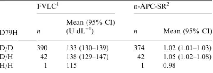
To investigate the effect of the D79H substitution on plasma FV levels, we used the data from the 433 healthy controls from the LETS. Carriers of the FV-R2 allele were excluded from this analysis (no compound heterozygous carriers of the FV-79H allele and FV-R2 were found in this group). Table 5 shows that the mean FVLC level in carriers
of the FV-79H allele (138 U dL)1) is similar to that in
non-carriers (133 U dL)1).
Fig. 1.Pedigree of FV deficient family. In this pedigree FV deficiency (FV Amersfoort) and reduced FV(HC)/FV(LC) antigen ratios (Table 1) are indicated together with the genotypes for the polymorphisms found in the exons coding for the heavy chain (Table 2). The proband of this family is child 1.
Table 3 Specific activity and FVHC/FVLCratio of recombinant FV-79D and FV-79H. FV activity and FV antigen (FVHCand FVLC) were meas-ured in conditioned medium containing either rFV-79D or rFV-79H
rFV FVa/FVLC1 FVHC/FVLC
rFV-79D 0.94 0.83
rFV-79H 1.04 0.06
1
FV activity was measured as described in Materials and methods.
Table 4 Alignment of amino acid sequence surrounding residue 79 in the heavy chain of FV. Conservation of the region surrounding position 79 (residue 71–85) in the heavy chain of FV was assessed by comparing the human amino acid sequence with the amino acid sequence of 6 different species. Position 79 is bold and underlined
Species FV amino acid sequence Homology
Theoretically, the reduced procoagulant FV activity reported by Bossoneet al. [24] for carriers of FV-79H may be caused by increased instability of the activity of FV-79H. To assess this possibility, the loss of (activated) rFV activity was followed as a function of time. The first-order rate constant for spontaneous inactivation (ks) of rFV-79D was similar to that for rFV-79H,
being around 3.0·10)6s)1(Table 6). Also theksfor activated
rFV-79D was similar to that for activated rFV-79H, being around 3.0·10)5s)1. The presence of phospholipids did not influence the stability of the activated rFV molecules.
Next, we investigated the effect of the D79H substitution on the APTT-based normalized-APC sensitivity ratio [31]. For this analysis carriers of FV Leiden were excluded. The n-APC-SRs in the absence of the FV Leiden mutation are summarized in Table 5 and demonstrate that carriership of the FV-79H allele is not associated with a lower n-APC-SR. Exclusion of FV-R2 carriers did not alter this result (data not shown). In this analysis, it was not possible to investigate whether carriership of FV-Leiden intranswith the FV-79H allele is associated with a further decrease of the n-APC-SR, because none of the healthy controls were compound heterzozygous for FV-Leiden and FV-79H. Therefore, we screened all patients in the LETS for compound heterozygosity for FV-Leiden and FV-79H. In total 8 patients were found compound heterozygous for FV-Leiden and FV-79H with a mean n-APC-SR of 0.57 (range 0.53–0.64). Finally, we analyzed the association of the D79H substitu-tion with risk of thrombosis. For this analysis all cases and
controls of the LETS were genotyped for this polymorphism. The allele frequency of the FV-79H allele was 5.0% in patients and 4.7% in controls (Table 7), which is in agreement with previously reported allele frequencies (5–15%) in different healthy Caucasian populations [23,24]. The odds ratio (OR), calculated as a measure of the relative risk of venous thrombosis, for subjects carrying the FV-79H allele (both heterozygous and homozygous) was 1.07 (CI 95: 0.7–1.7) compared to homozygous FV-79D carriers. Exclusion of all FV Leiden carriers did not alter this OR.
Discussion
In this study, we have analyzed a variant FV that was not recognized by a monoclonal antibody (mAb V-23) directed against the heavy chain of FV. Haplotype analysis of the variant FV allele suggested that an amino acid substitution at residue 79 (D79H) was responsible for the absence of the epitope for mAb V-23. This was confirmed by introducing this substitution in recombinant FV (rFV-79H). Functional tests with rFV-79H showed that this substitution did not affect the Xa-cofactor function or the stability of FV(a). Furthermore, carriership of the FV-79H allele (allele frequency5%) is not associated with FV levels (FVLC), APC sensitivity (using an
APTT-based test) or risk of venous thrombosis (OR 1.07, CI 95: 0.7–1.7). Together, these results indicate that the frequent D79H polymorphism in FV is neutral.
Initially, we were interested in the question whether truncated FV molecules were expressed in plasma from carriers of the FV Amersfoort allele, which refers to a 1102-A > T mutation in exon 7 ofF5resulting in a premature stop in codon 310 [22]. For this purpose plasma samples of the relatives of a heterozygous carrier of FV Amersfoort were screened with ELISAs for FV heavy chain (FVHC) and FV light chain
(FVLC). Recent data indicate that both antibodies used in the
ELISAHCrecognize an epitope at the amino-terminal end (1–
306) of FV (data not shown). Because no FVHC/FVLCratios
higher than 1 were observed (Table 1) it seems unlikely that substantial amounts of FV Amersfoort are expressed, although it can not excluded that the affinity of the truncated FV molecule for mAb V-23 or V-39 has been affected. This possibility could not be tested, because we have not generated recombinant FV Amersfoort. Instead, we have tested another truncated rFV molecule, labeled M-fragment, consisting of the first 909 amino acids of FV, which has been constructed before [29]. This showed that the affinity of the M-fragment for Mab V-23 and V-39 was not affected (data not shown). Taken together, it seems most likely that mRNA transcribed from the
Table 5FV antigen levels (U dL)1) and APC sensitivity ratios according to carriership of FV-79H. Carriership of FV-79H allele, FV light chain (FVLC) levels and n-APC-SR were determined in control subjects of the LETS
D79H
FVLC1 n-APC-SR2
n
Mean (95% CI)
(U dL)1) n Mean (95% CI)
D/D 390 133 (130–139) 374 1.02 (1.01–1.03)
D/H 42 138 (129–147) 42 1.05 (1.02–1.08)
H/H 1 115 1 0.98
1FV-R2 carriers (n
¼38) were excluded. 2FV Leiden carriers and individuals using oral anticoagulants were excluded (n¼54).
Table 6Stability of recombinant FV-79D and FV-79H in the absence or presence of phospholipids. Spontaneous inactivation rates (ks) were ob-tained by fitting time courses (30 h) of rFV(a) inactivation with a single exponential. Incubation conditions are described in Methods (1 nMFVa in 25 mMHepes (pH 7.5), 175 mMNaCl, 3 mMCaCl2, 5 mg mL)1 BSA ± 24.5lMphospholipids (10 : 90 DOPS:DOPC) at 37C)
FV +PL – PL
Spontaneous inactivation rates (s)1)
rFV-79D N.D. 3.0·10)6
rFV-79H N.D. 2.6·10)6
Activated
rFV-79D 5.1·10)5 2.6·10)5
Activated
rFV-79H 6.2·10)5 3.6·10)5
Table 7 Frequencies of D79H polymorphism in LETS
D79H Patients Controls
D/D 426 428
D/H 45 42
H/H 1 1
FV Amersfoort allele is degraded in the nonsense-mediated mRNA decay pathway [38].
Unexpectedly, we observed strongly reduced FVHC/FVLC
ratios in some family members (see Table 1). This observation suggested that in this family a variant FV was segregating, which was poorly recognized by one of the mAbs (V-23 or V-39) used in our ELISAHC. Further testing revealed that mAb
V-23 poorly recognized this variant FV and that the strongly reduced FVHC/FVLCratios cosegregated with a FV haplotype
containing one single missense mutation in the heavy chain (D79H). This made this mutation a strong candidate to affect the epitope for mAb V-23 (Fig. 1). Introduction of the D79H mutation in recombinant FV confirmed that it strongly reduced the affinity for mAb V-23 (FVHC/FVLCantigen ratio for
rFV-79H is0.06 compared to 0.83 for rFV-79D, Table 3). Further tests revealed that mAb V-23 did not bind synthetic peptides homologous to the region around residue 79 (71–85). This cannot easily be explained by the absence of post-translational modifications on these peptides, because in the region from 71 to 85 no modifications are known. This suggests that the epitope for mAb V-23 is not linear, which is in agreement with the observation that mAb V-23 did not detect FV in Western blots under reducing conditions (data not shown). Further-more, replacement of 79D into 79H in the experimental model of FVa [33] did not result in conformational changes in the heavy chain of FVa, which suggests that the D79H mutation rather has an effect on the electrostatic interactions between the epitope and mAb V-23.
Little is known about the effect of the D79H substitution on the function of FV. So far, this polymorphism has only been reported twice. The first time was in 1999 in a study by Cargill
et al. [23]. They included this polymorphism in an extensive catalog of single-nucleotide polymorphisms (SNPs) in the coding regions of human genes, which could be used in association studies. This report, however, gives no information on the functionality of this polymorphism. More recently, it has been reported that carriership of FV-79H is associated with slightly reduced procoagulant FV activity levels and possibly with a lower sensitivity to APC in heterozygous carriers of the FV Leiden allele [24]. This suggested that the D79H substitu-tion, in analogy to the FV-R2 mutasubstitu-tion, increases the APC-resistent phenotype of plasma from heterozygous carriers of the FV Leiden mutation via reduced FV expression of the FV-79H allele [34,39].
We did not observe an association between carriership of the FV-79H allele and lower FVLClevels in 433 healthy individuals
from the LETS (Table 5). This suggests that this allele is not associated with reduced FV antigen levels, which seems to be different from the results reported by Bossoneet al. [24]. T his may be explained by the relatively small size of the group studied by Bossone (n¼150 compared to 433 in our study). It also is possible that FV-79H has a slightly reduced specific procagulant activity. However, we found no indications that the D79H substitution has an effect on the Xa-cofactor activity of FVa: the specific activity (FVa/FVLC) of rFV-79H was similar to that of
rFV-79D (Table 3). This is in agreement with the results from
Zeibdawi et al. [40], who studied the effect of the D79A substitution on the cofactor function of FVa. Also, the D79H substitution did not affect the stability of the (activated) FV molecule (see Table 6), like the amino acid substitutions A221V and D111A, which have been reported to weaken the interaction between the heavy and light chain of activated FV [40,41].
Carriership of the FV-79H allele is not associated with a lower n-APC-SR after exclusion of FV Leiden carriers (Table 5), which is in line with the findings from Bossoneet al. [24]. We did not find that heterozygous carriers of FV Leiden, who carry the FV-79H allele on the other chromosome have a more APC-resistant phenotype than subjects (n¼8) carrying the alleles for FV Leiden and FV-79D (mean n-APC-SR¼0.57). This ratio is identical to the ratio of 0.57 (range 0.5–0.67) previously reported for heterozygous carriers of FV Leiden [11,37]. Finally, carriership of the FV-79H has no effect on the risk of venous thrombosis (Table 7), which is reflected in an odds ratio of 1.07 (CI 95: 0.7–1.7).
Taken together, our data suggest that the D79H substitution has no effect on FV(a) function or thrombosis risk. This is in agreement with the fact that so far no functional domains have been identified in the region surrounding residue 79 in FV (see also the review by Mann and Kalafatis [8]). Also the fact that the aspartic acid at position 79 is not fully conserved between the species aligned in Table 4, suggests that residue 79 is not critical for the function of FV.
We conclude that the D79H substitution should be consid-ered as a neutral polymorphism with an allele frequency of
5% in the Caucasian population. FV-79H is poorly recog-nized by mAb V-23. Therefore, it is recommended not to use mAb V-23 in diagnostic tests, because it would result in falsely reduced FV levels in 10% of the population.
References
1 Esmon CT. The subunit structure of thrombin-activated factor V. Isolation of activated factor V, separation of subunits, and reconsti-tution of biological activity.J Biol Chem1979;254: 964–73. 2 Kane WH, Majerus PW. Purification and characterization of human
coagulation factor V.J Biol Chem1981;256: 1002–7.
3 Suzuki K, Dahlba¨ck B, Stenflo J. Thrombin-catalyzed activation of human coagulation factor V.J Biol Chem1982;257: 6556–64. 4 Guinto ER, Esmon CT. Formation of a calcium-binding site on
bovine activated factor V following recombination of the isolated subunits.J Biol Chem1982;257: 10038–43.
5 Kalafatis M, Rand MD, Mann KG. The mechanism of inactivation of human factor V and human factor Va by activated protein C.J Biol Chem1994;269: 31869–80.
6 Shen L, Dahlba¨ck B. Factor V, protein S, as synergistic cofactors to activated protein C, in degradation of factor VIIIa.J Biol Chem1994; 269: 18735–8.
7 Varadi K, Rosing J, T ans G, Schwarz HP. Influence of factor V and factor Va on APC-induced cleavage of human factor VIII.Thromb Haemost1995;73: 730–1.
8 Mann KG, Kalafatis M. Factor V: a combination of Dr Jekyll and Mr Hyde.Blood2003;101: 20–30.
10 Montefusco MC, Duga S, Asselta R, Malcovati M, Peyvandi F, Santagostino E, Mannucci PM, Tenchini ML. Clinical and molecular characterization of 6 patients affected by severe deficiency of coagu-lation factor V. broadening of the mutational spectrum of factor V gene and in vitro analysis of the newly identified missense mutations. Blood2003;102: 3210–6.
11 Bertina RM, Koeleman BP, Koster T, Rosendaal FR, Dirven RJ, de Ronde H, van der Velden PA, Reitsma PH. Mutation in blood coagulation factor V associated with resistance to activated protein C. Nature1994;369: 64–7.
12 Ridker PM, Hennekens CH, Lindpaintner K, Stampfer MJ, Eisenberg PR, Miletich JP. Mutation in the gene coding for coagulation factor V and the risk of myocardial infarction, stroke, and venous thrombosis in apparently healthy men.N Engl J Med1995;332: 912–7. 13 Rosendaal FR, Koster T, Vandenbroucke JP, Reitsma PH. High risk
of thrombosis in patients homozygous for factor V Leiden (activated protein C resistance).Blood1995;85: 1504–8.
14 Owren PA. Parahaemophilia. haemorrhagic diathesis due to absence of a previously unknown clotting factor.Lancet1947;1: 446–51. 15 Girolami A, Simioni P, Venturelli U, Girolami B, Zanon E. Factor V
antigen levels in APC resistance, in factor V deficiency and in com-bined APC resistance and factor V deficiency (pseudohomozygosis for APC resistance).Blood Coagul Fibrinolysis1997;8: 245–8.
16 Guasch JF, Lensen RPM, Bertina RM. Molecular characterization of a type I quantitative factor V deficiency in a thrombosis patient that is Ôpseudo homozygousÕ for activated protein C resistance. Thromb Haemost1997;77: 252–7.
17 Kalafatis M, Simioni P, Bernardi F. Phenotype and genotype expression in pseudohomozygous R2 factor V.Blood2001;98: 1988– 9.
18 Lunghi B, Castoldi E, Mingozzi F, Bernardi F, Castaman G. A novel factor V null mutation detected in a thrombophilic patient with pseudo-homozygous APC resistance and in an asymptomatic unre-lated subject.Blood1998;92: 1463–4.
19 Kalafatis M, Bernardi F, Simioni P, Lunghi B, Girolami A, Mann KG. Phenotype and genotype expression in pseudohomozygous fac-tor VLEIDEN. the need for phenotype analysis.Arterioscler Thromb Vasc Biol1999;19: 336–42.
20 Castoldi E, Kalafatis M, Lunghi B, Simioni P, Ioannou PA, Petio M, Girolami A, Mann KG, Bernardi F. Molecular bases of pseudo-homozygous APC resistance: the compound heterozygosity for FV R506Q and a FV null mutation results in the exclusive presence of FV Leiden molecules in plasma.Thromb Haemost1998;80: 403–6. 21 Guasch JF, Cannegieter S, Reitsma PH. Van’t Veer-Korthof ET,
Bertina RM. Severe coagulation factor V deficiency caused by a 4 bp deletion in the factor V gene.Br J Haematol1998;101: 32–9. 22 van Wijk R, van den Nieuwenhuis KBM, Huizinga EG, van der
Meijden BB, Kraaijenhagen RJ, van Solinge WW. Five novel muta-tions in the gene for human blood coagulation factor V associated with type I factor V deficiency.Blood2001;98: 358–67.
23 Cargill M, Altshuler D, Ireland J, Sklar P, Ardlie K, Patil N, Shaw N, Lane CR, Lim EP, Kalyanaraman N, Nemesh J, Ziaugra L, Fried-land L, Rolfe A, Warrington J, Lipshutz R, Daley GQ, Lander ES. Characterization of single-nucleotide polymorphisms in coding regions of human genes.Nat Genet1999;22: 231–8.
24 Bossone A, Cappucci F, D’Andrea G, Brancaccio V, Cibelli G, Ian-naccone L, Grandone E, Margaglione M. The factor V (FV) gene ASP79HIS polymorphism modulates FV plasma levels and affects the activated protein c resistance phenotype in presence of the FV Leiden mutation.Haematologica2003;88: 286–9.
25 Kamphuisen PW, Rosendaal FR, Eikenboom JC, Bos R, Bertina RM. Factor V antigen levels and venous thrombosis: risk profile,
interaction with factor V leiden, and relation with factor VIII antigen levels.Arterioscler Thromb Vasc Biol2000;20: 1382–6.
26 Jenny RJ, Pittman DD, Toole JJ, Kriz RW, Aldape RA, Hewick RM, Kaufman RJ, Mann KG. Complete cDNA and derived amino acid sequence of human factor V.Proc Natl Acad Sci USA1987;84: 4846– 50.
27 Kaufman RJ. Vectors used for expression in mammalian cells.Meth Enzymol1990;185: 487–511.
28 van der Neut Kolfschoten M, Dirven RJ, Vos HL, Bertina RM. The R2-haplotype associated Asp2194Gly mutation in the light chain of human factor V results in lower expression levels of FV, but has no influence on the glycosylation of Asn2181.Thromb Haemost2003;89: 429–37.
29 van der Neut Kolfschoten M, Dirven RJ, Tans G, Rosing J, Vos HL, Bertina RM. The activated protein C (APC) -resistant phenotype of APC cleavage site mutants of recombinant factor V in a reconstituted plasma model.Blood Coagul Fibrinolysis2002;13: 207–15. 30 van der Neut Kolfschoten M, Dirven RJ, Tans G, Rosing J, Vos HL,
Bertina RM. Factor Va is inactivated by APC in the absence of cleavage sites at Arg306, Arg506 and Arg679.J Biol Chem2004;279: 6567–75.
31 Koster T, Rosendaal FR, de Ronde H, Briet E, Vandenbroucke JP, Bertina RM. Venous thrombosis due to poor anticoagulant response to activated protein C. Leiden Thrombophilia Study.Lancet1993; 342: 1503–6.
32 van der Meer FJ, Koster T, Vandenbroucke JP, Briet E, Rosendaal FR. The Leiden Thrombophilia Study (LETS). Thromb Haemost 1997;78: 631–5.
33 Villoutreix BO, Dahlba¨ck B. Structural investigation of the A domains of human blood coagulation factor V by molecular mode-ling.Protein Sci1998;7: 1317–25.
34 Bernardi F, Faioni EM, Castoldi E, Lunghi B, Castaman G, Sacchi E, Mannucci PM. A factor V genetic component differing from factor V R506Q contributes to the activated protein C resistance phenotype. Blood1997;90: 1552–7.
35 Alhenc-Gelas M, Nicaud V, Gandrille S, van Dreden P, Amiral J, Aubry ML, Fiessinger JN, Emmerich J, Aiach M. The factor V gene A4070G mutation and the risk of venous thrombosis.Thromb Hae-most1999;81: 193–7.
36 Faioni EM, Franchi F, Bucciarelli P, Margaglione M, De Stefano V, Castaman G, Finazzi G, Mannucci PM. Coinheritance of the HR2 haplotype in the factor V gene confers an increased risk of venous thromboembolism to carriers of factor V R506Q (factor V Leiden). Blood1999;94: 3062–6.
37 de Visser MC, Guasch JF, Kamphuisen PW, Vos HL, Rosendaal FR, Bertina RM. The HR2 haplotype of factor V. effects on factor V levels, normalized activated protein C sensitivity ratios and the risk of venous thrombosis.Thromb Haemost2000;83: 577–82.
38 Frischmeyer PA, Dietz HC. Nonsense-mediated mRNA decay in health and disease.Hum Mol Genet1999;8: 1893–900.
39 Lunghi B, Iacoviello L, Gemmati D, Dilasio MG, Castoldi E, Pinotti M, Castaman G, Redaelli R, Mariani G, Marchetti G, Bernardi F. Detection of new polymorphic markers in the factor V gene: associ-ation with factor V levels in plasma.Thromb Haemost1996;75: 45–8. 40 Zeibdawi AR, Grundy JE, Lasia B, Pryzdial EL. Coagulation factor Va Glu96-Asp111. A chelator-sensitive site involved in function and subunit association.Biochem J2004;377: 141–8.