Case Report
Rapid onset obesity and ondine’s curse: a deadly syndrome
Shalini Akunuri
1*, Dinesh Kumar Vuppu
2, Anil Kumar Sapare
1, Abhijith Bagde
1,
Rashmi Vasudeva Murthy
2, Subramanian Kannan
3INTRODUCTION
“Ondine's curse” is characterized by hypoventilation with normal respiratory rates and shallow breathing during sleep with adequate ventilation during wakefulness. Severely affected individuals hypoventilate also while awake. ROHHADNET syndrome is a rare disorder characterized by abnormalities of the endocrine system (hypothalamic dysfunction), autonomic nervous system, and respiratory control (ondine curse). These children appear “normal” until its onset between 1.5 and 10 years of age.1-3 The acronym ROHHAD describes the typical
sequence of symptoms experienced by most children, in order of their appearance. Often, the first sign is hyperphagia with dramatic weight gain. Hypoventilation develops few months later followed by hypothalamic dysfunction, diabetes insipidus, hyperprolactinemia,
precocious/delayed puberty, growth hormone (GH) deficiency, adrenocorticotropic hormone (ACTH) deficiency, central hypothyroidism, autonomic dysfunction, light-nonresponsive pupils, constipation, temperature dysregulation, sweating disorders and reduced pain sensation.4 Other features include
developmental/behavioral disorders and seizures.4
CASE REPORT
A 2-years-4-months old girl presented with inability to be aroused from sleep. She was obtunded, hypoventilating (SPO2-85% in room air). Physical examination revealed facio-truncal obesity and adipomastia, pre-puberta sexual maturity rating with normal appearing female external genitalia.
ABSTRACT
ROHHADNET syndrome is characterized by rapid-onset-obesity, hypoventilation, hypothalamic dysregulation, autonomic dysfunction and neural tumors. A 2.4-year-old girl presented with inability to be aroused from sleep. She was obese, obtunded, hypoventilating with severe hypercarbia. Non-invasive ventilation (NIV) was started following which her sensorium, hypercarbia normalized. History revealed hyperphagia, rapidly increasing weight after 1½ year of age with normal height centiles. Hormonal profile revealed hyperprolactinemia, central hypothyroidism, suboptimal growth hormone response and normal cortisol. She had presacral tumor, pain insensitivity, fluctuating blood pressure, constipation and strabismus. ROHHADNET syndrome was clinically diagnosed based on constellation of these features. She was discharged on nocturnal NIV. Over 8 months, her hypoventilation progressed requiring tracheostomy. She also underwent excision of the presacral tumor which proved to be ganglioneuroma. Whole exome sequencing was negative for CCHS and ROHHAD genes, which could be due to somatic mosaicism; variation in the genomic region not covered by the test; or large insertions, deletions, complex rearrangements. Arriving at the diagnosis is difficult due to its overlap with other hyperphagic obesity syndromes and lack of confirmatory genetic testing.
Keywords: Hypothalamic dysfunction, Hypoventilation, Neural crest tumor, Rapid onset obesity
1Department of Pediatric Critical Care, 2Department of Pediatrics, 3Department of Endocrinology, Diabetes and
Bariatric Medicine, Narayana Health, Bangalore, Karnataka, India
Received: 01 March 2017
Accepted: 03 April 2017
*Correspondence:
Dr. Shalini Akunuri,
E-mail: akunurishalini@gmail.com
Copyright: © the author(s), publisher and licensee Medip Academy. This is an open-access article distributed under the terms of the Creative Commons Attribution Non-Commercial License, which permits unrestricted non-commercial use, distribution, and reproduction in any medium, provided the original work is properly cited.
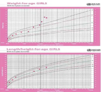
Figure 1: Growth chart depicting rapid weight gain after 1 ½ years of age and less than anticipated acceleration of height velocity in the
setting of rapid weight gain.
Her blood-pressure was labile and >95th centile. Blood
gas showed respiratory acidosis (pH:7.21, pCO2:80
mmHg). She was started on non-invasive ventilation (NIV) after which her sensorium and hypercarbia normalized (PCO2: 43 mmHg). On further interview,
child had hyperphagia and significant weight gain in preceding 6 months. Review of her growth chart revealed rapidly increasing weight after 1½ year of age, while her height centile remained within the normal range (Figure 1). Her developmental milestones were normal. She had several episodes of hypoventilation during the hospital stay. An attempted sedation for CT abdomen resulted in hypoventilation, hypercarbia (PCO2:115 mmHg, pH:6.9), and seizures, requiring brief period of invasive ventilation. A conventional respiratory physiological study was attempted, but could not be performed in view of hypoventilation and desaturation during sleep. Cardiac ultrasound revealed moderate pulmonary hypertension, in keeping with hypercarbic respiratory failure.
MRI brain showed non-enhancing T2/FLAIR hyperintensity in bilateral centrum semiovale, fronto-parietal subcortical white matter and right peri-trigonal region, with diffusion restriction. EEG was normal. She was noted to have diminished pain sensitivity as evidenced by absence of grimace/cry during insertion of IV cannulas/arterial catheters. She had normal pupillary light reflex with bilateral alternating exotropia and normal fundus.
Table 1: Summary of endocrine evaluation.
Test Result Normal Range
TSH 0.64 micro IU/ml (0.7 – 6.4)
T3 0.68 ng/ml (0.9 – 2.4)
Free T4 0.89 ng/ml (0.8 – 2.7)
Prolactin 27.82 ng/ml (2.5 - 15)
Random Cortisol 48.44 micro g/dl (0.49 to 58.6) 0.5 mg Dexamethasone suppression test 1.41 micro g/dl (4.3 – 22.4)
IGF 1 123 ng/ml (51-303)
IGF BP 3 3.45 micro g/ml (0.8-3.9)
5 mcg/kg Clonidine stimulation
GH Basal: 1.92 ng/ml GH 30 min: 0.27 ng/ml GH 60 min: 1.2 ng/ml
Peak GH: > 10 ng/ml
Fasting Leptin levels 4.64 ng/ml (3.63-11.09) 24 hour urine VMA 2 mg (<13.6) 24 hour metanephrines 0.4 mg (< 1) 24 hour epinephrine 6.04 μg/g creatinine (4-32) 24 hour nor-epinephrine 89 μg/g creatinine (20-108) 24 hour dopamine 1069 μg/g creatinine (295-1123)
75 g Oral glucose tolerance test
Fasting 64 mg/dl post 1 hour 72 mg/dl post 2 hours 82 mg/dl
(60-90) (80-120) (80-120) Total cholesterol 73 mg/dl (< 200)
Triglyceride 61 mg/dl (<150)
HDL (measured) 18 mg/dl (35 - 70) LDL (measured) 52 mg/dl (< 100) VLDL (calculated) 12.3 mg/dl (< 40)
She was evaluated for endocrine and genetic causes of obesity. Hormonal profile (Table 1) revealed normal
hypothyroidism; low IGF-1, and a sub-optimal GH response to 5 μg/kg of clonidine. Leptin levels were normal for age and weight. Oral 75 gm glucose tolerance test, lipid profile, creatine kinase, serum ammonia, lactate, liver function tests, renal function tests and chest X ray were normal. Her bone age corresponded to 2-3 years (Greulich-Pyle method).
Considering the constellation of symptoms, ROHHADNET syndrome was considered and screening thoracic and abdominal CT was performed to look for any neural crest tumor. CT revealed an FDG avid, homogenously enhancing mass in pre-sacral area on left side, posterior to rectum (Figure 2 and 3).
Figure 2: Contrast enhanced CT abdomen.
Figure 3: FDG-PET scan showing homogenously enhancing mass in pre-sacral region on left side
posterior to rectum.
In view of hypertension and neurogenic tumor, urinary catecholamines and metanephrines were done, which were normal (Table 1). She was discharged on thyroxine and nocturnal NIV. Parents were trained to provide basic life support. Eight months later she collapsed while
receiving nebulisation (? Hypoxia induced seizures) for respiratory illness at a nearby clinic where she was intubated. MRI Brain showed chronic infarcts in watershed areas with gliotic foci. Interim history revealed that the hypoventilation episodes have worsened (clinically and also evident with portable SP02 probe showing frequent desaturations) necessitating NIV support even for short naps during daytime and sometimes while awake also. She was extubated to BIPAP once awake. However, she developed hypoventilation soon after requiring re-intubation. She had 3 unsuccessful extubation attempts due to poor respiratory effort. Due to constant risk of apparent life threatening event, tracheostomy was planned. MRI Abdomen and pelvis showed mild interval increase in size and extension of the pre-sacral mass (Figure 4).
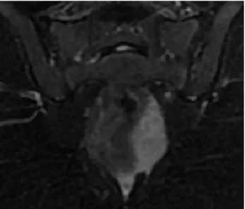
Figure 4: MRI Pelvis showing ill-defined presacral mass abutting rectum anteromedially, extending upto left mesorectal fascia and levator ani muscle laterally
and inferiorly respectively.
Figure 5: Histology of the presacral tumour showing monomorphic population of small, round cells with
She underwent tracheostomy and excision of the pre-sacral mass at the same setting. HPE of the pre-pre-sacral mass showed maturing ganglioneuroma (Figure 5).
Whole exome sequencing was done and 18 genes associated with CCHS and ROHHAD (ADCYAP1, ASCL1, BDNF, CD36, CD5, EDN3, GDNF, GFRA1, HTR1A, NDN, NTRK2, PDE11A, PHOX2B, RAI1, RELN, RET, TAF1, WDFY4) along with ‘pathogenic’ and ‘likely pathogenic’ variants were analysed, all of which were negative. She was discharged on BIPAP support via tracheostomy on need basis.
DISCUSSION
ROHHAD was first described in 1965 and was re-named in 2007 when it was shown to be distinct from congenital central hypoventilatory syndrome (CCHS) by absence of CCHS-related PHOX2B mutations.5,1 The disease is now
called ROHHADNET because of the accompanying ganglioneuroma in abdomen and lungs and neuroendocrinal tumors such as ganglioneuroblastoma in about 40% of the patients.6,7
Symptoms develop after 1½ year of age, with dramatic weight gain, hyperphagia; followed by hypoventilation. Before diagnosing central hypoventilation, it is important that diseases of lung and heart are ruled out.8 Other
hypothalamic abnormalities include diabetes insipidus and hyperprolactinemia.3,5 Autonomic dysfunction
include altered pupillary light reflex, strabismus, altered gastrointestinal motility, temperature dysregulation, decreased pain sensation. Few develop seizures, though this feature may be related to hypoxemia. About 40% develop neural crest tumors like ganglioneuromas or ganglioneuroblastomas.4 50% to 60% children ultimately
suffer cardiac arrest.9
Differential diagnosis includes Prader-Willi syndrome in which hyperphagia and obesity is associated with hypotonia, mental retardation, short stature, GH deficiency, hypogonadotropic hypogonadism, and sleep apnea. Bardet-Biedl syndrome is charecterised by obesity, mental retardation, dysmorphic extremities, pigmentary retinopathy, hypogonadism, renal abnormalities. Leptin deficiency, POMC gene mutation, and MCR4 gene mutation also have to be considered among monogenic causes of early-onset obesity.8,10,11
Normal levels of leptin and cortisol ruled out these causes in our patient. In contrast to most cases of exogenous obesity.2,9 where growth velocity and IGF-1 levels are
high normal, in ROHHAD patients IGF-1 levels are depressed with sub-optimal GH response to stimulatory tests.
Searches for a neuroanatomical pathology to explain symptoms of ROHHAD have not yielded consistent findings. Reported MRI pathologies include bilateral basal ganglia hypodensities, Rathke's cleft cyst and hypointensities in pons, midbrain.1,2 Hypothalamic
inflammation with lymphocytic infiltrates were found in two cases.12,13 MRI brain in our patient showed
hyperintensity in bilateral centrum semiovale, frontoparietal subcortical white matter and right peri-trigonal region with diffusion restriction, which is probably related to hypoxemia.
Sedation induced respiratory arrest is quite common.13,15
All children with ROHHAD require some form of ventilatory assistance. Few reports have suggested that early intervention with nocturnal artificial ventilation may improve daytime ventilation.14 Ventilator
management must be targeted to the child’s specific needs. The goal is to maintain adequate oxygenation and ventilation. Assistive breathing techniques like diaphragm pacing may have limited success due to associated obesity.
Previous studies postulated eight genes as candidate ROHHAD genes, but failed to identify any disease associated variants among their cohorts.1,2,9 There has
been a report of “Retinoic acid Induced-1” gene (RAI-1) receptor mutation in one case.16 It was later confirmed
that none of these 8 genes or RAI1 gene were major ROHHAD genes.17 In our case, none of the tested genes
were positive. A negative test could be due to variation in the genomic region not covered by the test; or due to large insertions, deletions, duplications, inversions, complex rearrangements which cannot be detected. As somatic mosaicism represents major mechanism, the major tissue carrying the mutation would not have been sampled. Use of patients’ neuroendocrine tumour represents one approach to address this challenge.17
Hence unambiguous identification of ROHHAD syndrome has been challenging; confirmatory laboratory testing is not yet available, and the patient population may represent heterogeneous group of underlying etiologies. Hence emphasis on diagnosis based on clinical findings.
CONCLUSION
ROHHADNET syndrome mimics genetic obesity syndromes and several endocrine disorders. Because of high prevalence of cardiorespiratory arrest and the probability of accompanying tumors, early diagnosis is important. Negative genetic test could be due to somatic mosaicism; variation in genomic region not covered by the test; or due to large insertions, deletions, complex rearrangements which cannot be detected. Unambiguous identification of ROHHAD syndrome is challenging; confirmatory laboratory testing is not yet available. Hence emphasis on diagnosis based on clinical findings. Multidisciplinary care and aggressive intervention is critical to optimise the neuro-developmental outcomes and to ensure a good quality of life.
REFERENCES
1. Ize-Ludlow D, Gray JA, Sperling MA, Berry-Kravis EM, Milunsky JM, Farooqi IS, et al. Rapid-onset obesity with hypothalamic dysfunction, hypoventilation, and autonomic dysregulation presenting in childhood. Pediatrics. 2007;120:179-88.
2. De Pontual L, Trochet D, Caillat-Zucman S, Abou Shenab OA, Bougneres P, Crow Y, et al. Delineation of late onset hypoventilation associated with hypothalamic dysfunction syndrome. Pediatr Res. 2008;64:689-94.
3. Onal H, Ersen A. A case of late-onset central hypoventilation syndrome with hypothalamic dysfunction: through a new phenotype. Turk J Pediatr. 2010;52:198-202.
4. Patwari PP, Rand CM, Berry-Kravis EM, Ize-Ludlow D, Weese-Mayer DE. Monozygotic twins discordant for ROHHAD phenotype. Pediatrics. 2011;128:711-5.
5. Fishman LS, Samson JH, Sperling DR. Primary Alveolar Hypoventilation Syndrome (Ondine's Curse). Am J Dis Child. 1965;110:155-61.
6. Bougneres P, Pantalone L, Linglart A, Rothenbuhler A, Le Stunff C. Endocrine manifestations of the rapid-onset obesity with hypoventilation, hypothalamic, autonomic dysregulation, and neural tumor syndrome in childhood. J Clin Endocrinol Metab. 2008;93:3971-80.
7. Sirvent N, Berard E, Chastagner P, Feillet F, Wagner K, Sommelet D. Hypothalamic dysfunction associated with neuroblastoma: evidence for a new Paraneoplastic syndrome? Med Pediatr Oncol. 2003;40:326-8.30
8. Abaci A, Catli G, Bayram E, Koroglu T, Olgun HN, Mutafoglu K, et al. A case of rapidonset obesity with hypothalamic dysfunction, hypoventilation, autonomic dysregulation, and neural crest tumor: ROHHADNET syndrome. Endocr Pract. 2013;19:12-6.
9. Rand CM, Patwari PP, Rodikova EA, Zhou L, Berry-Kravis EM, Wilson RJ, et al. Rapidonset obesity with hypothalamic dysfunction,
hypoventilation, and autonomic dysregulation: analysis of hypothalamic and autonomic candidate genes. Pediatr Res. 2011;70:375-8.
10. Farooqi IS, O'Rahilly S. Monogenic obesity in humans. Annu Rev Med. 2005;56:443-58.
11. Govaerts C, Srinivasan S, Shapiro A, Zhang S, Picard F, Clement K, et al. Obesityassociated mutations in the melanocortin 4 receptor provide novel insights into its function. Peptides. 2005;26:1909-19.
12. North KN, Ouvrier RA, McLean CA, Hopkins IJ. Idiopathic hypothalamic dysfunction with dilated unresponsive pupils: report of two cases. J Child Neurol. 1994;9:320-5.
13. Nunn K, Ouvrier R, Sprague T, Arbuckle S, Docker M. Idiopathic hypothalamic dysfunction: a paraneoplastic syndrome? J Child Neurol 1997;12:276-81.
14. Katz ES, McGrath S, Marcus CL. Late-onset central hypoventilation with hypothalamic dysfunction: a distinct clinical syndrome. Pediatr Pulmonol. 2000;29:62-8.
15. Chew HB, Ngu LH, Keng WT. Rapid-onset obesity with hypothalamic dysfunction, hypoventilation and autonomic dysregulation (ROHHAD): a case with additional features and review of the literature. BMJ Case Rep. 2011;2011;10.
16. Thaker VV, Esteves KM, Towne MC, Brownstein CA, James PM, Crowley L, et al. Whole exome sequencing identifies RAI1 mutation in a morbidly obese child diagnosed with ROHHAD syndrome. J Clin Endocrinol Metab. 2015;100:1723-30.
17. Barclay SF, Rand CM, Borch LA, Nguyen L, Gray PA, Gibson WT et al. rapid-Onset Obesity with Hypothalamic Dysfunction, Hypoventilation, and Autonomic Dysregulation (ROHHAD): exome sequencing of trios, monozygotic twins and tumours. Orphanet J Rare Dis. 2015;10:103.