ACC/AHA PRACTICE GUIDELINES—FULL TEXT
ACC/AHA 2002 Guideline Update for Exercise Testing
A Report of the American College of Cardiology/American Heart Association
Task Force on Practice Guidelines (Committee on Exercise Testing)
COMMITTEE MEMBERS
Raymond J. Gibbons, MD, FACC
, FAHA, Chair
TASK FORCE MEMBERS
Raymond J. Gibbons, MD, FACC, FAHA, Chair
Elliott M. Antman, MD, FACC
, FAHA, Vice Chair
Gary J. Balady, MD, FACC, FAHAJ. Timothy Bricker, MD, FACC
Bernard R. Chaitman, MD, FACC, FAHA Gerald F. Fletcher, MD, FACC, FAHA Victor F. Froelicher, MD, FACC, FAHA
Daniel B. Mark, MD, MPH, FACC, FAHA Ben D. McCallister, MD, FACC, FAHA Aryan N. Mooss, MBBS, FACC, FAHA Michael G. O'Reilly, MD, FACC
William L. Winters, Jr., MD, FACC, FAHA
Joseph S. Alpert, MD, FACC, FAHA David P. Faxon, MD, FACC, FAHA Valentin Fuster, MD, PhD, FACC, FAHA Gabriel Gregoratos, MD, FACC, FAHA
Loren F. Hiratzka, MD, FACC, FAHA Alice K. Jacobs, MD, FACC, FAHA Richard O. Russell, MD, FACC, FAHA* Sidney C. Smith, Jr., MD, FACC, FAHA
The ACC/AHA Task Force on Practice Guidelines makes every effort to avoid any actual or potential coflicts of interest that might arise as a result of an outside relationship or personal interest of a member of the writing panel. Specifically, all members of the writing panel are asked to provide disclosure statements of all such relationships that might be perceived as real or potential conflicts of interest. These statements are reviewed by the parent task force, reported orally to all members of the writing panel at the first meeting, and updated as changes occur.
This document was approved by the American College of Cardiology Board of Trustees in July 2002 and by the American Heart Association Science Advisory and Coordinating Committee in June 2002.
When citing this document, the American College of Cardiology Foundation and the American Heart Association request the following citation format be used: Gibbons RJ, Balady GJ, Bricker JT, Chaitman BR, Fletcher GF, Froelicher VF, Mark DB, McCallister BD, Mooss AN, O'Reilly MG, Winters WL Jr. ACC/AHA 2002 guideline update for exercise testing: a report of the American College of Cardiology/American Heart Association Task Force on Practice Guidelines (Committee on Exercise Testing). 2002. American College of Cardiology Web site. Available at: www.acc.org/clinical/guidelines/exercise/ dirIndex.htm.
This document is available on the World Wide Web sites of the American College of Cardiology (www.acc.org) and the American Heart Association (www.americanheart.org). Copies of this document (the complete guidelines) are available for $5 each by calling 800-253-4636 (US only) or writing the American College of Cardiology Resource Center, 9111 Old Georgetown Road, Bethesda, MD 20814-1699 (ask for No. 71-0231). To obtain a reprint of the shorter version (executive summary describing the changes to the guide-lines) planned for subsequent publication in the Journal of the American College of Cardiology and Circulation, ask for reprint No. 71-0232. To pur-chase additional reprints (specify version and reprint number): up to 999 copies, call 800-611-6083 (US only) or fax 413-665-2671; 1000 or more copies, call 214-706-1789, fax 214-691-6342, or email pubauth@heart.org.
*Former Task Force member during this writing effort.
TABLE OF CONTENTS
Preamble...2
I. Introduction... 2
Exercise Testing Procedure... 5
General Overview...5
Indications and Safety... 5
Equipment and Protocols... 5
Exercise End Points... 5
Interpretation of the Exercise Test... 6
Cost and Availability... 6
Clinical Context... 7
II. Exercise Testing to Diagnose Obstructive Coronary Artery Disease...7
Rationale... 8
Pretest Probability...8
Diagnostic Characteristics and Test Performance... 8
Believability Criteria for Diagnostic Tests... 9
Diagnostic Accuracy of the Standard Exercise Test...10
Confounders of Stress ECG Interpretation...11
Digoxin...12
Left Ventricular Hypertrophy With Repolarization Abnormalities... 12
Resting ST Depression...12
Left Bundle-Branch Block...12
Right Bundle-Branch Block...12
ST-Segment Interpretation Issues...13
III. Risk Assessment and Prognosis in Patients With Symptoms or a Prior History of Coronary Artery Disease... 14
2 ACC/AHA Practice Guidelines American Heart Association - www.americanheart.org
mates of expected health outcomes when data exist. Patient-specific modifiers, comorbidities, and issues of patient pref-erence that might influence the choice of particular tests or therapies are considered, as well as frequency of follow-up and cost-effectiveness.
The ACC/AHA Task Force on Practice Guidelines makes every effort to avoid any actual or potential conflicts of interest that might arise as a result of an outside relationship or personal interest of a member of the writing panel. Specifically, all members of the writing panel are asked to provide disclosure statements of all such relationships that might be perceived as real or potential conflicts of interest. These statements are reviewed by the parent task force, reported orally to all members of the writing panel at the first meeting, and updated yearly and as changes occur.
These practice guidelines are intended to assist physicians in clinical decision making by describing a range of gener-ally acceptable approaches for the diagnosis, management, or prevention of specific diseases or conditions. These guidelines attempt to define practices that meet the needs of most patients in most circumstances. The ultimate judgment regarding care of a particular patient must be made by the physician and patient in light of all of the circumstances pre-sented by that patient.
The summary article highlighting changes from the 1997 guideline to the 2002 guideline is published in the October 1 issue of Circulation and the October 16 issue of the Journal
of the American College of Cardiology. The full-text
guide-line is posted on the ACC and AHA Web sites. Copies of the full-text and summary article are available from both organ-izations.
The 1997 guidelines were officially endorsed by the American College of Sports Medicine, the American Society of Echocardiography, and the American Society of Nuclear Cardiology.
Raymond J. Gibbons, MD, FACC
Chair, ACC/AHA Task Force on Practice Guidelines
I. INTRODUCTION
The ACC/AHA Task Force on Practice Guidelines was formed to make recommendations regarding the appropriate use of testing in the diagnosis and treatment of patients with known or suspected cardiovascular disease. Exercise testing is widely available and relatively low cost. For the purposes of this document, exercise testing is a cardiovascular stress test that uses treadmill or bicycle exercise and electrocardio-graphic and blood pressure monitoring. Pharmacological stress and the use of imaging modalities (e.g., radionuclide imaging and echocardiography) are beyond the scope of these guidelines.
The current committee was given the task of reviewing and revising the guidelines for exercise testing published in September 1986. Since that report, many new studies have
Risk Stratification: General Considerations... 15
Prognosis of Coronary Artery Disease: General Considerations...15
Risk Stratification With the Exercise Test...16
Use of Exercise Test Results in Patient Treatment...20
IV. After Myocardial Infarction... 24
Exercise Test Logistics... 25
Risk Stratification and Prognosis... 26
Activity Counseling... 29
Cardiac Rehabilitation... 30
Summary... 30
V. Exercise Testing With Ventilatory Gas Analysis...31
VI. Special Groups: Women, Asymptomatic Individuals, and Postrevascularization Patients...33
Women... 33
Diagnosis of Coronary Artery Disease in the Elderly... 35
Exercise Testing in Asymptomatic Persons Without Known CAD...35
Valvular Heart Disease...39
Exercise Testing Before and After Revascularization...41
Investigation of Heart Rhythm Disorders... 42
Evaluation of Hypertension... 44
VII. Pediatric Testing: Exercise Testing in Children and Adolescents...44 Appendix 1...44 Appendix 2...44 Appendix 3...45 References ...45
PREAMBLE
It is important that the medical profession play a significant role in critically evaluating the use of diagnostic procedures and therapies in the management or prevention of disease states. Rigorous and expert analysis of the available data documenting relative benefits and risks of those procedures and therapies can produce helpful guidelines that improve the effectiveness of care, optimize patient outcomes, and impact the overall cost of care favorably by focusing resources on the most effective strategies.
The American College of Cardiology (ACC) and the American Heart Association (AHA) have jointly engaged in the production of such guidelines in the area of cardiovascu-lar disease since 1980. This effort is directed by the ACC/AHA Task Force on Practice Guidelines, whose charge is to develop and revise practice guidelines for important cardiovascular diseases and procedures. Experts in the sub-ject under consideration are selected from both organiza-tions to examine subject-specific data and write guidelines. The process includes additional representatives from other medical practitioner and specialty groups where appropriate. Writing groups are specifically charged to perform a formal literature review, weigh the strength of evidence for or against a particular treatment or procedure, and include
esti-tional registries. A lower rank (C) was given when expert consensus was the primary basis for the recommendation. When few or no data exist, this is noted in the text, and the recommendations are based on the expert consensus of the committee.
The ACC/AHA classifications I, II, and III are used to summarize indications as follows:
Class I: Conditions for which there is evidence and/or general agreement that a given procedure or treatment is useful and effective.
Class II: Conditions for which there is conflicting evi-dence and/or a divergence of opinion about the usefulness/efficacy of a procedure or treatment.
Class IIa: Weight of evidence/opinion is in favor of usefulness/efficacy.
Class IIb: Usefulness/efficacy is less well established by evidence/opinion.
Class III: Conditions for which there is evidence and/or general agreement that the procedure/treat-ment is not useful/effective and in some cases may be harmful.
A complete list of the hundreds of publications covering many decades of exercise testing is beyond the scope of these guidelines, and only selected references are included. The committee consisted of acknowledged experts in exer-cise testing, as well as general cardiologists and cardiolo-gists with expertise in the use of stress imaging modalities. Both the academic and private practice sectors, as well as both adult and pediatric expertise, were represented. This document was reviewed by two outside reviewers nominat-ed by the ACC and two outside reviewers nominatnominat-ed by the AHA, as well as by the ACC/AHA Task Force on Practice Guidelines. This document will be reviewed annually by the task force to determine whether a revision is needed. These guidelines will be considered current unless the task force revises or withdraws them from distribution.
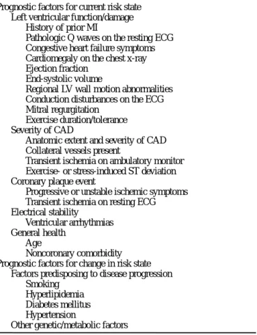
This report overlaps with several previously published ACC/AHA guidelines for patient treatment that potentially involve exercise testing, including guidelines for periopera-tive cardiovascular evaluation for noncardiac surgery (344), guidelines for management of patients with acute myocar-dial infarction (345), guidelines for percutaneous coronary intervention (346), guidelines and indications for coronary artery bypass graft surgery (347), and guidelines for man-agement of patients with chronic stable angina (348). The reader is referred to these other guidelines for a more com-plete description of the role of exercise testing in clinical decision making and a comparison of exercise electrocar-diography with noninvasive imaging modalities. The gener-al context for the use of exercise testing is outlined in Fig. 1. These guidelines are not intended to include information previously covered in guidelines for the use of noninvasive imaging modalities. This report does not include a discus-been published regarding the usefulness of exercise testing
for prediction of outcome in both symptomatic and asymp-tomatic patients. The usefulness of oxygen consumption measurements in association with exercise testing to identi-fy patients who are candidates for cardiac transplantation has been recognized. The usefulness and cost-effectiveness of exercise testing has been compared with more expensive imaging procedures in selected patient subsets. All of these developments are considered in these guidelines.
In considering the use of exercise testing in individual patients, the following factors are important:
1. The quality, expertise, and experience of the profession-al and technicprofession-al staff performing and interpreting the study
2. The sensitivity, specificity, and accuracy of the tech-nique
3. The cost and accuracy of the technique compared with more expensive imaging procedures
4. The effect of positive or negative results on clinical deci-sion making
5. The potential psychological benefits of patient reassur-ance
The format of these guidelines includes a brief description of exercise testing followed by a discussion of its usefulness in specific clinical situations. Usefulness is considered for 1) diagnosis; 2) severity of disease/risk assessment/prognosis in patients with known or suspected chronic coronary artery disease (CAD); 3) risk assessment of patients early after myocardial infarction; 4) specific clinical populations iden-tified by gender, age, other cardiac disease, or prior coronary revascularization; and 5) pediatric populations. The recom-mendations for particular situations are summarized in each section.
The committee reviewed and compiled all pertinent pub-lished reports (excluding abstracts) through a computerized search of the English-language literature since 1975 and a manual search of final articles. Specific attention was devot-ed to identification and compilation of appropriate meta-analyses. Detailed evidence tables were developed whenev-er necessary with specific critwhenev-eria detailed in the guidelines. The meta-analyses and evidence tables were reviewed exten-sively by an expert in methodologies. Inaccuracies and inconsistencies in the original publications were identified and corrected whenever possible. The recommendations made are based primarily on these published data. In the original guidelines, the committee did not rank the available scientific evidence in an A, B, or C fashion. The level of evi-dence is provided for new recommendations appearing in this update. The weight of evidence was ranked highest (1) if the data were derived from multiple randomized clinical trials that involved large numbers of patients and intermedi-ate (B) if the data were derived from a limited number of randomized trials that involved small numbers of patients or from careful analyses of nonrandomized studies or
observa-3 ACC/AHA Practice Guidelines
4 ACC/AHA Practice Guidelines American Heart Association - www.americanheart.org CAD diagnosis certain? Need for risk/prognostic assessment? Need to guide medical management? Contra-indications to stress testing? Symptoms warranting angiography? Can patient exercise? Is resting ECG interpretable*? Exercise test Continue/initiate/modify rx as appropriate Is diagnosis and prognosis certain? Is test result high risk?** Consider coronary angiography/revascularization
Consider coronary angiogram Continue/initiate/modify
medical rx
Pharmacologic imaging study
Exercise imaging study
Consider imaging study/angiography
Patient with stable chest pain
or unstable chest pain stabilized by therapy low-risk or intermediate-risk unstable angina or previous MI
or post-revascularization
Figure 1. Clinical context for exercise testing for patients with suspected ischemic heart disease.
*Electrocardiogram interpretable unless pre-excitation, electronically paced rhythm, left bundle branch block, or resting ST-segment depression greater than 1 mm. See text for discussion of digoxin use, left ventricular hypertrophy, and ST depression less than 1 mm. **For example, high-risk if Duke treadmill score predicts average annual cardiovascular mortality greater than 3% (see Fig 2 for nomogram). CAD indicates coronary artery disease; ECG,
electrocardiogram; MI, myocardial infarction; and rx, treatment. no yes yes yes yes yes yes yes yes yes no no no no no no no no
Patient with stable chest pain
or low-risk or intermediate-risk unstable angina or previous MI
5 ACC/AHA Practice Guidelines
sion of radionuclide angiography, myocardial perfusion imaging, or positron emission tomography, which are cov-ered in the published guidelines for clinical use of cardiac radionuclide imaging (5). This report also does not include any discussion of stress echocardiography, which is covered in the published guidelines for clinical application of echocardiography (349). For clarity, there are occasional ref-erences to the use of both radionuclide and echocardiograph-ic imaging techniques. However, these brief references are not intended to provide a comprehensive understanding of the use of these imaging modalities. For such an understand-ing, the reader is referred to the other published guidelines. These guidelines do apply to both adults and children.
Exercise Testing Procedure
General Overview
Exercise testing is a well-established procedure that has been in widespread clinical use for many decades. It is beyond the scope of this document to provide a detailed “how-to” description of this procedure. Such a description is available in previous publications from the AHA, including the state-ment on exercise standards (7), guidelines for clinical exer-cise testing laboratories (8), and guidelines for exerexer-cise test-ing in the pediatric age group (9), to which interested readers are referred. This section is intended to provide a brief overview of the exercise testing procedure.
Indications and Safety
Although exercise testing is generally a safe procedure, both myocardial infarction and death have been reported and can be expected to occur at a rate of up to 1 per 2500 tests (10). Good clinical judgment should therefore be used in deciding which patients should undergo exercise testing. Absolute and relative contraindications to exercise testing are summarized in Table 1.
Exercise testing should be supervised by an appropriately trained physician. As indicated in the American College of Physicians/ACC/AHA task force statement on clinical com-petence in exercise testing (11), exercise testing in selected patients can be performed safely by properly trained nurses, exercise physiologists, physician assistants, physical thera-pists, or medical technicians working directly under the supervision of a physician, who should be in the immediate vicinity and available for emergencies. The electrocardio-gram (ECG), heart rate, and blood pressure should be moni-tored carefully and recorded during each stage of exercise and during ST-segment abnormalities and chest pain. The patient should be monitored continuously for transient rhythm disturbances, ST-segment changes, and other electro-cardiographic manifestations of myocardial ischemia. Further details are provided in the AHA guidelines for clini-cal exercise testing laboratories (8).
Equipment and Protocols
Both treadmill and cycle ergometer devices are available for exercise testing. Although cycle ergometers are generally
less expensive, smaller, and less noisy than treadmills and produce less motion of the upper body, the fatigue of the quadriceps muscles in patients who are not experienced cyclists is a major limitation, because subjects usually stop before reaching their maximum oxygen uptake. As a result, treadmills are much more commonly used in the United States for exercise testing.
Commonly used treadmill protocols are summarized in a variety of published documents. Although much of the pub-lished data are based on the Bruce protocol, there are clear advantages to customizing the protocol to the individual patient to allow 6 to 12 minutes of exercise (12). Exercise capacity should be reported in estimated metabolic equiva-lents (METs) of exercise. If exercise capacity is also report-ed in minutes, the nature of the protocol should be specifireport-ed clearly.
Exercise End Points
Although exercise testing is commonly terminated when sub-jects reach an arbitrary percentage of predicted maximum heart rate, it should be recognized that other end points (summarized in Table 2) are strongly preferred. There is a wide spectrum of individual subject values around the regression line for maximum heart rate, which may therefore be beyond the limit of some patients and submaximal for oth-ers. The target heart rate approach has obvious additional limitations in patients receiving beta-blockers, those with heart rate impairment, and those with excessive heart rate response. The use of rating of perceived exertion scales, such as the Borg scale (Appendix 1) (13), is often helpful in
Table 1. Contraindications to Exercise Testing Absolute
•
Acute myocardial infarction (within 2 d)•
High-risk unstable angina*•
Uncontrolled cardiac arrhythmias causing symptoms or hemodynamic compromise•
Symptomatic severe aortic stenosis•
Uncontrolled symptomatic heart failure•
Acute pulmonary embolus or pulmonary infarction•
Acute myocarditis or pericarditis•
Acute aortic dissection Relative†•
Left main coronary stenosis•
Moderate stenotic valvular heart disease•
Electrolyte abnormalities•
Severe arterial hypertension‡•
Tachyarrhythmias or bradyarrhythmias•
Hypertrophic cardiomyopathy and other forms of outflow tract obstruction•
Mental or physical impairment leading to inability to exercise adequately•
High-degree atrioventricular block*ACC/AHA Guidelines for the Management of Patients With Unstable Angina/Non-ST-Segment Elevation Myocardial Infarction (350) (see Table 17).
†Relative contraindications can be superseded if the benefits of exercise outweigh the risks.
‡In the absence of definitive evidence, the committee suggests systolic blood pressure of >200 mm Hg and/or diastolic blood pressure of >110 mm Hg. Modified from Fletcher et
6 ACC/AHA Practice Guidelines American Heart Association - www.americanheart.org
ic response. The occurrence of ischemic chest pain consis-tent with angina is important, particularly if it forces termi-nation of the test. Abnormalities in exercise capacity, sys-tolic blood pressure response to exercise, and heart rate response to exercise are important findings. The most impor-tant electrocardiographic findings are ST depression and ele-vation. The most commonly used definition for visual inter-pretation of a positive exercise test result from an electro-cardiographic standpoint is greater than or equal to 1 mm of horizontal or downsloping ST-segment depression or eleva-tion for at least 60 to 80 milliseconds (ms) after the end of the QRS complex (347). The details of interpretation are covered elsewhere in these guidelines.
Cost and Availability
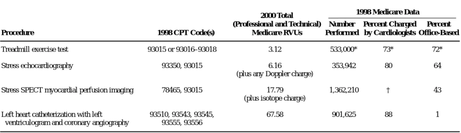
There are relatively few published studies comparing the cost-effectiveness of treadmill exercise testing with more expensive imaging procedures. Compared with imaging pro-cedures such as stress echocardiography, stress single-pho-ton emission computed tomography (SPECT) myocardial perfusion imaging, and coronary angiography, treadmill exercise testing can be performed at a much lower cost. Table 3 is a comparison of year 2000 Medicare RVUs (rela-tive value units, professional and technical) for treadmill exercise testing and selected imaging procedures. These RVUs provide an estimate of relative costs. Compared with the treadmill exercise test, the cost of stress echocardiogra-phy is at least 2.1 times higher, stress SPECT myocardial imaging 5.7 times higher, and coronary angiography 21.7 times higher. Lower cost of the treadmill exercise test alone does not necessarily result in a lower overall cost of patient care, because the sum of the cost of additional testing and interventions may be higher when the initial treadmill exer-cise test is less accurate than these more sophisticated pro-cedures.
Treadmill exercise testing is performed frequently ( Table 3). An estimated 72% of the treadmill exercise tests charged to Medicare in 1998 were performed as office procedures, and 27% of the charges were submitted by noncardiologists.
Table 2. Indications for Terminating Exercise Testing Absolute indications
•
Drop in systolic blood pressure of >10 mm Hg from baselineblood pressure despite an increase in workload, when accompanied by other evidence of ischemia•
Moderate to severe angina•
Increasing nervous system symptoms (eg, ataxia, dizziness, or near-syncope)•
Signs of poor perfusion (cyanosis or pallor)•
Technical difficulties in monitoring ECG or systolic blood pressure•
Subject’s desire to stop•
Sustained ventricular tachycardia•
ST elevation (≥1.0 mm) in leads without diagnostic Q-waves (other than V1or aVR)Relative indications
•
Drop in systolic blood pressure of (≥10 mm Hg from baseline blood pressure despite an increase in workload, in the absence of other evidence of ischemia•
ST or QRS changes such as excessive ST depression (>2 mm of horizontal or downsloping ST-segment depression) or marked axis shift•
Arrhythmias other than sustained ventricular tachycardia, includ-ing multifocal PVCs, triplets of PVCs, supraventricular tachycar-dia, heart block, or bradyarrhythmias•
Fatigue, shortness of breath, wheezing, leg cramps, or claudica-tion•
Development of bundle-branch block or IVCD that cannot be distinguished from ventricular tachycardia•
Increasing chest pain•
Hypertensive response**In the absence of definitive evidence, the committee suggests systolic blood pressure of >250 mm Hg and/or a diastolic blood pressure of >115 mm Hg.
ECG indicates electrocardiogram; PVCs, premature ventricular contractions; ICD, implantable cardioverter-defibrillator discharge; and IVCD, intraventricular conduction
delay. Modified from Fletcher et al.7
Table 3. Medicare Fees and Volumes of Commonly Used Diagnostic Procedures
2000 Total
(Professional and Technical) Number Percent Charged Percent
Procedure 1998 CPT Code(s) Medicare RVUs Performed by Cardiologists Office-Based
Treadmill exercise test 93015 or 93016–93018 3.12 533,000* 73* 72*
Stress echocardiography 93350, 93015 6.16 353,942 80 64
(plus any Doppler charge)
Stress SPECT myocardial perfusion imaging 78465, 93015 17.79 1,362,210 † 43
(plus isotope charge)
Left heart catheterization with left 93510, 93543, 93545, 67.58 901,625 88 1
ventriculogram and coronary angiography 93555, 93556
*These numbers are estimates, after excluding treadmill exercise tests performed with perfusion imaging. †There are no reliable data regarding this percentage.
CPT indicates current procedural terminology; RVUs, relative value units; and SPECT, single-photon emission computed tomography.
1998 Medicare Data
assessment of patient fatigue. Symptom-limited testing with the Borg scale as an aid is very important when the test is used to assess functional capacity. Rating of perceived exer-tion is less helpful in pediatric populaexer-tions.
Interpretation of the Exercise Test
Interpretation of the exercise test should include exercise capacity and clinical, hemodynamic, and
electrocardiograph-7 ACC/AHA Practice Guidelines
Thus, treadmill exercise tests are more widely performed, do not always require a cardiologist, and are convenient for the patient because they are often an office-based procedure.
Clinical Context
The vast majority of treadmill exercise testing is performed in adults with symptoms of known or suspected ischemic heart disease. Special groups who represent exceptions to this norm are discussed in detail in sections VI and VII. Sections II through IV reflect the variety of patients and clin-ical decisions (so-called nodal points) for which exercise testing is used. Although this document is not intended to be a guideline for the management of stable chest pain, the committee thought that it was important to provide an over-all context for the use of exercise testing to facilitate the use of these guidelines (Fig. 1).
Patients who are candidates for exercise testing may have stable symptoms of chest pain, may be stabilized by medical therapy after symptoms of unstable chest pain, or may be post-myocardial infarction or postrevascularization patients. Patients who are unable to exercise or who have uninter-pretable ECGs because of pre-excitation, electronically paced rhythm, left bundle-branch block, or ST depression greater than 1 mm require imaging studies and are beyond the scope of these guidelines. Imaging studies are consid-ered in other ACC/AHA guidelines (5,348-350). The clini-cian should first address whether the diagnosis of CAD is certain, given the patient’s history, ECG, and symptoms of chest pain. The important factors involved in addressing this question are covered in section II of this document, which focuses on the use of treadmill exercise testing for diagnosis.
Even in patients for whom the diagnosis of CAD is certain on the basis of age, gender, description of chest pain, and history of prior myocardial infarction, there usually is a clin-ical need for risk or prognostic assessment to determine the need for possible coronary angiography or revascularization. The potential role of treadmill exercise testing in such patients is detailed in section III.
Post-myocardial infarction patients represent a common first presentation of ischemic heart disease. They are a sub-set of patients who may need risk or prognostic assessment.
This subgroup is considered in detail in section IV, which includes a discussion of the implications of acute reperfu-sion therapy for interpretation of exercise testing in this pop-ulation.
II. EXERCISE TESTING TO DIAGNOSE
OBSTRUCTIVE CAD
Class I
Adult patients (including those with complete right bundle-branch block or less than 1 mm of resting ST depression) with an intermediate pretest probability of CAD (Table 4) on the basis of gender, age, and symptoms (specific exceptions are noted under Classes II and III below).
Class IIa
Patients with vasospastic angina. Class IIb
1. Patients with a high pretest probability of CAD by age, symptoms, and gender.
2. Patients with a low pretest probability of CAD by age, symptoms, and gender.
3. Patients with less than 1 mm of baseline ST depres-sion and taking digoxin.
4. Patients with electrocardiographic criteria for left ventricular hypertrophy (LVH) and less than 1 mm of baseline ST depression.
Class III
1. Patients with the following baseline ECG abnormali-ties:
•
Pre-excitation (Wolff-Parkinson-White) syndrome•
Electronically paced ventricular rhythm•
Greater than 1 mm of resting ST depression•
Complete left bundle-branch blockTable 4. Pretest Probability of Coronary Artery Disease by Age, Gender, and Symptoms* Age Typical/Definite Atypical/Probable Nonanginal
(y) Gender Angina Pectoris Angina Pectoris Chest Pain Asymptomatic
30–39 Men Intermediate Intermediate Low Very low
Women Intermediate Very low Very low Very low
40–49 Men High Intermediate Intermediate Low
Women Intermediate Low Very low Very low
50–59 Men High Intermediate Intermediate Low
Women Intermediate Intermediate Low Very low
60–69 Men High Intermediate Intermediate Low
Women High Intermediate Intermediate Low
*No data exist for patients <30 or >69 years, but it can be assumed that prevalence of CAD increases with age. In a few cases, patients with ages at the extremes of the decades listed may have probabilities slightly outside the high or low range. High indicates >90%; intermediate, 10%–90%; low, <10%; and very low, <5%.
Table 5. Definitions and Calculation of the Terms Used to Quantify the Diagnostic Accuracy of a Test
Sensitivity = [TP/(TP + FN)] × 100 Specificity = [TN/(FP + TN)] × 100 Predictive value of an abnormal test (PV+) =
Predictive accuracy = [Sensitivity × P(CAD)] + [Specificity × [1 – P(CAD)]]
TP indicates those with an abnormal test result and disease positives); TN, those with a normal test result and no disease (true-negatives); FP, those with an abnormal test result but no disease (false-positives); FN, those with a normal test result but disease (false-negatives); PV1, the percentage of those with an abnormal (1) test result who have disease; predictive accuracy, the percentage of correct classifications, both 1 and 2; and P(CAD), pretest probability.
Sensitivity × P(CAD)
[Sensitivity × P(CAD)] + [(1 – Specificity)[1 – P(CAD)]]
8 ACC/AHA Practice Guidelines American Heart Association - www.americanheart.org
rest and/or nitroglycerin. Atypical or probable angina can be defined as chest pain or discomfort that lacks one of the three characteristics of definite or typical angina (18). Other clinical scores have been developed that could better predict pretest probability (351).
Detailed nomograms are available that incorporate the effects of a history of prior infarction, electrocardiographic Q waves, electrocardiographic ST- and T-wave changes, dia-betes, smoking, and hypercholesterolemia (19). History and electrocardiographic evidence of prior infarction dramatical-ly affect pretest probability.
Diagnostic Characteristics and Test Performance
Sensitivity and Specificity
Sensitivity is the percentage of patients with a disease who will have an abnormal test. Specificity is the percentage of patients free of disease who will have a normal test. The method of calculating these terms is shown in Table 5.
Cut Point or Discriminant Value
A basic step in the application of any testing procedure for the separation of subjects without disease from patients with disease is to determine a value measured by the test that best separates the two groups. The problem with any diagnostic test is that there is a large overlap of measurement values of a test in the groups with and without disease. All tests used for diagnosis of CAD have considerable overlap in the range of measurements for the normal population and those with heart disease. A certain value (discriminant value) is used to separate these two groups (i.e., 1 mm of ST-segment depres-sion). If the value is set high (i.e., 2 mm of ST-segment depression) to ensure that nearly all subjects without the dis-ease have a normal test, giving the test a high specificity, then a substantial number of those with the disease appear to be normal, reducing the test’s sensitivity. There may be reasons for wanting to adjust a test to have a relatively higher sensi-tivity, but sensitivity and specificity are inversely related.
Population Effect
Sensitivity and specificity are inversely related, affected by the population tested, and determined by the choice of a cut point or discriminant value. Once a discriminant value that determines the specificity and sensitivity of a test is chosen, then the population tested must be considered. If the popu-2. Patients with a documented myocardial infarction or
prior coronary angiography demonstrating signifi-cant disease have an established diagnosis of CAD; however, ischemia and risk can be determined by test-ing (see sections III and IV).
Rationale
The exercise test may be used if the diagnosis of CAD is uncertain. Although other clinical findings, such as dyspnea on exertion, resting ECG abnormalities, or multiple risk fac-tors for atherosclerosis, may suggest the possibility of CAD, the most predictive clinical finding is a history of chest pain or discomfort. Myocardial ischemia is the most important cause of chest pain and is most commonly a consequence of underlying coronary disease. CAD that has not resulted in sufficient luminal occlusion to cause ischemia during stress (15) can still lead to ischemic events through spasm, plaque rupture, and thrombosis, but most catastrophic events are associated with extensive atherosclerosis. These nonobstruc-tive lesions explain some of the events that occur after a nor-mal exercise test (see section III). Although the coronary angiogram has obvious limitations (16), angiographic lesions remain the clinical gold standard. Results of correla-tive studies have been divided concerning the use of 50% or 70% luminal occlusion. Meta-analysis of the studies has not demonstrated that the criteria affect the test characteristics.
Pretest Probability
The clinician’s estimation of pretest probability of obstruc-tive CAD is based on the patient’s history (including age, gender, and chest pain characteristics), physical examination and initial testing, and the clinician’s experience with this type of problem. Table 4 is a modification of the literature review of Diamond and Forrester (17). Typical or definite angina makes the pretest probability of disease so high that the test result does not dramatically change the probability. However, the test can be performed in these patients for other reasons. Atypical or probable angina in a 50-year-old man or a 60-year-old woman is associated with approxi-mately a 50% probability of CAD. Diagnostic testing is most valuable in this intermediate pretest probability cate-gory, because the test result has the largest potential effect on diagnostic outcome. Typical or definite angina can be defined as 1) substernal chest pain or discomfort that is 2) provoked by exertion or emotional stress and 3) relieved by
9 ACC/AHA Practice Guidelines
lation is skewed toward persons with a greater severity of dis-ease, then the test will have a higher sensitivity for any cut point chosen. For instance, the exercise test has a higher sen-sitivity in the elderly and persons with three-vessel disease than in younger persons and those with one-vessel disease. A test can have a lower specificity if it is used in persons in whom false-positive results are more likely, such as those with valvular heart disease, LVH, resting ST depression, and patients taking digoxin.
Predictive Value
The predictive value of a positive test is another term that defines the diagnostic performance of a test and is deter-mined by sensitivity and specificity. Table 5 shows how pre-dictive value is calculated. Note that it is dependent on the prevalence of disease in the population tested. Table 6 demonstrates how disease prevalence affects the calculation.
The positive predictive value of an abnormal test result is the percentage of persons with an abnormal test result who have a disease. Predictive value cannot be estimated directly from the demonstrated specificity or sensitivity of a test, but it is dependent on disease prevalence (pretest probability of disease).
Probability Analysis
The information most important to a clinician attempting to make a diagnosis is the probability of the patient having or not having the disease once the test result is known. Such a probability cannot be estimated accurately from the test result and the diagnostic characteristics of the test alone. Knowledge of the probability of the patient having the dis-ease before the test is administered (i.e., pretest probability) is also required. Bayes’ theorem states that the probability of a patient having the disease after a test is performed will be the product of the disease probability before the test and the probability that the test provided a true result. The clinician often makes this calculation intuitively, for instance, when he or she suspects a false result when a 30-year-old woman with atypical angina has an abnormal exercise test result (low pretest probability). The same abnormal response would be
intuitively considered a true-positive result in a 60-year-old man with typical angina pectoris (high pretest probability).
Scores
Mathematical equations or scores developed from multivari-able analysis of clinical and exercise test varimultivari-ables provide superior discrimination compared with use of only the ST-segment response to diagnose CAD. Such scores can provide probabilities of CAD that are more accurate than ST meas-urements alone (20,21). However, diagnostic interpretation of the exercise test still centers around the ST response, because the clinician remains uncertain about which other variables to apply and how to include them in prediction. Although the statistical models proposed have proved supe-rior, the available equations have differed as to variables and coefficients chosen. In addition, the equations were usually derived in study populations with a higher prevalence of dis-ease than seen in clinical settings because of workup bias, e.g., the results of the exercise test were used to decide who would undergo cardiac catheterization. For these reasons, use of these equations remains controversial and limited. Several such equations are shown in Appendix 2. In addition, the Duke treadmill prognostic score has been shown to be better than ST depression alone for diagnosing angiographic coro-nary disease (352). When these computational techniques have been compared with the judgment of experienced clini-cal cardiologists, the predictions have been comparable (22,23). Physicians are often urged to “use” more than just the ST segment in interpreting the exercise test; these equa-tions provide the only scientific means to do so.
Believability Criteria for Diagnostic Tests
Studies validating diagnostic tests should include consecu-tive or randomly selected patients for whom the diagnosis is in doubt (24). Any diagnostic test appears to function well if obviously normal subjects are compared with those who obviously have the disease in question (a “limited chal-lenge”). The more relevant issue is to evaluate patients who are suspected but not known to have the disease of interest and to differentiate those who do from those who do not. If
Table 6. Effect of Disease Prevalence on Predictive Value of a Positive Test
Number With Number With
Prevalence of Test Abnormal Normal Test Predictive Value of
CAD (%) Subjects Characteristics Test Result Result a Positive Result
5 500 with CAD 50% sensitive 250 (TP) 250 (FN) 250/(250 + 950)
9500 without CAD 90% specific 950 (FP) 8550 (TN) = 21%
50 5000 with CAD 50% sensitive 2500 (TP) 2500 (FN) 2500/(2500 + 500)
5000 without CAD 90% specific 500 (FP) 4500 (TN) = 83%
Calculation of the predictive value of an abnormal test (positive predictive value) using a test with a sensitivity of 50% and a specificity of 90% in two populations of 10,000 patients, one with a CAD prevalence of 5% and the other with a prevalence of 50%. In a test with characteristics like the exercise ECG, the predictive value of 1 mm of ST depression increas-es from 21% when there is a 5% prevalence of disease to 83% when there is a 50% prevalence of disease. Thus, four timincreas-es as many of those with an abnormal tincreas-est rincreas-esult will be found to have coronary disease when the patient population increases from a 5% prevalence of CAD to a 50% prevalence. These calculations demonstrate the important influence that prevalence has on the positive predictive value. PV+ is the test performance characteristic most apparent to the clinician using the test. This explains the greater percentage of false-positive results found when the test is used as a screening procedure in an asymptomatic group (with a low prevalence of CAD) as opposed to when it is used as a diagnostic procedure in patients with symptoms most likely due to CAD (higher prevalence of CAD). For 5% prevalence: PV+ = 250/(250 + 950) = 21%. For 50% prevalence: PV+ = 2500/(2500 + 500) = 83%. CAD indicates coronary artery disease; TP, true-positive; FN, false-negative; FP, false-positive; and TN, true-negative.
10 ACC/AHA Practice Guidelines American Heart Association - www.americanheart.org
internist or family practitioner. As mentioned previously, sensitivity will be higher in patients with three-vessel disease and lower in patients with one-vessel disease. It is apparent that the true diagnostic value of the exercise ECG lies in its relatively high specificity. The modest sensitivity (about 50%) of the exercise ECG is generally less than the sensitiv-ity of imaging procedures (349); however, the multivariable scores discussed previously appear to make the tests compa-rable.
Sensitivity From Meta-Analysis
Sensitivity (percentage of those with coronary disease who had an abnormal ST response) was found to be significantly and independently related to two study characteristics: the patients enrolled in the study do not represent this
diag-nostic dilemma group, the test may perform well in the study but not in clinical practice. Problems arise when patients who most certainly have the disease (e.g., post-myocardial infarc-tion patients) are included in this diagnostic sample. Post-myocardial infarction patients may be included in studies to predict disease severity but should not be included in studies attempting to distinguish those with disease from those with-out disease.
Diagnostic Accuracy of the Standard Exercise Test
The variability of the reported diagnostic accuracy of the exercise ECG has been studied by meta-analysis (25,26). Criteria to judge the credibility and applicability of the results of studies evaluating diagnostic tests (27) were applied. Most of the studies failed to fulfill these criteria, par-ticularly removal of workup bias. Workup bias refers to the fact that most reported studies were affected by clinical prac-tice wherein test results were used to determine who should be included. However, this analysis provides the best description of the diagnostic accuracy of the exercise test. Meta-analysis of 147 consecutively published reports (Tables 7 through 13) involving 24,074 patients who underwent both coronary angiography and exercise testing revealed a wide variability in sensitivity and specificity (mean sensitivity was 68%, with a range of 23% to 100% and a standard deviation of 16%; mean specificity was 77%, with a range of 17% to 100% and a standard deviation of 17%). However, only the results in the 58 studies (which included 11,691 patients from this meta-analysis) that removed patients with a prior myocardial infarction, thus fulfilling one of the criteria for evaluating a diagnostic test, accurately portray the perform-ance of the test. These studies demonstrated a mean sensitiv-ity of 67% and a mean specificsensitiv-ity of 72%. In the few studies in which workup bias was avoided by having patients agree to undergo both procedures, thereby fulfilling the other major criterion, the approximate sensitivity and specificity of 1 mm of horizontal or downward ST depression were 50% and 90%, respectively (28,29,353). These latter studies pro-vide a true estimate of how standard electrocardiographic cri-teria perform in patients with chest pain typically seen by the
Table 8. Studies Including Resting ST Depression Total
Author Year Patients Sensitivity Specificity
Roitman46 1970 100 0.73 0.82 Erikssen74 1977 113 0.84 0.17 Silber75 1979 108 0.71 0.70 Dunn76 1979 125 0.70 0.65 Weiner77 1979 2045 0.79 0.69 Marcomichelakis78 1980 100 0.92 0.62 Morales-Ballejo79 1981 100 0.62 0.74 Machecourt80 1981 112 0.48 0.82 Guiteras81 1982 112 0.79 0.61 Santinga82 1982 113 0.56 0.86 Currie83 1983 105 0.77 0.82 Hlatky84 1984 3094 0.69 0.79 O’Hara85 1985 103 0.69 0.65 Machecourt86 1985 105 0.45 0.80 Huerta87 1985 114 0.90 0.60 Melin88 1985 135 0.61 0.79 Hung89 1985 171 0.85 0.63 Detry90 1985 284 0.64 0.72 Weiner91 1985 617 0.61 0.76 Ananich92 1986 111 0.55 0.92 Vincent93 1986 122 0.68 0.48 Detrano94 1986 303 0.69 0.73 Others (11)* 1974–1986 861 0.71 0.73 Averages with ST 9153 0.69 0.70 depression
*Eleven other studies, each with <100 subjects, combined.
Table 7. Meta-Analyses of Exercise Testing25,26
Number of Total Number Sens Spec Predictive
Grouping Studies of Patients (%) (%) Accuracy (%)
Meta-analysis of standard exercise test 147 24,047 68 77 73
Meta-analysis without MI 58 11,691 67 72 69
Meta-analysis without workup bias 3 >1000 50 90 69
Meta-analysis with ST depression 22 9153 69 70 69
Meta-analysis without ST depression 3 840 67 84 75
Meta-analysis with digoxin 15 6338 68 74 71
Meta-analysis without digoxin 9 3548 72 69 70
Meta-analysis with LVH 15 8016 68 69 68
Meta-analysis without LVH 10 1977 72 77 74
11 ACC/AHA Practice Guidelines
•
Sensitivity decreased when equivocal tests wereconsidered normal.
•
Comparison with a new, “better” test lowered thesensitivity of the exercise ECG (publication bias).
Specificity From Meta-Analysis
Specificity (percentage of those without coronary disease who had a normal ST response) was found to be significant-ly and independentsignificant-ly related to two variables:
•
When upsloping ST depression was classified asabnormal, specificity was lowered and sensitivity increased.
•
The use of pre-exercise hyperventilation wasassociat-ed with a decreasassociat-ed specificity, although there is no explanation for this association. Hyperventilation was once thought to reveal false-positive ST responders by bringing out ST depression with a stimulus other than ischemia; however, this has not been validated, and it is no longer recommended as a routine to be per-formed before standard testing (26).
Table 9. Studies Excluding Resting ST Depression Total
Author Year Patients Sensitivity Specificity
Sketch95 1980 107 0.64 0.81 Nair96 1983 280 0.66 0.93 Furuse97 1987 135 0.77 0.83 Others* 1971–1984 318 0.59 0.78 Averages w/o 840 0.67 0.84 ST depression
*Four other studies, each with <100 subjects, combined.
Table 10. Studies Including Digitalis Total
Author Year Patients Sensitivity Specificity
Roitman46 1970 100 0.73 0.82 Silber75 1979 108 0.71 0.70 Dunn76 1979 125 0.63 0.65 Marcomichelakis78 1980 100 0.92 0.62 Machecourt80 1981 112 0.48 0.82 Currie83 1983 105 0.77 0.82 Nair96 1983 280 0.66 0.93 Hlatky84 1984 3094 0.70 0.85 O’Hara85 1985 103 0.69 0.65 Machecourt86 1985 105 0.45 0.80 Huerta87 1985 114 0.90 0.60 Weiner91 1985 617 0.61 0.76 Ananich92 1986 111 0.55 0.92 Vincent93 1986 122 0.68 0.48 Detrano94 1986 303 0.69 0.73 Others* 1971 839 0.64 0.69 through 1986 Averages 6338 0.68 0.74 with digitalis
*Ten other studies, each with <100 subjects, combined.
Table 11. Studies Excluding Digitalis Total
Author Year Patients Sensitivity Specificity
Erikssen74 1977 113 0.84 0.17 Weiner77 1979 2045 0.79 0.69 Morales-Ballejo79 1981 100 0.62 0.74 Guiteras81 1982 112 0.79 0.66 Santinga82 1982 113 0.56 0.86 Melin88 1985 135 0.61 0.79 Hung89 1985 171 0.85 0.63 Detry90 1985 284 0.64 0.72 Furuse97 1987 135 0.77 0.83 Others* 1978 340 0.71 0.85 through 1986 Averages w/o 3548 0.72 0.69 digitalis
*Five other studies, each with <100 subjects, combined.
Table 12. Studies Including Left Ventricular Hypertrophy Total
Author Year Patients Sensitivity Specificity
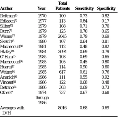
Roitman46 1970 100 0.73 0.82 Erikssen74 1977 113 0.84 0.17 Silber75 1979 108 0.71 0.70 Dunn76 1979 125 0.70 0.65 Weiner77 1979 2045 0.79 0.69 Sketch95 1980 107 0.64 0.81 Machecourt80 1981 112 0.48 0.82 Hlatky84 1984 3094 0.69 0.79 O’Hara85 1985 103 0.69 0.65 Machecourt86 1985 105 0.45 0.80 Huerta87 1985 114 0.90 0.60 Weiner91 1985 617 0.61 0.76 Ananich92 1986 111 0.55 0.92 Vincent93 1986 122 0.68 0.48 Detrano94 1986 303 0.69 0.73 Others* 1974 737 0.67 0.68 through 1986 Averages with 8016 0.68 0.69 LVH
*Nine other studies, each with <100 subjects, combined. LVH indicates left ventricular hypertrophy.
Confounders of Stress ECG Interpretation
Resting ST-segment depression is a marker for a higher prevalence of severe CAD and is associated with a poor prognosis; standard exercise testing continues to be diagnos-tically useful in these patients. Although specificity is low-ered in the presence of resting ST depression less than 1 mm, the standard exercise test is still a reasonable first test option because sensitivity is increased. There is a divergence of opinion regarding two specific patient groups: those who are taking digoxin and have less than 1 mm of ST depression and those with LVH with less than 1 mm of resting ST depres-sion. If the test result is negative, the likelihood of CAD is substantially reduced, but an abnormal response, has low specificity, and therefore further testing is indicated. In the
12 ACC/AHA Practice Guidelines American Heart Association - www.americanheart.org
true. The wide variability in test performance apparent from this meta-analysis can be explained by differing degrees of workup bias (354), but it also demonstrates that some of the variability is explained by improper methods for testing and analysis. Upsloping ST depression should be considered bor-derline or negative.
Digoxin
Digoxin produces an abnormal ST-segment response to exer-cise. This abnormal ST depression occurs in 25% to 40% of healthy subjects studied (30,31) and is directly related to age. Two weeks are required to alleviate the effect on the repolar-ization pattern (32).
Left Ventricular Hypertrophy With
Repolarization Abnormalities
This ECG abnormality is associated with a decreased speci-ficity of exercise testing, but sensitivity is unaffected. Therefore, a standard exercise test may still be the first test, with referrals for additional tests only indicated in patients with an abnormal test result.
Resting ST Depression
Resting ST-segment depression has been identified as a marker for adverse cardiac events in patients with and with-out known CAD (38-42). Miranda et al. (43) performed a ret-rospective study of 223 patients without clinical or electro-cardiographic evidence of prior myocardial infarction. Women, patients with resting ECGs showing left bundle-branch block or LVH, and those taking digoxin or with valvular or congenital heart disease were excluded. Ten per-cent of these selected male patients had persistent resting ST-segment depression that correlated with nearly twice the prevalence of severe coronary disease (30%) compared with those without resting ST-segment depression (16%). Diagnostic end points of two mm of additional exercise-induced ST-segment depression or downsloping depression of 1 mm or more in recovery were particularly useful mark-ers in these patients for diagnosis of any coronary disease (likelihood ratio, 3.4; sensitivity, 67%; specificity, 80%). Smaller studies by Kansal et al. (44) and Harris et al. (45), as well as a large study by Fearon et al. (355), had similar results.
Left Bundle-Branch Block
Exercise-induced ST depression usually occurs with left bun-dle-branch block and has no association with ischemia (36). Even up to 1 cm of ST depression can occur in healthy nor-mal subjects. There is no level of ST-segment depression that confers diagnostic significance in left bundle-branch block.
Right Bundle-Branch Block
Exercise-induced ST depression usually occurs with right
bundle-branch block in the anterior chest leads (V1through
V3) and is not associated with ischemia (37). However, in the
published data, there are few patients with resting ST depres-sion greater than 1 mm. It was the consensus of the commit-tee that exercise testing is unlikely to provide important diag-nostic information in such patients and that exercise imaging modalities are preferred in this subset of patients.
Tables 8 through 13 were developed to resolve the issues of LVH, resting ST depression, and digoxin use. Of the 58 stud-ies, only those that provided sensitivity, specificity, and total patient numbers were considered, and only those with more than 100 patients were considered separately. These studies can be summarized as follows:
•
Studies that included patients with LVH had a meansensitivity of 68% and a mean specificity of 69%; the studies that excluded them had a mean sensitivity of 72% and a mean specificity of 77%.
•
Studies that included patients with resting STdepres-sion had a mean sensitivity of 69% and a mean spec-ificity of 70%; studies that excluded them had a mean sensitivity of 67% and a mean specificity of 84%.
•
Studies that included patients taking digoxin had amean sensitivity of 68% and a mean specificity of 74%; studies that excluded patients taking digoxin had a mean sensitivity of 72% and a mean specificity of 69%.
When these results are compared with the average sensitiv-ity of 67% and specificsensitiv-ity of 72%, as well as to themselves, only LVH and resting ST depression appear to lower speci-ficity. However, other studies in apparently healthy persons (see below) have suggested that digoxin use also lowers specificity.
These meta-analyses provide only indirect evidence regard-ing these potentially important factors, because they assume that the study populations were otherwise equal with respect to characteristics that might influence test performance. This critical assumption has not been confirmed and may not be
Table 13. Studies Excluding Left Ventricular Hypertrophy Total
Author Year Patients Sensitivity Specificity
Marcomichelakis78 1980 100 0.92 0.62 Morales-Ballejo79 1981 100 0.62 0.74 Guiteras81 1982 112 0.79 0.66 Santinga82 1982 113 0.56 0.86 Currie83 1983 105 0.77 0.82 Nair96 1983 280 0.66 0.93 Melin88 1985 135 0.61 0.79 Hung89 1985 171 0.85 0.63 Detry90 1985 284 0.64 0.72 Furuse97 1987 135 0.77 0.83 Others* 1971 442 0.69 0.84 through 1983 Averages w/o 1977 0.72 0.77 LVH
*Six other studies, each with <100 subjects, combined. LVH indicates left ventricular hypertrophy.
13 ACC/AHA Practice Guidelines
left chest leads (V5and V6) or inferior leads (II and aVF), its
test characteristics are similar to those of a normal resting ECG. The presence of right bundle-branch block does not appear to reduce the sensitivity, specificity, or predictive value of the stress ECG for the diagnosis of ischemia.
Beta-Blocker Therapy
Despite the marked effect of beta-blockers on maximal exer-cise heart rate, when patients were subgrouped according to beta-blocker administration initiated by their referring physi-cian, no differences in test performance were found in a con-secutive group of men being evaluated for possible CAD (33). For routine exercise testing, it appears unnecessary for physicians to accept the risk of stopping beta-blockers before testing when a patient exhibits possible symptoms of ischemia or has hypertension. However, exercise testing in patients taking beta-blockers may have reduced diagnostic or prognostic value because of inadequate heart rate response. The decision to remove a patient from beta-blocker therapy for exercise testing should be made on an individual basis and should be done carefully to avoid a potential hemody-namic “rebound” effect, which can lead to accelerated angi-na or hypertension.
Other Drugs
Various medications, including antihypertensive agents and vasodilators, can affect test performance by altering the hemodynamic response of blood pressure. Acute administra-tion of nitrates can attenuate the angina and ST depression associated with myocardial ischemia. Flecainide has been associated with exercise-induced ventricular tachycardia (VT) (34,35).
Atrial Repolarization
Atrial repolarization waves are opposite in direction to P waves and may extend into the ST segment and T wave. Exaggerated atrial repolarization waves during exercise can cause downsloping ST depression in the absence of ischemia. Patients with false-positive exercise tests based on this finding have a high peak exercise heart rate, absence of exercise-induced chest pain, and markedly downsloping PR segments in the inferior leads (356,357).
ST-Segment Interpretation Issues
Lead Selection
Lead V5alone consistently outperforms the inferior leads and
the combination of lead V5with II, because lead II has a high
false-positive rate. In patients without prior myocardial infarction and with normal resting ECGs, the precordial leads alone are a reliable marker for CAD, and monitoring of inferior limb leads adds little additional diagnostic informa-tion. In patients with a normal resting ECG, exercise-induced ST-segment depression confined to the inferior leads is of lit-tle value for identification of coronary disease (48).
Right-Sided Chest Leads
In a new approach, Michaelides et al. (358) examined 245 patients who underwent exercise testing with standard 12 leads, right ventricular leads, and thallium-201 scintigraphy. They found sensitivities of 66%, 92%, and 93% and speci-ficities of 88%, 88%, and 82%, respectively, for the detection of CAD by angiography, i.e., comparable results to perfusion scanning when right-sided leads were added. However, their study was performed in a population with an abnormally high prevalence of coronary disease, and the committee would not recommend clinical use of right-sided chest leads until these results are confirmed by others.
Upsloping ST Depression
Downsloping ST-segment depression is a stronger predictor of CAD than horizontal depression, and both are more pre-dictive than upsloping depression. However, patients with slowly upsloping ST-segment depression, for example, when the slope is less than 1 mV/s, probably have an increased probability of coronary disease (49,50). If a slowly ascend-ing slope is used as a criterion for abnormal findascend-ings, the specificity of exercise testing will be decreased (more false-positive results), although the test becomes more sensitive. The committee favored the use of the more commonly used definition for a positive test: 1 mm of horizontal or downsloping ST depression (zero or negative slope visually).
ST Elevation
Early repolarization is a common resting pattern of ST vation in normal persons. Exercise-induced ST-segment ele-vation is always considered from the baseline ST level. ST elevation is relatively common after a Q-wave infarction, but ST elevation in leads without Q waves occurs in only 1 of 1000 patients seen in a typical exercise laboratory (51-57).
ST elevation on a normal ECG (other than in aVR or V1)
rep-resents transmural ischemia (caused by spasm or a critical lesion), is very rare (0.1% in a clinical laboratory), and, in contrast to ST depression, is very arrhythmogenic and
local-izes the ischemia. When it occurs in leads V2through V4, the
left anterior descending artery is involved; in the lateral leads, the left circumflex and diagonals are involved; and in leads II, III, and aVF, the right coronary artery is involved. When the resting ECG shows Q waves of an old myocardial infarction, the significance of ST elevation is controversial. Some studies have suggested that ST elevation is caused by wall-motion abnormalities (58,59); other studies have found it to be a marker of residual viability in the infarcted area (60-62). Accompanying ST depression in such patients can be caused by a second area of ischemia or reciprocal changes.
R-Wave Changes
Many factors affect the R-wave amplitude response to exer-cise (63), and the response does not have diagnostic signifi-cance (64,65). R-wave amplitude typically increases from
14 ACC/AHA Practice Guidelines American Heart Association - www.americanheart.org
approach could prove useful, such as in rendering a judgment concerning certain borderline or equivocal ST responses, e.g., ST-segment depression associated with a very high exercise heart rate.
Computer Processing
Although computer processing of the exercise ECG can be helpful, it can result in a false-positive indication of ST depression (73). To avoid this problem, the physician should always be provided with ECG recordings of the raw, unprocessed ECG data for comparison with any averages the exercise test monitor generates. It is preferable that averages always be contiguously preceded by the raw ECG data. The degree of filtering and preprocessing should always be pre-sented along with the ECG recordings and should be com-pared with the AHA recommendations (0 to 100 Hz with notched power line frequency filters). It is preferable that the AHA standards be the default setting. All averages should be carefully labeled and explained, particularly those that simu-late raw data. Simulation of raw data with averaged data should be avoided. Obvious breaks should be inserted between averaged ECG complexes. Averages should be checkmarked to indicate the PR isoelectric line and the ST measurement points. None of the computerized scores or measurements have been validated sufficiently to recom-mend their widespread use. At least one study in which these shortcomings have been addressed has shown that computer-ized measurements are comparable to visual measurements, and, when combined with scores, they can provide excellent test characteristics (366).
III. RISK ASSESSMENT AND PROGNOSIS
IN PATIENTS WITH SYMPTOMS OR A
PRIOR HISTORY OF CAD
Class I
1. Patients undergoing initial evaluation with suspected or known CAD, including those with complete right bundle-branch block or less than 1 mm of resting ST depression. Specific exceptions are noted below in Class IIb.
2. Patients with suspected or known CAD, previously evaluated, now presenting with significant change in clinical status.
3. Low-risk unstable angina patients (see Table 17) 8 to 12 hours after presentation who have been free of active ischemic or heart failure symptoms. (Level of
Evidence: B)
4. Intermediate-risk unstable angina patients (see Table 17) 2 to 3 days after presentation who have been free of active ischemic or heart failure symptoms. (Level of
Evidence: B)
Class IIa
Intermediate-risk unstable angina patients (see Table 17) who have initial cardiac markers that are normal, a repeat ECG without significant change, and cardiac rest to submaximal exercise, perhaps to a heart rate of 130
beats per minute (bpm), then decreases to a minimum at maximal exercise (66). If a patient were limited by objective signs or subjective symptoms, R-wave amplitude would increase from rest to such an end point. Such patients may be demonstrating a normal R-wave response but are classified as abnormal because of a submaximal effort. Exercise-induced changes in R-wave amplitude have no independent predictive power but are associated with CAD because such patients are often submaximally tested, and an R-wave decrease normally occurs at maximal exercise. Adjustment of the amount of ST-segment depression by the R-wave height has not been shown to consistently improve the diag-nostic value of exercise-induced ST depression.
ST-Heart Rate Adjustment
Several methods of heart rate adjustment have been proposed to increase the diagnostic accuracy of the exercise ECG. The maximal slope of the ST segment relative to heart rate is derived either manually (67) or by computer (68). A second technique, termed the ST/HR index, divides the difference between ST depression at peak exercise by the exercise-induced increase in heart rate (69,70). ST/HR adjustment has been the subject of several reviews since the last publication of these guidelines (359,360). The major articles that used this approach for diagnostic testing include Morise’s report (361) of 1358 individuals undergoing exercise testing (only 152 with catheterization data) and the report by Okin et al. (362) considering heart rate reserve (238 controls and 337 patients with coronary disease). Viik et al. considered the maximum value of the ST/HR hysteresis over a different number of leads for the detection of CAD (363). The study population consisted of 127 patients with coronary disease and 220 patients with a low likelihood of the disease referred for an exercise test. Neither the study by Okin et al. or that by Viik et al. considered consecutive patients with chest pain, and both had limited challenge. Limited challenge favors the ST/HR index, because healthy patients have relatively high heart rates and sick patients have low heart rates, thus lead-ing to a lower ST/HR index in those without disease and a higher index in sicker patients, the enrollment of relatively healthy patients in these studies presents a limited challenge to the ST/HR index. Likewise, the Morise study had a small number of patients who underwent angiography. The only study with neither of these limitations was QUEXTA (353). This large, multicenter study followed a protocol to reduce workup bias and was analyzed by independent statisticians. The ST/HR slope or index was not found to be more accurate than simple measurement of the ST segment. Although some studies in asymptomatic (and therefore very low likelihood) individuals have demonstrated additional prognostic value with the ST/HR adjustment, these data are not directly appli-cable to the issue of diagnosis in symptomatic patients (364,365). Nevertheless, one could take the perspective that the ST/HR approach in symptomatic patients has at least equivalent accuracy to the standard approach. Although not yet validated, there are situations in which the ST/HR


![Table 5. Definitions and Calculation of the Terms Used to Quantify the Diagnostic Accuracy of a Test Sensitivity = [TP/(TP + FN)] × 100 Specificity = [TN/(FP + TN)] × 100](https://thumb-us.123doks.com/thumbv2/123dok_us/8995914.2384544/8.918.182.763.984.1074/table-definitions-calculation-quantify-diagnostic-accuracy-sensitivity-specificity.webp)