Facts, Figures
and First-Hand
Experience
Four leading experts assess key performance
factors of IOLs and preloaded delivery systems.
Here they share their views on the Vivinex™
multiSert™ injector, the rotational stability of
the Vivinex™ IOL platform and associated toric
outcomes, as well as the comparative PCO
performance of the Vivinex™ IOL.
Featuring:
Stefanie Schmickler, Augen-Zentrum-Nordwest, Ahaus, Germany Rupert Menapace, University of Vienna, Austria
Hiroko Bissen-Miyajima, Tokyo Dental College Suidobashi Hospital, Tokyo, Japan
Michael Wormstone, University of East Anglia, UK
HOYA Surgical Optics names and logos are registered trademarks of HOYA Surgical Optics, Inc. © 2018 HOYA Surgical Optics, Inc. All rights reserved.
Impressive!
By Stefanie Schmickler, Augen-Zentrum-Nordwest, Ahaus, Germany
I work in a very large practice in Germany: the staff includes 19 ophthalmologists, 8 of whom do cataract surgery. We perform several thousand outpatient cataract procedures and only 500 inpatient cataract procedures a year. Most of our patients have very hard nuclear cataracts – not the so-called Californian cataracts, i.e. soft cataracts that some surgeons remove rather earlier than many other surgeons might consider necessary. The majority of our procedures are covered by taxpayer-funded social reimbursement, but some patients elect for premium IOLs or femto-laser assisted cataract surgery and so must pay for their care privately. In the past, we used the J&J TECNIS iTec® and Alcon
AcrySof® UltraSert™ preloaded systems
as standard, but 2 years ago, we adopted the HOYA iSert® system – and over the
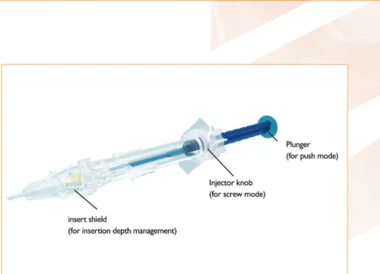
last 6 weeks, I have been working with the new Vivinex™ multiSert™ (Figure 1). A system for all surgeons
My immediate impression is that multiSert is an excellent system. I particularly like having the option of push or screw approaches. Usually, I employ the push mode – the lens comes out of the injector in the right orientation and is delivered very smoothly into the eye, without risk of tearing the posterior capsule. Also, the push option allows the surgeon to keep one hand free, which can be very useful during surgery. For example, I had a case where there was insufficient viscoelastic on the table and the single-handed push mode allowed me to deliver the lens with one hand and irrigate the anterior chamber with the other. That said, it’s good to have an alternative to the push delivery mode. For example, when I am dealing with a weak zonula case, I
usually prefer to have the additional control provided by the screw approach. Of course, others may prefer the screw mode as routine (some of my very skilled colleagues take this view!).
I also appreciate the broader flexibility of multiSert; it permits many variations on the cataract surgery theme. In particular, the adjustable insert shield limits the penetration depth according to the surgeon’s preference. For example, you can advance the insert shield if you want to dock the injector into the wound, or you can use it in the retracted position if you are doing a posterior incision or want to be able to place the tip inside the capsular bag. Personally, I prefer the deeper injection mode, but I know that those colleagues who routinely perform clear corneal incisions prefer the shallower injection format. So having these options is very helpful. MultiSert really is a system for all surgeons!
Whatever your preferred mode of use, the multiSert is very user-friendly. Perhaps the most impressive aspect of its use is the low level of resistance during lens delivery. In contrast, when using the J&J TECNIS iTec, I always felt that I needed to force the lens out of the
injector, but I never get that impression with multiSert. It is fantastic to be using a very modern, advanced system like this. By coincidence, we also work with Fritz Ruck GmbH, a company which was recently acquired by HOYA. We use Ruck’s Qube® PRO surgical system
and have enjoyed close contact with the manufacturer which has helped us to optimise how we use the machine. Given our experience, I think that the combined HOYA / RUCK portfolio will be an interesting proposition for surgeons. Last thoughts
In conclusion, multiSert is a system that has been developed with the surgeon in mind. It delivers the lens safely and consistently and its multiple modes of use provide unparalleled flexibility of operation. Like all preloaded systems, the Vivinex multiSert also makes life easier for the operating team: the nurses don’t need to think about matching a particular injector cartridge to a particular IOL, they just have a single item to prepare. And because multiSert is less stressful for our nurses, it makes the operating theatre a much calmer place!
www.hoyasurgicaloptics.com
Part 2: Stick or Twist?
People tend to prefer familiar products – it’s known as the “mere-exposure effect” – but how do surgeons know if their preferred toric IOL provides optimal rotational stability? To seek clarity, we have evaluated post-surgical rotation in the most frequently-used IOLs. Recently, our work with the HOYA VivinexTM IOL was published in
the British Journal of Ophthalmology.
By Rupert Menapace, University of Vienna, Austria
There aren’t many rigorous studies on the comparative rotational stability of different IOLs – which complicates rational IOL choice. We want to change that. How to deal with a rotational stability study Our study methodology is fine-tuned to minimise secondary rotation and it is standardised to ensure consistency between procedures (see sidebar: “Rotational stability study design”). We use only cohesive OVD, which helps anchor the lens, and allow 3 minutes between surgery completion and meridional
measurement so that the haptics can fully unfold. We gently flatten and reinflate the anterior chamber, without over-inflation, so that eyes are always normotonic.
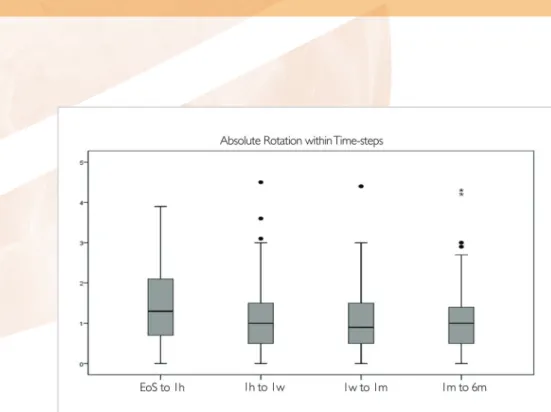
We examined the Vivinexlens. Our published data in the BJO (1) indicate that this IOL has an outstanding stability profile. We saw no rotations that exceeded 5 degrees at any individual time point (Figure 2), including at final follow-up; median and mean rotations were very low, as was the standard deviation. Interestingly, Vivinex rotations show no correlation between rotational stability and either white-to-white distance or axial length. By contrast, when we have applied the same methodology to some of the most popular IOL platforms available (data yet to be published), there were outliers with more significant rotation and these outliers correlated with longer eyes and larger capsular bags.
In all lenses that we have evaluated using this methodology, a notable feature of our data is that the greatest frequency and extent of IOL rotation occurs within the first week. This is probably because the bag is still patent in this period. Rotation decreases dramatically as the capsular bag closes. After a month,
Rotational stability
study design
• Evaluation is made using the monofocal IOL base platform. This has two benefits:
• recruitment is not limited to patients with astigmatism • any rotation does not
impair vision – and therefore patients do not leave the study for corrective surgery • For reference points to
measure rotation, we used: • haptic junctions
(on axle or shoulder) • scleral landmarks
(usually emissaria)
• Measured meridional position at: end of surgery (patient supine on operating table); 1 hour; 1 day; 1 week; and 4 to 6 months
Figure 2. The Vivinex IOL rotation values never exceeded 5 degrees at any post-surgery time-point.
“Vivinex rotation
values never
exceed 5 degrees
between the end
of surgery and
any post-surgery
time-point.”
Rupert Menapace
significant rotation can occur. Cleaning up
What have we learnt from our work on rotational stability? Firstly, the key period for rotational stability is the hour immediately after surgery. If the first measurements are taken later than hour 1, the great majority of lens rotation will be missed and lenses will appear more stable than they actually are.
Secondly, among the lenses we have studied, only the HOYA Vivinex IOL was free of any rotations significant enough to require surgical intervention. This finding might be accounted for by the textured surface on both the front and back of Vivinexhaptics, which may help anchor the lens, apparently independent of the capsular size.
Most importantly, it is clear that not all lenses are rotationally equal. Therefore, when deciding whether to “stick” with their favorite IOL or “twist” to another, surgeons should consider data from well-designed studies where measurements have been taken within the first hour, immediately after completion of surgery, with the patient still supine on the OR table.
Part 3: Total Success
By Hiroko Bissen-Miyajima, Dental College Suidobashi Hospital, Tokyo, Japan
This clinical evaluation shows that all eyes implanted with HOYA’s Preloaded Vivinex™ Toric have excellent rotational stability and astigmatic correction outcomes.
About one-third of cataract patients have corneal astigmatism of 1 D or more; 22 percent have astigmatism values over 1.5 D and 8 percent over 2 D (2, 3). For these patients, it makes sense to correct astigmatism at the same time as addressing the cataract. After all, when given the choice, most
people would opt for post-operative spectacle-independent distance vision. But, surgeons also have choices to make – which IOL is most likely to achieve the desired outcome?
Broadly speaking, toric IOLs are an option that should be considered for patients with ≥1 D astigmatism, particularly as technology advances have conferred significant improvements in rotational stability and visual outcomes (4). To choose between the different toric IOLs available, however, clinicians need data. This prospective, single-arm, single-centre study (August 2017 – March 2018) evaluated Vivinex Toric outcomes in aphakic eyes subsequent to cataract surgery. Study parameters were aimed at the assessment of (i) performance (visual acuity improvement and astigmatism reduction) and (ii) safety.
Incontrovertible Outcomes
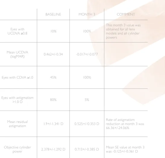
How did the lenses perform against the study hypothesis that at least 59.9 percent of subjects would have a decimal UCDVA value of ≥0.8 at the final follow-up (post-operative month 3)? The numbers are unarguable, particularly with regard to baseline versus post-operative month 3 outcomes (Table 1). Most tellingly, all 20 eyes achieved a decimal UCDVA ≥0.8 at post-operative month 3. Thus, the efficacy of Vivinex Toric is 100 percent and easily exceeds the pre-defined criteria for success. Since this target was chosen based on clinical results of competitor toric IOLs, it can be concluded that the performance of Vivinex Toric is certainly non-inferior to other approved toric IOLs.
The study findings (5) speak for themselves in other measures too: no eye required IOL repositioning and no other safety issues were observed.
Table 1. Dynamics of cataract surgery with simultaneous astigmatism correction by Vivinex Toric. powers Mean UCDVA
(logMAR) 0.462+/-0.34 -0.017+/-0.077
Eyes with CDVA ≥1.0 45% 100%
Eyes with astigmatism
>1.0 D 80% 5%
Mean residual
astigmatism 1.9+/-1.341 D 0.525+/-0.353 D
Rate of astigmatism reduction at month 3 was 66.36+/-24.06%
Objective cylinder
www.hoyasurgicaloptics.com
Conclusions
On the basis of these UCDVA outcomes and patient satisfaction data, the preloaded HOYA Vivinex XY1A Toric IOL is rated as ‘remarkably effective.’ The study (5) unequivocally demonstrates that Vivinex Toric is effective and safe in the correction of regular corneal astigmatism between ≥0.75 D and ≤3.5 D. In this population, the Vivinex Toric IOL should be the first choice for patients who seek spectacle-independent distance vision after cataract surgery; and for surgeons, the preloaded injector is a major benefit.
Part 4: Top Marks to the
Vivinex™ IOL
When assessing a process as complex
as posterior capsule opacification,
it pays to use a model that closely
reflects the human situation.
By Michael Wormstone, University of East Anglia, UK
Posterior capsule opacification (PCO) is a major complication of cataract surgery. Intraocular lenses (IOLs) do not cause PCO – but design aspects of a given IOL do influence PCO progress. How can we assess which IOLs are better able to suppress this problematic phenomenon? Laboratory models are critical for such evaluations – but not all models perform equally. First, do the hard work…
A good PCO model should reflect the cell biological events that occur in the clinical PCO context (see sidebar: “Key steps in PCO”).
Through a process of iterative improvement over many years, we have developed a model that recapitulates PCO etiology to a greater extent than ever before. The basic features of the model have remained constant: in brief, we perform cataract surgery on donor cadaver eyes to
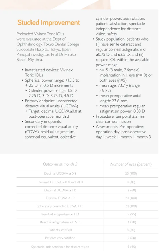
Studied Improvement
Preloaded Vivinex Toric IOLs were evaluated at the Dept of Ophthalmology, Tokyo Dental College Suidobashi Hospital, Tokyo, Japan. Principal investigator: Prof Dr Hiroko Bissen-Miyajima.
• Investigated devices: Vivinex Toric IOLs
• Spherical power range: +15.5 to + 25 D, in 0.5 D increments • Cylinder power range: 1.5 D,
2.25 D, 3 D, 3.75 D, 4.5 D • Primary endpoint: uncorrected
distance visual acuity (UCDVA) • Target: decimal UCDVA≥0.8 at
post-operative month 3 • Secondary endpoints:
corrected distance visual acuity (CDVA), residual astigmatism, spherical equivalent, objective
patient satisfaction, spectacle independence for distance vision, safety
• Study population: patients who (i) have senile cataract and regular corneal astigmatism of ≥0.75 D and ≤3.5 D; and (ii) require IOL within the available power range
• n=15 (8 male, 7 female) implantation in 1 eye (n=10) or both eyes (n=5)
• mean age: 73.7 y (range: 56-82)
• mean preoperative axial length: 23.61mm
• mean preoperative regular astigmatism power: 0.83 D • Procedure: temporal 2.2 mm
clear corneal incision • Assessments: Pre-operative;
operation day; post-operative day 1; week 1; month 1; month 3
Outcome at month 3 Number of eyes (percent)
Decimal UCDVA ≥ 0.8 20 (100)
Decimal UCDVA ≥ 0.8 and <1.0 8 (40)
Decimal UCDVA ≥ 1.0 12 (60)
Decimal CDVA >1.0 20 (100)
Spherically corrected CDVA >1.0 20 (100)
Residual astigmatism ≤ 1 D 19 (95)
Residual astigmatism ≤ 0.5 D 14 (70)
Patients satisfied 8 (40)
Patients very satisfied 12 (60)
Spectacle independence for distant vision 19 (95) Table 2. In all Vivinex Toric implanted eyes, post-operative uncorrected distance visual acuity meets or exceeds the target decimal value (0.8). This target was based on outcomes achieved with competitor IOLs. The study therefore demonstrates that Vivinex Toric is non-inferior to other approved IOLs (5).
produce lens capsular bags in which an IOL can be implanted. We isolate and transfer the IOL-bag system to a tissue culture dish and maintain in defined conditions in vitro (6). A matched-pair experimental design – where the eyes from a single donor each receive a different IOL and are compared in a single experiment – helps eliminate inter-donor variability. This model enables us to monitor PCO in the presence of different IOLs and under different environmental conditions (7), using standard microscopy techniques.
Successive improvements to the model have included: pinning the anterior capsule to the culture dish to improve capsule-IOL interaction (8); development of a system in which the
bag is kept suspended by retaining and pinning its ciliary body to a silicone ring in the culture dish (9); and further humanization of this system with human serum and recombinant human growth factors. Even so, these earlier versions of the model did not reflect the temporal pattern of protein flare seen in the clinic – namely, a rapid post-operative increase in the protein content of the aqueous humor, which is sustained for about a week before declining (10).
We have now developed a graded culture model (11), which mimics the clinical situation by providing high initial levels of human serum and TGF beta, and then reducing them over time. Our data show that the graded culture • Cataract surgery, however
skillful, causes physical trauma to ocular tissues
• Consequently, a compromised blood-aqueous barrier permits inflammatory proteins to reach the lens environment
• Inflammatory drivers stimulate lens epithelial cells (LECs), thereby enhancing proliferation, migration, and differentiation • LECs grow on the anterior
capsule, the intraocular lens surface, and, most importantly, on the posterior capsule • Over time, LEC can
transdifferentiation into myofibroblasts and enhance matrix deposition. In addition, differentiation can occur leading to the formation of structures such as Soemmering’s rings and Elschnig’s pearls
• Result: impaired vision
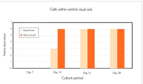
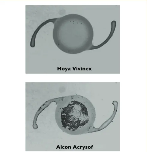
Figure 3. Cell growth on the central posterior capsule is suppressed by Vivinex relative to AcrySof.
Figure 4. (A) Cell coverage and (B) light scatter on the central posterior capsule (PC) after 28 days: Vivinex versus AcrySof. * Light scatter was significantly greater with AcrySof present than Vivinex.
A
B
“This model
indicates that
patients implanted
with the Vivinex IOL
are less likely to
develop PCO than
with an AcrySof
IOL
implant.” Michael
Wormstone
www.hoyasurgicaloptics.com
system mimics a number of changes observed in clinical specimens, which include cell migration, matrix contraction, matrix deposition, and higher levels of myofibroblast markers.
… then answer the big questions What has the graded culture model taught us about IOL influence on PCO? A comparison of the HOYA Vivinex IOL and the Alcon AcrySof® reveals some
interesting differences. Migration of cells from the periphery to the center of capsular bags occurs more slowly, on average, with the Vivinex IOL compared to an AcrySof IOL (Figure 3). At the 28-day end point, while cell coverage within the central visual axis of the central posterior capsule was not significantly different (Figure 4A), the difference in the degree of light scatter detectable in the central visual axis was significant: the Vivinex IOL demonstrated an advantage of more than 20% versus AcrySof, on average (Figure 4B).
Figure 5 shows cell growth on the anterior surface of the IOL. The significant reduction of cell outgrowth (Figures 5 and 6) on the anterior surface with the Vivinex IOL may explain the better optical performance observed in terms of there being less cell-induced light scattering.
The Vivinex IOL passes the test
In conclusion, our fully-humanized, graded culture system provides the best available model of PCO. This model indicates that patients implanted with the Vivinex IOL are less likely to develop PCO than with an AcrySof IOL implant. In addition, potential for cell growth on the anterior surface of the IOL to cause further light scatter is greater with the Acrysof than with the Vivinex IOL. Findings such as these may help surgeons make rational choices between IOLs – and ultimately provide better outcomes for patients.
Figure 5. Extent of cell growth on the anterior IOL surface of Vivinex versus AcrySof.
HOYA Surgical Optics GmbH De-Saint-Exupery-Strasse, 10 60549 Frankfurt am Main, Germany Tel: +49-(0)69-664-268-0
EC REP
HOYA Surgical Optics names and logos are registered trademarks of HOYA Surgical Optics, Inc. © 2019 HOYA Surgical Optics, Inc. All rights reserved. AcrySof, TECNIS and Ruck’s QUBE Pro are trademarks of their respective manufacturers.
1. D Schartmüller , S Schriefl, L Schartmueller, C Leydolt, R Menapace , “True rotational stability of a single-piece hydrophobic IOL”, Brit J
Ophthalmol, 103, 186-190 (2019). PMID: 29666120.
2. P Hoffman and W Hütz, “Analysis of biometry and prevalence data for corneal astigmatism in 23,239 eyes”, J Cataract Refract Surg, 36, 1479-85 (2010). PMID: 20692558. 3. T Ferrer-Blasco et al., “Prevalence of corneal
astigmatism before cataract surgery”, J Cataract Refract Surg, 35, 70-75 (2009). PMID: 19101427.
4. M Kaur, et al, “Optimizing outcomes with toric
1301-13 (2017). PMID: 29208810. 5. HOYA data on file: DoF_SP2Y-TC_
CSR_29012019. SP2Y-TC Clinical Study, Protocl Number: HCT_07.
6. C Liu et al., “A study of human lens cell growth in vitro: a model for posterior capsule opacification”, Invest Ophthalmol Vis Sci, 37, 906-14 (1996). PMID: 8603875.
7. G Duncan et al., “Thapsigargin-coated intraocular lenses inhibit human lens cell growth”, Nature Med, 3, 1026-1028 (1997). PMID: 9288732. 8. L Dawes, CD Illingworth, IM Wormstone, “A fully human in vitro capsular bag model to permit intraocular lens evaluation”, Invest
22125276.
9. G Cleary et al., “In vitro lens capsule model for investigation of posterior capsule
opacification”, J Cataract Refract Surg, 36, 1249-1252 (2010). PMID: 20656145. 10. M Pande et al., “Cellular reaction on the
anterior surface of poly(methylmethacrylate) intraocular lenses”, J Cataract Refract Surg, 22 Suppl 1, 811-17 (1996). PMID: 9279677. 11. J Eldred et al., “An in vitro human lens capsular
bag model adopting a graded culture regime to assess putative impact of IOLs on PCO formation”, Invest Ophthalmol Vis Sci, 60, 113-122 (2019). PMID: 30629726.