Copyright © 2015 European Crohn’s and Colitis Organisation (ECCO). Published by Oxford University Press. All rights reserved.
For permissions, please email: [email protected] 38
doi:10.1093/ecco-jcc/jjv173 Advance Access publication September 28, 2015 Original Article
Original Article
Image-guided Percutaneous Drainage for
Treatment of Post-Surgical Anastomotic Leak in
Patients with Crohn’s Disease
James Byrne,
aRyan Stephens,
aAri Isaacson,
bHyeon Yu,
bCharles Burke
baSchool of Medicine, University of North Carolina at Chapel Hill, Chapel Hill, NC, USA bDepartment of Radiology,
Division of Vascular Interventional Radiology, University of North Carolina at Chapel Hill, Chapel Hill, NC, USA Corresponding author: Ari Isaacson, MD, Department of Radiology, Division of Vascular Interventional Radiology, School of Medicine, University of North Carolina at Chapel Hill, Chapel Hill, NC 27599, USA. Tel: +1 [919] 966-6646; fax: +1 [919] 843-8740; email: [email protected]
Abstract
Background and Aims: Anastomotic leaks with abscess formation are a common complication
after bowel surgery in Crohn’s disease patients. Image-guided percutaneous drainage is an attractive alternative to reoperation because of decreased morbidity and length of hospital stay. Because data for this specific population are scarce, the purpose of this study is to determine the safety and efficacy of image-guided percutaneous drainage in the management of post-surgical anastomotic leak in patients with Crohn’s disease.
Methods: A total of 41 patients who underwent percutaneous drain placement for the treatment of
fluid collections due to anastomotic leak from September 2004 to November 2013 were retrospectively identified from the electronic medical record and picture archiving and communication system. Data recorded included number, size, and location of anastomotic leaks, number of drains placed, number of follow-up visits, post-drainage complications, abscess resolution, and subsequent surgeries.
Results: In all, 41 patients with 76 fluid collections were identified as having received percutaneous
drains. The mean number of targeted fluid collections per patient was 1.5, and the mean duration between surgery and percutaneous drain placement was 18.5 days. The mean number of drains placed was 1.6, and the median drain size was 10 French [range 8–16 French]. One of 41 [2.4%] patients experienced a minor complication from drain placement [injury to a superficial abdominal artery] and no major complications occurred. Two of 41 [4.9%] patients required repeat surgeries.
Conclusions: Image-guided percutaneous drainage for the treatment of post-surgical anastomotic
leaks in Crohn’s patients is effective and safe, with low rates of complications and reoperations.
Key Words: Crohn’s disease; bowel surgery; anastomotic leak; percutaneous drainage; abscess
1. Introduction
The majority of patients with Crohn’s disease will require bowel surgery during their lifetime.1 The postoperative complication rates
are higher in patients with Crohn’s disease than for other benign diseases.2 Anastomotic leak is a frequent and severe
postopera-tive complication after bowel resection and reanastomosis in these patients, occurring at rates between 1% and 30%.3,4,5,6 Defined as
the leak of luminal contents from a surgical join of two hollow vis-cera, anastomotic leak can be diagnosed using variations of clinical indicators like pain, peritonitis, and biochemical markers, as well as radiological studies showing fluid collections or gas-containing col-lections. Anastomotic leaks are detected anywhere from 3 to 45 days postoperatively.3 If untreated, anastomotic leaks can lead to abscess
formation, peritonitis, and sepsis.5,7,8,9,10,11,12,13
Historically, anastomotic leaks required additional surgical inter-vention. However, image-guided percutaneous drainage has become an attractive alternative to reoperation because of decreased mor-bidity and hospital stay.13,14,15,16,17,18 Currently, there are limited data
available as to the efficacy and rate of complication when image-guided percutaneous drainage is performed for anastomotic leak in this specific patient population. The purpose of this study was to report such data by retrospectively reviewing cases at a single aca-demic institution.
2. Methods
2.1. Study design
Institutional review board approval was obtained and all guidelines of the Health Insurance Portability and Accountability Act were fol-lowed. A retrospective review was initially performed that included all patients aged 18 years and older with Crohn’s disease who under-went bowel anastomosis at a single academic medical centre from September 2004 to November 2013. The patients were identified using the electronic medical record and picture archiving and com-munication systems. Eligible patients for this study were those who underwent image-guided percutaneous drain placement for treat-ment of post-surgical anastomotic leak. Patients were determined to have an anastomotic leak if they had a fluid collection adjacent to an anastomosis identified on contrast-enhanced computed tomography [CT] within 1 month of the surgery in which that anastomosis was created, 41 patients were identified who met the criteria of the study.
Once identified, the patient’s medical record was reviewed, and the following data points were recorded: [1] demographic data, [2] location of disease, [3] Montreal classification, [4] immunosuppres-sive medications, [5] date of procedure, [6] type of surgery, [7] indi-cation for surgery, [8] initial surgery vs repeat surgery, [9] loindi-cation of anastomosis, [10] location of anastomotic leak [left upper rant, right upper quadrant, left lower quadrant, right lower quad-rant, abdominal wall, psoas gluteal, pelvic], [11] stoma creation, [12] size of abscess, [13] days post-surgery that drains were placed, [14] size and number of drains placed, [15] time to first follow-up, [16] duration of drainage, [17] number of manipulations/exchanges, [18] need for total parenteral nutrition [TPN], [19] complications of treatment including complications of sedation, drug-related allergic reactions, cardiopulmonary complications, infectious complications, bleeding, and non-target access, and [20] number of a repeat surger-ies required to repair the anastomotic leak. The primary outcome was abscess resolution as determined by the lack of contrast leakage through drainage catheter under fluoroscopy. Other outcomes meas-ured included post-drainage complications and subsequent surgery required.
2.2. Procedure
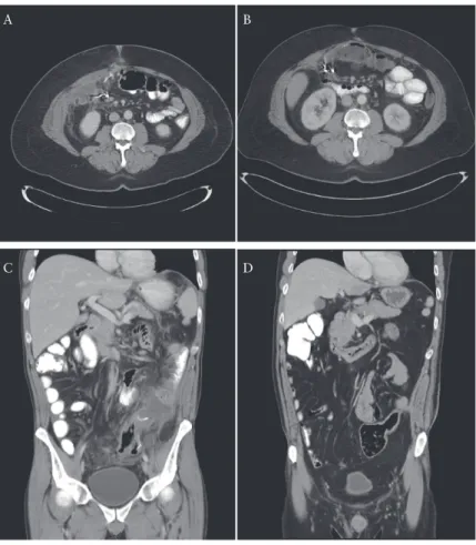
All image-guided drain placements were performed by or under the supervision of experienced academic interventional radiolo-gists. Drainage catheters were placed using either ultrasound [US], computed tomography [CT], or a combination of US and CT guid-ance [Figure 1]. Drainage catheters were placed using standard techniques.19 Once access to the fluid collection was obtained, a
self-locking pigtail drainage catheter, ranging from 8–16 French in size, was placed. Drainage catheter size was at the discretion of the operating physician.
The drains were flushed with normal saline, 5-10 ml once or twice a day, depending on operator preference. The patients returned every 2 weeks for drain study under fluoroscopy. The patient would
be evaluated earlier than 2 weeks if the output significantly declined or if there was concern that the drain may have become dislodged. During the follow up drain evaluation, the drains were exchanged if they appeared obstructed or malpositioned. Drains were removed when there was only minimal fluid cavity remaining on the drain study and there was no evidence for persistent fistula to the bowel [Figure 2].
All patients were initially treated with broad-spectrum antibi-otics that individually, or in combination, covered Gram-negative organisms, Gram-positive organisms, and anaerobes, for 2–14 days. Patients who did not initially tolerate enteral nutrition [EN] were placed on TPN, until they were able to eat again.
2.3. Statistical analysis
The relationship between size of fluid collection and duration of drain, type of surgery and location of fluid collection, size of drain-age tube and duration of draindrain-age, and location of fluid collection and duration of drain were analysed and compared using Student’s t-test or one-way analysis of variance [ANOVA] test. All other quan-titative data were expressed as mean, median, and range.
3. Results
A total of 127 potentially eligible patients were identified as patients with Crohn’s disease who underwent bowel surgery. Among these, 41 patients [32%] were identified as having an anastomotic leak managed with percutaneous drainage. Table 1 presents baseline patient charac-teristics, including age of patients, location of disease, age at diagno-sis, Montreal classification, immunosuppressive medications, smoking status, and previous bowel surgery; 25 of 41 patients had previously undergone bowel surgery for their Crohn’s disease at sites at or near the anastomotic site, whereas 16 patients had never undergone surgical intervention. Of the 25 patients who had previously undergone bowel surgery, 8 patients underwent ostomy takedowns. The indications for surgery included medication refractory disease [10 patients], enterocu-taneous fistula repair [9 patients], ostomy takedowns [8 patients], lysis of adhesions [6 patients], bowel obstruction [4 patients], ileocolic fis-tula repair [1 patient], ileal fisfis-tula repair [1 patient], ostomy revision [1 patient], and jejunoileal fistula repair [1 patient]. Twelve of 41 patients had ostomies prior to surgery. In addition, 9 of 41 patients underwent the creation of a stoma during their bowel surgery, and 9 of 9 patients who underwent stoma creation also had additional bowel procedures performed at the same time. There were a total of 76 independent fluid collections identified. )Of the 41 patients, 25 were taking at least one immunosuppressive medication during the course of surgery; these immunosuppressive medications included infliximab, prednisone, azathioprine, methylprednisolone, budesonide, and certolizumab. The immunosuppressive medications were witheld in 11 of 25 patients prior to drainage due to their acute infectious process but were contin-ued in 14 of 25 patients. The incidence of clinical leakage by location of anastomosis and type of anastomosis is shown in Tables 2 and 3. The most common locations for the anastomotic leak were in the pelvis and right lower quadrant of the abdomen, and the most common types of anastomosis were to-side ileum and colon anastomoses and side-to-side small bowel anastomoses. The mean number of targeted fluid collections per patient was 1.5 [median 1; range 1–4], and 15 of 41 [38.1%] patients were treated for multiple abscesses. The mean vol-ume of the abscesses resulting from anastomotic leak was 167.2 cm3
[median 59.5 cm3; range 1.8–1173.1 cm3], and the mean diameter of
the abscesses resulting from anastomotic leak was 8.5 cm [median 8.0 cm; range 2.0–20.0 cm].
The mean duration between surgery and percutaneous drain placement was 18.5 days [median 14 days; range 6–60 days]. Catheters ranged in size from 8 to 16 French [median 10 French]. The average number of drains placed was 1.6 [median 1; range 1–6]; multiple drains were placed because of a large fluid collec-tion, undrained portions of the fluid colleccollec-tion, or new fluid col-lection development. Overall, the mean duration of drainage was 70.4 days [median 29 days; range 2–732 days]. Fluid volumes of greater than 50 cm3 resulted in significantly longer duration of
drain-age [p < 0.0448]. Furthermore, drain sizes of 12 or greater resulted in significantly longer durations of drainage [p < 0.0001]. The mean number of drain manipulations/exchanges because of obstruction
was 1.2 [median 0; range 0–14]. Overall, there was no obvious rela-tionship between the location of the fluid collection and duration of drainage [p = 0.9154] nor type of anastomosis and duration of drainage [p = 0.8967]. Follow-up sinograms indicated fluid collec-tion resolucollec-tion, fistulas, undrained fluid pockets, or residual cavities.
Figure 2 shows fluoroscopic images from a sonogram showing an unresolved fluid pocket and the resolved drain endpoint.
The average time to first follow-up after drain placement was 9.7 days [median 8; range 2–25]. Of the 41 patients, 16 required TPN as a consequence of their bowel surgery, whereas the others maintained their regular diet. One patient [2.4%] experienced a minor complication from drain placement, injury to a superficial
A B
C D
Figure 1. Contrast-enhanced computed tomography [CT] images of two patients showing the [A, C] anastomotic leak [white arrow] and abscess [grey arrow], and [B, D] resolution.
A B
Figure 2. Anterior-to-posterior fluoroscopy images of a sinogram from one patient which shows an [A] unresolved fluid pocket and the [B] resolved fluid pocket.
abdominal artery, which resolved spontaneously without the need for transfusion or an additional procedure. No major complica-tions occurred during or after the procedure [no infectious seque-lae, major bleeding events, non-target access, or fistulas. Of the 41 patients, 39 [95.1%] patients experienced resolution of the anasto-motic leak, as confirmed by CT and drain study. Two of 41 [4.9%] patients required repeat surgeries to repair the anastomotic leak. The decision was made to operate because of the continued high output despite prolonged percutaneous drainage.
4. Discussion
The goal of our work was to determine the safety and efficacy of image-guided percutaneous drainage in the management of
post-surgical anastomotic leak in Crohn’s patients. We found that 95.1% of patients who underwent percutaneous drainage experi-enced resolution of the anastomotic leak, as confirmed by CT and drain study, and did not require a repeat surgery to repair their anas-tomotic leak. There was one minor complication, injury to a super-ficial abdominal artery, and no major complications that occurred as a result of percutaneous drainage. In addition, the use of immu-nosuppressive medications did not limit patients from receiving per-cutaneous drainage. Overall, our results suggest that perper-cutaneous drainage is safe and effective within this patient population.
Physicians are often confronted with the problem of how to treat post-surgical anastomotic leak in patients with Crohn’s disease. Previous authors have supported both surgery and percutaneous drainage to treat these leaks in patients with and without Crohn’s disease.20,21,22,23,24,25 In a retrospective study, Xie et al. found that
per-cutaneous drainage resulted in a lower rate of post-drainage compli-cations compared with the surgery group in patients with Crohn’s disease.22 Furthermore, Gutierrez et al. found that time to resolution
of abdominal or pelvic abscess in Crohn’s disease is similar between percutaneous drainage and surgery.21
Our results are further supported by studies evaluating the percutaneous drainage of spontaneously formed intra-abdominal abscesses in Crohn’s patients which do not arise from anastomotic leaks.18,21 Gutierrez et al. found that approximately two-thirds of
Crohn’s patients with intra-abdominal abscesses treated with percu-taneous drainage do not require follow-up surgery for treatment of their abscesses within 1 year.21 Additionally, in a prospective study
by Casola et al. in which percutaneous drainage was planned as the definitive therapeutic approach, 15 patients with Crohn’s dis-ease had successful abscess drainage percutaneously with no com-plications and no development of cutaneous fistula tracts. Drainage catheters were maintained from 8 days to 6 weeks, and only three Table 1. Baseline characteristics.
Characteristic Crohn’s patients with anastomotic
leak [n = 41]
Age, years
Mean 40.5
SD 14.0
Gender
Male 18
Female 23
Location of disease
Ileal 8
Colonic 2
Ileocolonic 31
Isolated upper disease 0
Age of diagnosis, years
Mean 21.1
SD 9.4
Montreal classification
A1l3b3 10
A2l3b3 7
A1l1b3 5
A2l3b1 4
A3l3b3 3
A2l3b2 2
A3l3b1 1
A3l1b2 1
A2l1b3 1
pa1l3b3 1
A2l2b1 1
A2l2b3 1
A3l3b2 1
pa2l3b1 1
pa2l3b3 1
A2l1b1 1
Immunosuppressive medications
Infliximab 7
Prednisone 10
Azathioprine 3
Methylprednisolone 3
Budesonide 1
Certolizumab 4
Smoker
Yes 5
No 36
Previous surgery
Yes 25
No 16
SD, standard deviation.
Table 2. Location and number of anastomotic leaks.
Location of anastomotic leak Number of leaks
RUQ 11
LUQ 8
RLQ 16
LLQ 6
Pelvis 21
Psoas gluteal 8
Abdominal wall 5
RUQ, right upper quadrant; LUQ, left upper quadrant; RLQ, right lower quadrant; LLQ, left lower quadrant.
Table 3. Type of anastomosis and number of anastomotic leaks.
Type of anastomosis Number of leaks
SB side-to-side 16
SB end-to-end 5
Ileum and colon side-to-side 23
Ileum and colon end-to-side 4
Colorectal side-to-end 1
Colorectal end-to-end 2
Colon side-to-side 2
Colon end-to-end 1
Ileum and proximal rectum side-to-side 1
Ileum J-pouch anal 1
End distal side proximal SB 2
SB, small bowel.
patients required surgery for complete resolution of the abscess in follow-up.18
Anastomotic leakage from colorectal surgery generates a consid-erable demand for hospital resources.26 Consequently, the economic
benefit of resolving leaks without surgery cannot be overstated. Studies by Ashraf et al. established that patients who underwent sur-gical repair of the anastomotic leak had significantly longer hospital stays compared with those managed with percutaneous drainage or antibiotics and were more likely to require intensive care.27 Thus,
resolving leaks using conservative management, including percutane-ous drainage, could reduce the economic burden of anastomotic leak. The main limitation of our study is its retrospective design. Second, despite the proximity of the fluid collections seen on CT to the recent bowel anastomoses, we could not be certain that all of these collections represented true anastomotic leaks. The leakage rate from our study was larger than in most previous studies, which may be attributed to the large number of immunosuppressive medications used in these patients.6 Our
study did not collect data regarding post-drainage fistula formation. In conclusion, image-guided percutaneous drainage for the treat-ment of post-surgical anastomotic leaks in Crohn’s patients is effec-tive and safe, with low rates of complications and reoperations.
Funding
None.
Conflict of Interest
None.
Author Contributions
JB collected data, analysed the data, and wrote the manuscript. RS collected data. AI, HY, and CB analysed the data, critically revised the manuscript, and approved the final version to be submitted. All the authors read and approved the final manuscript.
References
1. Hurst RD, Molinari M, Chung P, et al. Prospective study of the features, indications, and surgical treatment in 513 consecutive patients affected by Crohn’s disease. Surgery 1997;122:661–8.
2. Iesalnieks I, Kilger A, Glass H, et al. Intraabdominal septic complications following bowel resection for Crohn’s disease: detrimental influence on long-term outcome. Int J Colorectal Dis 2008;23:1167–74.
3. Kingham TP, Pachter HL. Colonic anastomotic leak: Risk factors, diagno-sis, and treatment. J Am Coll Surg 2009;209:269–78.
4. Hyman N, Manchester TL, Osler T, et al. Anastomotic leaks after intesti-nal anastomosis. Ann Surg 2007;245:254–8.
5. O’Riordan JM, O’Connor BI, Huang H, et al. Long-term outcome of colectomy and ileorectal anastomosis for Crohn’s colitis. Dis Colon Rec-tum 2011;54:1347–54.
6. El-Hussuna A, Krag A, Olaison G, et al. The Effect of Anti-Tumor Necro-sis Factor Alpha Agents on Postoperative Anastomotic Complications in Crohn’s Disease: a Systematic Review. Dis Colon Rectum 2013;56:1423–33. 7. Scarpa M, Martinato M, Bertin E, et al. Intestinal Surgery for Crohn’s Dis-ease: Role of Preoperative Therapy in Postoperative Outcome. Dig Surg 2015; 32:243–50.
8. Huang W, Tang Y, Nong L, Sun Y. Risk factors for postoperative intra-abdominal septic complications after surgery in Crohn’s disease: A meta-analysis of observational studies. J Crohn’s Colitis 2015;9:293–301. 9. Tzivanakis A, Singh JC, Guy RJ, et al. Influence of risk factors on the
safety of ileocolic anastomosis in Crohn’s disease surgery. Dis Colon Rec-tum 2012;55:558–62.
10. Resegotti A, Astegiano M, Farina EC, et al. Side-to-side stapled anastomo-sis strongly reduces anastomotic leak rates in Crohn’s disease surgery. Dis Colon Rectum 2005;48:464–8.
11. Guo Z, Li Y, Zhu W, et al. Comparing outcomes between side-to-side anas-tomosis and other anastomotic configurations after intestinal resection for patients with Crohn’s disease: a meta-analysis. World J Surg 2013;37:893– 901.
12. Shapiro M, Greenstein AJ, Byrn J, et al. Surgical management and out-comes of patients with duodenal Crohn’s disease. J Am Coll Surg 2008;207:36–42.
13. Post S, Betzler M, von Ditfurth B, et al. Risks of intestinal anastomosis in Crohn’s disease. Ann Surg 1991;213:37–42.
14. Burke LM, Bashir MR, Gardner CS, et al. Image-guided percutaneous drainage vs. surgical repair of gastrointestinal anastomotic leaks: is there a difference in hospital course or hospitalization cost? Abdom Imaging 2015;40:1279–84.
15. Gerzof SG, Robbins AH, Johnson WC, Birkett DH, Nabseth DC. Per-cutaneous catheter drainage of abdominal abscesses. N Engl J Med 1981;305:653–7.
16. vanSonnenberg E, Ferrucci JT, Mueller PR, et al. Percutaneous radio-graphically guided catheter drainage of abdominal abscesses. JAMA 1982;247:190–2.
17. Gerzof SG, Robbins AH, Birkett DH, et al. Percutaneous catheter drainage of abdominal abscesses guided by ultrasound and computed tomography. AJR Am J Roentgenol 1979;133:1–8.
18. Casola G, vanSonnenberg E, Neff CC, et al. Abscesses in Crohn disease: percutaneous drainage. Radiology 1987;163:19–22.
19. Rypens F, Dubois J, Garel L, et al. Percutaneous drainage of abdomi-nal abscesses in pediatric Crohn’s disease. AJR Am J Roentgenol 2006;188:579–85.
20. Felder SI, Barmparas G, Murrell Z, Fleshner P. Risk factors for failure of percutaneous drainage and need for reoperation following symptomatic gastrointestinal anastomotic leak. Am J Surg 2014;208:58–64.
21. Gutierrez A, Lee H, Sands BE. Outcome of surgical versus percutaneous drainage of abdominal and pelvic abscess in Crohn’s disease. Am J Gastro-enterol 2006;101:2283–9.
22. Xie Y, Zhu W, Ning L, et al. The outcome of initial percutaneous drainage versus surgical drainage for intra-abdominal abscesses in Crohn’s disease. Int J Colorectal Dis 2012;27:199–206.
23. Ananthakrishnan AN, McGinley EL. Treatment of intra-abdominal abscesses in Crohn’s disease: A nationwide analysis of patterns and out-comes of care. Dig Dis Sci 2013;58:2013–8.
24. Georgopoulos F, Mylonaki M, Malgarinos G, et al. Intraabdominal abscesses in patients with Crohn’s disease: Clinical data and therapeutic manipulations in 17 cases of a single hospital setting. Ann Gastroenterol 2008;21:188–92.
25. Longo WE, Milsom JW, Lavery IC, et al. Pelvic abscess after colon and rectal surgery – what is optimal management? Dis Colon Rectum 1993;36:936–41.
26. Frye J, Bokey EL, Chapuis PH, Sinclair G, Dent OF. Anastomotic leakage after resection of colorectal cancer generates prodigious use of hospital resources. Colorectal Dis 2009;11:917–20.
27. Ashraf SQ, Burns EM, Jani A, et al. The economic impact of anastomotic leakage after anterior resections in English NHS hospitals: are we ade-quately remunerating them? Colorectal Dis 2013;4:e190–8.