MEIOTIC CROSSOVER PATTERNING IN
DROSOPHILA MELANOGASTER
Michaelyn Ann Hartmann
A dissertation submitted to the faculty at the University of North Carolina at Chapel Hill in partial fulfillment of the requirements for the degree of Doctor of Philosophy in the Curriculum in Genetics
and Molecular Biology in the School of Medicine.
Chapel Hill 2019
ii ©2019
iii
ABSTRACT
Michaelyn Ann Hartmann: Meiotic Crossover Patterning in Drosophila melanogaster (Under the direction of Jeff Sekelsky)
Meiosis is an essential process to halve an organism’s genome in preparation for transmission to the next generation. Recombination between homologous chromosomes is necessary for the proper segregation of chromosomes, and allows the generation of genetic diversity. Mistakes in meiosis can lead to aneuploidy, therefore, to minimize mistakes, recombination is a highly regulated process. Crossovers are patterned along a
iv
To my mom, for your constant love and support. You are the strongest, most amazing woman I
v
ACKNOWLEDGEMENTS
I would first like to thank my advisor, Jeff Sekelsky. Thank you for always being there and striving to be the best mentor. You have been a wonderful teaching role model and you have definitely inspired me to be be a great teacher and mentor. Thank you for allowing me to explore my interests throughout graduate school, it made graduate school enjoyable and allowed me to reach my goals and obtain my dream job. I am truly grateful to have been a member of the Sekelsky lab.
Thank you also to my committee, Bob Duronio, Dan McKay, Jill Dowen, and Dale Ramsden, for your scientific guidance and support.
I want to thank the extraordinary people I have had the pleasure of working with in the Sekelsky Lab: Nicole Crown, Noelle Romero, Danielle Rognstad, Stephanie Bellendir, Simon Brady, Julie Korda Holsclaw, Greg Zapotoczny, Juan Carvajal Garcia, Leeza Santiago Millan, Carolyn Turcotte, Alex Stutzman, and Evan Dewey. I especially want to thank Susan Cheek for helping me stay sane and maintaining a great lab environment. I want to thank my two outstanding undergraduates that I was lucky enough to mentor: Caitlin Moffatt and Leela Wissmann. I also want to specially thank one of my very best friends and labmate, Talia Hatkevich. Talia has helped me grow and find myself throughout graduate school and I will always be grateful for our friendship. Working with all of these amazing people helped brighten every day and I will always be thankful for all of your love and support.
vi
vii
TABLE OF CONTENTS
LIST OF FIGURES ... XI LIST OF TABLES ... XIII LIST OF ABBREVIATIONS ... XIV
CHAPTER 1: INTRODUCTION ... 1
Meiotic crossover patterning phenomena ... 3
Crossover distribution modeling ... 5
Drosophila as a model for meiotic crossover control ... 6
Two pathways to crossover formation ... 7
Centromere proximal crossover suppression in Drosophila ... 10
Scope of this work ... 16
CHAPTER 2: EXAMINATION OF ANKLE1 AS A POTENTIAL NUCLEASE ... 18
Preface ... 18
Introduction ... 18
Materials and Methods ... 21
Yeast two-hybrid ... 21
CRISPR/Cas9 Deletion of Ankle1 ... 21
Sensitivity assays ... 22
Nondisjunction assay... 23
viii
Results ... 23
Nuclease Complex Interactions ... 23
Ankle1 deletion and characterization ... 26
Ankle1 sensitivity to DNA-damaging agents ... 26
Mitotic role of Ankle1 ... 27
Discussion ... 29
CHAPTER 3: MEIOTIC MCM PROTEINS PROMOTE AND INHIBIT CROSSOVERS DURING MEIOTC RECOMBINATION ... 31
Abstract... 31
Introduction ... 32
Figure 3.1. MCM protein structure and alignments... 33
Materials and Methods ... 39
Drosophila stocks ... 39
Generating mei-218 transgenic alleles ... 40
Generating recKA and recDA mutants ... 40
Nondisjunction assay... 40
Crossover distribution assay ... 41
Protein structure and alignment ... 41
Results and Discussion ... 43
The N-terminus of MEI-218 is dispensable for crossover formation ... 43
REC ATPase motifs are required for crossover formation ... 46
REC-dependent ATP hydrolysis is required for MEI-9-dependent crossovers ... 49
ix
CHAPTER 4: THE ABSENCE OF CROSSOVERS ON CHROMOSOME 4 IN DROSOPHILA MELANOGASTER: IMPERFECTION OR INTERESTNG
EXCEPTION? ... 56
Abstract... 56
Preface: Opposing Views of Drosophila Geneticists on Chromosome 4 ... 56
The Absence (and Presence) of Crossovers on 4 ... 57
Can Unique Physical Properties of 4 Explain the Absence of Crossovers? ... 58
The Centromere Effect and the Absence of Crossovers on 4... 61
Eliminating crossover patterning allows crossovers on 4 ... 63
Conclusions and Future Directions ... 65
CHAPTER 5: CENTROMERE-PROXIMAL MEIOTIC CROSSOVERS IN DROSOPHILA MELANOGASTER ARE SUPPRESSED BY BOTH HIGHLY-REPETITIVE HETEROCHROMATIN AND THE CENTROMERE EFFECT... 67
Abstract... 67
Introduction ... 68
Materials and Methods ... 71
Drosophila stocks ... 71
Phenotypic crossover distribution assay ... 71
SNP/indel crossover mapping ... 72
Drosophila whole mount ovary immunofluorescence ... 73
Generation of fluorescence in situ hybridization (FISH) probes ... 79
Drosophila whole mount ovary IF-FISH ... 80
Imaging and quantification ... 81
Statistical methods ... 81
Results ... 82
x
A centromere effect mutant separates the centromere effect and the
HR-heterochromatin effect ... 84
Heterochromatin alone does not produce a centromere effect ... 87
Examination of contributions to the centromere effect ... 89
Discussion ... 94
Two Contributions to Suppression of Proximal Crossovers ... 94
Heterochromatin effect suppresses crossovers ... 95
Recombination and genomic features ... 98
Conclusion ... 99
CHAPTER 6: DISCUSSION AND FUTURE DIRECTIONS ... 100
Highlighted Findings ... 100
Future Directions ... 102
Ankle1 ... 102
mei-MCM ... 103
Centromere-proximal crossovers ... 104
xi
LIST OF FIGURES
Figure 1.1. Two pathway DSB repair model. ... 9
Figure 1.2. Types of nondisjunction. ... 14
Figure 2.1. Nucleases and Ankle1 Model. ... 20
Figure 2.2. Yeast Two-Hybrid with proposed components of nuclease complexes. ... 25
Figure 2.3 Sensitivity Assays of Ankle1. ... 27
Figure 3.1. MCM protein structure and alignments. ... 33
Figure 3.2. Occurrence of Msh4, Msh5, MCM8, MCM9, MEI-217, and MEI-218 in Diptera. ... 34
Figure 3.3. Structures of MEI-217 and MEI-218 in Diptera... 36
Figure 3.4. Sequence conservation and divergence in MEI-218. ... 38
Figure 3.5. Reproducibility of recombination assays. ... 42
Figure 3.6. The role of MEI-218 N-terminus in crossover formation and distribution. ... 44
Figure 3.7. Cross scheme of mei-218 overexpression. ... 44
Figure 3.8. REC ATPase binding and hydrolysis requirements for crossover formation. ... 47
Figure 3.9. MEI-9-dependent crossovers in recKA and recDA mutants. ... 50
Figure 3.10. Requirements of REC ATPase activity in Blm function. ... 53
Figure 4.1. Comparison of proximal 2L and 4. ... 59
Figure 4.2. Representation of T(1;4)wm5. ... 63
Figure 4.3. Use of the pathway that generates Class I crossovers requires Blm... 65
Figure 5.1. Fine mapping of centromere-proximal crossovers. ... 85
Figure 5.2. Fine mapping of centromere-proximal crossovers in Blm mutants. ... 86
Figure 5.3. Insertion of a block of heterochromatin does not decrease crossovers. ... 88
Figure 5.4. Distribution of TE and gene density. ... 90
xii
xiii
LIST OF TABLES
Table 2.1. Nondisjunction of Ankle1. ... 26
Table 2.2. Mutagenizing agent assays. ... 28
Table 2.3. Mitotic crossovers in Ankle1. ... 29
Table 3.1. Meiotic crossovers on chromosome 2L in mei-218FL and mei-218∆N. ... 48
Table 3.2. Nondisjunction of WT, rec- / rec+, and recDA / rec+. ... 48
Table 3.3. Crossovers in each interval on chromosome 2L for all mutants discussed. ... 55
Table 5.1. Phenotypic crossover mapping in WT and Blm. ... 72
Table 5.2. Primers used in fine mapping of crossovers. ... 76
Table 5.3. Fine mapping of crossovers, gene density and TE density. ... 79
Table 5.4. Blm mutant fine mapping. ... 79
Table 5.5. Crossovers in WT and bwD.... 89
Table 5.6. Model averaged standardized effect sizes for each chromosome. ... 91
Table 5.7. 95% confidence set for wild type chromosome analysis. ... 92
Table 5.8. Modeled averaged parameters for mutant comparison. ... 93
xiv
LIST OF ABBREVIATIONS
AICc Corrected Akaike Information Criterion
BF Beam-Film
bp Basepair
Cis Cisplatin
CE Measure of the centromere effect
cM Centimorgan
CO Crossover
CoC Coefficient of Coincidence
dHJ Double Holliday junction
DSB Double strand break
FISH Fluorescence in-situ hybridization
HJ Holliday Junction
HR Highly-repetitive
Indel Insertion/deletion
IF Immuno-fluorescence
IR Ionizing radiation
LR Less-repetitive
xv
MI Meiosis I
MLR Mei-9 Interaction-like Domain
MMS Methyl Methanesulfonate
NCO Noncrossover
NDJ Nondisjunction
PBS Phosphate Buffered Saline
RT Room Temperature
SC Synaptonemal Complex
SSC Saline Sodium Citrate
SNP Single Nucleotide Polymorphism
1
CHAPTER 1: INTRODUCTION
Meiosis is the fundamental process of sexual reproduction. To maintain a population by sexual reproduction, organisms must pass on only half of their genetic material through their gametes so that two gametes can unite to recreate the diploid organism. In many organisms, exchange between homologous chromosomes is necessary for the physical connection and proper segregation of chromosomes at meiosis I (MI). This exchange requires breaks in the DNA and crossovers between homologous chromosomes. Breaking and rearranging DNA is a precarious process; therefore, meiotic recombination must be an exceptionally coordinated system.
The scientific community often describes crossover formation as having two purposes. The first is the proper segregation of chromosomes, as already stated. The second purpose of crossovers is to generate genetic diversity by generating new
combinations of alleles in a genome. However, this second purpose of crossovers stirs up an interesting debate. Is genetic diversity a reason why crossovers occur, or is it simply a result of crossovers? At this point, it is impossible to answer this question but it is an important example to remind us to always keep different perspectives in mind.
2
(trisomy 21). Aneuploidy often arises from mistakes that occur in chromosome segregation in maternal MI, usually due to recombination errors. Specifically, MI nondisjunction events occur primarily in chromosomes that do not experience a crossover. Alternatively, apparent MII nondisjunction events occur primarily in chromosomes that experience a crossover proximal to the centromere (Koehler et al. 1996a).
Precursors to crossovers are double strand breaks (DSBs), which can be repaired as crossovers (COs), where exchange of genetic information occurs between
chromosomes, or noncrossover gene conversions (NCOs), where smaller sequences are replaced by the homologous chromosome’s sequence. DSBs are created by the
evolutionarily conserved protein Spo11 (MEI-W68 in Drosophila) (Keeney et al. 1997; McKim and Hayashi-Hagihara 1998). A mechanism to explain how COs and NCOs are produced was first proposed by Robin Holliday (Holliday 1964). In Holliday’s model, strands from homologous chromosomes swap pairing partners; this intermediate is termed a
Holliday Junction (HJ) and is cleaved by resolvases. COs and NCOs could both be produced from this intermediate depending on which strand was nicked. This model has been subsequently revised throughout the years. Szostak et al. proposed that the process begins with a DSB on one chromatid, instead of nicks on homologous chromosomes as proposed to by Holliday (Szostak et al. 1983). Additionally, it was proposed that the intermediate was formed from two Holliday junctions instead of one, which is termed the double Holliday junction (dHJ). Allers and Lichten (2001) discovered that NCOs arose before COs and proposed an alternate model where NCOs arose from an earlier intermediate, a D-loop, and COs were preferentially formed from the dHJ.
3
are not selected to become COs become NCOs. DSBs are likely randomly distributed throughout the genome, including centromere-proximal regions, as evidenced by NCO distribution not being significantly different from a random distribution in whole-genome studies of CO and NCO events (Miller et al. 2016). The DSB fate choice of becoming a CO or NCO is a critically regulated process and is arguably one of the most important aspects of meiosis. Throughout this work, I discuss meiotic DSB repair and the mechanisms of DSB fate.
Meiotic crossover patterning phenomena
DSB fate is thought to be regulated by a few crossover patterning phenomena. First, assurance or the obligatory crossover ensures that there is at least one crossover per chromosome (or chromosome arm) (Wang et al. 2015). In most organisms, the frequency of chromosomes without a crossover is less than 1%, although there are a few exceptions (Charles 1938; Zhang et al. 2014). Additionally, studies have shown that chromosomes from oocytes that experienced nondisjunction more frequently include chromosomes without a crossover (Koehler et al. 1996a; Hatkevich et al. 2017). These results together support the idea that chromosomes must experience at least one crossover to have proper segregation in MI. Assurance is not achieved simply by having a large amount of DSBs along a
chromosome arm, therefore increasing the chance that one will become a crossover. In many cases, there are few DSBs and crossover designation is an important process to ensure the obligatory crossover. Conversely, some organisms have many DSBs but very few COs and assurance ensures that these limited crossovers reach each chromosome.
4
Interference is classically measured by the Coefficient of Coincidence (CoC) (Charles 1938). To calculate CoC, crossovers are scored within two adjacent intervals. Both single
crossovers (a crossover within just one of the intervals) and double crossovers (crossovers in both of the intervals) are scored. Then the observed frequency of double COs is
compared with the frequency of double COs predicted if crossovers occurred independently, which is calculated from the number of single crossovers. Then CoC is calculated as the ratio of these two numbers. A CoC that equals 1 means there is no interference. A CoC < 1 indicates interference because the observed frequency of double COs was less than that predicted by occurrence of independent crossovers. Interference and assurance are interesting phenomena because they suggest some sort of communication between
chromosomes and along a chromosome arm. The mechanisms of these phenomena are still largely unknown and a large area of interest.
The last crossover patterning phenomenon is known as the centromere effect, which was also discovered in Drosophila by Beadle (Beadle 1932). Beadle noticed that there were fewer crossovers between markers that were proximal to the “spindle-fibre” and so he therefore termed this the “spindle-fibre effect,” which is now referred to as the centromere effect. The centromere effect is seen in many organisms, but the mechanism of this
5
Crossover distribution modeling
Even though these crossover patterning mechanisms are not understood, we can use mathematical formulas to create models of crossover distribution. One such model that has been developed is the Beam-Film (BF) Model (Zhang et al. 2014). The Beam-Film model has parameters that are categorized as precursor parameters (number of precursors, precursor distribution among and along chromosomes, precursor density), and patterning parameters (designation driving force, interference, and end effects of interference). The resulting CO distribution results from interaction of designation driving force and
interference. The idea is that there is stress along the entire chromosome and that stress will be relieved by designating a CO, and that relief of stress emanates out from the CO,
dissipating with distance until a high amount of stress can again create a CO (Zhang et al. 2014; and reviewed in Wang et al. 2015). The parameter for “end effects of interference” represents the centromere effect, in which case this parameter can be set so that it behaves as if a crossover already occurred at that end and an interference signal will spread from that end. The BF model was able to accurately describe CO data sets from yeast, tomato, Drosophila, and grasshopper. Interestingly, the BF model suggests that the obligatory crossover is not a mechanism on its own, yet it is a result of the other crossover patterning parameters explained by the BF model. This is an interesting insight provided by the BF model and shows the value of modeling CO distribution.
6
distribution such as the polymer-based model and the counting model. The polymer-based model is explained by King and Mortimer (1990) where they propose that early structures randomly attach to chromosomes and some begin to polymerize and expand, thus inhibiting nearby early structures from polymerizing and dislodging other structures in the region. King and Mortimer (1990) suggest that these structures form the recombination nodules observed in electron microscopy images. The counting model proposes that COs are designated every “N” precursors. However, this model does not account for the fact that in some instances, number and distribution of COs will remain the same even if the number of precursors change (reviewed in Wang et al. 2015).
Modeling of crossover distribution gives us insight into the quantitative aspects of crossover designation, but it only allows us to speculate on the mechanisms of crossover distribution. Many labs are still focused on understanding the mechanisms involved in crossover distribution, and this will probably be an area of study for many years to come.
Drosophila as a model for meiotic crossover control
7
more mutants (Sandler et al. 1968; Baker and Carpenter 1972; Sekelsky et al. 1999; Fedorova et al. 2001; Page et al. 2007; Collins et al. 2012).
Drosophila is also a great visual model for meiotic recombination because the ovary is arranged in accordance with developmental time (Lake and Hawley 2012). The ovary consists of ovarioles, which contain strings of developing egg chambers. The anterior tip of each ovariole contains the germarium, where meiosis begins and meiotic recombination occurs. The germline stem cell is in the most anterior region of the germarium, and this cell divides to create a cystoblast that will divide incompletely four times, creating a 16 cell cyst. Some of the cells within the cyst will enter meiosis, designated by the formation of
synaptonemal complex (SC) between homologous chromosomes. By the end of meiotic recombination and the posterior end of the germarium, only one cell is designated as the oocyte. Thus, the germarium provides a snapshot of meiotic recombination and is a useful tool to visualize meiotic cells and progress of meiotic recombination.
Two pathways to crossover formation
8
mutants, suggesting that these two complexes work in different pathways (De Los Santos et al. 2003; Berchowitz et al. 2007). These two pathways of crossover formation have been termed Class I, which is dependent on Msh4 and Msh5, and Class II, which is dependent on Mus81-Mms4.
Additionally, it was observed that Class I crossovers exhibit interference, while Class II crossovers do not. This was first modeled using crossover data in A. thaliana that showed crossover distribution can be explained by having one set of crossovers that experience interference and the other set does not (Copenhaver et al. 2002). In accordance with this modeling, crossovers in budding yeast and A. thaliana msh4 and msh5 mutants do not exhibit interference (Novak et al. 2001; Argueso et al. 2004; Lu et al. 2008). Additionally, crossovers in budding yeast and A. thaliana lacking Mus81-Mms4 experience interference. These results suggest that the Class I pathway dependent on Msh4 and Msh5 produces crossovers that experience interference and the Class II pathway dependent on Mus81-Mms4 produces non-interfering crossovers.
Drosophila have lost Msh4 and Msh5, but are thought to have been functionally replaced by a pro-CO complex termed mei-MCM (Kohl et al. 2012). The mei-MCM complex consists of REC, MEI-217 and MEI-218. REC is the Drosophila ortholog of MCM8, and is an MCM protein based on its N-terminal MCM domain and C-terminal AAA+ ATPase domain. MEI-217 and MEI-218 together resemble one full MCM protein with MEI-217 carrying the MCM domain, and MEI-218 carrying the AAA+ ATPase domain. However, the ATPase domain in MEI-218 has differences in key conserved, catalytic residues. Crossovers in Drosophila are dependent on the mei-MCM complex and experience interference,
9
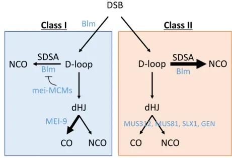
Major insight into the meiotic recombination pathways was gained when timing of CO and NCO product formation was studied in budding yeast. Allers and Lichten (2001)found that NCOs arose before COs did, but also that they arose before joint molecules (which are generally accepted to be dHJs). This result did not support the current model of meiotic recombination pathways, but instead suggested a pathway where NCOs form from an earlier intermediate and COs are preferentially formed from dHJs later in meiosis. McVey et al. showed that in Drosophila Blm is the helicase that processes the earlier intermediate, a D-loop, via SDSA into a NCO in mitotic DSB repair. (McVey et al. 2004). Yildiz et al. determined that MEI-9–ERCC1 together with the scaffolding protein MUS312, is the major meiotic resolvase that forms crossovers in Drosophila (Yildiz et al. 2002, 2004). MEI-9-ERCC1 is the Drosophila ortholog of XPF-ERCC1 and MUS312 is the Drosophila ortholog of SLX4.
Figure 1.1. Two pathway DSB repair model. A DSB can repaired by Blm directing repair down the Class I pathway in which a D-loop is preferentially directed into becoming a dHJ by the mei-MCM complex, and dHJs are resolved by MEI-9 into COs. If Blm is absent, repair will be directed down the Class II pathway, where D-loops are preferentially migrated
through SDSA to form NCOs and the remaining go through dHJs to be resolved in an unbiased manner as COs or NCOs by the endonucleases MUS81, SLX1, MUS312, and GEN.
DSB
D-loop
D-loop
SDSA
NCO
SDSA
NCO
dHJ
dHJ
CO
NCO
CO
NCO
Blm Blm
mei-MCMs Blm
MEI-9 MUS312, MUS81, SLX1, GEN
10
In S. cerevisiae, Bloom syndrome helicase mutants, sgs1, reveal COs and NCOs that form at the same time (De Muyt et al. 2012). These COs and NCOs are both formed from dHJs, but the repair of these dHJs is not biased toward COs. In the absence of Sgs1, the resolvases Mus81-Mms4, Yen1, and Slx1-Slx4 resolve dHJs into COs and NCOs (De Muyt et al. 2012; Zakharyevich et al. 2012). This was shown through the fact that crossovers in sgs1 mutants are resolved by these proteins. Mutants of sgs1 in combination with
mutants of these resolvases result in a buildup of intermediates and fewer COs and NCOs formed, which is worsened in sgs1 mms4 yen1 slx1 quadruple mutants (De Muyt et al. 2012).
Studies in Drosophila have shown that Blm mutants in combination with mutations in either mus81, mus312, or Gen (the Drosophila Yen1 ortholog) are synthetically lethal (Andersen et al. 2011). Additionally, in Blm mutants, all crossovers are MEI-9 independent, which supports the idea that Blm mutants experience only Class II crossovers (Hatkevich et al. 2017). Hatkevich et al. also showed that Blm mutants lose all crossover patterning: assurance, interference, and the centromere effect, suggesting that these three crossover patterning phenomena are characteristic of Class I meiotic crossovers (Hatkevich et al. 2017). All of these results lead us to the model that Blm is responsible for directing DSBs down the Class I meiotic recombination pathway.
Centromere proximal crossover suppression in Drosophila
In meiosis, DSBs are mostly processed through the Class I pathway. It is important that crossovers are formed via the Class I pathway because proper crossover patterning is needed for the correct segregation of chromosomes in meiosis. Together, assurance,
11
fact that Meiosis I (MI) nondisjunction events more often contain chromosomes that are lacking a crossover (Koehler et al. 1996a). Additionally, proper crossover placement is essential, as evidenced by the fact that apparent Meiosis II (MII) nondisjunction events more often contain chromosomes that have centromere-proximal crossovers (Koehler et al.
1996b).
12
MI nondisjunction events occur when homologous chromosomes do not segregate properly and go to the same pole in MI. Then in MII, the sisters segregate properly, so the resulting aneuploid gamete includes homologous chromosomes, identifiable by different centromeres as in Figure 1.2A. When MI proceeds normally and mistakes occur in MII, the two centromeres come from sisters, so they are the same, as shown in Figure 1.2B. As mentioned previously, apparent MII events from Trisomy 21 individuals more often have centromere-proximal crossover events (Koehler et al. 1996b). However, it was puzzling why these events appeared to be from MII nondisjunction when recombination occurs in MI.
Two hypotheses for how these events could have occurred were proposed by Lamb et al. (1996). The first explanation is termed “entanglement” where homologues are
entangled by the centromere-proximal crossover and the bivalent remains together until separation at MII where sisters remain together and homologues separate to opposite poles as shown in Figure 1.2C. The second hypothesis is termed precocious separation of sister chromatids (PSSC). In this case, pericentromeric crossovers disrupt the cohesion of sisters and cause premature separation of the sisters at MI and then are susceptible to random segregation at MII, where both sisters could segregate to the same pole (Figure 1.2D). Therefore, these mistakes both occur at MI due to the peri-centromeric exchange, but are scored as MII events because they contain the same centromere.
What is important to note is that centromere-proximal crossovers are correlated with nondisjunction events. As discussed previously, it is thought that the centromere effect suppresses these centromere-proximal crossovers but the mechanism is unknown and largely unstudied. However, there are ideas about what could be suppressing centromere-proximal crossovers including heterochromatin and transposable elements.
14
Figure 1.2. Types of nondisjunction. (A) Schematic of chromosomes experiencing normal meiotic segregation. (B) In Meiosis I (MI) nondisjunction (NDJ), homologues missegregate and go to the same cell, and then sisters separate, resulting in gametes containing
chromosomes with different centromeres (orange and purple). (C) in Meiosis II (MII) NDJ, homologues segregate properly in the first division, but then in one instance, sisters did not segregate properly in the second division (orange chromosomes), resulting in a gamete containing chromosomes with the same color centromere. (D) Entanglement begins with a peri-centromeric crossover causing both homologues to segregate to the same pole. Homologues remain together in a bivalent and separate in MII causing sisters to go to the same cell and resulting in gametes with the same centromeres, presenting as MII NDJ even though the mistake occurred in MI. (E) Precocious separation of sister chromatids (PSSC) occurs when peri-centromeric crossovers cause sister chromatid cohesion to be released early, resulting in random segregation of sisters into the same cell in MII (bottom).
show that when peri-centromeric heterochromatin is rearranged in a way that places euchromatin closer to the centromere, there is a greater decrease in crossovers in that euchromatin than a euchromatic region moved nearer to a large amount of heterochromatin (farther from the centromere than in the first case). Similarly, Yamamoto and Miklos (1978) showed that centromere-proximal suppression of crossovers moved farther into the
euchromatin when sections of X chromosome heterochromatin were deleted. These studies suggest that the centromere effect depends on distance from the centromere and not necessarily the amount of heterochromatin. However, there are also studies that suggest heterochromatin directly plays a role in suppressing crossovers.
In cytological studies of DSBs, Mehrotra and McKim (2006) show that DSB markers do not colocalize with heterochromatin marker HP1. This result suggests that DSB
machinery may not have access to the tightly packed chromatin, so crossovers are unable to form in this region. Nonetheless, the question still remains whether heterochromatin has the ability to decrease crossovers in adjacent euchromatic regions causing the suppression of crossovers we see in these regions.
15
that is adjacent to euchromatin, making these regions easier to assemble to the genome (Yamamoto et al. 1990; Carmena and González 1995). Transposable elements have been suggested to have an effect on crossover rate, by suppressing crossovers in
peri-centromeric regions (Bartolome et al. 2001; Bartolome and Maside 2004; Kent et al. 2017). The interplay of transposable elements and gene density with crossover rate is a highly debated phenomenon (reviewed in Kent et al. 2017). It has been proposed that an increase in crossover rate in gene dense regions is favored due to the resulting increase in genetic diversity. Additionally, it is thought that crossing over is suppressed in regions that have a high density of transposable elements (TEs) to repress harmful recombination within
repetitive elements. Recombination in repetitive regions can lead to insertions or deletions of repeats, or ectopic recombination events. Alternatively, it has been proposed that
transposable elements themselves repress recombination. Miller et al. (2016) reported that crossovers can occur within TEs, but less frequently than would be expected if they were freely able to form within TEs. It has been suggested that active silencing of TEs could lead to the silencing or suppression of recombination around those regions (Kent et al. 2017). It is still unknown whether TEs or gene density directly or even indirectly affect recombination rates, but many studies have at least suggested a correlation between these factors.
Crossover patterning is such a complex and interesting field; however, it is very difficult to gain insight into the mechanisms controlling crossover designation. My thesis work has addressed crossover patterning by both examining proteins involved in crossover formation as well as the mechanisms governing centromere-proximal crossover
16
Scope of this work
De Muyt et al. saw that even in the quadruple sgs1 mms4 yen1 slx1 mutant, there were still residual COs and NCOs, suggesting there were other resolvases yet to be identified that are able to resolve intermediates into COs and NCOs. MUS81 is an ERCC4 nuclease that has been shown to interact with SLX1 and MUS312 to resolve dHJs (Gaillard et al. 2003; Gaskell et al. 2007; Fekairi et al. 2009). MEI-9 is similar to MUS81 in that it is an ERCC4 nuclease and also interacts with MUS312. I hypothesized that there is another partner that interacts with MEI-9 and MUS312 to resolve HJs. This potential partner is Ankle1, which is similar to SLX1 because it contains a GIY-YIG nuclease domain. I examined Ankle1’s potential role as an endonuclease in Chapter 2.
Interestingly, mutations in both Blm and the mei-MCM complex genetically interact: in Blm rec double mutants, crossovers are increased compared to Blm single mutants, suggesting that the mei-MCM complex may play a role in inhibiting crossovers in the Class II pathway. The role of the mei-MCM complex in the Class II pathway is investigated and described in Chapter 3. Little is known about how individual members of the mei-MCM complex contribute to crossovers, so I investigate this in the work described in Chapter 3.
I was particularly interested in the centromere effect and understanding how centromere-proximal crossovers were suppressed became the focus of my thesis work. Chapter 4 contains work that was published as a review paper on chromosome 4, which delves into understanding why chromosome 4 does not experience crossing over. I
hypothesize that chromosome 4 does not have crossovers because the entire chromosome is subject to the centromere effect. This is supported by the fact that Blm mutants do
17
18
CHAPTER 2: EXAMINATION OF ANKLE1 AS A POTENTIAL NUCLEASE
Preface
This chapter includes preliminary data on the examination of the potential nuclease Ankle1. This project encompassed my rotation and part of my first year of research in the Sekelsky Lab. I provide results suggesting that Ankle1 interacts with MUS312 and MEI-9; however, studies of an Ankle1 deletion showed no meiotic defects or sensitivity to DNA-damaging agents. These results show support for a role of Ankle1 in creating mitotic crossovers in the absence of the Fancm helicase. I did not pursue this project as my
dissertation research to instead focus on the centromere effect. Here, I detail the preliminary results I obtained on the potential nuclease Ankle1, and suggest possible future studies.
Introduction
Throughout DNA replication and recombination, DNA forms different branched structures, which need to be resolved for proper completion of DNA replication and maintenance of genome integrity. SLX4 is a scaffolding protein known to assemble with multiple structure-specific nucleases that will aid in the resolution of Y forks, 3’ flaps, 5’ flaps, replication forks, and Holliday Junctions (HJs) (Muñoz et al. 2009). SLX4 interacts with nucleases such as GEN1, XPF-ERCC1 and MUS81-EME1 in distinct complexes (Fekairi et al. 2009; Muñoz et al. 2009; Wyatt et al. 2013).
19
complex formed with SLX4 contains XPF-ERCC1, which is involved in resolving interstrand crosslinks as well as HJs (Fekairi et al. 2009). MUS312 and MEI-9–ERCC1 are the
Drosophila homologs of this complex, whichis involved in meiotic DSB repair and thought to be the major meiotic resolvase that generates crossovers (Radford et al. 2005, 2007). However, in the absence of MEI-9 or MUS312, there are still some residual crossovers (Radford et al. 2005; Andersen et al. 2009). Therefore, I hypothesize that there is another resolvase involved in creating meiotic crossovers.
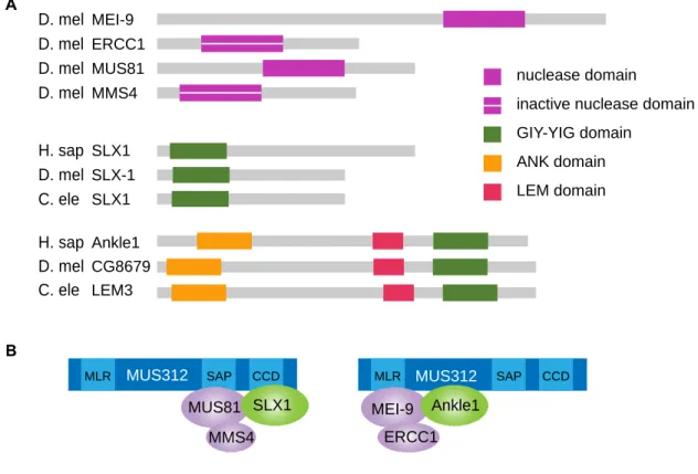
MEI-9 and MUS81 are both nucleases that belong to the ERCC4 nuclease family, and both have binding partners containing inactive nuclease domains (Figure 2.1A).
MUS81-EME1 pairs with SLX1-SLX4 to resolve junctions, so due to the similarities between MUS81 and MEI-9, I hypothesize there is another protein within the MUS312-MEI-9-ERCC1 resolvase complex similar to SLX1. SLX1 has a GIY nuclease domain (Coulon et al. 2004; Svendsen et al. 2009), so I set to identify another protein in Drosophila that contains a GIY nuclease domain that may form a similar complex with MUS312, MEI-9-ERCC1.
Besides Slx1, the mammalian genome includes only one other GIY-YIG nuclease domain containing protein, Ankle1 (Dunin-Horkawicz et al. 2006; Zlopasa et al. 2016). At the start of this study, not much was known about Ankle1. Brachner et al. showed that enriching Ankle1 induced DNA cleavage and the DNA damage response and that the GIY-YIG and LEM domains of this protein are required for that activity (Brachner et al. 2012). The
Caenorhabditis elegans homolog was identified as LEM-3, and was isolated in a screen for mutants sensitive to ionizing radiation (IR) (Dittrich et al. 2012). Dittrich et al. also found that lem-3 mutants were sensitive to other types of DNA-damaging agents suggesting a role for LEM-3/Ankle1 following DNA damage. We identified a candidate protein, CG8679 in
20
Figure 2.1. Nucleases and Ankle1 Model. (A) Schematic of domains within nucleases that assemble into complexes as shown in (B). Domains shown include the ERCC4 nuclease domain, predicted inactive nuclease domain, GIY-YIG domain, ANK domain, and LEM domain. (B) shows predicted nuclease complexes with the scaffolding protein, MUS312. We hypothesize that Ankle1 acts as part of the complex on the right as a nuclease with MEI-9 and ERCC1. Domains in MUS312 shown are the MUS312/MEI-9 interaction like domain (MLR), the SAF-A/B, Acinus, and PIAS (SAP) domain involved in substrate recognition, and the conserved C-terminal domain (CCD) involved in SLX1 interaction (Fekairi et al. 2009).
Ankle1 (Figure 2.1A) (Dunin-Horkawicz et al. 2006; Brachner et al. 2012). From now on, I refer to the candidate gene CG8679 as Ankle1 in Drosophila. SLX1 interacts with MUS312 near the C-terminal end (Fekairi et al. 2009; Svendsen et al. 2009; Castor et al. 2013), and MUS81-EME1 interacts with MUS312 through its SAP domain (Castor et al. 2013; Kim et al. 2013). Alternatively, MEI-9 interacts near the N-terminus of MUS312, in what Fekairi et al. have termed the MEI-9 interaction like domain (MLR) (Yildiz et al. 2002; Fekairi et al. 2009). I hypothesize that Ankle1 is part of a complex with MUS312 and MEI-9-ERCC1 and predict that Ankle1 will interact with MEI-9 and MUS312 near the MLR domain (Figure 2.1B). I
nuclease domain
inactive nuclease domain
GIY-YIG domain ANK domain LEM domain D. mel D. mel D. mel D. mel D. mel D. mel H. sap C. ele H. sap C. ele MEI-9 MUS81 MMS4 ERCC1 SLX1 SLX1 SLX-1 Ankle1 CG8679 LEM3 A B
MLR SAP CCD MLR SAP CCD
21
performed yeast two-hybrid studies to examine these interactions and to confirm other interactions within the MUS312 complexes. I created a deletion of Ankle1 in Drosophila using the CRISPR/Cas9 system and characterized this mutant by measuring meiotic nondisjunction, sensitivity to DNA damaging agents, and mitotic recombination.
Materials and Methods
Yeast two-hybrid
Vectors were created using either pGBD-DEST (James et al. 1996) or pACT2.2gtwy (Addgene plasmid 11346 deposited by Guy Caldwell) using the Gateway Vector Conversion System (Life Technologies, Carlsbad, CA). Full-length and truncated versions of mei-218, full-length Slx1, and full-length mei-9 were previously made (Radford et al. 2005; Andersen et al. 2009). mus-81 and Ankle1 were cloned into pGBD-DEST. mus-81 and mms-4 were cloned from other vectors and Ankle1 was cloned from the BacPac genomic DNA clone library (ID BACR34H23). Constructs were transformed into Saccharomyces cerevisiae strain PJ69-4A (James et al. 1996). Co-transformants were obtained by selecting for growth on plates with Yeast Extract-Peptone-Dextrose (YPD) media containing supplements lacking tryptophan (-trp) and leucine (-leu) after growth for 3 days at 30C. Single colonies were re-streaked onto fresh -trp -leu plates and grown for 3 days at 30C. Colonies were then streaked onto triple dropout (-trp -leu -his) plates, grown for 3 days at 30C and scored for
interaction. It was verified that single transformants did not self activate and did not grow on double and triple dropout plates. For serial dilutions, yeast cultures were grown in -trp -leu dropout media to saturation, diluted 4 times by 10 fold and all five solutions were plated on -trp -leu and --trp -leu -his plates.
CRISPR/Cas9 Deletion of Ankle1
Ankle1dsRED was created using the CRISPR/Cas9 technology (Gratz et al. 2013).
22
plasmid #49411) that targeted two locations near the 5’ and 3’ ends of the Ankle1 gene. Sequence of gRNAs were CCGTTTCGCATGCCGCACA-3’ and
5’-CTCCGCCAGATAGATGTGCA-3’. Ankle1 is 2,115 bps and the deletion is of 1,908 bps. A
repair template vector was co-injected, which contains approximately 1kb of homologous sequence on either side of the two cuts and a dsRED in between, with the goal of making a deletion of Ankle1 and inserting a copy of dsRED for easy screening of deletions. Injected flies were crossed to Pin/CyO and progeny were screened for red fluorescent eyes. Multiple dsRED flies were collected and stocks of Ankle1dsRED/ CyO were created. Stocks were
verified by PCR screening using one primer in the fly genome and one primer in dsRED to ensure that dsRED was inserted correctly and a partial deletion of Ankle1 was created. All stocks that experienced proper placement of dsRED in the Ankle1 deletion were kept and maintained at 25C on standard cornmeal medium.
Sensitivity assays
23
concentrations. MMS induces DNA alkylation and possibly double strand breaks, IR induces double strand breaks, and cisplatin induces inter-strand crosslinks (Radford 1985; Lundin et al. 2005; Sawant et al. 2017). Alleles used in this study were Ankle1dsRED, Df(2L)Exel6047
(deficiency chromosome with deletion of CG8679), mei-9a(Yildiz et al. 2002), Fancm0693
(Kuo et al. 2014), Fancmdel(Romero et al. 2016), mus312D1, and mus312Z1973(Andersen et
al. 2011). Treated and untreated classes were compared using unpaired t-test in Prism 8.
Nondisjunction assay
Approximately 5-6 virgin females of desired genotype (WT or
Ankle1dsRED/Df(2L)Exel6047) were crossed to three y cv v f / Bs Y y+males in 10 vials.
Parents were flipped to new vials after three days, then emptied after three more days. Progeny were scored for nondisjunction (NDJ). Parental genotypes were XX females (B+), XY males (B-), and exceptional progeny were XXY females (B-), and XO males (B+). Exceptional class numbers were multiplied by two to account for nonviable exceptional progeny (XXX, OY). WT nondisjunction data was obtained from Hartmann et al. (2019, in preparation). WT and Ankle1 NDJ rates were compared using fisher’s exact test.
Mitotic crossover assay
Mitotic crossovers were scored by crossing males of desired genotype with st Sb/+ to females homozygous for st Sb. Mitotic crossovers were scored between st and Sb. Fancm mus312 data is previously published (Kuo et al. 2014). Statistical analyses were performed using fisher’s exact test.
Results
Nuclease Complex Interactions
24
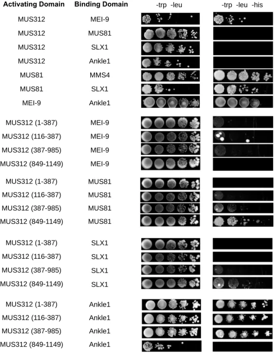
in complex with MUS312 using the yeast two-hybrid system. I confirmed interaction between MUS312 and MEI-9, MUS81 and MMS4, MUS81 and SLX1 (Figure 2.2). I was not able to recapitulate the interaction between MUS312 and MUS81, or MUS312 and SLX1.
Interaction between MUS312 and SLX1 was previously shown with Drosophila proteins in yeast two-hybrid, and MUS312 and MUS81 have been shown to interact in human cells, but not with Drosophila proteins (Andersen et al. 2009; Wyatt et al. 2013). However, in using truncations of MUS312, I show that SLX1 and MUS81 interact near the C terminus, as expected from previous studies and the model shown in Figure 2.2B.
To test if Ankle1 is a component of the complex, we tested interactions between Ankle1 with MUS312 and MEI-9. Via the yeast two-hybrid system, there is an interaction between Ankle1 and MEI-9 and the N-terminus of MUS312. There is no interaction between Ankle1 and full length MUS312, but we do see the interaction in N-terminal truncations of MUS312. We hypothesized that Ankle1 interacts in a complex with MEI-9, and since MEI-9 is known to interact with MUS312 near the N-terminus, we expected Ankle1 to also interact near the N-terminus. Full length MUS312 did not produce interactions with SLX1, MUS81, or Ankle1, even though truncated forms were able to produce an interaction. I predict that the full-length MUS312 may not be expressed properly or fold in the correct configurations to produce these interactions in the yeast two-hybrid system.
25
Figure 2.2. Yeast Two-Hybrid with proposed components of nuclease complexes. Genes of the proteins in the activating domain column were cloned into the pACT2.2gtwy vector to be expressed as a fusion protein with the Gal4 activating domain. Genes of the proteins in the binding domain column were cloned into the pGBD-DEST vector to be expressed as a fusion protein with the Gal4 binding domain. Serial dilutions are shown on -trp -leu plates and --trp -leu -his plates. Growth on triple dropout plates indicates interaction between the two proteins.
MUS312
Activating Domain Binding Domain
MEI-9 MUS81 MUS312 MUS312 MUS312 MUS81 MEI-9 Ankle1 SLX1 MMS4 Ankle1 SLX1 MUS81 MUS312 (1-387) MUS312 (116-387) MUS312 (387-985) MUS312 (849-1149) MUS312 (1-387) MUS312 (116-387) MUS312 (387-985) MUS312 (849-1149) MUS312 (1-387) MUS312 (116-387) MUS312 (387-985) MUS312 (849-1149) MUS312 (1-387) MUS312 (116-387) MUS312 (387-985) MUS312 (849-1149) MEI-9 MEI-9 MEI-9 MEI-9 MUS81 MUS81 MUS81 MUS81 SLX1 SLX1 SLX1 SLX1 Ankle1 Ankle1 Ankle1 Ankle1
26 Ankle1 deletion and characterization
I created a CRISPR deletion of Ankle1 with an insertion of dsRED as described in Materials and Methods. Since MEI-9 works with MUS312 to resolve HJs to create meiotic crossovers, I hypothesized that Ankle1 may also play a role in resolution of meiotic crossovers. If Ankle1 is required for meiotic crossovers, I hypothesize a decrease in crossovers in the mutant, and a subsequent increase in nondisjunction. I first screened for nondisjunction in Ankle1 mutants, which shows no significant increase in nondisjunction as compared to wild-type (p=0.56) (Table 2.1). I therefore conclude that Ankle1 either does not play a role in the meiotic function of this complex or its function is redundant.
Genotype
XX Females
XY Males
XXY Females
XO
Males Total
WT (yw) 1551 1481 0 1 3033
Ankle1 1107 766 2 0 1875
Table 2.1. Nondisjunction of Ankle1. Nondisjunction of Ankle1dsRED/ Df(2L)Exel6047 was
scored (see materials and methods). Parental classes are XX females and XY males,
exceptional classes are XXY females and XO males. WT data is from Hartmann et al. (2019, in preparation). WT is not significantly different from Ankle1 using fisher’s exact test
(p=0.56).
Ankle1 sensitivity to DNA-damaging agents
27
Figure 2.3 Sensitivity Assays of Ankle1. Flies heterozygous for Ankle or mei-9 mutations were mated and scored for the ratio of heterozygous progeny to homozygous mutant progeny. That ratio was compared between treated and untreated broods. Numbers and significance values between treated and untreated are shown in Table 2.2. *Note: These experiments were not done at the same time.
are not sensitive to IR. Mutants sensitive methyl methanosulfate (MMS) have defects in homologous recombination (HR), so we tested if Ankle1 mutants are sensitive to MMS (Lundin et al. 2005). Ankle1 mutants are not sensitive to two different concentrations of MMS, whereas the positive control, mei-9 is severely sensitive (Figure 2.3, Table 2.2) (Ankle1 0.05% MMS p=0.82, Ankle1 0.08% MMS p=0.58, mei-9 0.08% MMS p<0.0001). Ankle1 is also not sensitive Cisplatin, which induces interstrand crosslinks (Sawant et al. 2017) (Figure 2.3, Table 2.2, p=0.07 *note, Ankle1 experiences increased survival when exposed to Cisplatin, which may or may not be biologically relevant).
Mitotic role of Ankle1
Since Ankle1 does not experience meiotic nondisjunction or sensitivity to DNA-damaging agents, I sought to determine if Ankle1 has a role in mitotic recombination. It is thought that when helicases are absent, mitotic crossovers are more likely to occur because
28
Genotype Treatment Mutant
Progeny
Heterozygous Total Biological
Replicates
p Value
Ankle1 IR (1500 rads) 362 783 1145 10 0.11
Untreated 521 760 1281 10
Ankle1 IR (2000 rads) 228 475 703 20 0.48
Untreated 840 1638 2478 20
mei-9 IR (2000 rads) 74 81 155 10 0.16
Untreated 571 499 1070 10
Ankle1 MMS (0.05%) 304 559 863 10 0.82
Untreated 347 642 989 10
Ankle1 MMS (0.08%) 622 1074 1696 20 0.58
Untreated 848 1631 2479 20
mei-9 MMS (0.08%) 0 140 140 10 <0.0001
Untreated 450 355 805 10
Ankle1 Cis (0.25 mM) 331 512 843 10 0.07
Untreated 473 856 1329 10
Table 2.2. Mutagenizing agent assays. Genotype of each mutant is displayed, along with what treatment they were given and at what dose (see Materials and Methods for more information on implementation of assay). For each genotype and treatment, there was an untreated brood and a treated brood. Biological replicates represents how many vials were scored for each genotype/treatment. Numbers for mutant progeny, heterozygous progeny, and total progeny are shown. Treated and untreated classes were compared using unpaired t-test in Prism 8.
29
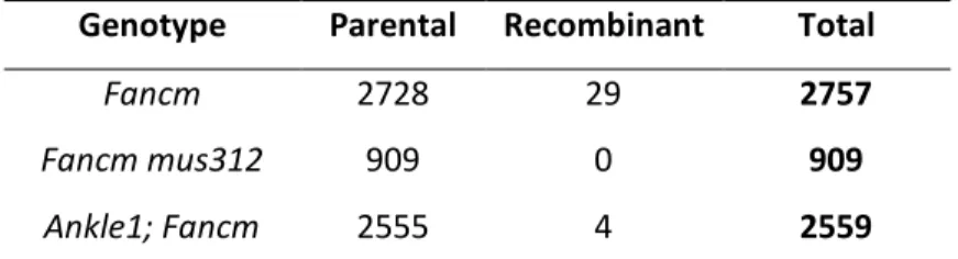
Genotype Parental Recombinant Total
Fancm 2728 29 2757
Fancm mus312 909 0 909
Ankle1; Fancm 2555 4 2559
Table 2.3. Mitotic crossovers in Ankle1. Mitotic crossovers scored as in Materials and Methods. Fancm mus312 data is from (Kuo et al. 2014). Fancm mus312 is not significantly different from Ankle1; Fancm (p=0.58), statistical analysis done by fisher’s exact test.
Discussion
Overall, these data show that Ankle1 has no visible meiotic defects or sensitivity to the DNA-damaging agents I tested. Therefore, Ankle1either does not have a role in creating meiotic crossovers or repair of DNA damage, or its role is redundant, so a mutant phenotype is not presented. However, Ankle1appears to have a role in creating mitotic crossovers in the presence of DNA stress due to absence of the helicase Fancm.
We hypothesized that Ankle1forms a complex with MUS312 and MEI-9-ERCC1. MEI-9 has shown to have roles in creating meiotic crossovers as well as repairing DNA after damage (Yildiz et al. 2004). I therefore hypothesized that we would see defects in meiotic recombination or sensitivity to DNA-damaging agents in Ankle1 mutants. I did not observe any defects in meiotic nondisjunction or sensitivity to MMS, IR, or Cisplatin in Ankle1 mutants, so I conclude that Ankle1either does not play a role in these processes or its role is redundant. It is puzzling that we see an interaction between Ankle1 and Mei-9, yet Ankle1 mutants do not show defects in activities that the MEI-9 complex is thought to play a role in. However, we can not rule out a complex with Ankle1, MEI-9, and MUS312 because
Ankle1’s activity may be redundant so we can not detect the defects.
30
phenotype seen in mus312 Fancm double mutants, suggesting that MUS312 repairs DNA stress as mitotic crossovers in Fancm mutants (Kuo et al. 2014). Kuo et al. also examined Fancm double mutants with other members of the complexes discussed, including MUS81, SLX1, and MEI-9. Fancm mutants did not have a significant change in mitotic crossovers compared to mei-9; Fancm double mutants (Kuo et al. 2014). Fancm mutants with Slx1 or mus81 also did not show any change mitotic crossovers, but mus81; Slx1 Fancm triple mutants had a significant decrease in crossovers as compared to Fancm single mutants, suggesting SLX1 and MUS81 act together to form mitotic crossovers and that MUS312 is needed for this function (Kuo et al. 2014). The fact that Ankle1; Fancm has a significant decrease in crossovers as compared to Fancm single mutants suggest that Ankle1 complexes with MUS81, SLX1, and MUS312 to form mitotic crossovers. This is partially supported by the observation that Ankle1 interacts with MUS312 truncations through yeast two-hybrid, and the interactions between Ankle1 with MUS81 and SLX1 should also be explored in future studies.
31
CHAPTER 3: MEIOTIC MCM PROTEINS PROMOTE AND INHIBIT CROSSOVERS
DURING MEIOTC RECOMBINATION
1Abstract
Crossover formation as a result of meiotic recombination is vital for proper segregation of homologous chromosomes at the end of meiosis I. In many organisms, crossovers are generated through two crossover pathways: Class I and Class II. To ensure accurate crossover formation, meiosis-specific protein complexes regulate the degree in which each pathway is used. One such complex is the mei-MCM complex, which contains MCM (mini-chromosome maintenance) and MCM-like proteins REC (ortholog of Mcm8), MEI-217, and MEI-218, collectively called the mei-MCM complex. The mei-MCM complex genetically promotes Class I crossovers and inhibits Class II crossovers in Drosophila, but it is unclear how individual mei-MCM proteins contribute to crossover regulation. In this study, we perform genetic analyses to understand how specific regions and motifs of mei-MCM proteins contribute to Class I and II crossover formation and distribution. Our analyses show that the long, disordered N-terminus of MEI-218 is dispensable for crossover formation, and mutations that disrupt REC’s Walker A and B motifs differentially affect Class I and Class II crossover formation. In Rec Walker A mutants, Class I crossovers exhibit no change, but Class II crossovers are increased. However, in rec Walker B mutants, Class I crossovers are severely impaired, and Class II crossovers are increased. These results suggest that REC
1 This chapter is adapted from previous work published in the journal Genetics. The original citation is as follows:
32
may form multiple complexes that exhibit differential REC-dependent ATP binding and hydrolyzing requirements. These results provide genetic insight into the mechanisms
through which mei-MCM proteins promote Class I crossovers and inhibit Class II crossovers.
Introduction
To reestablish the diploid genome upon sexual fertilization, the genome of progenitor germ cells must be successfully reduced by half through meiosis. Accurate reduction of the genome at the end of meiosis I requires crossover formation between homologous
chromosomes during meiotic recombination. Meiotic recombination is initiated by the formation of multiple double-strand breaks (DSBs); the majority of meiotic DSBs are repaired asnoncrossovers, while a selected subset are repaired as crossovers between homologs (Lake and Hawley 2016).
Two distinct types of meiotic crossovers have been described: Class I and Class II. First defined in budding yeast (De Los Santos et al. 2003), Class I and Class II crossovers exist in most sexually reproducing organisms, but the relative proportions of each crossover type vary among organisms (Hollingsworth and Brill 2004). In Drosophila, most – if not all – crossovers are generated through the Class I pathway (Hatkevich et al. 2017), as shown through their dependence on the putative catalytic unit of the Class I meiotic resolvase MEI-9 (Sekelsky et al. 1995; Yildiz et al. 2002) and their display of crossover interference (Hatkevich et al. 2017). Most crossovers in Drosophila are also dependent upon a group of MCM- or MCM-like proteins, called the mei-MCM complex (Baker and Carpenter 1972; Grell 1978; Liu et al. 2000; Kohl et al. 2012).
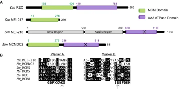
The mei-MCM complex consists of REC (the Drosophila ortholog of MCM8), MEI-217, and MEI-218. REC appears to be a bona fide MCM protein, based on
33
Figure 3.1. MCM protein structure and alignments. (A) Structural domains of Drosophila melanogaster REC, MEI-217, MEI-218 and Mus musculus MCMDC2. Structural domains identified using PHYRE 2 (Kohl et al. 2012). “MCM domain” corresponds to protein data bank ID #c2vl6C and the AAA ATPase domains identified correspond to protein data bank ID #d1g8pa. The X on Dm MEI-218 and Mm MCMDC2 represents predicted inactive AAA ATPase domains. (B) Consensus sequence for Walker A motif (Walker et al. 1982), and consensus sequence for Walker B motif (Forsburg 2004). Identical or conserved amino acids are denoted with black background. Arrows denote the conserved catalytic residues.
contrast, MEI-217 and MEI-218 are highly divergent MCM-like proteins, and together resemble one full MCM protein. MEI-217 is structurally similar to the MCM N-terminal
34
analysis and details regarding the evolution of the mei-MCM complex, see Supplemental Figures 3.2-3.4.
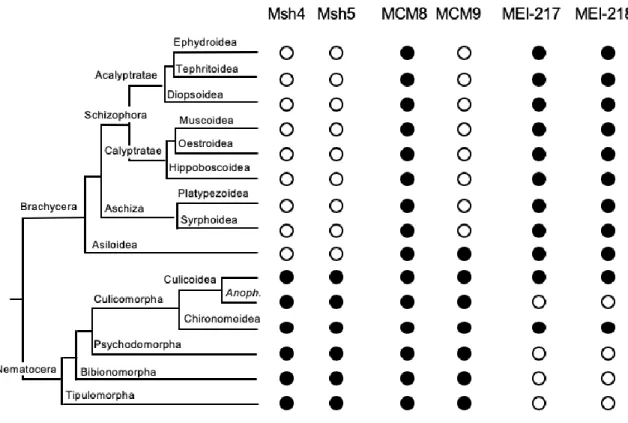
Figure 3.2. Occurrence of Msh4, Msh5, MCM8, MCM9, MEI-217, and MEI-218 in Diptera. The dendrogram on the left illustrates relationships among Dipteran taxa for which sufficient genome or transcriptome sequence is available to determine with reasonable confidence the presence or absence of genes encoding proteins relevant to this work. Circles to the right indicate presence (filled) or absence (open) of each gene/protein. For the suborder
36
Figure 3.3. Structures of MEI-217 and MEI-218 in Diptera. (A) The dendrogram is the same as in Figure S1, with additional species to illustrate the variation in domain
architectures. Domain architectures for representative species are to the right (the jagged N-terminal end in Ferdiandea cuprea indicates incomplete sequence). Domains were
determined by PHYRE2 alignment to Protein Data Bank entry c5udb7 (a cryo-electron microscopy structure of S. cerevisiae MCM7). Accession numbers for the sequences included are listed below. Accession numbers that start with J are from Ensemble Metazoa genomic assemblies (found at http://metazoan.ensemble.org). The Aedes, Musca, and Glossina sequences are genomic contigs from Vectorbase (http://vectorbase.org). All other sequences are from NCBI (http://ncbi.nih.nlm.gov); those starting with a G are from the transcriptome shotgun assembly (TSA) database.
Species Accession .
Drosophila melanogaster NM_167557.3
Drosophila grimshawi XM_001992187.1
Ceratitis capitate GAMC01014250.1
Teleopsis whitei GBBQ01026862.1
Musca domestica scf7180000644883
Lucilia cuprina JRES01000755:1975-11359
Glossina morsitans scf7180000644883
Platypeza anthrax GCGU01008763.1
Ferdinandea cuprea GCHQ01011487.1
Aedes aegypti AAGE02016621.1
Belgica Antarctica JPYR01000187:32247-35225
37
(B) Junctions between open reading frames (ORFs) for the N-terminal and AAA+ ATPase domains are shown. At the top are three species from Shizophora and one from Aschiza, showing overlapping ORFs. Amino acids at the end of the N-terminal domain are shown in green above the DNA sequence (the position of the stop codon is highlighted in green); amino acids for the beginning of the AAA+ ATPase domain are in purple below the DNA sequence (the position of the start codon is highlighted in purple). Below that are two Nematocera species, showing separate but non-overlapping ORFs. Non-coding sequence between the ORFs is in lowercase text. At the bottom is a non-Diptera representative, the moth Bombyx mori. In this case, as in all other non-Dipteran species with an Mcmdc2 ortholog and in replicative MCM proteins, the two domains are on the same polypeptide, separated by a short linker.
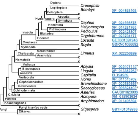
(C) Distribution of Mcmdc2 in Opisthokonta. Dendrogram shows phyla, some sub-phyla, and several orders within the sub-phylum Insecta in which we can find clear orthologs of
Mcmdc2 or in which there are sufficient genome or transcriptome sequences to suggest loss of Mcmdc2 with reasonable confidence. For those clades with an ortholog, a representative genus is listed, along with an accession number. Taxa in which we could not find any orthologs are indicated with an x. We have not found Mcmdc2 orthologs outside of Opisthokonta.
While most crossovers are generated through the Class I pathway in wild-type Drosophila and are mei-MCM dependent, mutants that lack the Bloom syndrome helicase (Blm) generate only Class II crossovers based on their independence of MEI-9 and lack of patterning (e.g., interference) that is associated with Class I crossovers (Hatkevich et al. 2017). Blm is an ATP-dependent 3’-5’ helicase that exhibits vital anti-crossover functions in both meiotic and somatic DSB repair (Hatkevich and Sekelsky 2017). Interestingly,
mutations in mei-MCM and Blm genes genetically interact. In Blm mutants, crossovers are reduced by 30% but in a Blm rec double mutant, crossovers are significantly increased compared to wild-type (Kohl et al. 2012). This suggests that the mei-MCMs may function to inhibit crossovers within the Class II pathway, in addition to their role promoting crossovers in the Class I pathway.
38
Figure 3.4. Sequence conservation and divergence in MEI-218. MEI-218 orthologs from 12 Drosophila species and two Bactrocera species (Tephritid fruit flies, also in the
39
Species are, from top to bottom and sorted by species group as indicated on the figure: melanogaster: Drosophila melanogaster
Drosophila sechelia Drosophila simulans Drosophila mauritiana Drosophila yakuba Drosophila erecta
obscura: Drosophila pseudoobscura pseudoobscura Drosophila persimilis
Drosophila miranda repleta: Drosophila navojoa
Drosophila mojavensis Drosophila arizonae Bactrocera: Bactrocera latifrons
Bactrocera dorsalis
Thin horizontal lines denote gaps introduced in the alignment process. Vertical lines indicate amino acid identity or similarity, using the Dayhoff PAM 200 matrix. Black is conserved among at least 11 sequences (e.g., one mismatch in the 12 Drosophila species). Pink indicates conservation within the melanogaster species group, aqua within the obscura group, and orange within the repleta group. The red arrow denotes the start codon for the truncated MEI-218 described in the text.
Sequences were aligned in MEGA (v. 10.0.4) using the MUSCLE algorithm. Manual adjustments were done in GeneDoc v. 2.7.000. This visualization is the summary view produced by GeneDoc, with species groups with conservation mode shading enabled.
formation and general crossover distribution. By mutating key residues in REC’s Walker A and B motifs (recKA and recDA,respectively), we found that recKA mutants exhibit no Class I
crossover defect, while Class II crossovers are significantly increased. Surprisingly, recDA
mutants exhibit a severe decrease in Class I crossovers and a significant increase in Class II crossovers. Our results suggest that the mei-MCMs function in multiple roles and may complex in a variety of configurations to properly regulate crossover formation.
Materials and Methods
Drosophila stocks
Flies were maintained on standard medium at 25°C. Some mutant alleles have been previously described, including mei-9a (Baker and Carpenter 1972; Yildiz et al. 2004),
40
(McVey et al. 2007), rec1and rec2 (Grell 1978; Matsubayashi and Yamamoto 2003; Blanton
et al. 2005). The maternal-effect lethality in BlmN1/BlmD2 mutants was overcome by the
UAS::GAL4 rescue system previously described (Kohl et al. 2012).
Generating mei-218 transgenic alleles
The transgenes for mei-218△Nand mei-218FLwere constructed by cloning cDNA for mei-218
into P{attBUASpW} (AddGene). Full-length mei-218 included codons 1-1186; the mei-218△N
transgene includedcodons 527-1186. Transgenics were made by integrating into a phiC31 landing site in 2A on the X chromosome.
Generating recKA and recDA mutants
Annealed oligonucleotides were inserted into BbsI-digested pU6-BbsI-chiRNA plasmid (Addgene). recKA: CTTCGCCGAGAAGGGATAGTAAAC; recDA:
CTTCGTTGCAGTGCCTACAATCAG. Resulting plasmids were co-injected with repair template plasmid, consisting of synthesized gBlocks (IDT DNA) cloned into pBlueScript plasmid (sequences available on request). Injected larvae were raised to adulthood, and their male progeny were crossed to TM3/TM6B females (Bloomington Stock Center) to generate stocks, after which DNA was extracted for screening through PCR and restriction digest.
Nondisjunction assay
X-chromosome nondisjunction (NDJ) was assayed by mating virgin females to y cv v f / T(1:Y)BSmales. Each cross was set up as a single experiment with 20-50 separate vials.
41 Crossover distribution assay
Crossover distribution on chromosome 2Lwas scored by crossing virgin net dppd-ho dp b pr
cn / + female flies with mutant background of interest to net dppd-ho dp b pr cn homozygous
males. Each cross was set up as a single experiment with at least 25 separate vials scored. The first set of vials was flipped after three days of mating into vials of a new batch, although these were counted as one experiment. Batch effects for recombination assays have not been observed in repeated studies for multiple genotypes used in this study (Figure 3.5). These include wild-type (unpublished data), Blm (unpublished data), rec (Blanton et al. 2005; Kohl et al. 2012), mei-9 (Sekelsky and Hawley 1995), and mei-9; rec (Blanton et al. 2005). All progeny were scored for parental and recombinant phenotypes. Crossover numbers in flies are shown as cM where cM = (number of crossovers / total number of flies) * 100. Chi-squared tests with Bonferroni correction were performed for each interval. For total cM, Fisher’s Exact Test was used to compare total crossovers to total number of flies. Crossover distribution is represented as cM/Mb where Mb is length of the interval without transposable elements (TEs) because crossovers rarely occur within TEs (Miller et al. 2016).
Protein structure and alignment