Acute HIV-1 Infection
Myron S. Cohen, M.D., George M. Shaw, M.D., Ph.D., Andrew J. McMichael, M.B., B.Ch., Ph.D., and Barton F. Haynes, M.D.
From the Institute of Global Health and Infectious Diseases, University of North Carolina at Chapel Hill, Chapel Hill (M.S.C.), and the Duke Human Vaccine Institute, Duke University School of Med-icine, Durham (B.F.H.) — both in North Carolina; the University of Alabama at Birmingham, Birmingham (G.M.S.); and the Weatherall Institute of Molecular Medicine and the National Institute for Health Research Biomedical Research Centre, Oxford University, Oxford, United Kingdom (A.J.M.). Address reprint re-quests to Dr. Cohen at 2031 Bioinformat-ics Bldg., 130 Mason Farm Rd., Chapel Hill, NC 27517, or at [email protected].
N Engl J Med 2011;364:1943-54.
I
n 2009, the United Nations estimated that 33.2 million peopleworldwide were living with human immunodeficiency virus type 1 (HIV-1) in-fection and that 2.6 million people had been newly infected.1 The need for
ef-fective HIV-1 prevention has never been greater. In this review, we address recent critical advances in our understanding of HIV-1 transmission and acute HIV-1
infec-tion. Fourth-generation HIV-1 testing, now available worldwide,2,3 will allow the
diagnosis of infection in many patients and may lead to new treatments and oppor-tunities for prevention.
The HIV-1 Tr ansmission Event
More than 80% of adults infected with HIV-1 became infected through the exposure of mucosal surfaces to the virus; most of the remaining 20% were infected by per-cutaneous or intravenous inoculations.1 The risk of infection associated with
differ-ent exposure routes varies,4 but no matter what the transmission route, the timing of
the appearance of viral and host markers of infection is generally uniform and fol-lows an orderly pattern.5 Immediately after exposure and transmission, as HIV-1 is
replicating in the mucosa, submucosa, and draining lymphoreticular tissues (Fig. 1),6,7
the virus cannot be detected in plasma; this so-called eclipse phase generally lasts 7 to 21 days.8,9 Once HIV-1 RNA reaches a concentration of 1 to 5 copies per milliliter
in plasma, the virus can be detected with the use of sensitive qualitative methods of nucleic acid amplification10; at concentrations of 50 copies per milliliter, HIV-1 can
be detected by means of quantitative clinical assays used to monitor viral load.11
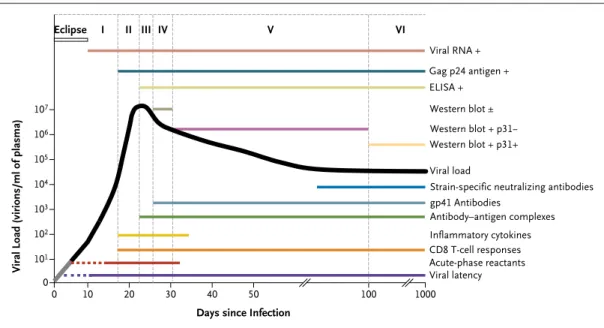
The stages that define acute and early HIV-1 infection are characterized by the
se-quential appearance of viral markers and antibodies in the blood (Fig. 2).5 More
sensitive, fourth-generation tests, which detect both antigens and antibodies, shrink
the virus-positive–antibody-negative window by about 5 days.12 Testing for viral
RNA in plasma closes this gap by an additional 7 days.
The characteristic appearance in the blood of viral markers of acute HIV-1 infec-tion belies an extremely complicated and still poorly understood series of virus–host cell interactions in the tissues (Fig. 1).4,13 Given the varied routes of viral
transmis-sion — cervicovaginal, penile, rectal, oral, percutaneous, intravenous, in utero — and the distinctly different histologic features of these tissues, it is not surprising that several cell types are candidates for early infection. More is known about vagi-nal transmission than about other routes, and the study of human tissue explants14,15
and the Indian rhesus macaque model of vaginal transmission of the simian
im-munodeficiency virus (SIV)13,16-18 have been informative (Fig. 1). The
preponder-ance of evidence implicates CD4 T cells and Langerhans’ cells as the first targets of the virus,14,15 but other dendritic cells may play an important accessory role.19
that monocyte-derived macrophages are generally poor targets for infection as compared with CD4 T cells.20,21
Regardless of the route of viral transmission and the first cells infected, within a few days, viral replication converges on the lymphoreticu-lar system of the gastrointestinal tract (i.e., gut-associated lymphoid tissue).22-25 In this tissue, in
both humans and macaques, the phenotype of most productively infected cells appears to be the resting CD4 T cell lacking activation markers and expressing low levels of the chemokine receptor CCR5.16-18 Many of these cells express α
4β7 inte-grin receptors and type 17 helper T (Th17)–cell surface markers.26,27 (Since these receptors are
also detected on T cells harvested from the geni-tal mucosa, they may play an important role in HIV acquisition.28) The rapid expansion of HIV-1,
first in gut-associated lymphoid tissue and then systemically,25,29 along with a sharp rise in
plas-ma levels of viral RNA, is clinically important be-cause of the coincident irreversible destruction of reservoirs of helper T cells and the establishment of viral latency (defined as the silent integration of HIV-1 DNA into the genomes of resting T cells, an effect that has stymied curative treatment ef-forts30,31).
Rather than being genetically homogeneous, RNA viruses, including HIV-1, consist of complex mixtures of mutant and recombinant genomes called quasi-species. Genetic studies of the HIV-1 quasi-species in patients with chronic infection as compared with patients with acute infection have brought some clarity to the qualitative and quantitative aspects of HIV-1 transmission.8
Fig-ure 3 depicts the HIV-1 transmission event,8,9,32,33
in which the inoculum (e.g., semen, cervicovagi-nal secretions, or blood) contains a complex ge-netic quasi-species of viruses, of which only a very small number are likely to broach mucosal bar-riers and establish infection. Lee and colleagues9
developed a model that allows transmitted viral genomes to be inferred from a phylogenetic analysis of the viral quasi-species that replicate in the weeks after infection. Empirical analyses based on single-genome amplification of HIV-1 RNA in plasma or HIV-1 DNA in blood lympho-cytes have provided robust evidence to support this model.8,20,21,33-38 A single virion is
respon-sible for HIV-1 transmission in approximately 80% of heterosexuals but in only about 60% of men who have sex with men and about 40% of
injection-drug users.8,20,21,33-35 In injection-drug
users, as many as 16 transmitted virions have been found to be responsible for productive
in-fection,36 which would be consistent with the
absence of a mucosal barrier to transmission. The phenotypes of cloned proviruses correspond-ing to transmitted (or founder) viruses are nearly always CD4 and CCR5 T-cell tropic variants and exhibit neutralization-sensitivity patterns that are typical of primary viral strains. These phenotypic properties are present at the moment of trans-mission, when the virus encounters the first tar-get cell; they are not the consequence of viral ad-aptation to the new host.8,20,21
Initial Innate Immune R esponses to HIV-1
The first signal of an immune response to HIV-1 infection is the appearance of acute-phase
1
Ingelfinger
4/28/11
AUTHOR PLEASE NOTE:
Figure has been redrawn and type has been reset Please check carefully
Author Fig # Title ME DE Artist
Issue date
COLOR FIGURE Draft 8
Cohen
Knoper
5/19/11 A HIV-1 translocation through female epithelium
Columnar epithelium in rectum and endocervix Mucus layer
B HIV-1 translocation through male epithelium
C Timing of HIV-1 infection events
Columnar epithelium in rectum Mucus
layer
Virus or virus-infected cells crossing mucosal barrier
Local propagation of infection on CD4 T cells
Dissemination into draining lymph nodes
Systemic dissemination
Establishment of CD4 T-cell virus reservoirs (? earlier)
Stratified squamous epithelium in vagina and ectocervix
Stratified squamous epithelium of inner side of penile foreskin
2–6 hours 3–6 days 6–25 days
HIV-1– infected cells
HIV-1 virions
X4
X4
R5
R5
CD4 T cell
CD4 T cell
Tissue macrophage
Tissue macrophage
Tissue macrophage
Dendritic cell
Dendritic cell Langerhans’
cell
HIV-1 virions
X4 R5
?
? ?
Dendritic cell CD4 T cell
Langerhans’ cell
Mucus layer
HIV-1– infected cells
X4 R5
CD4 T cell
Tissue macrophage
Dendritic cell
tants, including alpha 1-antitrypsin and serum amy-loid A, in plasma 3 to 5 days after transmission39
(Fig. 2). The steep rise in the HIV-1 viral load (ramp-up viremia) coincides with a large burst of inflammatory cytokines led by interferon-α and interleukin-1540 and a shower of plasma
micropar-ticles with surface phosphatidylserine, derived from infected and activated CD4 T cells undergo-ing apoptosis; these particles have immunosup-pressive properties.41
The earliest cytokines are produced by dendritic cells, but later in the infective process, multiple cell types (e.g., monocytes, macrophages, natural killer [NK] cells, and T cells) also produce these
mediators.42 Although cytokines enhance
protec-tive antiviral immune responses in acute HIV-1 infection, the cytokine storm probably also con-tributes to harmful immune activation and loss of CD4 T cells.
NK cells are activated in acute HIV-1 infection and, in vitro, kill cells infected with the virus.43
NK cells have a range of receptors that either en-hance or inhibit their function. NK-cell immu-noglobulin-like receptors interact with HLA mol-ecules with some specificity for the peptides they bind.44 This activity might explain the genetic
as-sociations between certain NK-cell immunoglob-ulin-like receptors and HLA types with more fa-vorable outcomes of infection.45
Adap tive Immune R esponses in Acute HIV-1 Infection
The initial antibody response to the viral enve-lope is non-neutralizing and does not select for viral escape46 (Fig. 2). Antibodies that neutralize
the transmitted founder virus are not detected until 3 months or more after infection.47 Although
Viral Load (virions/ml of plasma)
107
105 106
104
103
101 102
0
0 10 20 30 40 50 100 1000
Days since Infection
Viral RNA + Gag p24 antigen + ELISA +
Western blot ± Western blot + p31− Western blot + p31+
Viral latency Acute-phase reactants CD8 T-cell responses Inflammatory cytokines Antibody–antigen complexes gp41 Antibodies
Strain-specific neutralizing antibodies Viral load
Eclipse I II III IV V VI
Figure 2. Natural History and Immunopathogenesis of HIV-1 Infection.
Infected cell with defective virus Uninfected cells
Approximately 109infection
events
Less-fit virus (R0 approximately 1) Most fit virus (R0>>1)
Infected cell with defective virus
Less-fit virus, attenuated or stochastic event (R0<1)
>106 virions/ml of plasma
Inoculum Mucosa Recipient
3
Ingelfinger Koopman
4/22/11
AUTHOR PLEASE NOTE:
Figure has been redrawn and type has been reset Please check carefully
Author Fig # Title ME DE Artist
Issue date
COLOR FIGURE Draft 11
Cohen
Knoper
5/19/11 Figure 3. Model of HIV-1 Transmission.
many of the targets of neutralizing antibodies are on the glycoprotein-120 component of the HIV-1 envelope, the initial antibody response to HIV-1 is focused on non-neutralizing sites of the glyco-protein 41 envelope stalk.46 It is not known why
the initial HIV-1 antibody response is directed (or misdirected) to ineffective envelope sites, but the response may be related in part to the relative abundance of non-native HIV-1 envelope mole-cules when glycoprotein 41 is exposed, whereas the exposure of functional native envelope trimers is rare.48 Similarly, other potentially protective
an-tibodies directed against envelope proteins, such as antibodies that neutralize the founder viral strain or those that mediate antibody-dependent cellular cytotoxicity, do not arise until weeks af-ter transmission.47,49,50 By the time a potentially
effective antibody response has developed, it is much too late to influence the course of the in-fection (Fig. 2).
The first CD8 T-cell responses appear days be-fore the peak of viremia and focus on between one and three distinct epitopes (short antigenic peptides bound to HLA molecules that are de-rived from HIV-1 proteins) most commonly found in HIV-1 proteins nef and gag.51 These first T-cell
responses select escape mutants (which cannot be recognized by killer CD8 T cells), with complete replacement of the original viral amino acid
se-quence by the new sese-quence in 10 to 21 days.52
These initial T-cell responses are followed by new T-cell responses to other epitopes, which often escape as well. A combination of strong T-cell responses, producing chemokine (C-C motif) li-gand 4 (CCL4), and a focus on epitopes with high levels of variability (entropy) favors rapid escape.53
These CD8 T cells also express perforin — a pro-tein closely associated with cell-mediated cyto-toxicity — which suggests that they can kill in-fected cells.54
Other CD8 T-cell responses do not appear to select escape mutants — or must do so very slowly. Some of these T cells may be functionally deficient, but most appear to be effective, focus-ing on regions of the virus that can mutate, but at the cost of making the virus less efficient in replication.55 These latter T cells are likely to
con-tribute to the control of HIV-1. As this T-cell re-sponse evolves, the plasma viral load falls (Fig. 2). The rate of loss of virus containing the epitopes recognized by the early T-cell responses that drive escape provides a measure of the rate of killing
(or removal) of virus-infected cells in vivo.52 Other
factors, such as loss of susceptible cells (given the extreme depletion of activated CD4 T cells in gut-associated lymphoid tissue), probably also play a part in lowering the initial peak viral load.23,56
During acute HIV-1 infection, irrevocable de-pletion of CD4 T lymphocytes from the gastro-intestinal tract23 and some other lymphoid tissues
has been observed in humans and rhesus
ma-caques.25 In humans, adjunctive damage to the
mucosal barriers may allow leakage of gut bacte-rial products into otherwise sterile tissues and to the bloodstream, leading to further immune activation that can promote HIV replication and have other adverse consequences.57 The rapid,
early, and massive loss of CD4 T cells in lymphoid organs (which is poorly reflected in CD4 T-cell counts in blood) probably accounts for the weak CD4 T-cell responses in cases of acute HIV-1 in-fection.
The importance of CD8 T cells in controlling acute HIV-1 infection is consistent with studies in the rhesus macaque SIV model showing that in vivo depletion of CD8 T cells abrogated viral control in both acute infection and chronic in-fection.58 In addition, many studies in macaques
have shown that vaccines that stimulate SIV-spe-cific CD8 T-cell responses can attenuate subse-quent SIV infection.59 These data are also
consis-tent with extensive work showing that in patients with certain HLA types, particularly HLA-B57 (and the very closely related HLA-B58) and HLA-B27, viral control is often better than average, with a lower virus set point and longer survival in the absence of antiretroviral therapy.60 The B27, B57,
and B58 molecules present highly conserved parts of the virus to T cells, so that the virus can es-cape immune control only at the cost of replica-tive fitness.55
Detection of Acute HIV-1 Infection
In the absence of a high degree of clinical suspi-cion, the symptoms associated with acute HIV-1 infection are often too vague or nonspecific to lead to a diagnosis.61 In the absence of antibody
involves searching for HIV RNA in pooled, anti-body-negative samples has been used to increase
detection.61 This approach has been used to
de-tect acute HIV-1 infection, with a prevalence of 0.5 cases detected per 1000 persons tested, in North Carolina, to 4.0 cases per 1000, in San Francisco; acute infection accounted for 5 to 10% of all cas-es of HIV in both placcas-es.
As an alternative and more practical strategy, an enzyme-linked immunosorbent assay that can concomitantly detect viral p24 antigen and anti-viral antibodies has been developed and approved for clinical use.2,3,61 This test can increase the
number of patients with acute HIV-1 infection whose condition is diagnosed at a time when they are most infectious to others.3 It is anticipated
that a rapid point-of-care test will also be devel-oped for the purpose of detecting acute HIV-1 infection. The implementation of these tests across the United States in public health and commer-cial laboratories can be expected to dramatically increase the number of patients with acute HIV-1 infection who will require care.
Public He alth Consequences of Acute HIV-1 Infection
The per-person probability of transmitting HIV-1 is most closely correlated with the viral burden in blood; each time the viral burden in an HIV-1– infected person increases by a factor of 10, the risk of transmission is expected to increase by a
factor of 2.5.62 The risk of contagion from
pa-tients with acute, early infection appears to be much higher than that from patients with estab-lished infection,63 at least in part because of the
high viral load and the homogeneity of viral vari-ants clearly capable of causing infection. In the rhesus macaque SIV model, plasma from animals with acute infection is up to 750 times as infec-tious, on a per-virion basis, as plasma from
ani-mals with chronic infection.64 The reduced risk
of contagion from patients with chronic infection probably results from the presence of neutraliz-ing antibodies, which are not evident in acute in-fection.
Mathematical models used to estimate the role of patients with acute infection in the spread of HIV-1 have produced strikingly different results, depending on the population studied and the as-sumptions used (Fig. 4).65-77 The epidemic phase
used for modeling has been a critical
determi-nant.78 In communities subject to a new epidemic,
early infections are held to be responsible for a considerable share of HIV-1 transmission, since a larger proportion of infected persons have acute or early-stage disease rather than late-stage
dis-ease.79 Sexual behavior plays an important role
in rates of infection, with high rates of partner change increasing the chances of contact with a person who has acute HIV-1 infection. In a recent comprehensive study conducted in Lilongwe, Ma-lawi, in which both behavioral and biologic data were used, 38% of cases of HIV-1 were ascribed to sexual exposure to patients in the first 5 months of infection, even though there is a
long-estab-lished epidemic in Malawi.75 The results of the
Malawi study may be most relevant to the HIV-1 pandemic in sub-Saharan Africa.
The importance of acute HIV-1 infection can also be seen in studies of phylogenetically related cases, such as the study reported by Brenner and colleagues.79 They state that more than half the
patients with newly diagnosed early HIV-1 infec-tion in Montreal are infected with viral variants that can be linked through phylogenetic studies, which suggests the presence of clusters of trans-mission, perhaps from patients with acute and early infection.
Pr eventing HIV-1 Infection
Effective HIV preventive strategies must be in place before or immediately after the transmis-sion event. This is a tall order for antiviral prophy-laxis, administered before or after exposure, or a vaccine. Indeed, the antibody responses directed against the HIV-1 envelope after the administra-tion of vaccine regimens are not of long dura-tion.80,81 Nevertheless, there are several points in
the transmission event at which the founder virus may be vulnerable to inhibition by antibodies, ranging from the entry of virus or virus-infected cells into mucus in the genital tract to cell-to-cell transmission in genital tract submucosa (Fig. 1).82
Weakly neutralizing antibodies that mediate an-tibody-dependent, cell-mediated cytotoxicity or antibody-dependent cellular viral inhibition may have a protective effect by stimulating immune cells to produce anti–HIV-1 chemokines, such as CCL3, CCL4, and CCL5.81 In a recent vaccine
results could also be explained by the actions of one or more innate antiviral immune mechanisms.
An alternative prevention strategy that is more readily available is that of offering antiretroviral agents to people at risk before or immediately after HIV exposure or as a means of secondary prevention.84,85 Use of the antiretroviral drug
tenofovir as a topical prophylactic agent before viral exposure in women at high risk led to a 39% reduction in incident cases of HIV infection that was directly correlated with concentrations of the drug in mucosal tissue.86 Seven ongoing trials of
oral preexposure prophylaxis have been
under-taken.87 A multinational trial focused on men
who have sex with men87 showed that a
once-daily pill containing tenofovir plus emtricitabine provided an average of 44% protection over and above that conferred by the provision of compre-hensive preventive services, including the provision of condoms and counseling.88 The level of
protec-tion varied widely, depending on how consis-tently participants used preexposure prophylaxis. The Centers for Disease Control and Prevention has issued preliminary recommendations for the use of preexposure prophylaxis by men who have
sex with men.89 Work with rhesus macaques
suggests that achieving high levels of antiviral agents in mucosal tissues shortly after exposure to SIV–HIV viral chimeras is critical for protection from infection.90 This work, along with the further
Proportion of New Infections Caused by Early Infections
1.0
0.8 0.9
0.7
0.6
0.4
0.3
0.1 0.5
0.2
0.0
Sub-Saharan Africa
(heterosexuals) (MSM)U.S. Europe(MSM)
Population Hollingsworth
et al., 2008 Hayes and
White,
2006 Xiridou et al.,
2004
Abu-Raddad and Longini,
2008
Salomon and Hogan,
2008
U.S. (heterosexuals or MSM)
Pinkerton, 2007
Prabhu et al., 2009
Pinkerton and Abramson, 1996
Kretzschmar and Dietz, 1998
Jacquez et al.,
1994 Koopman et al., 1997 Powers et al.,
2010
Figure 4. Role of Acute and Early HIV-1 Infection in the Spread of HIV-1, According to Population Studies in Sub-Saharan Africa, the United States, and Europe.
exploration of new drug combinations, will prob-ably play a role in the further development of pre-exposure and postpre-exposure prophylaxis.
M anaging Acute HIV-1 Infection
The health care provider has three responsibili-ties with respect to acute HIV-1 infection: detec-tion; secondary prevention, which in some cases must include partner notification (and possibly postexposure prophylaxis with antiretroviral ther-apy); and initiation of antiretroviral therapy, if it is considered appropriate. Although the fourth-generation HIV tests have the capacity to detect acute HIV-1 infection, algorithms that will reduce the time to diagnosis and linkage to medical care must be put into place.3 Equally important,
strat-egies must be developed to offer the best possible counseling in order to reduce further spread of HIV-1 and to break up the sexual networks that can form around patients with acute HIV-1 infec-tion.91 However, partner notification in the
Unit-ed States has been limitUnit-ed92 and is only now
be-ing studied in resource-constrained countries.93
A recent study in Africa has highlighted the dif-ficulty of explaining acute HIV-1 infection to study subjects in a manner that is likely to reduce fur-ther transmission.94
Alternatively, antiretroviral therapy could be used to suppress viral replication in order to re-duce HIV-1 transmission. What constitutes the optimal use of antiretroviral therapy for patients with acute infection is unclear, in part because the extent of the personal health benefit derived from early use of antiretroviral therapy remains in question.95,96 There have been considerable
dif-ferences in reported results of the clinical bene-fits of antiretroviral therapy for patients with primary HIV infection because in many cases antiretroviral therapy was initiated weeks or even months after HIV-1 acquisition — probably much too late to influence the course of dis-ease. A few small studies have shown some bene-fit when therapy was provided before or during seroconversion, with some degree of immune
preservation96 and with sustained reduction of
blood viral load after antiretroviral therapy was discontinued. Moir et al. reported that patients treated soon after receiving the diagnosis had im-proved B-cell function.97 Very early
administra-tion of antiretroviral therapy may limit the size of the latent pool of HIV-1–infected CD4 T cells.98,99
Although encouraging, these results underscore the need for well-constructed clinical trials that will provide the basis for determining the over-all cost–benefit ratio of antiretroviral therapy for acute HIV-1 infection and for balancing the pub-lic health benefits with the benefits for individ-ual patients.
In the most recent treatment guidelines from the International Antiviral Society — USA, the authors argue that potential benefits to public and individual health may even now justify the treatment of patients with acute HIV infection,
particularly those who are symptomatic.100 The
authors of other guidelines have come to similar conclusions, setting the stage for regular treat-ment of persons with acute HIV infection. If anti-retroviral therapy is to be provided, the treatment regimen might include drugs that concentrate in
the genital tract of men and women85 and an
integrase inhibitor, the latter because of the ra-pidity with which this class of drugs lowers the viral load.101 Some pilot studies using multidrug
regimens, such as those administered before HIV seroconversion, are in progress.102
Conclusions
The earliest events in acute HIV-1 infection deter-mine the future health of the individual patient and the extent of transmission in the general pop-ulation. Recent studies have unraveled many of the initial immune events of acute infection. With im-proved diagnostic tests, greater numbers of per-sons with acute HIV-1 infection will come to the attention of practicing physicians and public health officials. Although considerable progress has been made in understanding the HIV-1 trans-mission event, more studies are needed to develop optimal treatment and prevention strategies for people in the earliest stages of HIV-1 infection.
Supported by the National Institutes of Health and the Center for HIV–AIDS Vaccine Immunology.
Dr. Cohen reports receiving consulting fees from GlaxoSmith-Kline and Merck; Dr. McMichael, receiving payment for the de-velopment of educational presentations from Henry Stewart Talks; and Dr. Haynes, receiving a research grant from Pere-grine Pharmaceuticals. No other potential conflict of interest relevant to this article was reported.
Disclosure forms provided by the authors are available with the full text of this article at NEJM.org.
References
1. Global report 2010. Geneva: UNAIDS, 2010. (http://www.unaids.org/globalreport/ documents/20101123_GlobalReport_full_ en.pdf.)
2. Branson BM. State of the art for diag-nosis of HIV infection. Clin Infect Dis 2007;45:Suppl 4:S221-S225.
3. Eshleman SH, Khaki L, Laeyendecker O, et al. Detection of individuals with acute HIV-1 infection using the ARCHITECT HIV Ag/Ab Combo assay. J Acquir Im-mune Defic Syndr 2009;52:121-4. 4. Hladik F, McElrath MJ. Setting the stage: host invasion by HIV. Nat Rev Im-munol 2008;8:447-57.
5. Fiebig EW, Wright DJ, Rawal BD, et al. Dynamics of HIV viremia and antibody seroconversion in plasma donors: impli-cations for diagnosis and staging of pri-mary HIV infection. AIDS 2003;17:1871-9. 6. Weiss HA, Dickson KE, Agot K, Han-kins CA. Male circumcision for HIV preven-tion: current research and programmatic issues. AIDS 2010;24:Suppl 4:S61-S69. 7. Emau P, Jiang Y, Agy MB, Tian B, Bekele G, Tsai CC. Post-exposure prophy-laxis for SIV revisited: animal model for HIV prevention. AIDS Res Ther 2006;3:29. 8. Keele BF, Giorgi EE, Salazar-Gonza-lez JF, et al. Identification and character-ization of transmitted and early founder virus envelopes in primary HIV-1 infec-tion. Proc Natl Acad Sci U S A 2008; 105:7552-7.
9. Lee HY, Giorgi EE, Keele BF, et al. Modeling sequence evolution in acute HIV-1 infection. J Theor Biol 2009;261:341-60.
10. Palmer S, Wiegand AP, Maldarelli F, et al. New real-time reverse transcriptase-initiated PCR assay with single-copy sensi-tivity for human immunodeficiency virus type 1 RNA in plasma. J Clin Microbiol 2003;41:4531-6.
11. Damond F, Avettand-Fenoel V, Collin G, et al. Evaluation of an upgraded version of the Roche Cobas AmpliPrep/Cobas TaqMan HIV-1 test for HIV-1 load quanti-fication. J Clin Microbiol 2010;48:1413-6. 12. Sickinger E, Jonas G, Yem AW, et al. Performance evaluation of the new fully automated human immunodeficiency vi-rus antigen-antibody combination assay designed for blood screening. Transfusion 2008;48:584-93.
13. Haase AT. Targeting early infection to prevent HIV-1 mucosal transmission. Na-ture 2010;464:217-23.
14. Hladik F, Sakchalathorn P, Ballweber L, et al. Initial events in establishing vagi-nal entry and infection by human immu-nodeficiency virus type-1. Immunity 2007; 26:257-70.
15. Boggiano C, Littman DR. HIV’s vagina travelogue. Immunity 2007;26:145-7. 16. Zhang Z, Schuler T, Zupancic M, et al. Sexual transmission and propagation of SIV and HIV in resting and activated CD4+
T cells. Science 1999;286:1353-7. [Erratum, Science 1999;286:2273.]
17. Zhang ZQ, Wietgrefe SW, Li Q, et al. Roles of substrate availability and infec-tion of resting and activated CD4+ T cells in transmission and acute simian immu-nodeficiency virus infection. Proc Natl Acad Sci U S A 2004;101:5640-5. 18. Li Q, Duan L, Estes JD, et al. Peak SIV replication in resting memory CD4+ T cells depletes gut lamina propria CD4+ T cells. Nature 2005;434:1148-52.
19. Lackner AA, Veazey RS. Current con-cepts in AIDS pathogenesis: insights from the SIV/macaque model. Annu Rev Med 2007;58:461-76.
20. Salazar-Gonzalez JF, Salazar MG, Keele BF, et al. Genetic identity, biological phenotype, and evolutionary pathways of transmitted/founder viruses in acute and early HIV-1 infection. J Exp Med 2009; 206:1273-89.
21. Li H, Bar KJ, Wang S, et al. High mul-tiplicity infection by HIV-1 in men who have sex with men. PLoS Pathog 2010; 6(5):e1000890.
22. Veazey RS, DeMaria M, Chalifoux LV, et al. Gastrointestinal tract as a major site of CD4+ T cell depletion and viral replica-tion in SIV infecreplica-tion. Science 1998;280: 427-31.
23. Brenchley JM, Schacker TW, Ruff LE, et al. CD4+ T cell depletion during all stages of HIV disease occurs predomi-nantly in the gastrointestinal tract. J Exp Med 2004;200:749-59.
24. Mehandru S, Poles MA, Tenner-Racz K, et al. Primary HIV-1 infection is associ-ated with preferential depletion of CD4+ T lymphocytes from effector sites in the gastrointestinal tract. J Exp Med 2004; 200:761-70.
25. Mattapallil JJ, Douek DC, Hill B, Nishimura Y, Martin M, Roederer M. Mas-sive infection and loss of memory CD4+ T cells in multiple tissues during acute SIV infection. Nature 2005;434:1093-7. 26. Arthos J, Cicala C, Martinelli E, et al. HIV-1 envelope protein binds to and sig-nals through integrin alpha4beta7, the gut mucosal homing receptor for periph-eral T cells. Nat Immunol 2008;9:301-9. 27. Kader M, Wang X, Piatak M, et al. Al-pha4(+)beta7(hi)CD4(+) memory T cells harbor most Th-17 cells and are preferen-tially infected during acute SIV infection. Mucosal Immunol 2009;2:439-49. 28. Cicala C, Martinelli E, McNally JP, et al. The integrin alpha4beta7 forms a com-plex with cell-surface CD4 and defines a T-cell subset that is highly susceptible to infection by HIV-1. Proc Natl Acad Sci U S A 2009;106:20877-82.
29. Schacker T, Little S, Connick E, et al. Productive infection of T cells in lym-phoid tissues during primary and early human immunodeficiency virus infection. J Infect Dis 2001;183:555-62.
30. Chun TW, Engel D, Berrey MM, Shea T, Corey L, Fauci AS. Early establishment of a pool of latently infected, resting CD4(+) T cells during primary HIV-1 in-fection. Proc Natl Acad Sci U S A 1998; 95:8869-73.
31. Richman DD, Margolis DM, Delaney M, Greene WC, Hazuda D, Pomerantz RJ. The challenge of finding a cure for HIV infection. Science 2009;323:1304-7. 32. Ribeiro RM, Qin L, Chavez LL, Li D, Self SG, Perelson AS. Estimation of the initial viral growth rate and basic repro-ductive number during acute HIV-1 infec-tion. J Virol 2010;84:6096-102.
33. Keele BF, Li H, Learn GH, et al. Low-dose rectal inoculation of rhesus ma-caques by SIVsmE660 or SIVmac251 reca-pitulates human mucosal infection by HIV-1. J Exp Med 2009;206:1117-34. 34. Abrahams MR, Anderson JA, Giorgi EE, et al. Quantitating the multiplicity of infection with human immunodeficiency virus type 1 subtype C reveals a non-Pois-son distribution of transmitted variants. J Virol 2009;83:3556-67. [Erratum, J Virol 2009;83:6974.]
35. Haaland RE, Hawkins PA, Salazar-Gonzalez J, et al. Inflammatory genital infections mitigate a severe genetic bottle-neck in heterosexual transmission of sub-type A and C HIV-1. PLoS Pathog 2009; 5(1):e1000274.
36. Bar KJ, Li H, Chamberland A, et al. Wide variation in the multiplicity of HIV-1 infection among injection drug users. J Virol 2010;84:6241-7.
37. Masharsky AE, Dukhovlinova EN, Verevochkin SV, et al. A substantial trans-mission bottleneck among newly and re-cently HIV-1-infected injection drug users in St Petersburg, Russia. J Infect Dis 2010; 201:1697-702.
38. Fischer W, Ganusov VV, Giorgi EE, et al. Transmission of single HIV-1 genomes and dynamics of early immune escape re-vealed by ultra-deep sequencing. PLoS One 2010;5(8):e12303.
39. Kramer HB, Lavender KJ, Qin L, et al. Elevation of intact and proteolytic frag-ments of acute phase proteins constitutes the earliest systemic antiviral response in HIV-1 infection. PLoS Pathog 2010;6(5): e1000893.
42. Borrow P, Bhardwaj N. Innate im-mune responses in primary HIV-1 infec-tion. Curr Opin HIV AIDS 2008;3:36-44. 43. Alter G, Martin MP, Teigen N, et al. Differential natural killer cell-mediated inhibition of HIV-1 replication based on distinct KIR/HLA subtypes. J Exp Med 2007;204:3027-36.
44. Hansasuta P, Dong T, Thananchai H, et al. Recognition of A3 and HLA-A11 by KIR3DL2 is peptide-specific. Eur J Immunol 2004;34:1673-9.
45. Martin MP, Gao X, Lee JH, et al. Epi-static interaction between KIR3DS1 and HLA-B delays the progression to AIDS. Nat Genet 2002;31:429-34.
46. Tomaras GD, Yates NL, Liu P, et al. Initial B-cell responses to transmitted hu-man immunodeficiency virus type 1: virion-binding immunoglobulin M (IgM) and IgG antibodies followed by plasma anti-gp41 antibodies with ineffective control of initial viremia. J Virol 2008;82:12449-63. 47. Wei X, Decker JM, Wang S, et al. Anti-body neutralization and escape by HIV-1. Nature 2003;422:307-12.
48. Moore PL, Crooks ET, Porter L, et al. Nature of nonfunctional envelope pro-teins on the surface of human immuno-deficiency virus type 1. J Virol 2006;80: 2515-28.
49. Pollara J, Landucci G, Trac C, et al. Early appearance of ADCC- and ADCVI-mediating antibody responses against au-tologous HIV-1 transmitted/founder virus. AIDS Res Hum Retroviruses 2010;26:A-12. abstract.
50. Richman DD, Wrin T, Little SJ, Petro-poulos CJ. Rapid evolution of the neutral-izing antibody response to HIV type 1 in-fection. Proc Natl Acad Sci U S A 2003; 100:4144-9.
51. Lichterfeld M, Yu XG, Cohen D, et al. HIV-1 Nef is preferentially recognized by CD8 T cells in primary HIV-1 infection despite a relatively high degree of genetic diversity. AIDS 2004;18:1383-92. 52. Goonetilleke N, Liu MK, Salazar-Gonzalez JF, et al. The first T cell re-sponse to transmitted/founder virus con-tributes to the control of acute viremia in HIV-1 infection. J Exp Med 2009;206:1253-72.
53. Ferrari G, Korber BT, Turnbull E, et al. High magnitude and multifunctional-ity epitope-specific CD8+ T-cell responses are associated with selection of escape mutants in acute HIV infection. AIDS Res Hum Retroviruses 2010;26:A-5. abstract. 54. Hersperger AR, Pereyra F, Nason M, et al. Perforin expression directly ex vivo by HIV-specific CD8 T-cells is a correlate of HIV elite control. PLoS Pathog 2010; 6(5):e1000917.
55. Leslie AJ, Pfafferott KJ, Chetty P, et al. HIV evolution: CTL escape mutation and reversion after transmission. Nat Med 2004;10:282-9.
56. Phillips AN. Reduction of HIV
con-centration during acute infection: inde-pendence from a specific immune re-sponse. Science 1996;271:497-9. 57. Brenchley JM, Douek DC. The muco-sal barrier and immune activation in HIV pathogenesis. Curr Opin HIV AIDS 2008;3: 356-61.
58. Schmitz JE, Kuroda MJ, Santra S, et al. Control of viremia in simian immunode-ficiency virus infection by CD8+ lympho-cytes. Science 1999;283:857-60. 59. Liu J, O’Brien KL, Lynch DM, et al. Im-mune control of an SIV challenge by a T-cell-based vaccine in rhesus monkeys. Nature 2009;457:87-91.
60. International HIV Controllers Study. The major genetic determinants of HIV-1 control affect HLA class I peptide presen-tation. Science 2010;330:1551-7. 61. Daar ES, Pilcher CD, Hecht FM. Clini-cal presentation and diagnosis of primary HIV-1 infection. Curr Opin HIV AIDS 2008;3:10-5.
62. Quinn TC, Wawer MJ, Sewankambo N, et al. Viral load and heterosexual trans-mission of human immunodeficiency vi-rus type 1. N Engl J Med 2000;342:921-9. 63. Wawer MJ, Gray RH, Sewankambo NK, et al. Rates of HIV-1 transmission per co-ital act, by stage of HIV-1 infection, in Rakai, Uganda. J Infect Dis 2005;191: 1403-9.
64. Ma ZM, Stone M, Piatak M Jr, et al. High specific infectivity of plasma virus from the pre-ramp-up and ramp-up stag-es of acute simian immunodeficiency vi-rus infection. J Virol 2009;83:3288-97. 65. Hollingsworth TD, Anderson RM, Fraser C. HIV-1 transmission, by stage of infection. J Infect Dis 2008;198:687-93. 66. Pinkerton SD. How many sexually-acquired HIV infections in the USA are due to acute-phase HIV transmission? AIDS 2007;21:1625-9.
67. Idem. Probability of HIV transmission
during acute infection in Rakai, Uganda. AIDS Behav 2008;12:677-84.
68. Jacquez JA, Koopman JS, Simon CP, Longini IM Jr. Role of the primary infec-tion in epidemics of HIV infecinfec-tion in gay cohorts. J Acquir Immune Defic Syndr 1994;7:1169-84.
69. Koopman JS, Jacquez JA, Welch GW, et al. The role of early HIV infection in the spread of HIV through populations. J Ac-quir Immune Defic Syndr Hum Retrovirol 1997;14:249-58.
70. Kretzschmar M, Dietz K. The effect of pair formation and variable infectivity on the spread of an infection without recov-ery. Math Biosci 1998;148:83-113. 71. Coutinho FA, Lopez LF, Burattini MN, Massad E. Modelling the natural history of HIV infection in individuals and its epidemiological implications. Bull Math Biol 2001;63:1041-62.
72. Xiridou M, Geskus R, de Wit J, Coutinho R, Kretzschmar M. Primary HIV infection as source of HIV
transmis-sion within steady and casual partner-ships among homosexual men. AIDS 2004; 18:1311-20.
73. Hayes RJ, White RG. Amplified HIV transmission during early-stage infection. J Infect Dis 2006;193:604-6.
74. Abu-Raddad LJ, Longini IM Jr. No HIV stage is dominant in driving the HIV epi-demic in sub-Saharan Africa. AIDS 2008; 22:1055-61.
75. Powers K, Kamanga G, Mapanje C, et al. Efficient detection of acute HIV infec-tion through targeted HIV RNA screening in a Malawian STI clinic. Presented at the XVIII International AIDS Conference, Vi-enna, Austria, July 18–23, 2010. abstract. 76. Salomon JA, Hogan DR. Evaluating the impact of antiretroviral therapy on HIV transmission. AIDS 2008; 22:Suppl 1:S149-S159.
77. Pinkerton SD, Abramson PR. Implica-tion of increased infectivity in early-stage HIV infection: application of a Bernoulli-process model of HIV transmission. Eval Rev 1996;20:516-40.
78. Miller WC, Rosenberg NE, Rutstein SE, Powers KA. Role of acute and early HIV infection in the sexual transmission of HIV. Curr Opin HIV AIDS 2010;5:277-82. 79. Brenner BG, Roger M, Routy JP, et al. High rates of forward transmission events after acute/early HIV-1 infection. J Infect Dis 2007;195:951-9.
80. Morris L, Binley JM, Clas BA, et al. HIV-1 antigen-specific and -nonspecific B cell responses are sensitive to combina-tion antiretroviral therapy. J Exp Med 1998;188:233-45.
81. Bonsignori M, Moody MA, Parks RJ, et al. HIV-1 envelope induces memory B cell responses that correlate with plasma antibody levels after envelope gp120 pro-tein vaccination or HIV-1 infection. J Im-munol 2009;183:2708-17.
82. McElrath MJ, Haynes BF. Induction of immunity to human Immunodeficiency virus type-1 by vaccination. Immunity 2010;33:542-54.
83. Haynes B, Liao HX, Tomaras GD. Is developing an HIV-1 vaccine possible? Curr Opin HIV AIDS 2010;5:362-7. 84. Mayer KH, Venkatesh KK. Antiretro-viral therapy as HIV prevention: status and prospects. Am J Public Health 2010; 100:1867-76.
85. Cohen MS, Gay C, Kashuba AD, Blower S, Paxton L. Narrative review: antiretrovi-ral therapy to prevent the sexual trans-mission of HIV-1. Ann Intern Med 2007; 146:591-601.
86. Karim QA, Karim SS, Frohlich JA, et al. Effectiveness and safety of tenofovir gel, an antiretroviral microbicide for the prevention of HIV infection in women. Science 2010;329:1168-74.
88. Grant RM, Lama JR, Anderson PL, et al. Preexposure chemoprophylaxis for HIV prevention in men who have sex with men. N Engl J Med 2010;363:2587-99. 89. Pre-exposure prophylaxis (PrEP) for HIV prevention: promoting safe and ef-fective use in the United States. Atlanta: Centers for Disease Control and Preven-tion, 2010. (http://www.cdc.gov/hiv/prep.) 90. Garcia-Lerma JG, Cong ME, Mitchell J, et al. Intermittent prophylaxis with oral truvada protects macaques from rectal SHIV infection. Sci Transl Med 2010;2: 14ra4.
91. Hightow LB, MacDonald PD, Pilcher CD, et al. The unexpected movement of the HIV epidemic in the Southeastern United States: transmission among col-lege students. J Acquir Immune Defic Syndr 2005;38:531-7.
92. Golden MR, Dombrowski JC, Wood RW, Fleming M, Harrington RD. A con-trolled study of the effectiveness of public health HIV partner notification services. AIDS 2009;23:133-5.
93. Brown LB, Miller WC, Kamanga G, et
al. HIV partner notification is effective and feasible in sub-Saharan Africa: op-portunities for HIV treatment and preven-tion. J Acquir Immune Defic Syndr 2011; 56:437-42.
94. Pettifor A, Macphail C, Corneli A, et al. Continued high risk sexual behavior following diagnosis with acute HIV infec-tion in South Africa and Malawi: implica-tions for prevention. AIDS Behav 2010 October 27 (Epub ahead of print). 95. Fidler S, Fox J, Porter K, Weber J. Pri-mary HIV infection: to treat or not to treat? Curr Opin Infect Dis 2008;21:4-10. 96. Bell SK, Little SJ, Rosenberg ES. Clini-cal management of acute HIV infection: best practice remains unknown. J Infect Dis 2010;202:Suppl 2:S278-S288. 97. Moir S, Buckner CM, Ho J, et al. B cells in early and chronic HIV infection: evidence for preservation of immune function associated with early initiation of antiretroviral therapy. Blood 2010;116: 5571-9.
98. Chun TW, Justement JS, Moir S, et al. Decay of the HIV reservoir in patients
re-ceiving antiretroviral therapy for extend-ed periods: implications for eradication of virus. J Infect Dis 2007;195:1762-4. 99. Archin N, Cheema M, Sackmann R, Sugarbaker A, Ngo L, Kuruc J. Correlation of peak and duration of viremia with rest-ing CD4+ T cell infection in acute HIV infection. Presented at the 17th Confer-ence on Retroviruses and Opportunistic Infections, San Francisco, February 16–19 2010. abstract.
100. Thompson MA, Aberg JA, Cahn P, et al. Antiretroviral treatment of adult HIV infection: 2010 recommendations of the International AIDS Society-USA panel. JAMA 2010;304:321-33.
101. Murray JM, Emery S, Kelleher AD, et al. Antiretroviral therapy with the inte-grase inhibitor raltegravir alters decay kinetics of HIV, significantly reducing the second phase. AIDS 2007;21:2315-21. 102. Burns DN, Dieffenbach CW, Ver-mund SH. Rethinking prevention of HIV type 1 infection. Clin Infect Dis 2010; 51:725-31.