A S S O C I A T I O N S T U D I E S A R T I C L E
Genome-wide association study of dental caries in the
Hispanic Communities Health Study/Study of Latinos
(HCHS/SOL)
Jean Morrison
1
, Cathy C. Laurie
1
, Mary L. Marazita
2,3,4
, Anne E. Sanders
5
,
Steven Offenbacher
6
, Christian R. Salazar
7,8
, Matthew P. Conomos
1
,
Timothy Thornton
1
, Deepti Jain
1
, Cecelia A. Laurie
1
, Kathleen F. Kerr
1
,
George Papanicolaou
9
, Kent Taylor
10
, Linda M. Kaste
11
, James D. Beck
5
and John R. Shaffer
2,
*
1
Department of Biostatistics, University of Washington, Seattle, WA 98077, USA,
2Department of Human Genetics,
Graduate School of Public Health, University of Pittsburgh, 130 De Soto Street, Pittsburgh, PA 15261, USA,
3
Department of Oral Biology, School of Dental Medicine, Center for Craniofacial and Dental Genetics and
4
Department of Psychiatry, Clinical and Translational Science Institute, School of Medicine, University of
Pittsburgh, Pittsburgh, PA 15261, USA,
5Department of Dental Ecology and
6Department of Periodontology, Center
for Oral and Systemic Diseases, School of Dentistry, University of North Carolina at Chapel Hill, Chapel Hill, NC
27599, USA,
7Department of Epidemiology and
8Department of Population Health, Albert Einstein College of
Medicine and Monte
fi
ore Medical Center, New York City, NY 10461, USA,
9National Heart, Lung, and Blood
Institute, Bethesda, MD 20892, USA,
10Institute for Translational Genomics and Population Sciences, Los Angeles
Biomedical Research Institute Harbor-UCLA Medical Center, Torrance, CA 90502, USA and
11College of Dentistry
and School of Public Health, University of Illinois at Chicago, Chicago, IL 60162, USA
*To whom correspondence should be addressed. Tel: +1 4126243020; Fax: +1 4126243018; Email: [email protected]
Abstract
Dental caries is the most common chronic disease worldwide, and exhibits profound disparities in the USA with racial and ethnic minorities experiencing disproportionate disease burden. Though heritable, the specific genes influencing risk of dental caries remain largely unknown. Therefore, we performed genome-wide association scans (GWASs) for dental caries in a population-based cohort of 12 000 Hispanic/Latino participants aged 18–74 years from the HCHS/SOL. Intra-oral examinations were used to generate two common indices of dental caries experience which were tested for association with 27.7 M genotyped or imputed single-nucleotide polymorphisms separately in the six ancestry groups. A mixed-models approach was used, which adjusted for age, sex, recruitment site,five principal components of ancestry and additional features of the sampling design. Meta-analyses were used to combine GWAS results across ancestry groups. Heritability estimates ranged from 20–53% in the six ancestry groups. The most significant association observed via meta-analysis for both phenotypes was in the region of the
NAMPTgene (rs190395159;P-value = 6 × 10⁻¹⁰), which is involved in many biological processes including periodontal healing. Another significant association was observed for rs72626594 (P-value = 3 × 10⁻⁸) downstream ofBMP7, a tooth development
Received:August 13, 2015.Revised:November 11, 2015.Accepted:December 7, 2015
© The Author 2015. Published by Oxford University Press. All rights reserved. For Permissions, please email: [email protected]
doi: 10.1093/hmg/ddv506
Advance Access Publication Date: 11 December 2015 Association Studies Article
gene. Other associations were observed in genes lacking known or plausible roles in dental caries. In conclusion, this was the largest GWAS of dental caries, to date and was thefirst to target Hispanic/Latino populations. Understanding the factors influencing dental caries susceptibility may lead to improvements in prediction, prevention and disease management, which may ultimately reduce the disparities in oral health across racial, ethnic and socioeconomic strata.
Introduction
Dental caries (i.e. tooth decay) is the most prevalent chronic dis-ease affecting all populations, worldwide, and if left untreated, leads to serious concomitants and comorbidities. In the USA, over 90% of adults are affected by dental caries with over 25% hav-ing untreated decay (1). Furthermore, enormous disparities in disease burden persist, with vulnerable populations such as racial/ethnic minorities, low-income groups, and residents of rural areas suffering disproportionate rates of untreated disease and greater barriers to accessing oral health care. These disparities in disease burden persist despite the fact that prevalence of caries (which includes treated decay) is higher in non-Hispanic whites than minority groups (2). In fact, Hispanic and Latino Amer-icans count among the highest-risk populations; for example, the National Health and Nutrition Examination Survey (NHANES) 1999–2004 survey showed that Mexican Americans have nearly twice as many teeth with untreated decay, have more missing teeth due to caries, and are far more likely to report poor perceived dental health as compared with non-Hispanic whites (1). Similar disparities were observed for Hispanic children, adolescents and adults in the NHANES 2011–2012 survey (2,3).
The core disease mechanism leading to dental caries is well-known: the dissolution of the mineralized dental tissues due to acidic byproducts of bacterial metabolism. In the non-disease state, this demineralization is counteracted by natural remineraliz-ing processes. Therefore, whether or not dental caries develops depends on the balance between mineral dissolution and re-precipitation, which is greatly influenced by a number of moder-ating factors. Among these factors are characteristics of the saliva (including buffering capacity, anti-microbial agents, remineraliza-tion-promoting agents and rate of saliva production), characteris-tics of the teeth (including morphology and position of the teeth, and structure and composition of the enamel), microbialflora, host immunity and exogenousfluoride exposures. These factors are in turn influenced by many environmental exposures (e.g. tobacco, diet and medications), behaviors (e.g. oral hygiene) and socioeconomic conditions (e.g. access to oral health care, cultural attitudes toward oral health). In all, the etiology of dental caries is extremely multifactorial (7).
Many of the factors influencing risk of dental caries are hypothesized to include a genetic component, and indeed, indi-ces of dental caries experience are highly heritable (30–60%) (4–6). Previous genetic studies have identified some variants influen-cing dental caries. Notably, enamel matrix and related genes have been implicated in candidate gene approaches [as reviewed in Vieiraet al. (8) and Opalet al. (9)], and a variety of loci have been nominated via genome-wide association studies (GWASs) (10–
14). For example,MPPED2andACTN2, were among the top signals from thefirst GWAS of dental caries, which was limited to chil-dren of European ancestry (10), and have subsequently shown evidence of association in some, but not all, independent replica-tion samples (15). Other GWAS studies in adults have yielded significant and‘suggestive’associations within or near genes with roles in tooth development and host defense (11–13). The most significant among these areLYZL2, a bacteriolytic agent that may affect oral pathogens, andAJAP1, a gene implicated in
independent GWAS studies of both adults (12) and children (14). To date, none of the dental caries loci nominated in GWAS stud-ies in adults have been followed up infine-mapping or replication studies. Altogether, previously identified genetic variants explain only a fraction of the disease heritability suggesting that add-itional genetic contributors have yet to be identified. Moreover, given the complexity of caries etiology, and the spectrum of con-tributing environmental factors, we speculate the effects of some genetic variants may vary across different populations.
No GWAS studies of dental caries have been performed in non-white populations. Therefore, it remains unknown whether the same or different variants are important across ancestry groups. Here, we report thefirst GWAS of dental caries in samples from six Hispanic and Latino US populations as part of the Hispanic Community Health Study/Study of Latinos (HCHS/ SOL) initiative. The aim of the present study, which is the largest GWAS of dental caries to date, was to nominate genetic loci in this understudied and growing population. Our ultimate goal of understanding the genetic contributors to dental caries may lead to insights into preventive or intervention strategies for alleviating disparities in disease burden and improving dental health.
Results
Characteristics of the HCHS/SOL sample are given in Table1. In general, dental caries prevalence, and two indices of dental car-ies experience, decayed, missing andfilled tooth surfaces (DMFS) and decayed, missing andfilled teeth (DMFT) (see the‘Materials and Methods’section for phenotype definition), were higher than national averages, which was expected given the large proportion of foreign-born immigrants and their status as ethnic minorities with relatively low socioeconomic status (14). Differences across ancestry groups were observed, with Mexicans and Central Americans having lower caries prevalence and lower DMFT and DMFS indices than other ancestry groups [due in large part to lower rates of missing teeth (17)]. Cuban and Dominican groups had higher numbers of missing teeth.
such as GWAS are suitable for identifying specific variants influ-encing disease.
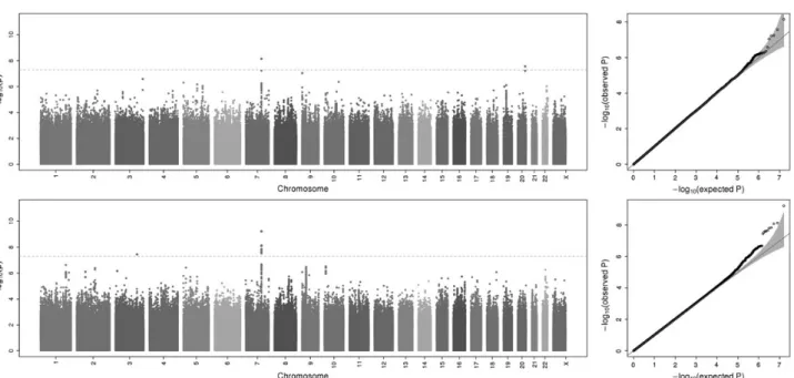
GWAS scans were performed for each ancestry group and meta-analysis was used to combine results across the six sam-ples. Overall, GWAS scans for DMFT and DMFS yielded multiple associations meeting genome-wide significance (i.e.P-values <5 × 10⁻⁸; Fig.1), and no evidence of genomic inflation was observed [see quantile–quantile(Q–Q) plots in Fig.1;λ= 1.006 and 1.017, re-spectively, for DMFT and DMFS] indicating that model assump-tions were adequately satisfied. For both dental caries phenotypes, the strongest association signal was observed for the single-nucleotide polymorphism (SNP) rs190395159 just up-stream of the geneNAMPT(P-value = 7.14 × 10⁻⁹for DMFT and
P-value = 5.97 × 10⁻¹⁰for DMFS; Fig.2A). This SNP (A/G) has a minor allele (G) frequency of 0.7% in HCHS/SOL sample, whereas in the 1000 Genomes Project the G allele is absent from Asian and South Asian populations, and occurs at frequencies of 19, 2 and ∼0% in African, American and European populations, respective-ly. Effect sizes for rs190395159 were similar within each of the six ancestry groups (Fig.3), and there was no evidence of heterogen-eity (Cochran’sQ P-value = 0.9). Multiple intronic SNPs within
NAMPT, which were uncorrelated with the top SNP rs190395159, showed associations havingP-values in the 1 × 10⁻⁷to 5 × 10⁻⁶ range; however, no associations with coding variants were ob-served. Of note, data from large-scale efforts to identify function-al non-coding elements [ENCODE (19), Roadmap Epigenomics (20) and FANTOM5 (21) projects] show that three SNPs (rs6947923, rs73409446 and rs73411544) in high-linkage disequi-librium (r2≥0.7) with rs190395159 occur within putative enhan-cers upstream ofNAMPT(Fig.4).
NAMPTis thought to be a pro-inflammatory adipokine, and increased serum levels ofNAMPThave been observed for a num-ber of diseases including obesity, metabolic syndrome, type 2 dia-betes and cardiovascular diseases (25). Though there is currently no direct evidence for the role ofNAMPTin dental caries, previous studies have implicatedNAMPTin supporting tissues. For ex-ample, gingivalfibroblasts have been shown to constitutively produceNAMPT, and to increase synthesis in response to oral bacteria (26,27). IncreasedNAMPTlevels have also been observed in gingival biopsies from patients with periodontal disease (26), andin vitrowork has shownNAMPTinhibits periodontal healing (28).NAMPThas also been shown to mediate osteoarthritic cartil-age destruction in a mouse model by regulating matrix-degrad-ing enzymes [matrix metalloproteinases (MMPs)] (29); MMPs, in turn, are known to be important for the progression of tissue damage in both dental caries and periodontitis (30). Overall, the confluence of several lines of evidence suggests thatNAMPTis expressed in the periodontal tissue, up-regulated in response to oral bacteria and in periodontitis patients, and operates through pro-inflammatory and pro-tissue destructive mechanisms. How
NAMPTmay affect dental caries is currently unknown, although its critical roles in epithelial-mesenchymal interaction and in-flammatory response in a variety of biological contexts supports its candidacy as a susceptibility gene.
Its possible role in dental caries notwithstanding, given that
NAMPTis a putative gene for periodontitis, which also ultimately leads to tooth loss, we tested the association of this locus with two additional caries-related phenotypes: DFS, which included only current decay and restorations, but not missing teeth, and MS, which included only missing teeth (reportedly lost due to dental caries or periodontitis). The purpose of testing these add-itional phenotypes was to explore whether tooth loss was driving the observed association. Indeed, MS showed strong evidence of association with this locus, whereas DFS did not (Fig.2B–C),
suggesting that the missing teeth component of DMFT and DMFS is largely responsible for the association. Because there is uncer-tainty regarding the cause of tooth loss we explored the possibil-ity that the association was due to periodontitis rather than dental caries by testing three measures of periodontal disease,
per se: (i) mean attachment loss, a quantitative phenotype, (ii) moderate periodontal disease, a dichotomous affection status defined by the Centers for Disease Control (CDC) and (iii) severe periodontal disease, a dichotomous affection status defined by the CDC. None of these three periodontal disease phenotypes showed association with theNAMPTlocus (results not shown).
Additional loci showing genome-wide significant evidence of association are summarized in Table2. One notable association was observed via meta-analysis between DMFT and rs72626594 (P-value = 2.75E−8), downstream ofBMP7. BMP7is a known tooth development gene (31,32), the deletion of which causes craniofa-cial manifestations including teeth and salivary gland defects in
mice (33). In humans, expression ofBMP7was down-regulated in dental pulp of human third molars that were experimentally injuredin vivo(in patients having the teeth extracted for orthodon-tic reasons within 15 days of injury) (34).BMP7has not previously been implicated in dental caries, but its role in promoting growth and repair of mineralized tissues is well-known. Indeed, BMP7 protein currently is used in orthopedic care, and its therapeutic po-tential for tooth repair/re-growth has been investigated, with mul-tiple animal studies showing that BMP7 (or crude BMP extract) stimulates dentin formation (35,36). It is currently unknown whether BMP7 is involved in dental caries, although plausible mechanisms by which genetic variation inBMP7may influence risk of dental caries include its role in tooth development or possible role in remineralizing pre-cavitated dental lesions.
Another associated locus observed in the meta-analysis of DMFS was the region on chromosome 3 spanning IGSF10, MIR5186,
MIR548H2andAADACL2(leading SNP rs138769355;P-value = 3.59E
Figure 2.Genetic association forNAMPTand (A) DMFS, (B) DFS and (C) MS. LocusZoom plots show the association (lefty-axis; log10-transformedP-values) with dental
caries-related phenotypes of SNPs in the region aroundNAMPT. The purple triangle represents the top SNP, rs190395159, from the genome-wide scans (which was
imputed with information quality score = 0.918 andR2= 0.947). Genotyped SNPs are indicated by circles and imputed SNPs are indicated by Xs. The blue overlay
represents recombination rate (righty-axis) from reference data. Positions of genes are indicated below the plot. Linkage disequilibrium (r2) values were calculated
from the HCHS/SOL sample.
−8). None of these genes/miRNAs have known functions that may impact dental caries. In the Mexican ancestry subsample, a few sig-nificant loci were observed, notablyANK3(leading SNP rs116717469,
P-value = 3.23E−8), a gene associated with bipolar disorder (37), which is in turn a risk factor for dental caries (38). Likewise, a region on chromosome 17 spanning several genes includingCACNA1Gwas observed (leading SNP rs71381322,P-value = 3.72E−8). Calcium ion channels are important for mineral homeostasis during tooth devel-opment, and disruptions of many ion channel genes lead to channe-lopathies affecting dentition (39). Genetic variants in some ion channels, but not specificallyCACNA1G, have been previously
shown to be associated with dental caries (Lewiset al.2014 ASHG ab-stract; manuscript under review). Moreover,in vitrostudies of the ef-fects ofBMP2on ameloblast differentiation showed up-regulation of
CACNA1G(40). Likewise,CACNA1Gwas up-regulated in mouse tooth germ in normal teeth compared withPPARα-associated hypoplastic teeth (41).
Other genome-wide significant associations were observed for Mexican and Puerto Rican groups in loci lacking genes with clear biological roles in oral health or in‘gene deserts’(Table2). Many suggestive associations, some in or near (based on physical proximity) genes with plausible biological roles, were also observed and are reported in the Supplementary Material.
The generalizability of genetic association results and effects of acculturation were addressed by repeating analyses while in-cluding years of residency in the USA and its interaction with age as predictors in our model. As indicators of acculturation, these variables were significantly associated with caries indices (P= 0.0086) and improved thefit of our model [change in Akaike’s information criterion (AIC) =−5.5]. Strong concordance among genetic associationP-values were observed for both DFMT and DMFS for models with and without adjustment for acculturation (see Supplemental Material). Top ranking SNPs and interpreta-tions of results were unchanged.
In addition to the quantitative caries indices, DMFS and DMFT, we also performed GWAS scans of a dichotomous caries pheno-type contrasting adults with and without any dental caries. The analysis across all groups recapitulated the association signal on chromosome 20 downstream of BMP7 (lead SNP rs62208680;
P-value = 8.20E−7), but not the signals on chromosomes 3 or 7 (see Supplementary Material).
Figure 4.Three SNPs, rs6947923, rs73409446 and rs73411544, in high-linkage disequilibrium (r2≥0.7) with the lead SNP (rs190395159) overlap with putative enhancers. The
rug plot shows positions relative toNAMPTof SNPs in linkage disequilibrium (indicated by color) with the lead SNP: red (r2= 1), blue (r2= 0.8), gold (r2= 0.7), light blue
(r2= 0.6) and gray (r2= 0.5
–0.4). Signal tracks are shown for biochemical signatures typical of upstream enhancer elements: DNase I hypersensitivity (22,23) (an
indicator of chromatin accessibility; blue), transcription factor CTCF binding (red), Histone mark H3k4me1 (yellow) and Histone mark H3K27ac (magenta). The
ENCODE (19) Txn factor ChIP track shows transcription factor-binding sites as gray boxes where the darkness of the shading is proportional to the maximum value
seen in any ENCODE cell line in that region. A green highlighted region indicates the highest scoring site of a Factorbook-identified (24) canonical motif for the
corresponding factor. The black boxes shown in the FANTOM5 enhancers track representin vivo-transcribed enhancers identified using cap analysis gene expression
in the FANTOM5 (21) project. HGF: human gingivalfibroblasts; HPdLF: normal human periodontal ligamentfibroblasts; AG09319: gum tissuefibroblasts from
apparently healthy 24-year old; PKFPC: penis foreskinfibroblast primary cells; CfMdMS: chondrocytes from bone marrow-derived mesenchymal stem cell cultured cells.
Figure 3.Forest plot for rs190395159.β-Values for DMFS models are shown for each ancestry group as squares proportional to the sample size. Error bars
indicate the 99.999995% (i.e. genome-wide significant) confidence intervals.
Discussion
In this study, we identified multiple novel genetic loci associated with dental caries. Moreover, this was thefirst GWAS of dental caries in a Hispanic and Latino sample (and thefirst GWAS of dental caries in a non-European ancestry sample) as well as the largest GWAS of dental caries, to date. The top association observed for both DMFS and DFMT was in the locus harboring the periodontal health-related geneNAMPT, which appeared to be due to the missing teeth component of the caries indices. Fur-thermore, the follow-up analyses suggested that this association was not due to periodontitis, but rather due to carious tooth loss. The up-regulation ofNAMPTin gingivalfibroblasts in response to oral bacterial provides a plausible mechanism by whichNAMPT
may influence dental caries (27,28). Another significant associ-ation was nearBMP7, a growth factor important for mineralized tissues. Both of these genes have plausible roles in the host re-sponse to carious injury. One could speculate that genes mitigat-ing the progression of active caries through a tooth could impact DMFT and DMFS scores even if their effects are in response to car-ies rather than in preventing initial decay. Such a mechanism is most plausible forNAMPTand BMP7based on their known biology.
Previous analyses have shown that the Hispanic and Latino groups included in the HCHS/SOL Study are heterogeneous with respect to their genetic composition (42,43) as well as their oral health status (17) and diet (44). Here, we showed that Hispan-ic and Latino groups also differ with respect to the cumulative role of genetics on dental caries experience (as indicated by the range of heritability estimates across ancestry groups and the lower heritability estimate in the total sample compared with all individual ancestry groups). While this study focused on the genetic risk factors common to all ancestry groups, the mélange of factors influencing susceptibility to dental caries and their relative effect sizes may differ across groups; additional work contrasting the ancestry groups is needed to extricate these differences.
To assess the role of acculturation on our analyses and deter-mine whether results were generalizable to other US Hispanic and Latino Americans, we repeated our analysis while including acculturation-related variables (i.e. number of years of residency in the USA and its interaction with age) in our model. Accultur-ation was indeed associated with dental caries indices, although genetic association results were robust to these effects. This
suggests that genetic associations identified in our study may be generalizable to other Hispanic and Latino populations. Given the admixed nature of our sample, genetic associations identified here may not be generalizable to other ancestry groups. For example, the minor allele of the top SNP rs190395159 up-stream ofNAMPTis absent or very rare in individuals of Euro-pean, Asian, and Southeast Asian ancestry and therefore its role in dental caries is precluded from these groups.
Strong a priori candidate genes such as the enamel matrix and related genes, as well as associated loci identified in previous GWAS studies, were notably absent from the list of associations observed in the HCHS/SOL samples. Moreover, associations iden-tified herein were not observed in previous GWAS (11–13). Expla-nations for this inconsistency include low power to detect weak genetic effects, heterogeneity across populations due to differ-ences in genetic composition or environment and the possibility of false-positive results from previous candidate gene and GWAS studies. Lack of replication is not entirely unexpected, as previous-ly identified associations are likeprevious-ly impacted by the‘winner’s curse’(e.g. the effect sizes were inflated in the data set in which they were discovered, which facilitated their discovery in the first place and explains subsequent failure to replicate similar ef-fects in other data sets). In fact, one of the challenges of the GWAS approach is the extensive multiple testing and need to accommo-date for this by setting a very lowP-value threshold for claiming statistical significance. Therefore, we anticipate identifying some number of false positives, and missing a large number of true as-sociations (i.e. many false negatives). Multi-marker tests and Bayesian methods that incorporate outside information about bio-logical functions of genes may be useful for mitigating these issues in the follow-up analyses. Studies of other diseases have also benefited from meta-analyses across large consortia, including or-ders of magnitude more participants than can be collected in any single study. Such an approach may also be possible for dental car-ies as genetic information is collected for more cohorts with dental phenotypes.
Another challenge in identifying the risk factors for dental car-ies is measuring the state of disease and defining a meaningful phenotype to analyze. Traditional DMFT and DMFS indices com-bine evidence of both active and past decay without distinguishing between the two, which is reasonable for a genetics study. How-ever, DMFT does not consider multiple lesions per tooth, and DMFS may misrepresent the number of carious surfaces if tooth extraction is used as a treatment option for otherwise restorable Table 2.Genetic loci significantly (P< 5 × 10⁻⁸) associated with dental caries indices
Sample Phenotype Chr. SNP BP Allele MAC β SE P-value Nearby gene(s)a
Metab DMFT 7 rs190395159 105 964 857 A 162 −1.24 0.21 7.14E−9 SYPL1, NAMPT
DMFT 20 rs72626594 55 535 521 G 65 2.61 0.47 2.75E−8 BMP7, MIR4325, SPO11
DMFS 3 rs138769355 151 367 491 A 36 −12.07 2.19 3.59E−8 IGSF10, MIR5186, MIR548H2, AADACL2
DMFS 7 rs190395159 105 964 857 A 162 −5.56 0.90 5.97E−10 SYPL1, NAMPT
Mexican DMFS 1 rs138642966 235 695 611 T 99 −11.01 1.96 1.94E−8 GNG4, LYST, B3GALNT2, TBCE, GGPS1,
ARIB4D
DMFS 10 rs116717469 62 237 360 C 39 −17.01 3.08 3.23E−8 ANK3, CDK1, RHOBTB1
DMFS 17 rs71381322 48 900 824 G 53 −15.15 2.75 3.72E−8 CACNA1G, ABCC3, ANKRD40, LUC7L3,
MIR8059, WFIKKN2, TOB1, SPAG9
DMFS 18 rs16946661 26 946 870 T 31 −19.23 3.50 4.02E−8 None
Puerto Rican DMFT X rs141563584 23 757 150 G 36 −2.90 0.53 3.89E−8 ACOT9, PRDX4, SAT1, APOO
aNearby genes were determined based on physical proximity (<400 kb) to the associated SNP while simultaneously considering the linkage disequilibrium structure in the
genomic region.
bMeta-analysis of all six sub-samples combined. Chr.: chromosome; BP: base pair position; allele: the effect allele in the model; MAC: minor allele count;β: the
lesions. Moreover, the missing tooth/surfaces components of these scores may conflate caries and periodontitis due to uncer-tainty in the reason for extraction. Therefore, both caries indices may misrepresent the true disease state to some degree. Another issue is that access to oral healthcare can counter-intuitively lead to increased DMFT and DMFS scores through aggressive restora-tions of pre-cavitated lesions and two-surface restorarestora-tions of in-terproximal lesions. Indeed, population surveys have shown that majority racial groups and those with higher socio-economic sta-tus or greater access to dental care often exhibit greater DMFT and DMFS indices than do minority racial or low SES groups. This is due to the greater number of restorations in the privileged groups, whereas disadvantaged groups may have lower overall DMFT and DMFS, although have more untreated decay and missing teeth (1). These and other sources of noise in the phenotype could reduce statistical power.
Conclusions
In thefirst GWAS of dental caries in Hispanic and Latino partici-pants, we discovered several novel associations including mul-tiple loci containing genes with compelling biological stories. This study demonstrated the utility of the GWAS approach, although additional work is required to fully identify and under-stand the genetic contributors of this multifactorial disease. Moreover, we showed evidence of heterogeneity across Hispan-ic/Latino groups, which suggests that future efforts investigating the differences among the groups, in addition to their similarities addressed herein, may benefit gene-mapping efforts for dental caries. Ultimately, understanding the factors influencing dental caries susceptibility may lead to improvements in prediction, prevention and disease management.
Materials and Methods
Participant recruitment and data collection
The HCHS/SOL is a multicenter prospective cohort study designed to investigate numerous indices of health and disease in the US Hispanic/Latino population. HCHS/SOL was funded by the Nation-al Heart, Lung and Blood Institute with support from six additionNation-al institutes including the National Institute of Dental and Craniofa-cial Research. Institutional Review Boards of all participating entities approved this study. All participants provided informed writ-ten consent. The present study reports cross-sectional analyses of the dental assessments collected at baseline.
Study recruitment followed a two-stage census block group-and household-based design in four US cities (Bronx borough of NYC, Chicago, Miami and San Diego) chosen based on the geo-graphical distribution and the place of origin of their respective Hispanic and Latino residents. Details regarding study design and recruitment of participants are available elsewhere (45,46). In all, 16 415 participants were recruited.
Intra-oral examinations by trained and calibrated examiners were performed for 15 848 participants based on the protocols used by the NHANES (1). In brief, dental examiners performed tooth-level and tooth surface-level assessments which included determining the presence or absence of each tooth, the reported cause of any missing teeth and the evidence of coronal decay and restorations of each tooth surface. Agreement among exami-ners was high for missing teeth (98%), decayed surfaces (99%) and decayed orfilled surfaces (86%) (17). Based on these assessments, several dental caries phenotypes were derived including indices that correspond to the counts of decayed, missing due to decay
or periodontal disease and restored (i.e.filled) Teeth, and tooth surfaces. DMFT and DMFS are the two most widely used indices of dental caries, capturing both current and past evidence of decay. To follow-up our results, we also considered the DFS index, corresponding to the count of decayed and restored sur-faces, and the MS index, corresponding to the count of missing tooth surfaces due to decay or periodontitis. All dental caries indi-ces were calculated while excluding the third molars, and indiindi-ces were not calculated for edentulous participants (4.1% of the sam-ple). In addition to quantitative dental caries phenotypes, we also considered a dichotomous measure of lifetime caries experience where participants with DMFS≥1 were defined as affected by caries, and participants with DMFS = 0 were defined as caries-free. For this caries presence/absence phenotype, edentulous indi-viduals were considered affected.
Genotyping, imputation and ancestry
A total of 12 803 participants providing consent for genetic ana-lyses were genotyped for over 2.5 million single-nucleotide poly-morphisms (SNPs) using a custom Illumina (San Diego) array consisting of the HumanOmni2.5-8v1-1 array content along with a panel of∼150 000 investigator-chosen SNPs. The custom content was selected to include ancestry-informative markers, variants distinctive of Amerindian populations and candidate polymorphisms. Genotyping was performed by Illumina Micro-array Services and all genotype data were extensively cleaned and quality-checked by Illumina Microarray Services, LA Biomed, and the SOL Genetic Analysis Center (GAC) at the University of Washington using analysis pipelines developed by the GAC (47). These analyses were standard for thefield and included scrutin-izing the participant samples for genetic sex, chromosomal anomalies, relatedness, population structure, Mendelian errors among relatives, concordance among duplicates, batch effects and genotyping call rates. SNP probes were scrutinized for poor performance in inter-sample comparisons (such as checks of Mendelian errors, concordance among sample duplicates etc.), missing call rates, separation of clusters during genotype calling, deviations from Hardy–Weinberg equilibrium and genotype con-cordance among duplicate probes. Recommended SNPfilters were established by the GAC based on quality control analyses.
components of ancestry that reflect the population structure free from the influence of the family structure and KC estimates ad-justed for ancestry.
Genetic analysis groups were defined using the self-identified Hispanic heritage group and thefirstfive PCs of ancestry in a multivariate outlier detection procedure based on the minimum covariant determinant estimation (42,52). The concordance be-tween self-reported ancestry and genetic ancestry was high (94–98% for each of the six ancestry groups). Nineteen partici-pants with genetically determined Asian ancestry and 37 Central American outliers with unusual ancestry were excluded from analyses. Genetic ancestry was used to stratify participants for genetic association analyses. The topfive principal components of ancestry were included in genetic association models.
Statistical analyses
Overview
GWAS scans of DMFT and DMFS were performed separately for six genetic analysis groups: Cuban (N= 1950), Dominican (N= 1083), Puerto Rican (N= 2006), Mexican (N= 4578), Central American (N= 1294) and South American (N= 843). Results from the six ancestry groups were combined via inverse variance-weighted meta-analysis. Cochran’sQwas used to test for hetero-geneity in SNP effects across samples. SNPs with minor allele counts of≥30 were included in analyses. The threshold for deter-mining statistical significance wasP-value <5 × 10⁻⁸. Cluster plots (of allele intensity data used for genotype calling) were visually scrutinized for all significant SNPs to confirm reliable genotyping. Given the strong correlational structure of the genome, this sig-nificance threshold may be considered conservative. Therefore, we also noted‘suggestive’loci havingP-values <1 × 10⁻⁷in the Supplemental Material.
Association models
The two caries indices, DMFT and DMFS, approximate continuous variables and were analyzed as such. A mixed-models approach was used to interrogate each SNP for evidence of association while adjusting forfixed effects, including sex, age, recruitment center, sampling weights andfive PCs of ancestry (calculated sep-arately for each group), and random effects, including census block group within each recruitment site, household and kinship. The distributions of DMFT and DMFS are non-normal; however, model residuals were approximately normal, indicating that the phenotypes were reasonably well-suited for our analytic frame-work. Environmental factors that may explain some phenotype variance (e.g. tobacco use, diet etc.) were not included in the mod-els. Note that such factors cannot confound the analyses because they do not alter constitutional genetic variants; however, by not modeling environmental effects, our GWAS scans may detect as-sociations that are mediated through environments (e.g. genes that influence diet and therefore could impact dental caries). This analysis strategy was chosen to cast the widest net possible given the multifactorial nature of the disease. Genomic inflation was estimated and visualized usingQ–Qplots. Analyses were per-formed in the R statistical environment (R Foundation for Statistic-al Computing, Vienna, Austria). Associated loci were visuStatistic-alized using LocusZoom (53).
Heritability estimation
Heritability was defined as the proportion of phenotype variance attributable to the observed kinship component from the‘base’
mixed-model, which accounts for the genetic relatedness among all participants in the sample or subsample. This is
similar to heritability estimates using the variance components approach in families, except here we estimate kinship from genomic data rather than using expected sharing due to known familial relationships.
Functional annotation
Identification of biochemical signals associated with non-coding functional elements was performed with the UCSC Genome Browser (https://genome.ucsc.edu/) using the GRCh37/hg19 assembly (54).
Sensitivity analysis and effect of acculturation
To explore the generalizability of results and address the possible role of acculturation on our analyses, we considered several vari-ables related to nativity and acculturation [short acculturation scale (55), MESA nativity subscore (56),first versus second gener-ation immigrant, the USA versus foreign born and years of resi-dence in the USA] by adding them individually to the association model described above. Based on change in AIC, we chose the number of years of residence in the USA and its interaction with age as indices of acculturation. Genetic association analyses were repeated while adjusting for these acculturation variables.
Supplementary Material
Supplementary Material is available atHMGonline.
Acknowledgements
We express our gratitude to the participants and staff of the HCHS/SOL study for their contribution to our goals of identifying the risk factors and protective factors impacting health and dis-ease in US Hispanic and Latino populations. We thank Marston Youngblood of the University of North Carolina at Chapel Hill for his role in managing and facilitating access to the data, Drs Jane Atkinson and Emily Harris of the National Institute for Den-tal and Craniofacial Research for their involvement in facilitating this work and Dr Bruce Weir of the University of Washington for his role in overseeing the Genetic Analysis Center and the devel-opment of the analysis pipelines utilized in this study.
Conflict of Interest statement. None declared.
Funding
References
1. Dye, B.A., Tan, S., Smith, V., Lewis, B.G., Barker, L.K., Thorn-ton-Evans, G., Eke, P.I., Beltran-Aguilar, E.D., Horowitz, A.M. and Li, C.H. (2007) Trends in oral health status: United States, 1988–1994 and 1999–2004. Vital Health Stat..Series 11, Data from theNationalHealthSurvey, 1–92.
2. Dye, B., Thornton-Evans, G., Li, X. and Iafolla, T. (2015) Dental caries and tooth loss in adults in the United States, 2011–
2012.NCHS Data Brief, 197.
3. Dye, B.A., Thornton-Evans, G., Li, X. and Iafolla, T.J. (2015) Dental caries and sealant prevalence in children and adoles-cents in the United States, 2011–2012.NCHS Data Brief, 1–8. 4. Boraas, J.C., Messer, L.B. and Till, M.J. (1988) A genetic
contri-bution to dental caries, occlusion, and morphology as de-monstrated by twins reared apart.J. Dent. Res.,67, 1150–1155. 5. Shaffer, J.R., Wang, X., Desensi, R.S., Wendell, S., Weyant, R.J., Cuenco, K.T., Crout, R., McNeil, D.W. and Marazita, M.L. (2012) Genetic susceptibility to dental caries on pit andfissure and smooth surfaces.Caries Res.,46, 38–46.
6. Wang, X., Shaffer, J.R., Weyant, R.J., Cuenco, K.T., DeSensi, R.S., Crout, R., McNeil, D.W. and Marazita, M.L. (2010) Genes and their effects on dental caries may differ between primary and permanent dentitions.Caries Res.,44, 277–284.
7. Fisher-Owens, S.A., Gansky, S.A., Platt, L.J., Weintraub, J.A., Soobader, M.J., Bramlett, M.D. and Newacheck, P.W. (2007) In-fluences on children's oral health: a conceptual model. Pediat-rics,120, e510–520.
8. Vieira, A.R., Modesto, A. and Marazita, M.L. (2014) Caries: re-view of human genetics research.Caries Res.,48, 491–506. 9. Opal, S., Garg, S., Jain, J. and Walia, I. (2015) Genetic factors
af-fecting dental caries risk.Aust. Dent. J.,60, 2–11.
10. Shaffer, J.R., Wang, X., Feingold, E., Lee, M., Begum, F., Weeks, D.E., Cuenco, K.T., Barmada, M.M., Wendell, S.K., Crosslin, D.R.
et al. (2011) Genome-wide association scan for childhood caries implicates novel genes.J. Den. Res.,90, 1457–1462. 11. Wang, X., Shaffer, J.R., Zeng, Z., Begum, F., Vieira, A.R., Noel, J.,
Anjomshoaa, I., Cuenco, K.T., Lee, M.K., Beck, J.et al.(2012) Genome-wide association scan of dental caries in the per-manent dentition.BMC Oral Health,12, 57.
12. Shaffer, J.R., Feingold, E., Wang, X., Lee, M., Tcuenco, K., Weeks, D.E., Weyant, R.J., Crout, R., McNeil, D.W. and Marazi-ta, M.L. (2013) GWAS of dental caries patterns in the perman-ent dperman-entition.J. Dent. Res.,92, 38–44.
13. Zeng, Z., Shaffer, J.R., Wang, X., Feingold, E., Weeks, D.E., Lee, M., Cuenco, K.T., Wendell, S.K., Weyant, R.J., Crout, R.et al.
(2013) Genome-wide association studies of pit-and-fissure-and smooth-surface caries in permanent dentition.J. Dent. Res.,92, 432–437.
14. Zeng, Z., Feingold, E., Wang, X., Weeks, D.E., Lee, M., Cuenco, D.T., Broffitt, B., Weyant, R.J., Crout, R., McNeil, D.W.et al.
(2014) Genome-wide association study of primary dentition pit-and-fissure and smooth surface caries.Caries Res.,48, 330–338.
15. Stanley, B.O., Feingold, E., Cooper, M., Vanyukov, M.M., Maher, B.S., Slayton, R.L., Willing, M.C., Reis, S.E., McNeil, D.W., Crout, R.J.et al.(2014) Genetic association of MPPED2 and ACTN2 with dental caries.J. Dent. Res.,93, 626–632. 16. Cruz, G.D., Chen, Y., Salazar, C.R. and Le Geros, R.Z. (2009) The
association of immigration and acculturation attributes with oral health among immigrants in New York City.Am. J. Public Health,99(Suppl. 2):S474–S480.
17. Beck, J.D., Youngblood, M. Jr., Atkinson, J.C., Mauriello, S., Kaste, L.M., Badner, V.M., Beaver, S., Becerra, K. and Singer,
R. (2014) The prevalence of caries and tooth loss among par-ticipants in the Hispanic Community Health Study/Study of Latinos.J. Am. Dent. Assoc.,145, 531–540.
18. Visscher, P.M., Hill, W.G. and Wray, N.R. (2008) Heritability in the genomics era—concepts and misconceptions.Nat. Rev. Genet.,9, 255–266.
19. Gerstein, M.B., Kundaje, A., Hariharan, M., Landt, S.G., Yan, K.K., Cheng, C., Mu, X.J., Khurana, E., Rozowsky, J., Alexander, R.et al.
(2012) Architecture of the human regulatory network derived from ENCODE data.Nature,489, 91–100.
20. Roadmap Epigenomics, C., Kundaje, A., Meuleman, W., Ernst, J., Bilenky, M., Yen, A., Heravi-Moussavi, A., Kheradpour, P., Zhang, Z., Wang, J.et al.(2015) Integrative analysis of 111 ref-erence human epigenomes.Nature,518, 317–330.
21. Andersson, R., Gebhard, C., Miguel-Escalada, I., Hoof, I., Born-holdt, J., Boyd, M., Chen, Y., Zhao, X., Schmidl, C., Suzuki, T.
et al.(2014) An atlas of active enhancers across human cell types and tissues.Nature,507, 455–461.
22. Thurman, R.E., Rynes, E., Humbert, R., Vierstra, J., Maurano, M.T., Haugen, E., Sheffield, N.C., Stergachis, A.B., Wang, H., Vernot, B.et al.(2012) The accessible chromatin landscape of the human genome.Nature,489, 75–82.
23. John, S., Sabo, P.J., Thurman, R.E., Sung, M.H., Biddie, S.C., Johnson, T.A., Hager, G.L. and Stamatoyannopoulos, J.A. (2011) Chromatin accessibility pre-determines glucocorticoid receptor binding patterns.Nat. Genet.,43, 264–268.
24. Wang, J., Zhuang, J., Iyer, S., Lin, X.Y., Greven, M.C., Kim, B.H., Moore, J., Pierce, B.G., Dong, X., Virgil, D.et al.(2013) Factor-book.org: a Wiki-based database for transcription factor-binding data generated by the ENCODE consortium.Nucleic Acids Res.,41, D171–D176.
25. Chang, Y.H., Chang, D.M., Lin, K.C., Shin, S.J. and Lee, Y.J. (2011) Visfatin in overweight/obesity, type 2 diabetes melli-tus, insulin resistance, metabolic syndrome and cardiovas-cular diseases: a meta-analysis and systemic review.
Diabetes Metab. Res. Rev.,27, 515–527.
26. Damanaki, A., Nokhbehsaim, M., Eick, S., Gotz, W., Winter, J., Wahl, G., Jager, A., Jepsen, S. and Deschner, J. (2014) Regula-tion of NAMPT in human gingivalfibroblasts and biopsies.
Mediat. Inflamm.,2014, 912821.
27. Nogueira, A.V., Nokhbehsaim, M., Eick, S., Bourauel, C., Jager, A., Jepsen, S., Cirelli, J.A. and Deschner, J. (2014) Regulation of visfatin by microbial and biomechanical signals in PDL cells.
Clin. Oral Invest.,18, 171–178.
28. Nokhbehsaim, M., Keser, S., Jager, A., Jepsen, S. and Desch-ner, J. (2013) Regulation of regenerative periodontal healing by NAMPT.Mediat. Inflamm.,2013, 202530.
29. Yang, S., Ryu, J.H., Oh, H., Jeon, J., Kwak, J.S., Kim, J.H., Kim, H.A., Chun, C.H. and Chun, J.S. (2015) NAMPT (visfatin), a direct target of hypoxia-inducible factor-2alpha, is an essential catabolic regulator of osteoarthritis. Ann. Rheum. Dis., 74, 595–602.
30. Sorsa, T., Tjaderhane, L. and Salo, T. (2004) Matrix metallopro-teinases (MMPs) in oral diseases.Oral Dis.,10, 311–318. 31. Helder, M.N., Karg, H., Bervoets, T.J., Vukicevic, S., Burger, E.
H., D’Souza, R.N., Woltgens, J.H., Karsenty, G. and Bronckers, A.L. (1998) Bone morphogenetic protein-7 (osteogenic pro-tein-1, OP-1) and tooth development.J. Dent. Res.,77, 545–554. 32. Tasli, P.N., Aydin, S., Yalvac, M.E. and Sahin, F. (2014) Bmp 2 and bmp 7 induce odonto- and osteogenesis of human tooth germ stem cells.Appl. Biochem. Biotechnol.,172, 3016–
3025.
and other ectodermal appendages of the orofacial complex.J. Exp. Zool. B Mol. Dev. Evol.,312B, 361–374.
34. Cevallos Gutierrez, F.A., Del Portillo Obando, P.D.P., Rodriguez Castillo, J.G. and Tibata Rodriguez, V.M. (2005) Expression Pat-terns of BMP7 of Healthy and Injured Human Teeth.J. Dent. Res.,84A, 0137.
35. Sloan, A.J., Rutherford, R.B. and Smith, A.J. (2000) Stimulation of the rat dentine-pulp complex by bone morphogenetic pro-tein-7 in vitro.Arch. Oral Biol.,45, 173–177.
36. Harichane, Y., Dimitrova-Nakov, S., Poliard, A., Veis, A., Den-Besten, P., Kellermann, O. and Goldberg, M. (2010) In Gold-berg, M. (ed),Amelogenins: Multifaceted Proteins for Dental and Bone Formation and Repair. Bentham Science Publishers, pp. 174–190.
37. Ferreira, M.A., O’Donovan, M.C., Meng, Y.A., Jones, I.R., Ruder-fer, D.M., Jones, L., Fan, J., Kirov, G., Perlis, R.H., Green, E.K.
et al.(2008) Collaborative genome-wide association analysis supports a role for ANK3 and CACNA1C in bipolar disorder.
Nat. Genet.,40, 1056–1058.
38. Friedlander, A.H., Friedlander, I.K. and Marder, S.R. (2002) Bi-polar I disorder: psychopathology, medical management and dental implications.J. Am. Dent. Assoc.,133, 1209–1217. 39. Duan, X. (2014) Ion channels, channelopathies, and tooth
for-mation.J. Dent. Res.,93, 117–125.
40. Miyoshi, K., Nagata, H., Horiguchi, T., Abe, K., Arie Wahyudi, I., Baba, Y., Harada, H. and Noma, T. (2008) BMP2-induced gene profiling in dental epithelial cell line.J. Med. Investig., 55, 216–226.
41. Sehic, A., Khuu, C., Risnes, S. and Osmundsen, H. (2009) Dif-ferential gene expression profiling of the molar tooth germ in peroxisome proliferator-activated receptor-alpha (PPAR-alpha) knockout mouse and in wild-type mouse: molar tooth phenotype of PPAR-alpha knockout mouse.Eur. J. Oral Sci.,117, 93–104.
42. Conomos, M.P., Laurie, C.A., Stilp, A.M., Gogarten, S.M., McHugh, C.P., Nelson, S.C., Sofer, T., Fernandez-Rhodes, L., Justice, A.E., Graff, M.et al.(2015) Genetic diversity and asso-ciation studies in U.S. Hispanic/Latino populations: Applica-tions in the Hispanic Community Health Study/Study of Latinos.Am. J. Hum. Genet., In Press.
43. Manichaikul, A., Palmas, W., Rodriguez, C.J., Peralta, C.A., Di-vers, J., Guo, X., Chen, W.M., Wong, Q., Williams, K., Kerr, K.F
et al.(2012) Population structure of Hispanics in the United States: the multi-ethnic study of atherosclerosis. PLoS Genet.,8, e1002640.
44. Siega-Riz, A.M., Sotres-Alvarez, D., Ayala, G.X., Ginsberg, M., Himes, J.H., Liu, K., Loria, C.M., Mossavar-Rahmani, Y., Rock, C.L., Rodriguez, B.et al.(2014) Food-group and nutrient-density
intakes by Hispanic and Latino backgrounds in the Hispanic Community Health Study/Study of Latinos.Am. J. Clin. Nutr., 99, 1487–1498.
45. Sorlie, P.D., Aviles-Santa, L.M., Wassertheil-Smoller, S., Ka-plan, R.C., Daviglus, M.L., Giachello, A.L., Schneiderman, N., Raij, L., Talavera, G., Allison, M.et al.(2010) Design and imple-mentation of the Hispanic Community Health Study/Study of Latinos.Ann. Epidemiol.,20, 629–641.
46. Bovbjerg, D.H. (2013) Genome-wide analysis of BMI in adoles-cents and young adults reveals additional insight into the ef-fects of genetic loci over the life course.J. Pain,22, 3597–3607. 47. Laurie, C.C., Doheny, K.F., Mirel, D.B., Pugh, E.W., Bierut, L.J., Bhangale, T., Boehm, F., Caporaso, N.E., Cornelis, M.C., Eden-berg, H.J.et al.(2010) Quality control and quality assurance in genotypic data for genome-wide association studies.Genet. Epidemiol.,34, 591–602.
48. Howie, B.N., Donnelly, P. and Marchini, J. (2009) Aflexible and accurate genotype imputation method for the next gener-ation of genome-wide associgener-ation studies.PLoS Genet.,5, e1000529.
49. Manichaikul, A., Mychaleckyj, J.C., Rich, S.S., Daly, K., Sale, M. and Chen, W.M. (2010) Robust relationship inference in gen-ome-wide association studies.Bioinformatics,26, 2867–2873. 50. Conomos, M.P., Miller, M.B. and Thornton, T.A. (2015) Robust
inference of population structure for ancestry prediction and correction of stratification in the presence of relatedness.
Genet. Epidemiol.,39, 276–293.
51. Conomos, M.P., Reiner, A.P., Weir, B.S. and Thornton, T.A. (2015) Model-free estimation of recent genetic relatedness.
Am. J. Hum. Genet., In Press.
52. Rousseeuw, P.J. and Van Driessen, K. (1999) A fast algorithm for the minimum covariance determinant estimator. Techno-metrics,41, 121–223.
53. Pruim, R.J., Welch, R.P., Sanna, S., Teslovich, T.M., Chines, P.S., Gliedt, T.P., Boehnke, M., Abecasis, G.R. and Willer, C.J. (2010) LocusZoom: regional visualization of genome-wide associ-ation scan results.Bioinformatics,26, 2336–2337.
54. Kent, W.J., Sugnet, C.W., Furey, T.S., Roskin, K.M., Pringle, T.H., Zahler, A.M. and Haussler, D. (2002) The human genome browser at UCSC.Genome Res.,12, 996–1006.
55. Marin, G., Sabogal, F., VanOss Marin, B., Otero-Sabogal, R. and Perez-Stable, E.J. (1987) Development of a short acculturation scale for Hispanics.Hispanic J. Behav. Sci.,9, 183–205. 56. Kandula, N.R., Diez-Roux, A.V., Chan, C., Daviglus, M.L.,