Molecular Determinants of Tbx20 Activity during
Cardiac Development
Erin Kaltenbrun
A dissertation submitted to the faculty of the University of North Carolina at Chapel Hill in partial fulfillment of the requirements for the degree of Doctor of Philosophy in
the Department of Biology.
Chapel Hill 2013
Approved by: Frank Conlon Kenneth Poss Jason Lieb
ABSTRACT
ERIN KALTENBRUN: Molecular Determinants of Tbx20 Activity during
Cardiac Development
(Under the direction of Dr. Frank L. Conlon)
The formation of the heart is a complex process that requires the
combinatorial activity of a number of critical cardiogenic transcription factors that function to drive distinct gene subprograms in the developing heart. Tbx20 is a potent activator of cardiac gene expression, and has also been shown to act as a transcriptional repressor in the heart. Precise regulation of Tbx20 expression levels and activity is crucial for normal heart development, as alterations in Tbx20 levels and activity lead to a spectrum of cardiac defects in humans and in animal models. Despite the importance of Tbx20 in heart development, very little is known about how Tbx20 expression is regulated in the heart. Further, the mechanisms by which Tbx20 acts to regulate its target genes are not understood. Here we explore the Tbx20 transcription network, both at the gene and protein level, to identify critical determinants of Tbx20 activity during heart development.
of Tbx20 in Xenopus, zebrafish, and mouse, indicating that this regulatory pathway is evolutionarily conserved among vertebrates. To begin to decipher how Tbx20 regulates its target genes, we have undertaken a proteomic screen of Tbx20 transcription complexes, both in human cells and mouse embryonic stem cell-derived cardiomyocytes. These studies resulted in the identification of a broad chromatin remodeling network that includes both co-activators and co-repressors. Specifically, Tbx20 directly recruits a Groucho/TLE and histone deacetylase repressor complex through an N-terminal eh1 domain. Additionally, Tbx20 recruits multiple chromatin remodelers including components of the SWI/SNF complex, the NuRD complex, and the INO80 complex. Collectively, these studies suggest that Tbx20 controls gene expression in the developing heart through selective
Acknowledgments
There are many people that have been critical to the success of my graduate studies. I would first like to thank current and past members of the Conlon Lab for helpful discussions, advice, support, and technical know-how: Elizabeth Mandel, Kathleen Christine, Chris Showell, Panna Tandon, Lauren Kuchenbrod, Lauren Waldron, Nirav Amin, Stephen Sojka, Kerry Dorr, Marta Charpentier, Chris Slagle, Michelle Villasmil, and Leslie Kennedy. Special thanks to Kathleen Christine and Chris Showell for being excellent mentors and friends my first few years in the lab, and to Kerry Dorr for being my lab BFF for the last five years. This experience would not have been the same without her friendship and unyieldingly positive attitude.
I cannot thank or acknowledge Frank Conlon enough. He has been so supportive of my career. He has been enthusiastic when I was down, always
high and there is a lot of camaraderie, qualities that have made coming to work a pleasure even when science wasn’t going so well.
I would also like to thank my committee members Bob Duronio, Jason Lieb, Steve Crews, and Ken Poss. One of the things that drew me to UNC was the faculty commitment to good training, and my committee has demonstrated this commitment throughout my graduate training. I am grateful for their insights and helpful
discussions on my work.
Table of Contents
LIST OF FIGURES………..xiii
LIST OF TABLES………..xv
LIST OF ABBREVIATIONS………xvi
CHAPTER 1. Introduction………..1
A CORE CARDIAC TRANSCRIPTION FACTOR NETWORK………4
Positive Regulation of cardiac cell fate: Evidence for a Transcription Factor Collective……….4
Nkx2.5………..5
Gata4………8
Tbx5………10
Tbx20………..12
Cardiac Transcription Factors and chromatin remodelers: Regulation at the chromatin level………….………...18
BAF Complexes………19
INO80 Complex………21
Histone Deacetylases………..22
Dissertation Goals………27
References………34
2. The BMP pathway acts to directly regulate Tbx20 in the developing heart………49
Preface………...49
ABSTRACT………50
INTRODUCTION………..51
MATERIALS AND METHODS………53
RESULTS………..59
A Tbx20-EGFP transgene recapitulates endogenous expression of Tbx20 in mid-tadpole stage embryos………...…59
A 334 bp regulatory element is sufficient for cardiac Tbx20 expression……….60
Tbx20 reporter expression is conserved in mouse and regulated by SMAD1/SMAD4 but not SMAD3………60
Tbx20 and SMAD1 are co-localized during cardiac chamber formation………...………....62
SMAD signaling is required for the maintenance of Tbx20 expression in vivo……….63
SMAD activation of Tbx20 occurs through direct binding of SMAD1………....63
Canonical SMAD sites alone are not sufficient for Tbx20 activation by SMAD1………...66
DISCUSSION………68
Tbx20 cardiac expression requires canonical and non-canonical SMAD1 binding sites………..………68
Cardiac-specific Tbx20 expression………70
ACKNOWLEDGMENTS………..72
REFERENCES……….91
3. The T-box transcription factor Tbx20 recruits a unique TLE-HDAC2-Tbx18 co-repressor complex………..95
Preface………...95
ABSTRACT………96
INTRODUCTION………..97
MATERIALS AND METHODS……….100
RESULTS………108
Tbx20-EGFP is localized to the nucleus and transcriptionally active…...………108
Directed proteomics of Tbx20-EGFP interactions reveals association with a unique transcription repression network……….109
Tbx20 forms protein complexes with TLE1/3, Tbx18, and HDAC2………114
Quantitative mass spectrometry reveals that Tbx20 recruits a Groucho-dependent repressive complex………..…...116
Endogenous Tbx20 interacts with TLE factors in mouse embryonic hearts………...………117
Tbx20 interacts with a Gro/TLE repressor complex……….119
Transcriptional repression by Tbx20 and Tbx18………...121
ACKNOWLEDGMENTS………124
REFERENCES………...154
4. A novel method to detect cardiac-specific Tbx20 protein interactions reveals association with a broad chromatin remodeling network…..……….160
ABSTRACT……….160
INTRODUCTION………161
MATERIALS AND METHODS……….163
RESULTS………167
Generation of a Tbx20Avitag knock-in mouse ESC line……….167
The Avitag-BirA system successfully isolates known Tbx20 protein-protein interactions….……….169
Tbx20 interacts with a chromatin remodeling network in cardiac progenitors……….………..170
DISCUSSION……….170
ACNOWLEDGMENTS………..174
REFERENCES………...182
5. Discussion and Future Directions………185
Tbx20 cardiac expression is regulated by BMP signaling………...186
A role for Groucho/TLE in cardiac development………...187
Future Directions………196
REFERENCES………...202
APPENDIX A1. Xenopus: An emerging model for studying congenital heart disease………211
Preface……….211
ABSTRACT……….211
INTRODUCTION………212
Xenopus as a Model System for Human Congenital Heart Disease………212
METHODS FOR STUDYING HEART DEVELOPMENT AND DISEASE IN XENOPUS………….………..214
Protein Depletion and Overexpression………..214
Xenopus Explants for Cardiogenic Assays………216
Xenopus Transgenesis………..219
CONGENITAL HEART DISEASE………...222
Atrial Septal Defects: Nkx2.5 and Gata4………...222
DiGeorge Syndrome: Tbx1………...………224
Holt-Oram Syndrome: Tbx5………..227
Spectrum of Congenital Heart Defects: Tbx20………..229
Noonan Syndrome: Shp-2………232
Heterotaxy and Cardiac Looping Defects: Zic3……….234
Axenfeld-Reiger Syndrome: Pitx2 and FoxC1………...237
FUTURE DIRECTIONS AND EMERGING TECHNOLOGIES IN
XENOPUS………...242
Investigating a Role for the Epicardium in Congenital Heart Disease………242
In Vivo Imaging of the Developing Xenopus Heart………...245
Protein Interactions and Biochemical Function……….247
Genetic Approaches in Xenopus tropicalis………248
REFERENCES………...251
A2. Immunoisolation of protein complexes from Xenopus……….275
Preface……….275
ABSTRACT……….276
INTRODUCTION………276
METHODS AND EQUIPMENT………277
METHODS AND PROCEDURES………283
Obtaining Xenopus laevis embryonic tissue………..283
Tissue lysis and protein extraction………..284
Cryogenic tissue disruption………..285
Optimization of lysis buffer and isolation conditions………….286
Immunoaffinity purification of protein complexes………..288
Conjugation of magnetic beads………288
Immunoaffinity purification: Basic elution of Immunoisolates………..291
Assessment of immunoaffinity purification: Sample
preparation………..296
Assessment of immunoaffinity purification: SDS-PAGE and western blot analysis………..297
Appropriate controls………...300
GFP-tagged……….300
FLAG-tagged………..301
Endogenous, non-tagged protein………301
NOTES……….302
List of Figures
Figure
1.1. The core cardiac transcription factor network………..29
1.2. The Nkx2.5 transcription network………..30
1.3. The Gata4 transcription network………31
1.4. The Tbx5 transcription network………..32
1.5. The Tbx20 transcription network………33
2.1. A regulatory element 5’ to the Tbx20 genomic locus is sufficient to drive gene expression in the cement gland and heart………74
2.2. A 334 bp regulatory element recapitulates the endogenous expression of Tbx20 throughout the X. laevis heart………76
2.3. XTbx20 5’ regulatory elements are activated by TGFβ/BMP signaling via SMAD1 and SMAD4 but not SMAD3………..78
2.4. XTbx20 is expressed throughout the myocardium and endocardium of the X. laevis heart………80
2.5. SMAD1 activation is required for cardiac specific expression of Tbx20 in X. laevis………81
2.6. SMAD1 binds to seven regions within the 334 bp Tbx20 regulatory element in vitro and occupies a combination of canonical and non-canonical SMAD1 binding sites in vivo………..83
2.7. SMAD1 activation is mediated through non-canonical SMAD1 binding sites………85
2.8. The Xenopus Tbx20 334 bp cardiac regulatory element is expressed in a cardiac-specific manner in zebrafish………..86
S2.2. Further deletion of the Tbx20(-334)-EGFP reporter leads to a decrease in activity in response to SMAD4 and an
increase in non-specific Tbx20 expression………..88 S2.3. SMAD1 inhibition during cardiac chamber differentiation
does not affect expression of the cardiac markers
tropomyosin and Tbx5……….89
3.1. Tbx20-EGFP is nuclear-localized and transcriptionally active………125 3.2. Directed proteomics of Tbx20-EGFP protein complexes
reveals association of Tbx20 with an HDAC-containing chromatin remodeling and Groucho transcriptional
protein network………...127
3.3. Tbx20 interacts with TLE1/3, HDAC2, and Tbx18………129 3.4. Tbx20 assembles a Groucho-Tbx20 repression complex
via the eh1 binding motif………...131
3.5. Endogenous Tbx20 interacts with TLE1/3 in mouse
embryonic hearts….……….…..133
4.1. Generation of Tbx20Avitag allele………175 4.2. Directed cardiac differentiation of Tbx20Avi; BirA ESCs
recapitulates normal cardiogenesis……….176 4.3. Endogenous Tbx20 is isolated from Tbx20Avi; BirA
cardiac progenitor cells……….178
4.4. Model of the cardiac Tbx20 chromatin remodeling network…………179 A2.1. Immunoisolation of protein complexes from Xenopus……….306 A2.2. Assessment of isolation efficiency and specificity of
List of Tables
Table
S2.1. ChIP Primer Sequences………..90 S2.2. Dissociation constants (Kd), standard deviation and
nucleotide sequence for each oligo analyzed in fluorescence
polarization studies………..90 3.1. Tbx20-associated proteins identified by LC-MS/MS……….…134
S3.1. Nuclear-enriched DNA-independent Tbx20 interactions from
three independent immunoisolations………..136 S3.2. Proteins excluded from nuclear-enriched DNA-independent
Tbx20 interactions identified in three independent isolations……….140 S3.3. GO analysis of nuclear-enriched interactions by
biological functions……….…148
S3.4. Enrichment analysis of nuclear-enriched Tbx20 associations………149 S3.5. Label-free quantitative mass spectrometric analyses of
nuclear-enriched Tbx20- and Tbx20eh1mut-associated
proteins………152
4.1. Tbx20 interacting proteins in cardiac progenitor cells (N=1)………..181 A1.1. Xenopus models of human congenital heart disease………..250 A2.1. Examples of detergents commonly used for cell lysis and
their properties………308
A2.2. Examples of lysis buffers used for immunoaffinity purification
List of Abbreviations
Affinity Purification- Mass Spectrometry AP-MS
Alpha-cardiac Myosin Heavy Chain 6 Myh6
Alpha/Beta-Myosin Heavy Chain α/β-MHC
Amino Enhancer of Split Aes
Atrial Natriuretic Factor ANF
Atrial Septal Defects ASD
Avitag Avi
Axenfeld-Rieger Syndrome ARS
B-type Natriuretic Peptide BNP
basic Helix Loop Helix bHLH
Bone Morphogenetic Protein BMP
BRG1-Associated Factor BAF
Cardiac alpha-actin αCA
Cardiac Troponin T cTnT
Coloboma, Heart Defects, choanal Atresia, Retarded growth and development,
Genital abnormalities, and Ear anomalies CHARGE
Congenital Heart Disease CHD
Connexin Cx
DiGeorge Syndrome DGS
Embryoid Bodies EBs
Embryonic day E
Engrailed Homology 1 eh1
Epicardium-derived cells EPDCs
Fibroblast Growth Factor FGF
Groucho Gro
Histone Deacetylase HDAC
Histone Methyltranserase HMT
Holt-Oram Syndrome HOS1
Human Embryonic Kidney HEK
Lymphoid Enhancer Binding Factor LEF
Metastasis-associated Protein MTA
Methyl-CpG Binding Domain Protein 3 MBD3
Morpholino MO
Myocyte Enhancer Factor 2 MEF2
Neuromancer nmr
Nucleolin NCL
Nucleophosmin NPM1
Nucleosome Remodeling and Deacetylase NuRD
Outflow Tract OFT
Polybromo- and BAF-containing PBAF
Pro-epicardium PEO
Restriction Enzyme Mediated Integration REMI
Serum Response Factor SRF
T-box containing protein Tbx Transcription Factor 3 TCF3
Transducin-like enhancer of split TLE
Transforming Growth Factor Beta TGFβ
Ventricular Septal Defects VSD
Wolf-Hirschorn Syndrome WHS
Xenopus Brachyury Xbra
Xenopus Dorsal Marginal Zone DMZ
Chapter 1
Introduction
The heart is one of the first structures to form during embryonic development,
and cardiac precursor cells are among the first cells of the epiblast to ingress during
gastrulation. Fate-mapping studies in mice have determined that prospective heart
mesoderm is localized in the anterior portion of the primitive streak (Lawson et al.,
1991; Parameswaran and Tam, 1995). In the early to mid-streak stages, heart
mesoderm progenitors begin to migrate from the anterior primitive streak to the
anterior proximal region of the epiblast (future anterior ventral midline of the embryo)
and come to lie underneath the head folds, forming a structure known as the cardiac
crescent (Parameswaran and Tam, 1995; Tam et al., 1997). By the late primitive
streak stages (E7-7.5), cells of the cardiac crescent begin to express the first
markers of heart mesoderm including Nkx2.5, Gata4/5, Tbx5, and Tbx20 (Arceci et
al., 1993; Komuro and Izumo, 1993; Horb and Thomsen, 1999; Carson et al., 2000).
Myocardial precursor cells undergo a number of sequential, but overlapping
processes to become terminally differentiated cardiomyocytes. Studies have
specification including the BMP, FGF, Hedgehog, and Wnt families (Reifers et al.,
2000; Zhang et al., 2001; Gadue et al., 2006; Foley et al., 2007; Kattman et al.,
2011). Integration of these signals by prospective cardiac precursors during
gastrulation and anterolateral migration triggers heart field patterning and
commitment to a myocardial fate. Expression of Mesp1, a bHLH transcription factor,
is transiently expressed in prospective cardiac mesoderm and is thought to act as a
molecular switch during cardiac specification by activating many of the key genes
within the core cardiac transcription network and repressing genes that promote
early mesoderm and endoderm cell fates (Bondue et al., 2008). Combinatorial
expression of the transcription factors Nkx2.5, Gata4/5, Tbx5, and Tbx20 within the
early heart field marks cardiac progenitors. Recent work in this field supports the
idea that these transcription factors are core components of a large regulatory
network that drives myocardial lineage development and morphogenesis of the early
embryonic heart. Additionally, the importance of Nkx2.5, Gata4/5, Tbx5, and Tbx20
as critical modifiers of cardiac cell fate and morphogenesis is underscored by the
identification of mutations within these genes that are associated with a variety of
congenital heart malformations in humans (Basson et al., 1997; Schott et al., 1998;
Pehlivan et al., 1999; Kirk et al., 2007). Therefore, it is critical to identify and
understand the protein-protein interactions, transcriptional targets, and upstream
regulators of these core cardiac transcription factors.
Here, we investigate the molecular regulators of Tbx20 expression and
factors, Tbx20 is unique in that it is expressed uniformly throughout all layers
(epicardium, myocardium, and endocardium) of the forming heart during
development and adulthood. Additionally, this expression pattern is conserved from
flies to humans implying a fundamental role for Tbx20 in proper heart development
and function (Ahn et al., 2000; Griffin et al., 2000; Meins et al., 2000; Iio et al., 2001;
Brown et al., 2003; Huang et al., 2012; Sakabe et al., 2012). We provide evidence
that BMP signaling is required to maintain Tbx20 expression in the heart during
cardiac chamber formation, suggesting a critical role for the BMP pathway in driving
proper regionalization of cardiac chambers (Mandel et al., 2010). We also show that
Tbx20 interacts with a unique transcription repression network that includes
components of both the Nucleosome Remodeling and Deacetylase (NuRD) complex
and Groucho/TLE co-repressors. We predict that the Tbx20 repression network is
essential to prevent inappropriate gene activation within the developing heart.
Finally, we have developed a novel approach to identify endogenous
cardiac-specific Tbx20 interaction partners over the course of cardiomyocyte development,
which has resulted in the discovery of new components of the Tbx20 regulatory
network. First, I will introduce the current understanding of relationships within the
core cardiac transcription factor network, highlighting the roles of critical activating
and repressing transcription factor protein complexes in specifying and maintaining
A CORE CARDIAC TRANSCRIPTION FACTOR NETWORK
In addition to the core factors Mesp1, Nkx2.5, Tbx5, Tbx20, and Gata4, other
transcription factors are expressed and have critical functions during cardiogenesis.
In particular, MEF2 factors and SRF, additional T-box proteins including Tbx1, Tbx2,
Tbx3, and Tbx18, as well as the homeodomain protein Isl1 have each been shown
to be important for proper heart development (Lin et al., 1997; Wang et al., 2001a;
Vitelli et al., 2002; Cai et al., 2003; Harrelson et al., 2004; Hoogaars et al., 2004;
Christoffels et al., 2006). Collectively, these transcription factors act in
interconnected pathways to regulate the expression of each other, as well as
downstream gene targets to control heart development. The transcription factors
Nkx2.5, Tbx5, Tbx20, Gata4, and Mef2C also physically interact with each other,
implying that the transcriptional control of heart development is directed by
cardiogenic transcription factors that are linked genetically and biochemically via
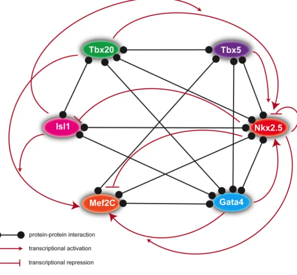
large multimeric protein complexes (Figure 1.1) (Morin et al., 2000; Hiroi et al., 2001;
Sepulveda et al., 2002; Stennard et al., 2003; Vincentz et al., 2008; Munshi et al.,
2009; Junion et al., 2012).
Positive regulation of cardiac cell fate: Evidence for a Transcription Factor Collective
Over the last two decades, studies regarding the relationships between
cardiac transcription factors support the notion that core cardiac transcription factors
promote specification, proliferation, and differentiation of cardiac precursor cells, as
well as morphogenetic movements of the primitive heart tube, via protein-protein
protein-protein interactions appear to be critical to ensure activation of multiple cardiac genes, and thus point to a “transcription factor collective” model in which cardiogenic transcription factors bind to and activate cardiac enhancers
cooperatively. I present the following examples in support of this model:
Nkx2.5
Nkx2.5 encodes a homeobox transcription factor that was originally cloned from Drosophila (msh-2, tinman) in a screen looking for new mesoderm-specific homeobox genes (Bodmer et al., 1990). Tinman is expressed in mesoderm
than repression of XNkx2.5 alone with a complete loss of differentiated myocardium (Fu et al., 1998; Grow and Krieg, 1998). This finding suggests that there may be some functional redundancy among the NK/tinman family members and helps to explain why Nkx2.5 knockout mice are able to generate a primitive heart tube. Early studies defined Nkx2.5 as a transcriptional activator capable of binding novel
homeodomain sites as well as sites that resemble serum response elements (Chen and Schwartz, 1995), which serve as binding sites for the MADs box transcription factor SRF (Treisman, 1986).
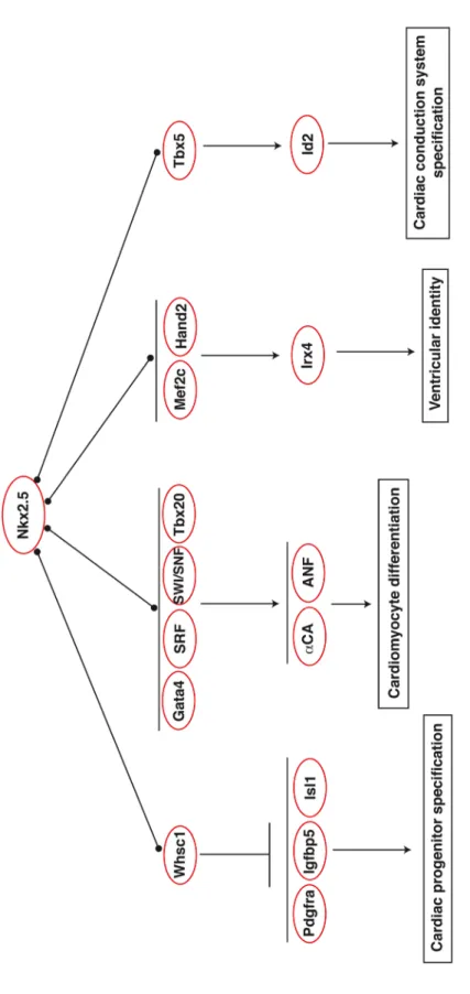
promote ventricular differentiation (Yamagishi et al., 2001). Nkx2.5-/-; Hand2 -/-mutants have a single atrial chamber, and expression of Irx4, a ventricle-specific gene, is completely abolished, thereby suggesting that Nkx2.5 and Hand2 promote ventricle differentiation in part through cooperative regulation of Irx4 (Yamagishi et al., 2001). Aside from its role in promoting normal differentiation and morphogenesis of chamber myocardium, Nkx2.5 is also critical for development of the cardiac
conduction system. Nkx2.5 dosage is directly proportional to the number of cells in the cardiac conduction system pointing to a role for Nkx2.5 in promoting the genetic conduction program (Jay et al., 2004). Nkx2.5 was later found to promote
specification of ventricular myocytes into ventricular conduction system cells through a genetic interaction with Tbx5, which together cooperatively regulate Id2 and other conduction system genes (Moskowitz et al., 2007). Collectively, these studies demonstrate that interaction of Nkx2.5 with other cardiogenic transcription factors is critical for proper regulation of downstream cardiac genes, and subsequent
activation of the appropriate cardiac gene program (Figure 1.2). The choice of protein co-factor may also provide an additional level of specificity for Nkx2.5-mediated gene regulation.
thus far, most of which are truncations or missense mutations in the homeodomain, are predicted to augment DNA binding. These data suggest that the principal
determinant of the congenital heart defects associated with NKX2.5 mutations is the total dosage of NKX2.5 capable of binding to DNA (Schott et al., 1998; Kasahara et al., 2000; Kasahara and Benson, 2004).
Gata4
Gata4 is a member of the GATA transcription factor family, which contains two zinc fingers that are required for binding to the GATA binding sequence [(A/T)GATA(A/G)] (Molkentin, 2000). Gata4 was identified in a screen of a mouse embryo cDNA library searching for new GATA-binding factors and was enriched in heart tissue (Arceci et al., 1993). A second group independently identified Gata4 in a Xenopus tadpole liver cDNA library and demonstrated that Gata4 is expressed in presumptive cardiac ventral mesoderm (Kelley et al., 1993). In the primitive heart, Gata4 is expressed in the developing atria and ventricles, as well as the
endocardium. Expression in the heart persists through gestation and after birth (Kelley et al., 1993; Heikinheimo et al., 1994). Early studies in cell culture models of cardiac differentiation suggest that Gata4 plays a role in cardiomyocyte
the cardiomyocytes fail to migrate to the ventral midline and instead generate cardiac structures in the dorsolateral regions of the embryo indicating that Gata4 is essential for migration of the presumptive heart fields (Kuo et al., 1997; Molkentin et al., 1997).
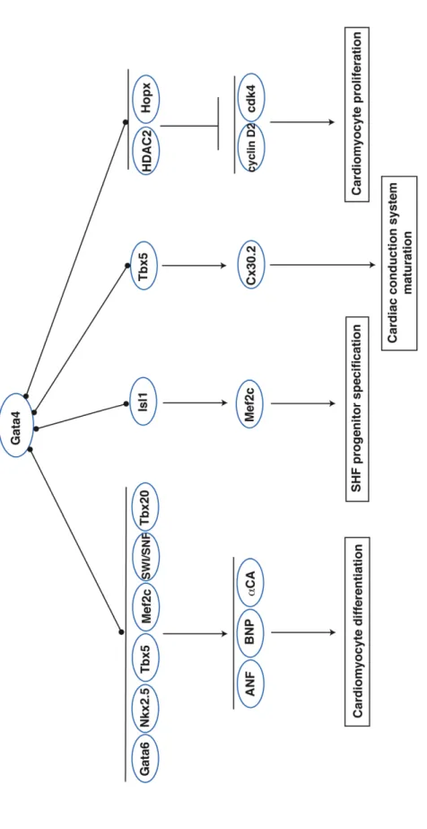
Gata4 acts as a potent transactivator of cardiac genes (Figure 1.3). Gata4 physically interacts with Gata6 in the myocardium to cooperatively activate
importance of the GATA4-TBX5 interaction in human heart development and supporting the idea that disruption of the core cardiac transcription factor network leads to major cardiac anomalies (Garg et al., 2003). GATA4 haploinsufficiency also leads to congenital heart defects in humans: patients with monosomy 8p23.1
(deletion of the distal arm of chromosome 8p) and congenital heart disease were found to have deletions at the GATA4 locus (Pehlivan et al., 1999). A del(8)(p23.1) patient who lacked cardiac anomalies did not have a GATA4 deletion, indicating that GATA4 deficiency directly contributes to congenital heart disease (Pehlivan et al., 1999).
Tbx5
mutations revealed that different mutations are predicted to perturb distinct target DNA interactions (binding to the major groove of DNA versus the minor groove), indicating that TBX5 may interact with DNA differently in the developing heart compared to the limbs (Basson et al., 1999). In vitro binding assays have identified an octamer sequence [AGGTGTG(A/G)] to which Tbx5 binds that is part of the Brachyury consensus half site (Ghosh et al., 2001; Macindoe et al., 2009). Tbx5 can also bind the full palindromic Brachyury binding site indicating there is some
flexibility in Tbx5 target sequence grammar (Ghosh et al., 2001).
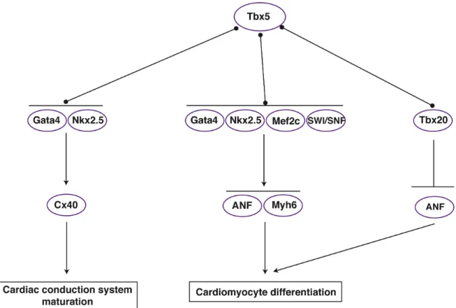
Gata4 to synergistically promote ANF/Nppa expression (Bruneau et al., 2001; Hiroi et al., 2001; Garg et al., 2003) and modulates Cx40 expression in combination with Nkx2.5 and Gata4 (Linhares et al., 2004). Additionally, Tbx5 and Gata4 genetically interact; mice heterozygous for both alleles have atrioventricular septal defects and myocardial thinning, defects distinct from the isolated ASD observed in Tbx5
heterozygotes (Maitra et al., 2009). Tbx5 also forms a complex with Mef2C on the alpha-cardiac myosin heavy chain (Myh6) promoter to drive Myh6 expression and normal heart patterning (Ghosh et al., 2009). Collectively, these studies have identified Tbx5 as a crucial node within the cardiac transcription network (Figure 1.4). Recent studies have continued to investigate the functions of Tbx5 in different cardiac cell populations. This work has revealed an essential function for Tbx5 in directing ventricular and atrial septation (Takeuchi et al., 2003; Xie et al., 2012), specifying the proepicardium (Liu and Stainier, 2010), patterning of the conduction system (Moskowitz et al., 2004; Moskowitz et al., 2007), and regulating cardiac cell cycle progression (Goetz et al., 2006). Further studies are required to explore how Tbx5 intersects with other transcription factor pathways to regulate these diverse processes.
Tbx20
of these species, Tbx20 transcipts are enriched in the anterior lateral plate
mesoderm and gradually become restricted to the cardiac primordial prior to ventral migration. Expression is maintained in the primary heart field throughout the process of migration, looping, and cardiac chamber formation. Thus, Tbx20 is expressed at the same time, and in many of the same regions as the transcription factors Tbx5, Nkx2.5, and Gata4. Morpholino knockdown of the Tbx20 protein in Xenopus embryos results in a loss of cardiomyocytes and unlooped hearts, but cardiac specification and migration proceeds normally (Brown et al., 2005). In agreement with these studies, deletion of Tbx20 in mice also results in a loss of
cardiomyocytes, failure to loop, and defects in cardiomyocyte maturation and chamber specialization (Cai et al., 2005; Singh et al., 2005; Stennard et al., 2005; Takeuchi et al., 2005). Moreover, the role of Tbx20 in heart formation appears to be evolutionarily ancient, with Drosophila having two Tbx20 orthologues, neuromancer (nmr1) and neuromancer2 (nmr2), which are also referred to as H15 and midline, respectively. Like Tbx20, this pair of genes is required for proper development of the dorsal vessel, a structure thought to be homologous to the vertebrate heart
(Miskolczi-McCallum et al., 2005; Qian et al., 2005; Reim et al., 2005).
Recent studies have identified a number of patients with dilated
missense mutations within the T-box domain, and two are identical mutations in unrelated individuals. Of the remaining four TBX20 mutations, one is a missense mutation leading to truncation of TBX20 in the T-box domain and the other three are missense mutations mapping to two different regions of unknown function.
Interestingly, one of the missense mutations in the T-box domain is a
gain-of-function mutation that enhances transcriptional activity and increases occupancy of DNA (Posch et al., 2010). These genetic studies imply that precise regulation of both TBX20 expression and transcriptional activity is critical for normal heart
development.
Similar to Tbx5, the optimal binding site for Tbx20 corresponds to a T-half-site (Macindoe et al., 2009). The Tbx20 binding site differs from that of Tbx5 in that it does not display any variation in the last nucleotide position (AGGTGTGA). Additionally, Tbx5 and Tbx20 exhibit different binding affinities and kinetics on T-half-sites, thus implying that there may be some degree of T-box competition on cardiac gene promoters (Macindoe et al., 2009).
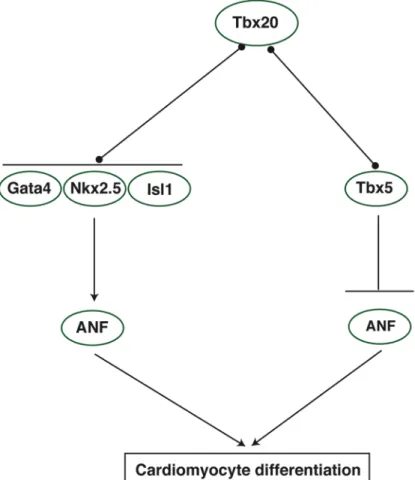
transcriptional activity is enhanced in the presence of Gata4/5 and Nkx2.5 (Posch et al., 2010) Consistent with these data, a recent study identified GATA and NKX binding motifs in Tbx20-bound genomic regions. Further, these binding motifs were over-represented in Tbx20-bound regions associated with genes that were down-regulated in Tbx20 knockout hearts, indicating that these genes represent targets of Tbx20-mediated activation (Sakabe et al., 2012). Tbx20 also interacts with Tbx5, and this interaction may be important for the regulation of genes associated with cell polarity or adhesion as depletion of Tbx20 and Tbx5 protein from Xenopus embryos results in severe cardiac defects compared to single morphants but cardiomyocyte marker expression is maintained (Brown et al., 2005). The effect of protein-protein interactions on Tbx20 transcriptional activity appears to be context- and dose-dependent. For instance, in the presence of Nkx2.5 and Gata4, Tbx20 activates the ANF/Nppa promoter; however, in the presence of Tbx5, Tbx20 appears to repress ANF/Nppa activation by Tbx5 in a dose-dependent manner. These results suggest that Tbx20 functions as both a co-activator and co-repressor of cardiac transcription factors (Figure 1.5), and this decision may be based on local levels of Tbx20 as well as on protein partner choice (Plageman and Yutzey, 2004; Brown et al., 2005). More studies are needed to shed light on this discussion. In particular, very little is known about the role of Tbx20 as a transcriptional repressor.
“enhanceosome model” suggests that transcription factors are recruited and bind to the enhancer as an intact complex. In this model, the exact positioning of bound transcription factors within the complex is important, as the complex must bind to a very specific motif grammar within the enhancer (Arnosti and Kulkarni, 2005). The second model, called the “billboard model” presupposes that each transcription factor is recruited independently to the cardiac enhancer and binds to its own sequence motif. This model predicts that cardiac enhancers will contain individual binding motifs for all bound transcription factors (Arnosti and Kulkarni, 2005). A recent study assessed genome-wide cardiac transcription factor binding in
cardiomyocytes, specifically examining regions bound by the factors Nkx2.5, Gata4, Tbx5, SRF, and Mef2A (He et al., 2011). The authors found that regions bound by one transcription factor were often highly enriched for binding motifs of factors that are known to interact with the first transcription factor. Of the chromatin regions bound by multiple transcription factors, many were enriched for genes that are expressed in a cardiac-specific manner and represent bona fide cardiac enhancers, confirming that binding of multiple cardiac transcription factors is predictive of
factors, they found much higher enrichment of Doc and Pnr motifs compared to the other transcription factor motifs. These data indicate that Doc and Pnr may bind in a sequence-specific manner and other cardiac transcription factors may be recruited independently of sequence grammar (Junion et al., 2012). These studies have led to the development of a third enhancer model, the “transcription factor collective
model”. This model suggests that cardiogenic transcription factors cooperatively activate cardiac enhancers; however, binding of the cohort of factors does not require a specific sequence motif. This model suggests that it may not be necessary for all of the factors to directly bind the enhancer. Instead, it may suffice for a subset of the transcriptions factors to bind their sequence motifs, which results in
recruitment of the rest of the complex via protein-protein interactions with the DNA-bound transcription factors.
Collectively, these studies reveal the complexity of transcription factor pathways within the cardiac regulatory network. Mutations in many of the core transcription factors in this network have been identified in humans, and these mutations have been linked to a variety of cardiac malformations (reviewed in Table A1.1). These data emphasize the importance of understanding how these
Though recent studies have demonstrated that the adult heart retains some
regenerative capacity, this capacity is extremely limited, and a large portion of adult cardiomyocytes are quiescent and incapable of generating new cardiomyocytes to replace injured myocardium (Bergmann et al., 2009; Kajstura et al., 2010; Walsh et al., 2010; Senyo et al., 2013). Recent work has demonstrated that the addition of Gata4, Mef2c, and Tbx5 to adult cardiac fibroblasts is sufficient to reprogram these cells to cardiomyocytes, in vitro and in vivo (Ieda et al., 2010; Qian et al., 2012). This provides additional evidence that cardiac cell fate is driven by the combinatorial activities of multiple core transcription factors and highlights the human health applications if the relationships between these factors are precisely defined.
Cardiac transcription factors and chromatin remodelers: Regulation at the chromatin level
BAF Complexes
The SWI/SNF ATP-dependent chromatin remodeling complexes PBAF and BAF are large protein complexes that share eight common subunits. The PBAF complex is distinguished from BAF by the presence of the unique subunit Baf180 (Xue et al., 2000), whereas BAF complexes contain Baf250 (Nie et al., 2000). SWI/SNF complexes facilitate chromatin remodeling through nucleosome
mobilization and allow transcription factors to access the DNA template (Kwon et al., 1994). Both SWI/SNF complexes have been demonstrated to be required for proper heart development (Lickert et al., 2004; Wang et al., 2004). Ablation of the PBAF subunit Baf180 results in hypoplastic ventricles and ventricular septal defects indicating that the PBAF complex is required for ventricular chamber development (Wang et al., 2004). Similarly, knockdown of Baf60c, a subunit common to both BAF complexes, results in impaired expansion of the early heart fields and defects in cardiomyocyte maturation (Lickert et al., 2004). Interestingly, expression of Baf60c with Tbx5, Nkx2.5, and Gata4 increased ANF/Nppa reporter activity compared to control in the presence of just the transcription factors, suggesting that BAF
cardiac genes, leading to the hypothesis that interactions between transcription factors and chromatin remodeling complexes may be a common feature of heart-specific chromatin activation.
Several groups have characterized the role of Brg1 in cardiac gene regulation (Stankunas et al., 2008; Hang et al., 2010; Takeuchi et al., 2011). Endocardial-specific deletion of Brg1 leads to trabeculation defects that arise as a result of
derepression of the secreted matrix metalloproteinase, ADAMTS1 (Stankunas et al., 2008). This study suggests that BAF complexes may be involved in both activation and repression of gene expression in the heart. Indeed, a later study examining the role of Brg1 in the myocardium found that Brg1 directly represses α-MHC, the MHC isoform that is predominantly expressed in adult heart, thereby maintaining
myocardial cells in an embryonic state (Hang et al., 2010). The authors also showed that Brg1 interacts with and requires HDACs and PARPs to transcriptionally repress α-MHC. Interestingly, Brg1 also activates β-MHC, the MHC isoform present in embryonic myocardium, indicating that Brg1 regulates parallel pathways to control MHC isoform expression (Hang et al., 2010). It is unclear precisely how
BAF/HDAC/PARP complexes are recruited to the MHC promoter; however, one potential mechanism is recruitment by DNA sequence-specific cardiac transcription factors. One recent study supports this hypothesis and demonstrated a genetic interdependence between cardiac transcription factors and BAF complexes.
(Takeuchi et al., 2011). Additionally, Brg1 occupancy at the ANF/Nppa and Gja promoters was markedly reduced in a Tbx5 heterozygous background, and further reduced in a Tbx5; Brg1 double heterozygous background, indicating that BAF complex occupancy of cardiac promoters relies on interactions with cardiac transcription factors on the target promoter (Takeuchi et al., 2011).
INO80 Complex
The INO80 chromatin remodeling complex is a very large protein complex with 11 to 16 members including the ATP-dependent helicases Ino80 and SRCAP, the DNA helicases Pontin (Ruvbl1, Tip49, Tip49a) and Reptin (Ruvbl2, Tip48, Tip49b), actin, and various actin-related proteins (Arp4, Arp5, Arp8, β-actin, Arp7, Arp9) (Shen et al., 2000; Jin et al., 2005). The INO80 complex alters chromatin accessability resulting in activation or repression of target genes (Cai et al., 2007; Ford et al., 2007; Klopf et al., 2009). Two members of the complex, Pontin and Reptin, have opposing acitivities within the INO80 complex, and this antagonistic relationship has been shown to play a role in cardiac growth in zebrafish (Rottbauer et al., 2002). An ENU-induced mutation in Reptin leads to cardiac hyperplasia and embryonic lethality. This mutation is an activating mutation in Reptin, increasing the ATPase activity of Reptin complexes, thus overriding Pontin diminution of Reptin activity, and increasing the transcriptional repressor activity of the complex. Mutant zReptin was subsequently shown to have a stronger repressive effect on
D and c-Myc to control the balance between proliferation and differentiation in the developing zebrafish heart (Rottbauer et al., 2002). Further studies are needed to investigate the role of the INO80 complex in mammalian heart development and to determine how Pontin, Reptin, or other members of the INO80 complex might intersect with additional transcription factor pathways in the heart.
Histone Deacetylases
HDACs are a class of histone-modifying enzymes that promote chromatin condensation by removing acetyl groups from conserved lysine residues of histone tails, resulting in transcriptional repression (Vidal and Gaber, 1991; Vidal et al., 1991; Rundlett et al., 1996; Hassig et al., 1997; Rundlett et al., 1998). HDACs
interact with a variety of DNA-binding transcription factors and are often components of larger repression complexes (Ayer et al., 1995; Heinzel et al., 1997; Zhang et al., 1997; Wade et al., 1998). HDACs are divided into 3 classes based on their
homology with the 3 yeast HDACs: class I HDACs consist of HDAC1, -2, -3, and-8, class II HDACs include HDAC4, -5, -7, and -9, and class III HDACs, which are termed sirtuins (Ekwall, 2005; Haberland et al., 2009). Several HDAC proteins have been shown to be critical for transcription repression during normal heart
development (Zhang et al., 2002; Chang et al., 2004; Montgomery et al., 2007; Montgomery et al., 2008; Trivedi et al., 2008; Trivedi et al., 2010).
HDAC1 and HDAC2 have partially redundant roles in developing
cardiac phenotype; however, mice mutant for both HDAC1 and -2 die neonatally of cardiac arrhythmias and dilated cardiomyopathy (Montgomery et al., 2007). Heart failure in these mice likely results from upregulation of calcium channel and contractile genes, indicating that HDAC1 and -2 function to negatively regulate genes involved in calcium flux and contraction. Additional studies are needed to address the mechanism by which HDAC1 and -2 are targeted to cardiac genes. Interestingly, HDAC2 also regulates Gata4 transcriptional activity via deacetylation its lysine resides, suggesting that Gata4 is a non-histone target of HDAC2 in the heart (Trivedi et al., 2010). HDAC2 deacetylation of Gata4 requires the adaptor protein Hopx and results in a suppression of Gata4-dependent transactivation of cyclinD2 and cdk4. Consistent with the requirement for HDAC2 in Gata4-mediated regulation of cell cycle genes, HDAC2; Hopx knockout hearts display increased cardiomyocyte proliferation indicating that HDAC2, Hopx, and Gata4 interact to regulate myocyte proliferation (Trivedi et al., 2010). It will be interesting to determine if HDACs are responsible for deacetylating other transcription factors in the heart.
Myocardium-specific deletion of HDAC3 results in lethality at 3-4 months of age due to cardiac hypertrophy and severe defects in cardiac metabolism,
HDAC3 may also be important for promoting postnatal cardiomyocyte proliferation, as myocardium-specific overexpression of HDAC3 results in increased
cardiomyocyte proliferation and inhibition of cyclin-dependent kinase inhibitors (Trivedi et al., 2008). Interestingly, the effects of HDAC3 overexpression on cardiomyocyte proliferation are limited to birth until 2 months of age, when
proliferation returns to normal. It is unclear whether this occurs because the neonatal cardiomyocytes are no longer competent to respond to proliferation cues, or is a result of temporal HDAC3 activity on cell cycle genes. The mechanism by which HDAC3 activity in the heart is regulated has yet to be determined; however, it is likely that HDAC3 exists as part of a larger repressive complex that is recruited temporally in a tissue-restricted manner by tissue-specific DNA-binding factors.
Similar to the coordinated functions of HDAC1/2, the class II HDACs HDAC5 and -9 redundantly regulate embryonic cardiac growth and hypertrophy. Mouse embryos that are double null for HDAC5 and HDAC9 begin to die at E15.5 with ventricular septal defects and thinned ventricular walls, highlighting a role for these HDACs in cardiac growth (Chang et al., 2004). Mice lacking only HDAC5 or HDAC9 are born in normal Mendelian ratios but go on to display age-dependent hypertrophy indicating a defect in the cardiac stress response (Zhang et al., 2002; Chang et al., 2004). Indeed, mice mutant for either HDAC5 or -9 develop enlarged hearts in
and these interactions result in the recruitment of HDACs to MEF2 target genes (McKinsey et al., 2002). In particular, one isoform of HDAC9 termed
MEF2-interacting transcription repressor (MITR) encodes a truncated form of HDAC9 that does not have HDAC activity but inhibits MEF2 target genes by association with other HDACs and transcriptional repressors (McKinsey et al., 2002). The precise role that HDAC-MEF2 complexes play in embryonic heart development is unclear and warrants investigation.
Histone Methyltransferases
The effect of histone methylation on gene expression is context dependent and can both activate and repress target genes. Histone methyltransferases (HMTs) catalyze the transfer of methyl groups to lysine and arginine residues of histones (Strahl et al., 1999; Wang et al., 2001b). Typically, methylation of K4, K36, and K79 of histone H3 results in transcriptional activation (Strahl et al., 1999; Rao et al., 2005; Vakoc et al., 2006), whereas methylation of K9 and K27 is associated with transcriptional repression (Nielsen et al., 2001; Vakoc et al., 2006). Several HMTs have been implicated in heart development as both targets and functional partners of cardiac transcription factors (Gottlieb et al., 2002; Sims et al., 2002; Phan et al., 2005; Tan et al., 2006; Nimura et al., 2009; Park et al., 2010).
in embryonic death with defects in cardiomyocyte differentiation and right ventricular development (Gottlieb et al., 2002). Additional studies in zebrafish conclude that Smyd1 is required for myofibril organization and skeletal and cardiac muscle contraction (Tan et al., 2006). Subsequent work has placed Smyd1 expression downstream of Mef2c, thereby suggesting that Mef2c induction of Smyd1 is
necessary for development of the second heart field, which gives rise to the outflow tract and right ventricle (Phan et al., 2005). In the heart, Smyd1 interacts with the muscle-specific transcription factor skNAC to regulate the transcriptional program governing ventricular identity (Sims et al., 2002; Park et al., 2010). Interestingly, expression of the Smyd1-dependent gene Hand2 is unaffected in skNAC null hearts. These results imply that Smyd1 regulates Hand2 and possibly other cardiac genes in an skNAC-independent manner, likely through interactions with other cardiac transcription factors.
A second HMT with implications in heart development, WHSC1 (MMSET), is associated with the dominant disorder Wolf-Hirschorn syndrome (WHS) (Wright et al., 1997; Marango et al., 2008). WHS is characterized by a constellation of
symptoms that include growth deficiency, mental retardation, craniofacial
including congenital heart defects, observed in WHS patients (Nimura et al., 2009). Additionally, Whsc1 physically interacts with Nkx2.5 in embryonic hearts to
transcriptionally repress the Nkx2.5 targets Pdgfra, Igfbp5, and Isl1, presumably via H3K36 trimethylation (Nimura et al., 2009). A functional interaction between Whsc1 and Nkx2.5 was also confirmed genetically. Mice double heterozygous for Whsc1 and Nkx2.5 display atrial and ventricular septal defects, providing additional
evidence that Whsc1-Nkx2.5 complexes negatively modulate cardiac transcriptional networks.
DISSERTATION GOALS
The interconnectivity of core cardiac transcription factors, as well as the interface between these transcription factors and the chromatin remodeling machinery of the cell represent critical relationships that ensure the proper
specification, differentiation, and morphogenesis of cardiac cells. Therefore, defining the precise interactions and mechanisms of action for each core transcription factor is critical for a full understanding of the entire transcription network. Although
maturation. We go on to show that this enhancer is downstream of BMP signaling and directly bound by phospho-Smad1/5/8. Because Tbx20 is required to ensure proper regionalization of the chambers, this work implies a critical role for BMP signaling in this process. To identify and characterize Tbx20 protein-protein interactions that are important in Tbx20-mediated gene regulation, Chapter 3 focuses on a proteomics-based approach to isolate Tbx20 transcription complexes using Human Embryonic Kidney 293 (HEK 293) cells as a model system. Here, we demonstrate that Tbx20 interacts with a unique transcription repression network that includes chromatin remodelers, Groucho/TLE co-repressors, and the cardiac T-box transcription factor Tbx18. In Chapter 4, we develop a method to isolate endogenous Tbx20 protein complexes from mouse embryonic stem cell (ESC)-derived
Ieda,!M.,!Fu,!J.!D.,!DelgadoPOlguin,!P.,!Vedantham,!V.,!Hayashi,!Y.,!Bruneau,!B.!G.!and! Srivastava,!D.!(2010)!'Direct!reprogramming!of!fibroblasts!into!functional!
cardiomyocytes!by!defined!factors',!Cell!142(3):!375P86.! !
Iio,!A.,!Koide,!M.,!Hidaka,!K.!and!Morisaki,!T.!(2001)!'Expression!pattern!of!novel!chick!TP box!gene,!Tbx20',!Development*genes*and*evolution!211(11):!559P62.!
! Jay,!P.!Y.,!Harris,!B.!S.,!Maguire,!C.!T.,!Buerger,!A.,!Wakimoto,!H.,!Tanaka,!M.,! Kupershmidt,!S.,!Roden,!D.!M.,!Schultheiss,!T.!M.,!O'Brien,!T.!X.!et!al.!(2004)!'Nkx2P5! mutation!causes!anatomic!hypoplasia!of!the!cardiac!conduction!system',!The*Journal*of* clinical*investigation!113(8):!1130P7.! ! Jin,!J.,!Cai,!Y.,!Yao,!T.,!Gottschalk,!A.!J.,!Florens,!L.,!Swanson,!S.!K.,!Gutierrez,!J.!L.,!Coleman,! M.!K.,!Workman,!J.!L.,!Mushegian,!A.!et!al.!(2005)!'A!mammalian!chromatin!remodeling! complex!with!similarities!to!the!yeast!INO80!complex',!The*Journal*of*biological* chemistry!280(50):!41207P12.! ! Junion,!G.,!Spivakov,!M.,!Girardot,!C.,!Braun,!M.,!Gustafson,!E.!H.,!Birney,!E.!and!Furlong,! E.!E.!(2012)!'A!transcription!factor!collective!defines!cardiac!cell!fate!and!reflects! lineage!history',!Cell!148(3):!473P86.!
!
Kajstura,!J.,!Gurusamy,!N.,!Ogorek,!B.,!Goichberg,!P.,!ClavoPRondon,!C.,!Hosoda,!T.,! D'Amario,!D.,!Bardelli,!S.,!Beltrami,!A.!P.,!Cesselli,!D.!et!al.!(2010)!'Myocyte!turnover!in! the!aging!human!heart',!Circulation*research!107(11):!1374P86.!
!
Kasahara,!H.!and!Benson,!D.!W.!(2004)!'Biochemical!analyses!of!eight!NKX2.5! homeodomain!missense!mutations!causing!atrioventricular!block!and!cardiac! anomalies',!Cardiovascular*research!64(1):!40P51.!
!
Kasahara,!H.,!Lee,!B.,!Schott,!J.!J.,!Benson,!D.!W.,!Seidman,!J.!G.,!Seidman,!C.!E.!and!Izumo,! S.!(2000)!'Loss!of!function!and!inhibitory!effects!of!human!CSX/NKX2.5!homeoprotein! mutations!associated!with!congenital!heart!disease',!The*Journal*of*clinical*investigation! 106(2):!299P308.! ! Kattman,!S.!J.,!Witty,!A.!D.,!Gagliardi,!M.,!Dubois,!N.!C.,!Niapour,!M.,!Hotta,!A.,!Ellis,!J.!and! Keller,!G.!(2011)!'StagePspecific!optimization!of!activin/nodal!and!BMP!signaling! promotes!cardiac!differentiation!of!mouse!and!human!pluripotent!stem!cell!lines',!Cell* stem*cell!8(2):!228P40.! ! Kelley,!C.,!Blumberg,!H.,!Zon,!L.!I.!and!Evans,!T.!(1993)!'GATAP4!is!a!novel!transcription! factor!expressed!in!endocardium!of!the!developing!heart',!Development!118(3):!817P27.! !
valvulogenesis!and!cardiomyopathy',!American*journal*of*human*genetics!81(2):!280P 91.! ! Klopf,!E.,!Paskova,!L.,!Sole,!C.,!Mas,!G.,!Petryshyn,!A.,!Posas,!F.,!Wintersberger,!U.,! Ammerer,!G.!and!Schuller,!C.!(2009)!'Cooperation!between!the!INO80!complex!and! histone!chaperones!determines!adaptation!of!stress!gene!transcription!in!the!yeast! Saccharomyces!cerevisiae',!Molecular*and*cellular*biology!29(18):!4994P5007.! ! Komuro,!I.!and!Izumo,!S.!(1993)!'Csx:!a!murine!homeoboxPcontaining!gene!specifically! expressed!in!the!developing!heart',!Proceedings*of*the*National*Academy*of*Sciences*of* the*United*States*of*America!90(17):!8145P9.! ! Kuo,!C.!T.,!Morrisey,!E.!E.,!Anandappa,!R.,!Sigrist,!K.,!Lu,!M.!M.,!Parmacek,!M.!S.,!Soudais,! C.!and!Leiden,!J.!M.!(1997)!'GATA4!transcription!factor!is!required!for!ventral!
morphogenesis!and!heart!tube!formation',!Genes*&*development!11(8):!1048P60.! !
Kwon,!H.,!Imbalzano,!A.!N.,!Khavari,!P.!A.,!Kingston,!R.!E.!and!Green,!M.!R.!(1994)! 'Nucleosome!disruption!and!enhancement!of!activator!binding!by!a!human!SW1/SNF! complex',!Nature!370(6489):!477P81.!
!
Lawson,!K.!A.,!Meneses,!J.!J.!and!Pedersen,!R.!A.!(1991)!'Clonal!analysis!of!epiblast!fate! during!germ!layer!formation!in!the!mouse!embryo',!Development!113(3):!891P911.! !
Lee,!Y.,!Shioi,!T.,!Kasahara,!H.,!Jobe,!S.!M.,!Wiese,!R.!J.,!Markham,!B.!E.!and!Izumo,!S.! (1998)!'The!cardiac!tissuePrestricted!homeobox!protein!Csx/Nkx2.5!physically! associates!with!the!zinc!finger!protein!GATA4!and!cooperatively!activates!atrial! natriuretic!factor!gene!expression',!Molecular*and*cellular*biology!18(6):!3120P9.! !
Li,!Q.!Y.,!NewburyPEcob,!R.!A.,!Terrett,!J.!A.,!Wilson,!D.!I.,!Curtis,!A.!R.,!Yi,!C.!H.,!Gebuhr,!T.,! Bullen,!P.!J.,!Robson,!S.!C.,!Strachan,!T.!et!al.!(1997)!'HoltPOram!syndrome!is!caused!by! mutations!in!TBX5,!a!member!of!the!Brachyury!(T)!gene!family',!Nature*genetics!15(1):! 21P9.!
!
Lickert,!H.,!Takeuchi,!J.!K.,!Von!Both,!I.,!Walls,!J.!R.,!McAuliffe,!F.,!Adamson,!S.!L.,!
Henkelman,!R.!M.,!Wrana,!J.!L.,!Rossant,!J.!and!Bruneau,!B.!G.!(2004)!'Baf60c!is!essential! for!function!of!BAF!chromatin!remodelling!complexes!in!heart!development',!Nature! 432(7013):!107P12.!
!
!
Xue,!Y.,!Canman,!J.!C.,!Lee,!C.!S.,!Nie,!Z.,!Yang,!D.,!Moreno,!G.!T.,!Young,!M.!K.,!Salmon,!E.!D.! and!Wang,!W.!(2000)!'The!human!SWI/SNFPB!chromatinPremodeling!complex!is!related! to!yeast!rsc!and!localizes!at!kinetochores!of!mitotic!chromosomes',!Proceedings*of*the* National*Academy*of*Sciences*of*the*United*States*of*America!97(24):!13015P20.! !
Yamagishi,!H.,!Yamagishi,!C.,!Nakagawa,!O.,!Harvey,!R.!P.,!Olson,!E.!N.!and!Srivastava,!D.! (2001)!'The!combinatorial!activities!of!Nkx2.5!and!dHAND!are!essential!for!cardiac! ventricle!formation',!Developmental*biology!239(2):!190P203.!
!
Zhang,!C.!L.,!McKinsey,!T.!A.,!Chang,!S.,!Antos,!C.!L.,!Hill,!J.!A.!and!Olson,!E.!N.!(2002)!'Class! II!histone!deacetylases!act!as!signalPresponsive!repressors!of!cardiac!hypertrophy',!Cell! 110(4):!479P88.!
!
Zhang,!X.!M.,!RamalhoPSantos,!M.!and!McMahon,!A.!P.!(2001)!'Smoothened!mutants! reveal!redundant!roles!for!Shh!and!Ihh!signaling!including!regulation!of!L/R!symmetry! by!the!mouse!node',!Cell!106(2):!781P92.!
!
Zhang,!Y.,!Iratni,!R.,!ErdjumentPBromage,!H.,!Tempst,!P.!and!Reinberg,!D.!(1997)!'Histone! deacetylases!and!SAP18,!a!novel!polypeptide,!are!components!of!a!human!Sin3!
Chapter 2
The BMP pathway acts to directly regulate Tbx20 in the
developing heart
Preface
This work was published as a co-first author publication in the journal
Development. Graduate student Elizabeth Mandel carried out the initial identification
and characterization of the Tbx20 cardiac enhancer by Xenopus transgenesis and
performed all of the cell culture transcription assays in collaboration with the Wang
lab. Elizabeth also performed the PHERAstar protein-DNA binding assay. I
generated the XTbx20-EGFP transgenic mice and performed all of the
BMP-inhibition assays in Xenopus embryos and explants. I also performed the
phospho-Smad1/5/8 chromatin immunoprecipitation in Xenopus tropicalis embryos. The
zebrafish experiments were carried out in Deborah Yelon’s lab. The project was
conceived by Frank Conlon, and the manuscript was written with contributions by
Mandel E.M.# , Kaltenbrun E.#, Callis, T.E., Zeng X.X.I., Marques S.R.,
Yelon D., Wang D.Z., and Conlon F.L. (2010) The BMP pathway acts to
directly regulate Tbx20 in the developing heart. Development Jun
137(11):1919-29. # These authors contributed equally to this work.
ABSTRACT
TBX20 has been shown to be essential for vertebrate heart development.
Mutations within the Tbx20 coding region are associated with human congenital
heart disease, and the loss of Tbx20 in a wide variety of model systems leads to
cardiac defects and eventually heart failure. Despite the critical role of TBX20 in a
range of cardiac cellular processes, the signal transduction pathways that act
upstream of Tbx20 remain unknown. Here we have identified and characterized a
conserved 334bp Tbx20 cardiac regulatory element that is directly activated by the
BMP/SMAD1 signaling pathway. We demonstrate that this element is both
necessary and sufficient to drive cardiac-specific expression of Tbx20 in Xenopus
and that blocking SMAD1 signaling in vivo specifically abolishes Tbx20 transcription,
but not that of other cardiac factors such as Tbx5 and MHC, in the developing heart.
We further demonstrate that activation of Tbx20 by SMAD1 is mediated by a set of
novel, non-canonical, high-affinity SMAD-binding sites located within this regulatory
element and that phospho-SMAD1 directly binds a non-canonical SMAD1 site in
vivo. Finally, we show that these non-canonical sites are necessary and sufficient
for Tbx20 expression in Xenopus and that reporter constructs containing these sites
our findings define Tbx20 as a direct transcriptional target of the BMP/SMAD1
signaling pathway during cardiac maturation.
INTRODUCTION
A series of clinical studies has provided direct evidence of a role for T-box
genes in heart development and human disease, as mutations in at least three
T-box genes, Tbx1, Tbx5, and Tbx20, have been linked to human congenital heart
disease (CHD) (Kirk et al., 2007; Hammer et al., 2008; Liu et al., 2008; Qian et al.,
2008). Specifically, mutations in Tbx20 have been associated with a wide array of
congenital abnormalities, including dilated cardiomyopathy (DCM), atrial septal
defects (ASD), and mitral valve disease. Moreover, upregulation of Tbx20 has been
reported in patients with tetralogy of Fallot (Kirk et al., 2007; Hammer et al., 2008;
Liu et al., 2008; Qian et al., 2008). These findings are consistent with studies of
Tbx20 orthologues in a wide range of model systems including mouse (Tbx12/20)
(Carson et al., 2000; Kraus et al., 2001), zebrafish (Tbx20/HrT) (Ahn et al., 2000;
Griffin et al., 2000), chick (Iio et al., 2001), and Xenopus (Brown et al., 2003; Showell
et al., 2006), which have shown a requirement for Tbx20 in a number of cardiac
cellular processes. The effects of loss of Tbx20 appear to be in part mediated
through its endogenous role in restricting expression of Tbx2, a T-box containing
protein required for the repression of chamber specific genes (Singh et al., 2009).
Despite the essential role of Tbx20 in cardiac development, little is known about the
signal transduction pathways that function upstream to regulate Tbx20 expression in
Members of the bone morphogenetic protein (BMP) family and their
downstream mediators, the SMADs, have also been shown to be required for many cellular events in early heart development, including cardiac progenitor specification, proliferation, and differentiation. The role of BMPs in cardiac development is
evidenced by the cardiac-associated defects in mouse mutants for components of the BMP pathway and by the observation that SMAD proteins, mediators of BMP signaling, are upregulated in response to cardiac stress or injury. However,
identification of a specific cellular role for any single component of the BMP pathway in cardiac development is frequently confounded by genetic redundancy within the BMP and SMAD families and temporal and spatial differences in the activities of individual pathway components (reviewed in Klaus and Birchmeier, 2009; Euler-Taimor and Heger, 2006; Wijk et al., 2007). An alternative means of dissecting the roles of BMPs in early heart development would be to identify the direct
transcriptional targets of BMP signaling; however, the cardiac targets of the BMP pathway remain poorly characterized.
In efforts to define the direct targets of growth factor pathways in heart development, we have identified a 334bp regulatory element that is both necessary and sufficient for Tbx20 expression during cardiac chamber formation in Xenopus. We further show that the Tbx20 cardiac element is a direct transcriptional target of
the BMP/SMAD1 arm of the transforming growth factor-β (TGF-β) pathway and that
demonstrate that Tbx20 is co-expressed with nuclear SMAD1 in cardiomyocytes during cardiac chamber formation and that blocking SMAD1 activity in vivo leads to a specific loss of cardiac Tbx20 but not other markers of cardiac tissue. We go on to demonstrate that the minimal cardiac Tbx20 element contains four critical
non-canonical, high-affinity SMAD-binding sites, which are directly bound by phospho-SMAD1 and are necessary for proper combinatorial regulation of Tbx20. Finally, we demonstrate that the ability to recognize the non-canonical SMAD1 sites is not specific to Xenopus by showing that reporter constructs containing these elements are expressed in a cardiac-specific manner in zebrafish and mouse. Collectively, our studies define a direct target of the BMP/SMAD1 signaling pathways in heart development and imply a role for BMP signaling in cardiac maturation.
MATERIALS AND METHODS
BAC Library Screen, RLM-RACE
The ISB-1 X. tropicalis bacterial artificial chromosome (BAC) library
(Children’s Hospital Oakland Research Institute (CHORI)) was screened with the 5’ terminus of the X. laevis Tbx20 coding region, and BAC DNA prepared according to CHORI. DNA was initially characterized by field inversion gel electrophoresis (FIGE) and Southern blot analysis using a panel of Tbx20 specific probes. The
tissues (approximately 250 embryos for each) at stage 28. Primer sequences and
details available upon request.
Tbx20-EGFP and Xenopus Transgenesis
Tbx20-EGFP reporter constructs were generated by introducing EGFP
in-frame into exon 1 of Tbx20 at position +142. A Tbx20-EGFP deletion series was
generated by substituting elements of Tbx20 ranging from 471-2106bp, each containing a 5’ EcoRI linker and a 3’ BamHI linker, for the original 2601bp of the Tbx20-EGFP construct. Details and primer sequences available upon request. All
Tbx20 reporter constructs were linearized by KpnI and transgenesis performed
according to Kroll and Amaya (Kroll and Amaya, 1996). For each construct greater
than 10 EGFP-positive embryos were examined from at least three independent sets of injections.
XTbx20-EGFP Transgenic Mice
The XTbx20(-2464)-EGFP plasmid was prepared for microinjection by
digestion with SacII and KpnI to release the linear transgene. The transgene DNA was purified by agarose gel electrophoresis and injected into the pronuclei of C57BL/6 x DBA2 hybrid embryos at the UNC Animal Model core facility. Fertilized
ovum were subsequently implanted into pseudo-pregnant females and offspring were analyzed for the presence of the transgene. Founders were identified by PCR
between one of the male founders and a wild-type C57Bl6 female were screened for EGFP expression on a Leica MZ16F stereomicroscope. Animal care and animal experiments were in accordance with the Animal Care Committee at the University of North Carolina-Chapel Hill.
Xenopus Embryo and Explant Culture
Xenopus embryos were obtained and staged according to Nieuwkoop and Faber (Nieuwkoop and Faber, 1967). For tissue explants, tissue posterior to the cement gland and including the heart field was excised at stage 35/36 and cultured in 1X MBS (Chemicon) at 23°C until stage 40. The cardiac explants include
overlying pharyngeal endoderm and some foregut endoderm. Anterior regions of whole embryos were excised and cultured in identical conditions as cardiac explants. Explants were treated at stage 40 with either 7µM DMSO or 5µM
dorsomorphin (also referred to as Compound C; Calbiochem) in 1X MBS for 6 hours at 23°C (Hao et al., 2008; Yu et al., 2008). Explants were then fixed for 2 hours at room temperature in either Dent’s Fix (80% MeOH in DMSO) for whole-mount antibody staining, MEMFA for in situ hybridization, or 4% PFA for
immunohistochemistry.
Zebrafish Embryo Culture and Transgenesis
capped mRNA encoding Tol2 transposase and 50-100pg of the transgene plasmid.
Injected embryos were examined and photographed at 48 hours post-fertilization
(hpf) on Zeiss M2Bio and Axioplan microscopes.
Cell Culture and Luciferase Assays
Transient transfections were conducted as previously described (Wang et al.,
2001). Each assay conducted in triplicate at least two times in 12-well plates using
the following expression plasmids: Myocardin (Wang et al., 2001), SRF(Wang et al.,
2001), Mef2c (Wang et al., 2001), SMAD3 (Feng et al., 2000), SMAD4 (Feng et al.,
2000), pRK5 N-Flag Smad1 (Liu et al., 1996), pGL3-Nkx2.5 (Lien et al., 2002),
Gata4 (Oh et al., 2004), and SM22 (Li et al., 1996). Fold induction was calculated as
induction compared to that of reporter alone, and error bars refer to the standard
deviation of fold induction.
In Situ Hybridization and Immunohistochemistry
In situ hybridization and immunohistochemistry were conducted as previously
described (Goetz et al., 2006) with the following addition:
anti-Phospho-Smad1(Ser463/465)/ Smad5(Ser463/465)/ Smad8(Ser426/428) (1: 100; Cell
Signaling).
Protein-DNA Binding Assays
base -1 (Figure 2.6A) and XVent and SRF binding site oligos were designed as positive and negative controls respectively, based on previously published work (Henningfeld et al., 2000; Chang et al., 2001). All fluorescence polarization experiments were performed in a PHERAstar microplate reader (BMG
Labtechnologies) with reactions performed in a 50µL volume containing 250nM
5’-FAM oligo and increasing concentrations of GST-SMAD1 (0-7416.67nM, information available upon request) in 10mM Tris-HCl, pH 8.0, 100mM NaCl. Each assay was performed in triplicate at 25oC. Anisotropy was measured by excitation with
vertically polarized light, using 490nm excitation and 520nm emission filters with the gain optimized for maximum signal and normalized to “no protein” controls. Data analysis was performed using SigmaPlot 8.0 software, and dissociation constants (Kd) determined for each oligo using the single rectangular I, 3 parameter equation y=yo + ax/(b+x) where b is equal to Kd.
Chromatin Immunoprecipitation in Xenopus tropicalis Embryos
Stage 41 embryos (n=30) were cross-linked in 1% Formaldehyde in PBS for 60 minutes and washed in 0.125M Glycine for 10minutes and then three times in PBS. Embryos were homogenized in 500µL Cell Lysis Buffer (50mM Tris-HCl pH8, 2mM EDTA, 0.1% NP-40, 10% glycerol, and protease/phosphatase inhibitors), centrifuged, and the pellet rinsed twice in cold PBS. Nuclei were lysed in 200µL Nuclei Lysis Buffer (50mM Tris-HCl pH8, 10mM EDTA, 1% SDS, and