Neuroprotective effects of cannabinoid
ligands in a model of early Alzheimer
pathogenesis
By
Emily Boesch
Honors Thesis
UNC Eshelman School of Pharmacy
University of North Carolina at Chapel Hill
February 17
th, 2020
Approved:
Neuroprotective effects of cannabinoid ligands in a model of early Alzheimer
pathogenesis
Emily Boesch, PharmD Candidate,1 Tom DeMarse, PhD2 Braxton Harris2, Ashley Suchy2, Anita Amin2, Paul Carney, Rick Meeker, PhD,2
1Eshelman School of Pharmacy, University of North Carolina, Chapel Hill, NC 27599
2Department of Neurology, University of North Carolina, Chapel Hill, NC 27599
5,460 words, 6 figures, 1 table, 52 references
Abstract
Introduction: Alzheimer’s disease (AD) is a devastating neurodegenerative disorder, and there
are currently no disease-modifying therapies available. Inflammation and network dysfunction
precede the development of Alzheimer pathology and are thought to contribute to the
development of pathology. Early intervention at this preliminary stage has the potential to modify
the course of the disease. Cannabidiol (CBD) has both anti-inflammatory and network stabilizing
effects which may be mediated through cannabinoid-like receptors such as GPR55. Our
objective was to determine if CBD has neuroprotective properties in an in vitro model of
Alzheimer’s disease pathogenesis and to identify the receptors through which CBD mediates its
effects. Methods: To better understand the potential neuroprotective properties of CBD in early
AD pathology, we evaluated calcium signaling and regulation of intracellular calcium in
CBD-treated neurons challenged with an inflammatory stimulus. We also investigated the role of CBD
as an anti-inflammatory agent by pretreating microglia with CBD before exposure to Aβo and
then measuring the effects of the conditioned medium on neurons. We measured network
stabilizing properties using microelectrode arrays to record the activity of neurons treated with
CBD and challenged with an inflammatory stimulus. To investigate which receptors mediate the
effects of CBD, we treated neurons and macrophages with cannabinoid ligands and measured
the effect on calcium signaling. Results: CBD treatment of neurons offered partial protection
against Aβ-induced inflammatory damage while CBD treatment of microglia conferred greater
protection. In neurons, CBD increased and synchronized neuronal calcium signaling and
network activity. The agonist stimulation profiles suggested that the neuroprotective effects of
CBD may be induced by modulating GPR55 receptors. CBD induced an increased number of
shorter electrophysiological bursts in neuronal network activity on the MEA indicating control
over bursting activity that may contribute to protection. Network analysis of the effects of CBD
treated with CBD, the release of toxins from microglia was inhibited, indicating that an
“anti-inflammatory” effect may confer neuroprotection than could exceed the effects on neurons.
Cannabinoid ligands each induced unique calcium signaling profiles on macrophages with CBD
and NAGly inducing similar effects on calcium spike frequency suggesting modulation of the
GPR18 receptor on macrophages. In addition, LPI induced strong and persistent activation of
the macrophages, whereas 2-AG suppressed signaling indicating complex cannabinoid control
of these cells. Conclusions: Our findings support the hypothesis that CBD has neuroprotective
potential for AD patients through the suppression of inflammation and regulation of network
activity. The mechanisms of CBD action are not well understood, but the findings indicate that
actions at GPR55, GPR18, and other receptors are candidates for novel therapies that suppress
Introduction
Alzheimer’s disease (AD) is a devastating neurodegenerative disorder, currently
affecting 5.4 million Americans and estimated to increase to 13.8 million Americans by 2050
(Gaugler et al. 2016). Current treatments for AD provide limited alleviation of cognitive decline in
early stages of the disease, are ineffective against disease progression, and are designed to
manage symptoms without targeting the underlying neuropathology (Karl et al. 2012; Briggs et
al. 2017). Thus, new treatment strategies are needed for the growing multitude of patients
suffering from this disease.
Disease-modifying treatments will likely be most effective during the early stages of AD
where the pathogenic process can be modified. Early targets for intervention include both
inflammation and a hyperexcitable state that are thought to contribute to disease progression.
Inflammatory activity destabilizes calcium regulation in neurons, which promotes cytoskeletal
damage, loss of transport and focal accumulation of proteins and organelles that set the stage
for synaptic dysfunction, hyperexcitability, and pathogenesis (Born 2015; Cook et al. 2015;
Horváth et al. 2016; Nicastro et al. 2016). The functional impact of inflammation includes
enhanced neural sensitivity. Basic and clinical studies support the presence of hyperexcitable
phenotypes in AD demonstrated as an increased prevalence of both clinical and subclinical
seizures (Born 2015; Cook et al. 2015; Horváth et al. 2016; Nicastro et al. 2016).
Observations in AD patients are similar in many respects to studies in epilepsy (Ravizza
et al. 2011; Galic et al. 2012; Devinsky et al. 2013; Auvin et al. 2015; Vezzani 2015), where the
link between inflammation and neural activity is well developed. Efforts to control this activity
have shown that inflammation and seizures are reduced by CBD (Devinsky et al. 2018b, a;
Thiele et al. 2018) and CBD has been approved for treatment of patients with uncontrolled
https://www.accessdata.fda.gov/drugsatfda_docs/nda/2018/210365Orig1s000Approv.pdf).
Since hyperactive neuronal activity is linked to cognitive dysfunction, interventions that
normalize network activity have the potential to reduce cognitive deficits. Thus, the unique ability
of CBD to reduce inflammation and stabilize network activity (Iuvone et al. 2009; Scuderi et al.
2009) offers promise to slow AD pathogenesis (Aso and Ferrer 2016).
In spite of the clinical successes with CBD, it is unclear which receptors mediate the
beneficial effects of CBD. CBD has a low affinity for both cannabinoid receptor types 1 and 2
(CB1 and CB2, IC50 ~3 and 30 µM, respectively). In addition, the role of various target cells is
unclear. CB1 is primarily located in the CNS with a higher prevalence on GABAergic
interneurons versus excitatory neurons (Busquets-Garcia et al. 2018), and CB2 is located
primarily on cells of the immune system (Turcotte et al. 2016a) including brain microglia. Some
studies report CBD acting as an antagonist of CB1 and a reverse agonist of CB2 (Pertwee 2008;
Ibeas Bih et al. 2015). In microglia, CBD acts as a CB2 inverse agonist which inhibits cell
migration and may contribute to its anti-inflammatory properties (Kim et al. 2008; Pertwee
2008). The low affinity of CBD has raised questions regarding the role of CB1 and CB2 as
circulating concentrations in vivo, in the mid to high nanomolar range (Taylor et al. 2018; Millar
et al. 2018), would provide relatively low receptor occupancy.
In addition to CB1 and CB2, GPR55 may be target for CBD, as growing evidence
suggests that GPR55 may mediate the anti-inflammatory effects (Anavi-Goffer et al. 2012;
Kallendrusch et al. 2013; Mellini et al. 2014; Chiurchiù et al. 2015; Turcotte et al. 2016b).
GRR55 is a non-endocannabinoid G protein-coupled receptor that shares similarities in
structure to cannabinoid receptors (Pertwee 2008). CBD has been shown to have a much
higher affinity (445 nM) for GPR55 and antagonistic effects have been reported, which would be
consistent with the antiepileptic effects of CBD based on the role of GPR55 in modulating
inhibited by CBD although the affinity for GPR18 is relatively low (51 µM) (McHugh 2012).
However, much less is known about the potential role of GPR55 or GRP18.
This research was designed to determine whether CBD has neuroprotective effects in an
in vitro model of early AD pathogenesis and to identify which receptors and actions might
modulate the effects. More specifically, we investigated whether CBD protects from neuronal
damage due to AD-like inflammatory changes and if these neuroprotective effects were
mediated through direct effects on neurons or via anti-inflammatory effects at microglia.
Additionally, we investigated the ability of CBD to stabilize neuronal networks and the possible
role of GPR55 receptors.
Methods
Neuronal Cultures
Neurons were cultured in Neurobasal medium + B27 based on published protocols
(Meeker et al. 2016). To collect neurons, a pregnant (E16) female mouse was euthanized in
isoflurane and the fetuses placed in ice cold Hank’s balance salt solution. After transecting the
brainstem, the brain was removed, and the cortex and hippocampus were collected and placed
in papain at 10 U/ml with 2.5 U/ml DNase. To produce a single cell suspension, tissue was
gently triturated and then diluted in Neurobasal Plus+B27 Plus+5% FBS+glutamine+20 µg/ml
gentamicin. Cells were seeded onto poly-D-lysine coated coverslips at 10,000-20,000 cells/cm2
for calcium imaging studies. For MEA studies, the electrode grid was coated with 0.005%
polyethyeneimine (PEI) and laminin (10 µg/ml) and the dissociated neurons were seeded at
50,000 cells/cm2. Once seeded and attached, the almost pure (>95%) neuronal cultures were
Microglial cultures
Microglial cultures were initiated as micro explants of E16 fetal brain grown in
suspension culture on ultra low adhesion plastic. Microglia were fed three times a week with
DMEM-H+10%FBS+20 µg/ml gentamicin. Microglia initially grow within the explants but as they
mature over ~2 weeks they migrate from explants and are the only cells that adhere to the low
adhesion plastic. After ~3 weeks, a monolayer of highly purified microglia was established on
the plastic. Cells in suspension were washed from the well prior to use leaving a pure layer of
microglia. Human monocyte derived macrophages were grown from isolated peripheral blood
mononuclear cells in complete DMEM according to standard protocols.
Microglial conditioned medium
To recapitulate pathological conditions associated with inflammation, primary cultured
neurons were treated with microglial conditioned medium (MGCM), which was produced by
exposing microglia to Aβ oligomers (Aβo) freshly prepared from a 1 μM stock of monomeric Aβ
stock. Microglia were washed and treated in serum free, phenol red free DMEM (2 mls/60 mm
dish) for 1 hour. This medium was then replaced with medium containing Aβo and the microglia
were incubated for 1 hour to produce MGCM from the activated microglia. The medium was
then collected and centrifuged at 1000 x g to remove any particulate material and the
supernatant was ultrafiltered at 300 kDa MWCO.
Exposure to Aβo triggered the secretion of factors that are toxic to neurons. MGCM was
delivered at dilution of 1:5 for live cell calcium imaging and electrophysiological recording
(MEAs). The response of neurons to these factors in vitro was similar to that evoked in vivo
Live cell calcium imaging
Since the actions of cannabinoid ligands and inflammatory stimuli often signal via
increases in intracellular calcium, we used live cell calcium imaging to monitor changes in
cellular neuronal calcium over time. In addition, dysregulation of calcium in neurons is believed
to be an initial step contributing to neurodegenerative pathologies (Bragg et al. 2002; Meeker et
al. 2005, 2012, 2016; Meeker 2007) thereby providing insight into the potential role of
cannabinoid signaling in early pathogenesis. Neurons or macrophages cultured on
poly-D-lysine coated coverslips were loaded with 1µM Fluo-4 and transferred to a specialized
microscopic chamber for imaging at a final magnification of 674X. MetaMorph Image Analysis
Software was used to capture time lapse images for both acute (every 6 sec for 6 min) and
delayed (every min for 40 min) calcium changes. For the analysis, each neuron cell in the field
was traced and the fluorescence was recorded at baseline and post challenge. The frequency
and height of smallintrinsic calcium spikes were quantified for each neuron as well as the acute
peak and extent of delayed calcium accumulation.
Microelectrode arrays
Optically transparent 60 electrode microelectrode arrays (MEAs) coated with 0.005%
polyethyleneimine (PEI) were seeded with neurons at a concentration of 50,000 cells/cm2. The
MEAs were used to record network wide electrophysiological bursts of neural activity which
provided measures of network connectivity and communication efficiency. Studies have
suggested that network dysfunction may appear early in AD in the form of hyperactivity and
subclinical seizures (Born 2015; Cook et al. 2015; Horváth et al. 2016; Nicastro et al. 2016) yet
little work has been done to characterize the changes in network function and the effects of
cannabinoids that may afford protection. We therefore assessed the effects of various
cannabinoid ligands on network hyperexcitability, neural network synchronization, and network
Statistical analyses
Prism 5 software was used to perform analysis of variance with planned multiple
comparisons and paired and unpaired t tests, depending on the design of the experiment.
Statistical significance was determined using Holm-Sidak and Tukey corrections for multiple
comparisons with alpha=0.05.
Cannabinoid ligands used to examine the pharmacology of CBD actions
As CBD displays high affinity for GPR55 and low affinity for CB1 and CB2, we
hypothesized that the beneficial effects of CBD at typical plasma concentrations were likely to
be due to interactions between CBD and GPR55 and/or GPR18. To test this hypothesis, we
treated both neurons and macrophages with the ligands outlined in Table 1.
Table 1. Cannabinoid ligands. Cannabinoid agonists and antagonists used to stimulate either neurons
or macrophages in live cell calcium imaging experiments. aConcentration based on estimates of
Results
CBD treatment of neurons offers partial protection against Aβ-induced inflammatory
damage
Previous studies have indicated that the beneficial actions of CBD could be due to either
direct stabilization of neural activity or via anti-inflammatory effects on microglia. To test the
effect of CBD on neurons under conditions that recapitulate features of early AD pathogenesis
(Kuchibhotla et al. 2008; Bittner et al. 2010), we treated primary mouse microglia with Aβo and
collected the conditioned medium (microglial conditioned medium; MGCM). After a 10 minute
baseline period with either CBD or vehicle, MGCM was applied to neurons (1:5 dilution in aCSF)
to recapitulate “inflammatory” conditions associated with early AD pathology.
Figure 1A shows images of neurons loaded with the calcium-sensitive dye Fluo-4 before
applying MGCM (0 minutes) and immediately after treatment with MGCM (0.1 minutes)
illustrating the acute rise in intracellular calcium. The acute calcium response recovers
substantially but remains higher than baseline intracellular calcium (0.5 minutes). After 27
minutes with the MGCM challenge, a delayed increase in intracellular calcium was seen that
failed to recover and was accompanied by beading of dendritic processes. Figure 1B illustrates
the average temporal changes in neuronal intracellular calcium when neurons were pretreated
with CBD and then challenged with MGCM at 0 minutes. No significant effects of CBD on
calcium accumulation were seen during the 10-minute pre-challenge baseline. After the MGCM
challenge, CBD significantly enhanced the acute calcium response and suppressed long term
calcium accumulation. The average changes in intracellular calcium are summarized in Figure
1C, for the acute peak (0-3 minutes) and the delayed calcium rise (22-30 minutes). Since the
delayed accumulation of calcium is correlated with neural damage, the modest 33%
suppression by CBD treatment of the neurons indicated partial protection due to stabilization of
the neuronal calcium homeostasis in response to the inflammatory challenge.
In addition to intracellular calcium accumulation, we looked at the effect of CBD on
individual neuronal calcium signaling events (not shown, but see below) after challenging
neurons with MGCM. These small calcium spikes reflect intrinsic signaling events in single cells
that are masked in the averaged data. Figure 1D illustrates that CBD had no effect on normal
signaling but partially prevented the loss of calcium signaling after the MGCM challenge.
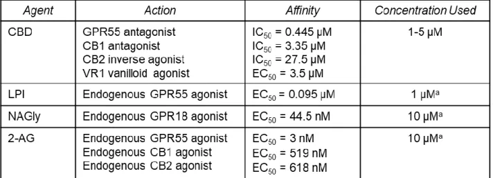
CBD synchronized neuronal calcium signaling and network activity
As CBD showed a direct effect on the neurons in our experiments, we treated neurons
directly with both CBD and cannabinoid agonists to determine which receptors CBD may be
acting on to modulate calcium signaling of neurons. We compared the effects of CBD and each
demonstrate the variable changes seen in neuronal calcium signaling during treatment with
cannabinoid receptor agonists as compared to baseline (vehicle) activity (Figure 2A). CBD
induced calcium spikes of high intensity (note scale change in Figure 2B) as compared to
vehicle. Notably, the pattern of spikes induced by CBD was characterized by a high degree of
synchrony, illustrated in Figure 2B by the superimposed peaks throughout the run. LPI induced
smaller calcium spikes but also showed a tendency toward synchronous activity (Figure 2E).
2-AG increased the frequency of calcium spikes relative to pre-treatment (Figure 2C) but did not
show synchronized activity. Treatment with NAGly had no effect on calcium signaling.
Quantification of the extent of synchronous activity under the various conditions in Figure 2F
illustrates the significantly higher proportion of synchronized calcium spikes in the presence of
CBD and LPI. Average spike rates were significantly increased for CBD and 2-AG but not LPI
(Figure 2G), whereas an increased intensity of the calcium response was only seen with CBD
(Figure 2H). Thus, CBD has complex properties that overlap with several different cannabinoid
agonists.
Since CBD has been shown to stabilize epileptiform activity and, in our experiments,
synchronize calcium signaling of neurons, we investigated the changes in neural network
communication induced by CBD. We used 60 channel microelectrode arrays (MEAs) to record
network-wide electrophysiological bursts of neural activity and measured network connectivity
and communication efficiency of neurons. Figure 3A shows the MEA seeded with neurons, and
Figure 3B shows examples of the recording on a subset of electrodes in the absence or
presence of CBD. Each rectangle in Figure 3B corresponds to a single electrode. These
recordings were converted to raster plots, shown in Figures 3C-D which show the activity of
each electrode (Y axis) over time (X axis). We recorded the electrophysiological activity of
neurons at baseline for 10 minutes, then added CBD and recorded for an additional 10 minutes.
The raster plots illustrate the synchronous bursts of activity which appear as a vertical line.
Treatment with CBD shifted the pattern toward an increased number of bursts with a regular
properties of the burst over the entire 10 minute run for three independent MEAs showed that
CBD did not change the frequency of spiking in each burst as compared to control (Figure 3E),
Figure 3. CBD induced a greater number of shorter bursts in neuronal
while the number of bursts increased significantly (Figure 3F) and the length of the bursts was
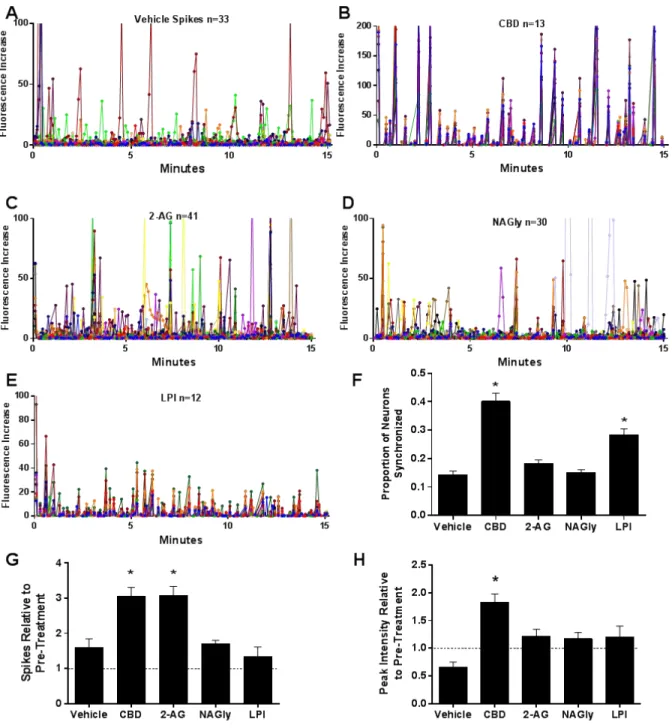
shorter (Figure 3G). To examine the translation of these effects to the inflammatory setting,
neurons on the MEA were challenged with MGCM in the presence or absence of 1 µM CBD.
Analysis of the network activity, summarized in Figure 4, showed that the hyperactive response
to the MGCM was significantly diminished in the presence of CBD. Both treated and
non-treated MEAs had a subset of cells that deceased firing in response to the MGCM. CBD also
had a small normalization effect on these cells, bringing them back toward baseline
characteristics. Thus, CBD appeared to modulate the activity of the neurons in a fashion that
retains normal firing and network activity.
CBD treatment of microglia offers greater protection against Aβ-induced inflammatory
damage
As anti-inflammatory effects of CBD could also contribute to its beneficial effects, we
treated microglia with CBD to determine if it could reduce the level of neurotoxic activity. We
measured the calcium signaling and accumulation in neurons challenged with MGCM collected
from microglia treated with CBD prior to exposure to Aβo. Figure 5A illustrates the temporal Figure 4. CBD normalized and stabilized neuronal networks during an inflammatory challenge. A network analysis of the percent change in firing rates of neurons on the MEA after challenging with MGCM showed that CBD significantly decreased the hyperactive
changes in neuronal intracellular calcium following a challenge with MGCM at 0 minutes. The
accumulation of neuronal calcium in response to MGCM from the CBD-treated microglia was
greatly reduced in both the acute (0-6 minutes) and delayed (6-46 minutes) phases relative to
vehicle treated microglia (Figure 5B). CBD resulted in a 48% reduction in delayed calcium
accumulation as compared to the 33% reduction when the neurons were pretreated with CBD,
indicating that the ability to suppress the release of neurotoxic factors may be of equal or
greater importance than the effects directly on neurons.
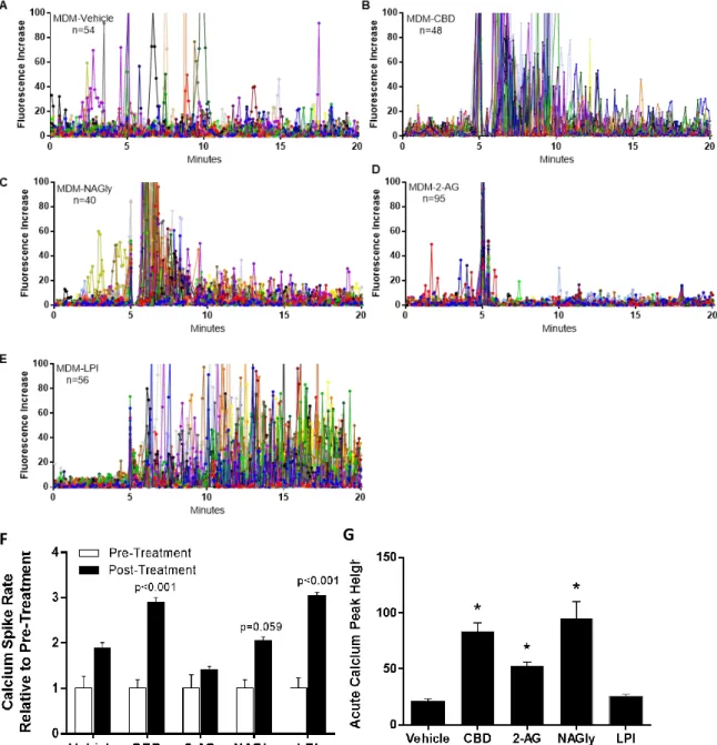
Cannabinoid agonists induce unique calcium signaling profiles in macrophages.
Since the MGCM studies showed that CBD has an effect on mouse microglia, we
treated human monocyte-derived macrophages with the same cannabinoid ligands used for the
analysis of neuronal activity. The effects of the cannabinoid ligands on macrophages
contrasted significantly with the effects on neurons. Figure 6 demonstrates the variable changes
in macrophage calcium signaling in response to treatment with cannabinoid receptor agonists
as compared to baseline (vehicle) activity (shown in Figure 6A). CBD induced an acute increase
NAGly treatment largely recapitulated the effect of CBD, with an acute increase which
recovered with a similar time course (Figure 6C). 2-AG induced a brief calcium spike which was
Figure 6. Agonists targeting different cannabinoid receptors produce distinctively different calcium signaling in primary human monocyte-derived macrophages. Composite profiles of calcium spikes in cultured macrophages illustrate baseline activity (vehicle), acute increases in calcium signaling frequency induced by CBD and LPI, a more acute increase with NAGly and a decrease with 2-AG indicating substantial
diversity in the regulation of macrophage activity by the cannabinoid ligands. F. CBD and LPI yielded the greatest mean increase in spike frequency. G. NAGly, CBD, and 2-AG increased intensity of the calcium spikes. * = p<0.05, analysis of variance with post-hoc multiple comparisons
rapidly followed by suppression of calcium signaling activity in the macrophages (Figure 6D).
LPI induced a modest acute spike followed by sustained activity in the macrophages (Figure
6E). Figure 6F displays the calcium spike frequency of macrophages before and after treatment
with the agonists, showing that CBD and LPI yielded the greatest mean increase relative to
pre-treatment. The average intensity of calcium spikes is summarized in Figure 6G. NAGly, CBD,
and 2-AG all increased calcium spike intensity in macrophages as compared to vehicle. Thus,
CBD may be acting through both GPR18 and GPR55 receptors on macrophages to affect
calcium signaling and inflammatory activity.
The response of macrophages showed some similarities to neurons but also dramatic
differences. CBD increased the calcium spike rate and the peak intensity in both neurons and
macrophages. However, LPI and NAGly did not change the calcium spike rate in neurons but
increased the calcium spike rate in macrophages. 2-AG and NAGly did not change the calcium
peak height in neurons but increased calcium peak height in macrophages. Thus, CBD and the
cannabinoid agonists have unique effects on neurons and macrophages. While our data
supports CBD actions at GPR55 on neurons, it appears that CBD may be acting through both
GPR18 and GPR55 receptors on macrophages.
Discussion
Inflammation appears early in neurodegenerative diseases and may enhance the
vulnerability of neurons to disease progression. Therapeutic intervention at this early stage will
likely have the greatest impact, as the pathology is not well developed and may still be
reversible. Successful intervention therefore could have disease modifying potential. Our study
found that CBD has complex actions that could offer protection against Aβ-induced
Neuroprotective potential of CBD
CBD treatment of neurons caused a modest stabilization of neuronal calcium
accumulation in response to an inflammatory challenge indicating suppression of pathogenesis.
This suppression of calcium accumulation demonstrates the ability of CBD to stabilize neuronal
calcium regulation in response to an inflammatory challenge. In our model of early AD
pathogenesis, the acute intracellular calcium accumulation that follows a challenge with MGCM
quickly recovers. The delayed calcium accumulation that slowly increases over time is more
toxic to the neurons, as these cells generally do not recover from the chronic increase in
intracellular calcium. This delayed calcium dysregulation increases the neuronal vulnerability to
further AD pathogenesis. The in vitro calcium dysregulation closely mimics in vivo
destabilization of neuronal calcium in mouse models of AD where chronically elevated calcium
is seen in neurons near amyloid plaques and activated microglia (Kuchibhotla et al. 2008;
Bittner et al. 2010). The dysregulation leads to cytoskeletal damage, posttranslational
modifications of tau protein, neuronal death and synapse loss. The ability of CBD to suppress
calcium accumulation could protect neurons by inhibiting the development of this pathology. In
this way, direct stimulation of neurons with CBD may be neuroprotective in the setting of early
AD pathology. How CBD induces these beneficial effects is not clear but some clues have
emerged from the examination of the effects of agonists targeting the receptors, CB1/CB2,
GPR55 and GPR18. CBD had unique effects on neuronal calcium signaling, inducing strong
calcium spikes that were highly synchronized between neurons. The GPR55 agonist, LPI,
increased the synchronization of calcium activity but the magnitude of signaling was relatively
weak. The mixed agonist, 2-AG, increased the intensity of the calcium spikes but not the
synchronization. Together these effects suggest that CBD may be acting through GPR55
receptors on neurons but may also have additional targets. GPR18 does not appear to play a
CBD promoted increased efficiency of neuronal network communication
The increased synchronization of calcium spikes suggested increased efficiency of calcium
signaling. Analysis of action potential firing and network electrophysiological activity on the
MEAs showed that CBD induced a greater number of shorter bursts. This suggests that CBD
restricts the generation of long bursts of action potentials. Neurons recovered more quickly
after each electrophysiological burst of neural activity, allowing the generation of short, highly
synchronized bursts of activity that may allow for more efficient network communication.
Destabilized, hyperactive network activity may be a treatable cause of cognitive dysfunction in
AD. Hyperactive network behavior may be responsible for the increased subclinical epileptiform
activity seen in AD (Born 2015; Cook et al. 2015; Horváth et al. 2016; Nicastro et al. 2016). CBD
has well established network stabilizing effects in patients with seizures and these properties
may reduce hyperactivity and prove beneficial in AD. The ability of CBD to normalize network
activity in the presence of an inflammatory stimulus (MGCM) indicated that it somehow prevents
the development of aberrant activity under inflammatory conditions. Overall, these findings
indicate that CBD may help to keep neural activity in check by regulating the properties of
network bursts, in part, due to stabilization of calcium homeostasis.
CBD suppressed inflammation
While beneficial effects of CBD were seen on neurons, the ability to suppress the
release of neurotoxic factors from activated microglia may provide “anti-inflammatory” effects
that confer even greater neuroprotection. In our model, microglia were exposed to Aβo, which
triggered the secretion of pro-inflammatory soluble factors and cytokines, mimicking early
events associated with the development of AD. In neurons, these microglial products induce a
dysregulation of intracellular calcium (Guo et al. 1999; Abramov et al. 2004; Kuchibhotla et al.
2008; Bittner et al. 2010; Um et al. 2013), as well as sensitization of NMDA glutamate receptors
Molokanova et al. 2014; Ferreira et al. 2015; Hamilton et al. 2015), mislocalization/accumulation
of Tau in dendrites (Ittner et al. 2010), focal swellings,(Yoshiyama et al. 2007; Kuchibhotla et al.
2008), loss of cytoskeletal structure and impaired transport (Yoshiyama et al. 2007; Meeker et
al. 2016). These changes are analogous to those seen in early AD pathogenesis. The
decreased ability of the conditioned medium to induce calcium dysregulation after exposure to
CBD demonstrated an “anti-inflammatory” effect which may restrict the ability of Aβo to generate
a toxic inflammatory response. Suppression of the release of neurotoxic factors from microglia
by CBD could therefore modify disease progression in the early stages of AD pathogenesis.
Previous studies have suggested that CBD could have anti-inflammatory effects through a
variety of mechanisms, such as the ability to inhibit the migration of immune cells and the
activation of microglia (Karl et al. 2012). Our data confirmed beneficial actions of CBD at
microglia, but showed an effect that would be independent of migration. The anti-inflammatory
properties demonstrated through our studies highlight the ability of CBD to decrease the
neurotoxicity of the microglial secretome in early AD pathogenesis. As inflammation may
represent an early event that alters the function of neuronal networks and contributes to
cognitive decline, these findings underscore the impact of the inflammatory system in the early
pathology of AD and the possible benefit to be realized with CBD through its anti-inflammatory
properties.
CBD induced increased calcium signaling of macrophages
As seen in the neurons, cannabinoid ligands each induced unique calcium signaling
profiles on macrophages. However, the actions were distinctively different in the two cell types.
Notably, NAGly-induced calcium signaling in macrophages largely recapitulated the effect of
CBD, suggesting that CBD may be acting through GPR18 receptors on macrophages to
increase calcium signaling. Macrophages express both GPR18 and GPR55 receptors and both
signaling but was characterized by a continuous activation. In contrast, 2-AG potently
suppressed activity after a very brief calcium spike. Although both 2-AG and LPI are agonists of
GPR55, it is noteworthy that they induced opposite effects on calcium signaling in
macrophages. This suggests that effects of 2-AG at CB1 and CB2 may contribute to the
regulation of calcium signaling in macrophages. These data indicate that CBD has actions on
macrophages consistent with activation of GPR18 but that GPR55 and CB1/CB2 receptors also
can exert significant effects on calcium signaling that may play a role in the regulation of
inflammatory activity.
Conclusion
CBD treatment of neurons caused a modest stabilization of neuronal calcium
accumulation in response to an inflammatory challenge indicating suppression of pathogenesis.
CBD had a larger effect on the release of toxins from microglia indicating that the
“anti-inflammatory” effects may confer greater neuroprotection. CBD promoted greater
synchronization of neuronal calcium signaling and a greater number of shorter bursts on the
MEA suggesting more efficient network communication and decreased network excitability
which may contribute to protection. These effects may be modulated by GPR55 on neurons.
Cannabinoid ligands each induced unique calcium signaling profiles on macrophages with CBD
and NAGly inducing similar effects, suggesting that CBD may modulate the GPR18 receptor on
macrophages. However, the robust and persistent calcium signaling induced by LPI and the
strong suppression by 2-AG indicated complex effects at multiple cannabinoid receptors
including GPR55 that regulate macrophage calcium signaling. Overall, our findings support the
hypothesis that CBD has neuroprotective potential for AD patients through both the suppression
of inflammation and the stabilization of network activity. The precise mechanisms underlying
these effects are not clear but cannabinoid-like receptors, such as GPR18 and GPR55, appear
inflammation and network activity have the potential to reveal novel therapies that suppress
References
Abramov AY, Canevari L, Duchen MR (2004) Calcium signals induced by amyloid β peptide and
their consequences in neurons and astrocytes in culture. In: Biochimica et Biophysica Acta
- Molecular Cell Research
Anavi-Goffer S, Baillie G, Irving AJ, et al (2012) Modulation of
L-α-lysophosphatidylinositol/GPR55 mitogen-activated protein kinase (MAPK) signaling by
cannabinoids. J Biol Chem. doi: 10.1074/jbc.M111.296020
Aso E, Ferrer I (2016) CB2 Cannabinoid Receptor As Potential Target against Alzheimer’s
Disease. Front Neurosci 10:243. doi: 10.3389/fnins.2016.00243
Auvin S, Cilio MR, Vezzani A (2015) Current understanding and neurobiology of epileptic
encephalopathies. Neurobiol. Dis.
Bittner T, Fuhrmann M, Burgold S, et al (2010) Multiple events lead to dendritic spine loss in
triple transgenic Alzheimer’s disease mice. PLoS One. doi: 10.1371/journal.pone.0015477
Born HA (2015) Seizures in Alzheimer’s disease. Neuroscience
Bragg DC, Boles JC, Meeker RB (2002) Destabilization of neuronal calcium homeostasis by
factors secreted from choroid plexus macrophage cultures in response to feline
immunodeficiency virus. Neurobiol Dis. doi: 10.1006/nbdi.2001.0459
Briggs DI, Defensor E, Memar Ardestani P, et al (2017) Role of Endoplasmic Reticulum Stress
in Learning and Memory Impairment and Alzheimer’s Disease-Like Neuropathology in the
PS19 and APP Swe Mouse Models of Tauopathy and Amyloidosis . eneuro. doi:
10.1523/eneuro.0025-17.2017
Busquets-Garcia A, Bains J, Marsicano G (2018) CB 1 Receptor Signaling in the Brain:
Chiurchiù V, Lanuti M, De Bardi M, et al (2015) The differential characterization of GPR55
receptor in human peripheral blood reveals a distinctive expression in monocytes and NK
cells and a proinflammatory role in these innate cells. Int Immunol. doi:
10.1093/intimm/dxu097
Cook M, Baker N, Lanes S, et al (2015) Incidence of stroke and seizure in Alzheimer’s disease
dementia. Age Ageing. doi: 10.1093/ageing/afv061
Devinsky O, Patel AD, Cross JH, et al (2018a) Effect of Cannabidiol on Drop Seizures in the
Lennox–Gastaut Syndrome. N Engl J Med. doi: 10.1056/NEJMoa1714631
Devinsky O, Patel AD, Thiele EA, et al (2018b) Randomized, dose-ranging safety trial of
cannabidiol in Dravet syndrome. Neurology. doi: 10.1212/WNL.0000000000005254
Devinsky O, Vezzani A, Najjar S, et al (2013) Glia and epilepsy: Excitability and inflammation.
Trends Neurosci.
Ferreira IL, Ferreiro E, Schmidt J, et al (2015) Aβ and NMDAR activation cause mitochondrial
dysfunction involving ER calcium release. Neurobiol Aging. doi:
10.1016/j.neurobiolaging.2014.09.006
Floden AM (2005) -Amyloid-Stimulated Microglia Induce Neuron Death via Synergistic
Stimulation of Tumor Necrosis Factor and NMDA Receptors. J Neurosci. doi:
10.1523/jneurosci.4998-04.2005
Galic MA, Riazi K, Pittman QJ (2012) Cytokines and brain excitability. Front. Neuroendocrinol.
Gaugler J, James B, Johnson T, et al (2016) 2016 Alzheimer’s disease facts and figures.
Alzheimer’s Dement. doi: 10.1016/j.jalz.2016.03.001
Guo Q, Fu W, Sopher BL, et al (1999) Increased vulnerability of hippocampal neurons to
Hamilton A, Zamponi GW, Ferguson SSG (2015) Glutamate receptors function as scaffolds for
the regulation of β-amyloid and cellular prion protein signaling complexes. Mol. Brain
Horváth A, Szcs A, Barcs G, et al (2016) Epileptic Seizures in Alzheimer Disease. Alzheimer
Dis. Assoc. Disord.
Ibeas Bih C, Chen T, Nunn AVW, et al (2015) Molecular Targets of Cannabidiol in Neurological
Disorders. Neurotherapeutics
Ittner LM, Ke YD, Delerue F, et al (2010) Dendritic function of tau mediates amyloid-β toxicity in
alzheimer’s disease mouse models. Cell. doi: 10.1016/j.cell.2010.06.036
Iuvone T, Esposito G, De Filippis D, et al (2009) Cannabidiol: A promising drug for
neurodegenerative disorders? CNS Neurosci. Ther.
Kallendrusch S, Kremzow S, Nowicki M, et al (2013) The G protein-coupled receptor 55 ligand
l-α-lysophosphatidylinositol exerts microglia-dependent neuroprotection after excitotoxic
lesion. Glia. doi: 10.1002/glia.22560
Karl T, Cheng D, Garner B, Arnold JC (2012) The therapeutic potential of the endocannabinoid
system for Alzheimer’s disease. Expert Opin Ther Targets. doi:
10.1517/14728222.2012.671812
Kaul M, Lipton SA (1999) Chemokines and activated macrophages in HIV gp120-induced
neuronal apoptosis. Proc Natl Acad Sci U S A
Kim HJ, Waataja JJ, Thayer SA (2008) Cannabinoids Inhibit Network-Driven Synapse Loss
between Hippocampal Neurons in Culture. J Pharmacol Exp Ther. doi:
10.1124/jpet.107.131607
Kuchibhotla K V., Goldman ST, Lattarulo CR, et al (2008) Aβ Plaques Lead to Aberrant
Disruption of Neuronal Networks. Neuron. doi: 10.1016/j.neuron.2008.06.008
McHugh D (2012) GPR18 in microglia: Implications for the CNS and endocannabinoid system
signalling. Br. J. Pharmacol.
Meeker RB (2007) Feline immunodeficiency virus neuropathogenesis: From cats to calcium. J.
Neuroimmune Pharmacol.
Meeker RB, Boles JC, Robertson KR, Hall CD (2005) Cerebrospinal fluid from human
immunodeficiency virus-infected individuals facilitates neurotoxicity by suppressing
intracellular calcium recovery. J Neurovirol. doi: 10.1080/13550280590922757
Meeker RB, Poulton W, Clary G, et al (2016) Novel p75 neurotrophin receptor ligand stabilizes
neuronal calcium, preserves mitochondrial movement and protects against HIV associated
neuropathogenesis. Exp Neurol. doi: 10.1016/j.expneurol.2015.09.012
Meeker RB, Poulton W, Feng WH, et al (2012) Suppression of immunodeficiency
virus-associated neural damage by the p75 neurotrophin receptor ligand, LM11A-31, in an in
vitro feline model. J Neuroimmune Pharmacol. doi: 10.1007/s11481-011-9325-0
Mellini P, Valente S, Mai A (2014) Sirtuin modulators: an updated patent review (2012 – 2014).
Expert Opin Ther Pat. doi: 10.1517/13543776.2014.982532
Millar SA, Stone NL, Yates AS, O’Sullivan SE (2018) A systematic review on the
pharmacokinetics of cannabidiol in humans. Front. Pharmacol. 9:1365
Molokanova E, Akhtar MW, Sanz-Blasco S, et al (2014) Differential Effects of Synaptic and
Extrasynaptic NMDA Receptors on A -Induced Nitric Oxide Production in Cerebrocortical
Neurons. J Neurosci. doi: 10.1523/jneurosci.2907-13.2014
Nicastro N, Assal F, Seeck M (2016) From here to epilepsy: The risk of seizure in patients with
Paula-Lima AC, Brito-Moreira J, Ferreira ST (2013) Deregulation of excitatory
neurotransmission underlying synapse failure in Alzheimer’s disease. J. Neurochem.
Pertwee RG (2008) The diverse CB 1 and CB 2 receptor pharmacology of three plant
cannabinoids: Δ 9-tetrahydrocannabinol, cannabidiol and Δ 9-tetrahydrocannabivarin. Br. J.
Pharmacol.
Ravizza T, Balosso S, Vezzani A (2011) Inflammation and prevention of epileptogenesis.
Neurosci. Lett.
Roberson ED, Halabisky B, Yoo JW, et al (2011) Amyloid-β/Fyn-induced synaptic, network, and
cognitive impairments depend on tau levels in multiple mouse models of Alzheimer’s
disease. J Neurosci. doi: 10.1523/JNEUROSCI.4152-10.2011
Scuderi C, De Filippis D, Iuvone T, et al (2009) Cannabidiol in medicine: A review of its
therapeutic potential in CNS disorders. Phyther. Res.
Taylor L, Gidal B, Blakey G, et al (2018) A Phase I, Randomized, Double-Blind,
Placebo-Controlled, Single Ascending Dose, Multiple Dose, and Food Effect Trial of the Safety,
Tolerability and Pharmacokinetics of Highly Purified Cannabidiol in Healthy Subjects. CNS
Drugs 32:1053–1067. doi: 10.1007/s40263-018-0578-5
Thiele EA, Marsh ED, French JA, et al (2018) Cannabidiol in patients with seizures associated
with Lennox-Gastaut syndrome (GWPCARE4): a randomised, double-blind,
placebo-controlled phase 3 trial. Lancet. doi: 10.1016/S0140-6736(18)30136-3
Turcotte C, Blanchet MR, Laviolette M, Flamand N (2016a) The CB2 receptor and its role as a
regulator of inflammation. Cell. Mol. Life Sci. 73:4449–4470
Turcotte C, Blanchet MR, Laviolette M, Flamand N (2016b) The CB 2 receptor and its role as a
Um JW, Kaufman AC, Kostylev M, et al (2013) Metabotropic Glutamate Receptor 5 Is a
Coreceptor for Alzheimer Aβ Oligomer Bound to Cellular Prion Protein. Neuron. doi:
10.1016/j.neuron.2013.06.036
Vezzani A (2015) Anti-inflammatory drugs in epilepsy: does it impact epileptogenesis? Expert
Opin Drug Saf. doi: 10.1517/14740338.2015.1010508
Xiong H, McCabe L, Costello J, et al (2004) Activation of NR1a/NR2B receptors by soluble
factors from APP-stimulated monocyte-derived macrophages: Implications for the
pathogenesis of Alzheimer’s disease. Neurobiol Aging. doi:
10.1016/j.neurobiolaging.2003.09.007
Yoshiyama Y, Higuchi M, Zhang B, et al (2007) Synapse Loss and Microglial Activation Precede
Tangles in a P301S Tauopathy Mouse Model. Neuron. doi: 10.1016/j.neuron.2007.01.010
(2003) Cannabis-based medicines - GW Pharmaceuticals. High CBD, high THC, medicinal
cannabis - GW Pharmaceuticals, THC:CBD. Drugs R D. doi:
Report Addendum
Acknowledgements
The authors would like to thank Braxton Harris and Ally Dombroski for their assistance with the analysis of the MEA data and Anita Amin for her assistance with the live cell calcium imaging experiments.
Funding Support
Supported by NIH Grant R01 NS108808, NC TraCS ECBR004 and the UNC Department of Neurology
Conflicts of Interest