GENETICS OF IMMUNITY
Several Critical Cell Types, Tissues, and Pathways
Are Implicated in Genome-Wide Association Studies
for Systemic Lupus Erythematosus
Lu Liu,*,†,‡,§,††,1Xianyong Yin,*,†,‡,§,**,1Leilei Wen,*,†,‡,§,††,1Chao Yang,*,†,‡,§,††,1
Yujun Sheng,*,†,‡,§Yan Lin,*,†,‡,§Zhengwei Zhu,*,†,‡,§Changbing Shen,*,†,‡,§Yinjuan Shi,*,†,‡,§,†† Yajie Zheng,*,†,‡,§,††Sen Yang,*,†,‡,§Xuejun Zhang,*,†,‡,§and Yong Cui††,2
*Institute of Dermatology, Department of Dermatology, The First Affiliated Hospital,†Key Laboratory of Dermatology,
Ministry of Education, State Key Lab of Dermatology Incubation Center, and§Collaborative Innovation Center for Complex and Severe Dermatosis, Anhui Medical University, Hefei, Anhui Province, 230032, China,‡Key Laboratory of Gene Resource Utilization for Complex Diseases, Hefei, Anhui Province, 230032, China,**Department of Genetics, The Renaissance Computing Institute, University of North Carolina at Chapel Hill, North Carolina 27517, and††Department of Dermatology, China-Japan Friendship Hospital, Beijing, 100029, China
ABSTRACT We aimed to elucidate the cell types, tissues, and pathways influenced by common variants in systemic lupus erythematosus (SLE). We applied a nonparameter enrichment statistical approach, termed SNPsea, in 181 single nucleotide polymorphisms (SNPs) that have been identified to be associated with the risk of SLE through genome-wide association studies (GWAS) in Eastern Asian and Caucasian populations, to manipulate the critical cell types, tissues, and pathways. In the two most significant cells’findings (B lymphocytes and CD14+ monocytes), we subjected the GWAS association evidence in the Han Chinese population to an enrichment test of expression quantitative trait locus (QTL) sites and DNase I hypersensitivity, respectively. In both Eastern Asian and Caucasian populations, we observed that the expression level of SLE GWAS implicated genes was significantly elevated in xeroderma pigentosum B cells (P # 1.00 · 1026), CD14+ monocytes (P # 2.74 · 1024) and CD19+ B cells (P # 2.00 · 1026), and plasmacytoid dendritic cells (pDCs) (P # 9.00 · 1026). We revealed that the SLE GWAS-associated variants were more likely to reside in expression QTL in B lymphocytes (q1/q0 = 2.15,P = 1.23 · 10244) and DNase I hypersensitivity sites (DHSs) in CD14+ monocytes (q1/q0 = 1.41,P = 0.08). We observed the common variants affected the risk of SLE mostly through by regulating multiple immune system processes and immune response signaling. This study sheds light on several immune cells and responses, as well as the regulatory effect of common variants in the pathogenesis of SLE.
KEYWORDS
systemic lupus erythematosus genome-wide
association studies SNPsea Genetics of
Immunity
Systemic lupus erythematosus (SLE) is a common autoimmune disease. It is well characterized by the production of a variety of antinuclear
antibodies, which then result in a wide spectrum of clinical symptoms in skin, blood, kidney, and lung(Lauet al.2006; Olsonet al.2002). Environmental factors and genetic predisposition both contribute to the risk of SLE (Tsokos 2011). In recent years, the genetic basis of SLE has been advanced remarkably, mostly through genome-wide associa-tion studies (GWAS) in diverse populaassocia-tions (Remmers et al. 2007; Cunninghame Grahamet al.2008; Grahamet al.2008; International Consortium for Systemic Lupus Erythematosuset al.2008; Kozyrev et al.2008; Homet al.2008; Musoneet al.2008; Nathet al.2008; Gateva et al.2009; Jacobet al.2009; Webbet al.2009; Yanget al.2009b, 2009a; Changet al.2009; Cunninghame Grahamet al.2011; Zhaoet al.2011; Luoet al.2011; Soet al.2011). Multiple GWAS have been conducted for SLE in Eastern Asian populations, identifying 63 single-nucleotide polymorphisms (SNPs) with genome-wide significant evidence (Zhang et al. 2014; Yanget al. 2013; Liet al.2013; Shenget al.2011, 2015; Copyright © 2016 Liuet al.
doi: 10.1534/g3.116.027326
Manuscript received January 17, 2016; accepted for publication March 14, 2016; published Early Online March 23, 2016.
This is an open-access article distributed under the terms of the Creative Commons Attribution 4.0 International License (http://creativecommons.org/ licenses/by/4.0/), which permits unrestricted use, distribution, and reproduction in any medium, provided the original work is properly cited.
Supplemental material is available online atwww.g3journal.org/lookup/suppl/ doi:10.1534/g3.116.027326/-/DC1
1These authors contributed equally to this work.
Hanet al.2009; Zhanget al.2011; Yuet al.2013; Sunet al.2016). Meanwhile,118 susceptibility SNPs have been established robustly in the Caucasian populations (Grahamet al.2008; International Consor-tium for Systemic Lupus Erythematosuset al.2008; Kozyrevet al.2008; Homet al.2008; Gatevaet al.2009; Lessardet al.2011; Chunget al. 2011; Cunninghame Grahamet al.2011; Fernandoet al.2012; Lessard et al.2012; Leeet al.2012; Martinet al.2013; Armstronget al.2014; Kariukiet al.2015; Vaughnet al.2014; Demirciet al.2016; Bentham et al.2015; Suarez-Gestalet al.2009). Thesefindings together promote genetic etiology research for SLE. However, the mechanism underlying each variant is still largely unknown. It is usually found that the variants with robust association evidence in complex diseases GWAS are tag SNPs that are linked to the true putative variants, rather than the putative variant itself. In addition, the vast majority of these variants reside in the noncoding genomic region, which therefore poses a great challenge for interpretation and follow-up biological studies (Cuiet al. 2013). Nevertheless, it is widely indicated that these variants contribute to the risk of diseases mainly through regulating gene expression levels by expression quantitative trait loci (eQTL) effects, or by changing chromatin accessibility, rather altering protein function directly (Edwardset al.2013). In order to disentangle the functional role of each variant in the disease, well-designed biological experiments are warranted. It is strongly believed that each variant contributes to the etiology of disease by affecting a typical tissue and cell through a complicated biological pathway. Hence, in order to dissect the func-tionality of each GWAS-identified variant, we have to use the most relevant cells and tissues in biological experiments. Nowadays, there are several integrative bioinformatics methods dedicated to identify-ing the cell types, tissues, and pathways affected by GWAS-implicated susceptibility variants (Wenget al.2011). Some critical cells, tissues, and pathways have been revealed for Crohn disease, rheumatoid arthritis, and multiple sclerosis (Huet al.2011). Among them, Hu et al.(2011) provided the evidence to underpin the key role of tran-sitional B lymphocyte cells in the etiology of SLE.
In the present study, we employed a nonparameter enrichment statistical approach to evaluate the specificity of conditions (i.e., cells, tissues and pathways) in SLE (Slowikowskiet al.2014), and tested the enrichment of eQTL sites and DNase I hypersensitivity sites (DHSs) in SLE GWAS common variants.
MATERIALS AND METHODS
The study design and analytical process are diagrammed in Figure 1. The enrichment test was implemented in SNPsea (Slowikowskiet al.2014). We used 63 and 118 SNPs that had been reported previously with genome-wide evidence in Eastern Asian and Caucasian populations, respectively, as input (Supplemental Material,Table S1). It was noted that 13 of the 118 SNPs from Caucasian populations reside in the HLAregion. In SNPsea, we identified genes implicated by each input SNP using linkage disequilibrium (LD) information in the CHB or CEU reference panel, respectively, from the 1000 Genomes Project. To include all potential genes, we extended the SNP interval to the nearest recombination hotspots with recombination rate. 3 cM/Mb (HapMap 3) (Myerset al.2005). If a SNP interval overlapped with no gene intervals, we extended the SNP interval to 10,000 base pairs. If one unified gene was implicated by at least two different SNPs, we merged these SNPs into one single locus. Usually, multiple genes would be implicated in each locus. But we assumed that there would be only one single gene truly associated with SLE in each locus. We normalized the expression of each gene through dividing its expression value in a specific condition (i.e., the cell types, tissues, and pathways) by the L2
norm of gene expression values for that gene in all different conditions, thus we would get an expression matrix with values between zero and one, which was used to indicate the specificity of the gene to a condition. For each condition, we ranked the normalized expression values in descending order, and divided them by the number of implicated genes. As a result, we got a score between zero and one, with smaller values representing a higher specificity for a given condition (Huet al.2011). We assigned the lowest score for each locus. For each condition, we used a sampling approach to build a null SNP set, and then calculated an empiricalP-value through comparing its gene expression distribu-tion with that of the SLE GWAS SNP set. This empiricalP-value was used to indicate the tail probability of observing a condition-specificity score greater than, or equal to, the sum across all SNPs (Slowikowski et al. 2014). The significance threshold was defined by Bonferroni correction with the number of conditions used in each test. We applied 533 cell types expression matrix forHomo sapiensfrom FANTOM5 (Bonferroni P threshold = 0.05/533), 249 cell types expression matrix forMus musculus(GEO dataset: GSE15907, Bonferroni Pthreshold = 0.05/249), 79 tissues expression matrix forH. sapiens (BonferroniPthreshold = 0.05/79) (Suet al.2004), and 1751 gene expression ontology (GO) matrix (data-version: 2013-06-29, CVS re-vision: 9700, BonferroniPthreshold = 0.05/1751), to test the specifi c-ity of cell types, tissues and pathways, respectively.
The role of B lymphoblastoid cells in the pathogenesis of SLE is well known. Our results in the SNPsea test also identified the significant evidence of B lympholastoid cells in both Eastern Asian and Caucasian populations. In addition, this test revealed the most significant evidence for CD14+ monocytes in the pathophysiology of SLE. In an attempt to unravel which functional part the SLE associated variant most likely resides in, we incorporated the SLE GWAS summary statistics in Han Chinese population, eQTL from B lymphoblastoid cells and DHSs functional genomics data from CD14+ cells. We retrieved the GWAS summary statistics for SLE described preciously in the Han Chinese (Hanet al. 2009). These contained the association evidence (single variantPvalue) for 494,559 autosomal variants. The eQTL results for whole blood B lymphocytes in 5311 Caucasian individuals were freely accessed (Westraet al.2013). In total, there were 923,021 and 4733 SNPs annotated to becis-eQTL andtrans-eQTL sites, respectively, in whole blood B lymphocytes. The DHSs for CD14+ primary cells were downloaded from the Encyclopedia of DNA Elements (ENCODE) consortium database (accession: ENCFF001SRV), in which there were 159,457 DHSs identified for CD14+ monocytes. Each of the 494,559 autosomal variants SNP in SLE GWAS was then annotated by whether it resided in the DHSs in CD14+ monocytes, or the eQTL site in whole blood B lymphocytes, respectively. The enrich-ment of DHSs and eQTL effect were tested through genetic analysis incorporating pleiotropy and annotation (GPA) packages in R 3.12 (Chung et al.2014). The enrichment size of DHS or eQTL site was characterized by estimating the enrichment fold (q1/q0), which was calculated as odds of proportional
anno-tated by CD14+ DHS, or whole blood lymphocytes, eQTL effect in non-null- and null-associated SNPs (Chunget al.2014).q1and Q0
were defined as the probability of being annotated as DHS or eQTL sites for non-null- and null-associated SNPs, respectively. The statistical significance for the enrichment effect was evaluated by likelihood ratio test. The maximum iteration was set to 10,000 in the GPA package.
Data availability
RESULTS
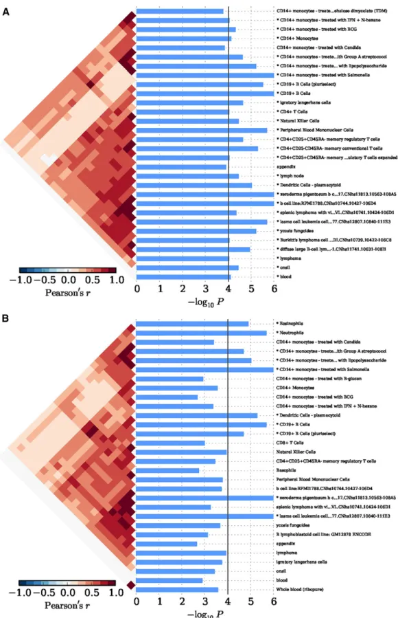
In Eastern Asian populations, it has been shown that the expression of the implicated genes were significantly enriched in 19 types of cells, for example, xeroderma pigentosum B cells (P = 1.00 · 1026), CD14+
monocytes (P = 7.33 · 1025), CD19+ B cells (P = 1.00 · 1026),
plasmacytoid dendritic cells (pDCs) (P = 9.00 · 1026), and
CD4+CD25+CD45RA–memory regulatory T cells (P = 2.20 · 1025)
(Figure 2A andTable S2). And significant evidence regarding B cells (e.g., B.T1.sp, B.T2.sp, B.T3.sp, B.Fo.sp, B.MZ.Sp, B1a.sp, B.FrF.BM, and B1a.pc etc.) was achieved consistently with using 249 cell types expression matrix forMus musculus(P # 4.88 · 1025;Figure S1A
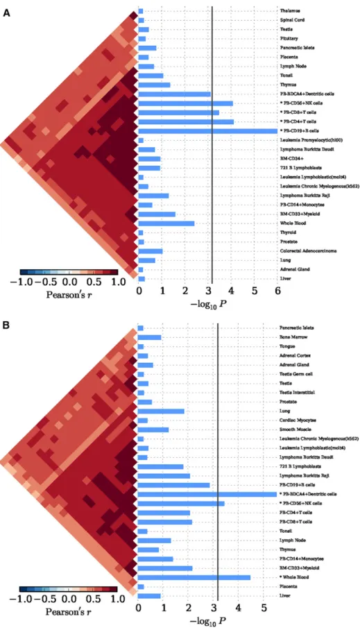
andTable S3). In peripheral blood, we achieved corrected significance at CD19+ B cells (P = 1.00 · 1026), CD4+ T cells (P = 7.57 · 1025),
CD56+NK cells (P = 8.30 · 1025) (Figure 3A andTable S4). We
only observed GO:0002684 positive regulation of immune system pro-cess (P= 1.60·10-5) was significantly elevated, although a series of the
immune response pathways achieved moderate significance (Figure S2A andTable S5). In Caucasian populations, the cell enrichment test
also indicated that xeroderma pigentosum B cells (P = 1.00 · 1026),
CD14+ monocytes (P = 1.00 · 1026), CD19+ B cells (P = 2.00 · 1026),
and plasmacytoid dendritic cells (pDCs) (P = 5.00 · 1026) were
significantly activated (Figure 2B andTable S2). Likewise, significant evidence for B cells (e.g., B.T2.sp and B.T3.sp) was also achieved, consistent with using 249 cell types expression matrix for Mus musculus(P # 6.23 · 1025;Figure S1B andTable S3). In
periph-eral blood, we also discovered corrected significance at BDCA4+ dentritic cells (P = 3.00 · 1026) and CD56+NK cells (P = 3.62 · 1024)
(Figure 3B andTable S4). The GO pathway enrichment test showed that 38 GO pathways relevant to immune process, cytokine pro-duction and cell activation (P # 1.90 · 1025) were significantly
regulated (Figure S2B andTable S5).
pigentosum B cells (P = 1.00 · 1026), CD14+ monocytes
(P = 1.00 · 1026), CD19+ B cells (P = 1.00 · 1026), plasmacytoid
dendritic cells (pDCs) (P = 6.10 · 1025) remained significantly involved
in the SLE (Figure S3andTable S2). Meanwhile, we achieved significant and consistent evidence for B cells (e.g., B.T2.sp, B.T3.sp, CD19+ Control,
etc.) using 249 cell types expression matrix for Mus musculus (P , 6.59 · 1025;Figure S1C andTable S3), and for BDCA4+ dentritic
cells in peripheral blood (P = 1.00 · 1026;Figure S4andTable S4). We
In our SLE GWAS results in the Han Chinese population, of the 494,559 common variants, 110,893 and 16,981 SNPs were annotated as eQTL site and DHS in human whole blood B lymphocytes and CD14+ monocytes, respectively. We observed a significant enrichment of eQTL effect in the SLE GWAS association evidences (q1/q0 = 2.15,
P = 1.23 · 10244). And we found an enrichment effect of DHS in
CD14+ monocytes (q1/q0 = 1.41), and this effect achieved
approxi-mately nominal significance (P = 0.08).
DISCUSSION
In the past decades, the genetic susceptibility of SLE has been advanced dramatically through GWAS in diverse populations. However, the exact mechanism for each individual variant is still unclear. In order to ultimately dissect the mechanism, biological experiments must be carefully conducted in the most relevant pathogenic cell types and tissues. In the present study, we observed that several immune cells (e.g., B cells, monocytes, and pDCs), and several immune regulating signaling pathways, were significantly activated in the pathogenesis of SLE in both Eastern Asian and Caucasian populations. Ourfindings indicated that common SLE-associated genetic variants are more likely to reside in eQTL sites and DHSs in whole blood B lymphocytes and CD14+ monocytes, respectively. This result will help the identification of SLE susceptibility variants in future studies.
SLE is a typical autoimmune disease. One of the remarkable hall-marks of SLE is the generation of various immunological abnormalities (Tsokos 2011). It is well accepted that B cells play an important role in its mechanism (Chanet al.1999). Our study underpins the critical role of B cells, especially CD19+ B cells, in the etiology of SLE. In a pre-liminary study, patients with SLE and secondary antiphospholipid syn-drome (APS) showed depletion of CD3-CD19+ B cells, and their decreasing number correlated with the severity of SLE (Dal Benet al. 2014). Using the SLE GWAS summary statistics, we discovered that the associated variants were significantly likely to confer an eQTL effect in whole blood B cells. The results not only confirm the critical role of B cells in the pathology of SLE, but also indicate that the genetic common variants probably contribute to the risk of SLE, mainly by regulating gene expression levels through eQTL effects. It has been broadly im-plicated that GWASfindings in several common diseases are enriched in eQTL sites (Nicolaeet al.2010). In addition, we revealed that the expression level of SLE GWAS implicated genes was significantly elevated in CD14+ monocytes. CD14+ monocytes have been demon-strated to impair phagocytosis of autologous apoptotic polymorpho-nuclear leukocytes (PMNs) (Mikolajczyket al.2014). Zhuet al.(2014) detected a series of severe disease clinical manifestations and laboratory features in SLE patients [e.g., presence of autoantibodies, 24-hr pro-teinuria excretion or systemic lupus erythematosus disease activity index (SLEDAI)$ 10] were associated with the decreased mAxl ex-pression on CD14+ monocytes. Axl, which is responsible for clearance of apoptotic cells and immune homeostasis maintenance, is a trans-membrane receptor tyrosine kinase (RTK) expressed on the surface of monocytes (mAxl) (Seitz et al. 2007). Our study revealed that SLE GWAS association evidence was more likely to be enriched in the DHSs in CD14+ monocytes. DHSs are considered as the mark of transcriptionally active regions in the genome, and have cell specificity (Thurmanet al.2012). Thefinding of enrichment of DHSs in GWAS has been found in several other common diseases (Disantoet al.2014; Mauranoet al.2012; Handelet al.2013; Chenet al.2015). Additionally, it is particularly noteworthy that we detected pDCs playing a pivotal role in SLE. Studies using various experimental lupus models have revealed that pDCs play an indispensable role in stimulating autoanti-body response and facilitate lupus progression, which bolsters the
rationale of targeting pDCs to alleviating SLE (Baccala et al.2013; Sisirak et al. 2014; Rowland et al. 2014; Davison and Jorgensen 2015). Patients with SLE frequently have aberrant expression of genes that are stimulated by type 1 interferons (IFN-a, IFN-b, IFN-v, IFN-t, and IFN-I), a family of pluripotent cytokines that are important for antiviral immune response, and this expression profile is correlated with anti-dsDNA antibody levels and disease severity (Bennettet al. 2003; Banchereau and Pascual 2006). In SLE patients, pDCs are be-lieved to be a major cellular source of IFN-I, primarily because they readily produce IFN-I when exposed to SLE immune complexes or other lupus-related, nucleic acid-containing compounds (Gillietet al. 2008; Colonnaet al.2004; Reiziset al.2011). In the present study, we also detect that Treg cells and NK cells are moderately activated, and confirm the role of these types of cells in the etiology of SLE. Regulatory T (Treg) cells are a subset of CD4+ T cells (Ohl and Tenbrock 2014). So far, it has been found that SLE may occur in connection with reduced numbers or impaired function of circulating Treg cells (Ohl and Tenbrock 2014). Only a minority of human CD4+ T cells expressing the highest levels of CD25 (termed as CD4+CD25+ T cells) main-tains self-tolerance by suppressing autoreactive lymphocytes (Ohl and Tenbrock 2014). Seddikiet al.(2006) found that CD45RA+ cells are members of the natural TREG lineage. To date, the literature has disregarded NK cells as relevant modulators in the pathogenesis of SLE, and they are rarely observed in SLE, but show dysfunction in patients with active SLE (Spadaet al.2015). Last, but not the least, it is notable that the Xeroderma pigentosum (XP) B cell line has been revealed in our study, although there is currently no evidence available linking XP B cells with SLE. It is well known that XP is an autosomal recessive disease, and shares the typical performance of solar sensitivity with SLE patients (Tsokos 2011; Yanget al.2015), which hence makes us believe that one unique mechanism potentially accounts partially for these two diseases. However, further studies are needed to elucidate the exact mechanism. Recently, Sun et al.(2016) suggested that certain genes implicated by SLE GWAS SNPs in Eastern Asian samples are signifi -cantly expressed in multiple immune cell types (such as XP B cells, CD19+ B cells, CD4+ T cells, and NK cells). In the present study, we implicated these cells types in either Eastern Asian or Caucasian pop-ulations. A comparison of the results suggests the important roles of these cell types in the pathogenesis of SLE. Our study underpins the key role of the immune regulating signaling pathways in the pathogenesis of SLE. By analyzing 17 alleles attaining an extremely high bar of statistical significance in thefirst round of GWAS, investigators demonstrated an important role for several pathways contributing to SLE susceptibility, including B-cell signaling and development, signaling through toll-like receptors 7 and 9, and neutrophil function (Grahamet al.2009).
Although we found many similar results between Caucasian and Eastern Asian populations, some differences were also observed. For example, we detected spleen lymphocytes with villous lymphocytes cell line (P = 4.27 · 1025) only in Eastern Asian populations. We
There are several weaknesses in our study. First, the condition expression matrices used in our study are less comprehensive and lack population, tissue and cell specificity. For example, the public data we used to generate these results was mostly from Caucasian ancestry. Second, the enrichment of DHSs in CD14+ monocytes did not achieve significance. We assume this is due to the incomplete annotation of DHSs in CD14+ cells. Third, the number of genes implicated with each susceptibility variants relies on the LD information used in the SNPsea algorithm. Thus, more complete LD information will help improve the accuracy in future.
In summary, we implicated several immune cells and immune systems in the pathogenesis of SLE. Thefindings will help guide the design of future biological experiments for SLE.
ACKNOWLEDGMENTS
The authors thank all participants and patients in this study. This study was partially supported by the National Key Basic Research Program of China (2014CB541901), the Research Project of Chinese Ministry of Education (No. 2013018A), the Program for New Century Excellent Talents in University (NCET-12-0600), the Anhui High Education Young Talent Fund (2014, X.Y.Y.), and the Anhui Medical University Ph.D. Fund (XJ201429).
LITERATURE CITED
Armstrong, D. L., R. Zidovetzki, M. E. Alarcon-Riquelme, B. P. Tsao, L. A. Criswellet al., 2014 GWAS identifies novel SLE susceptibility genes and explains the association of the HLA region. Genes Immun. 15(6): 347– 354.
Baccala, R., R. Gonzalez-Quintial, A. L. Blasius, I. Rimann, K. Ozatoet al., 2013 Essential requirement for IRF8 and SLC15A4 implicates plasma-cytoid dendritic cells in the pathogenesis of lupus. Proc. Natl. Acad. Sci. USA 110(8): 2940–2945.
Banchereau, J., and V. Pascual, 2006 Type I interferon in systemic lupus erythematosus and other autoimmune diseases. Immunity 25(3): 383– 392.
Bennett, L., A. K. Palucka, E. Arce, V. Cantrell, J. Borvaket al., 2003 Interferon and granulopoiesis signatures in systemic lupus erythematosus blood. J. Exp. Med. 197(6): 711–723.
Bentham, J., D. L. Morris, D. S. Cunninghame Graham, C. L. Pinder, P. Tomblesonet al., 2015 Genetic association analyses implicate aberrant regulation of innate and adaptive immunity genes in the path-ogenesis of systemic lupus erythematosus. Nat. Genet. 47: 1457–1464. Chan, O. T., M. P. Madaio, and M. J. Shlomchik, 1999 The central and multiple roles of B cells in lupus pathogenesis. Immunol. Rev. 169: 107– 121.
Chang, Y. K., W. Yang, M. Zhao, C. C. Mok, T. M. Chanet al.,
2009 Association of BANK1 and TNFSF4 with systemic lupus erythe-matosus in Hong Kong Chinese. Genes Immun. 10(5): 414–420. Chen, H., H. Yu, J. Wang, Z. Zhang, Z. Gaoet al., 2015 Systematic
en-richment analysis of potentially functional regions for 103 prostate cancer risk-associated loci. Prostate 75(12): 1264–1276.
Chung, D., C. Yang, C. Li, J. Gelernter, and H. Zhao, 2014 GPA: a statistical approach to prioritizing GWAS results by integrating pleiotropy and annotation. PLoS Genet. 10(11): e1004787.
Chung, S. A., K. E. Taylor, R. R. Graham, J. Nititham, A. T. Leeet al., 2011 Differential genetic associations for systemic lupus erythematosus based on anti-dsDNA autoantibody production. PLoS Genet. 7(3): e1001323.
Colonna, M., G. Trinchieri, and Y. J. Liu, 2004 Plasmacytoid dendritic cells in immunity. Nat. Immunol. 5(12): 1219–1226.
Cui, Y., Y. Sheng, and X. Zhang, 2013 Genetic susceptibility to SLE: recent progress from GWAS. J. Autoimmun. 41: 25–33.
Cunninghame Graham, D. S., R. R. Graham, H. Manku, A. K. Wong, J. C. Whittakeret al., 2008 Polymorphism at the TNF superfamily gene
TNFSF4 confers susceptibility to systemic lupus erythematosus. Nat. Genet. 40(1): 83–89.
Cunninghame Graham, D. S., D. L. Morris, T. R. Bhangale, L. A. Criswell, A. C. Syvanenet al., 2011 Association of NCF2, IKZF1, IRF8, IFIH1, and TYK2 with systemic lupus erythematosus. PLoS Genet. 7(10): e1002341.
Dal Ben, E. R., C. H. do Prado, T. S. Baptista, M. E. Bauer, and H. L. Staub, 2014 Patients with systemic lupus erythematosus and secondary anti-phospholipid syndrome have decreased numbers of circulating CD4+CD25+Foxp3+Treg and CD32CD19+B cells. Rev. Bras. Reumatol. 54(3): 241–246.
Davison, L. M., and T. N. Jorgensen, 2015 Sialic acid-binding immuno-globulin-type lectin H-positive plasmacytoid dendritic cells drive spon-taneous lupus-like disease development in B6.Nba2 mice. Arthritis Rheumatol. 67(4): 1012–1022.
Demirci, F. Y., X. Wang, J. A. Kelly, D. L. Morris, M. M. Barmadaet al., 2016 Identification of a new susceptibility locus for systemic lupus er-ythematosus on chromosome 12 in individuals of European ancestry. Arthritis Rheumatol. 68(1): 174–183.
Disanto, G., G. Kjetil Sandve, V. A. Ricigliano, J. Pakpoor, A. J. Berlanga-Taylor
et al., 2014 DNase hypersensitive sites and association with multiple sclerosis. Hum. Mol. Genet. 23(4): 942–948.
Edwards, S. L., J. Beesley, J. D. French, and A. M. Dunning, 2013 Beyond GWASs: illuminating the dark road from association to function. Am. J. Hum. Genet. 93(5): 779–797.
Fernando, M. M., J. Freudenberg, A. Lee, D. L. Morris, L. Botevaet al., 2012 Transancestral mapping of the MHC region in systemic lupus erythematosus identifies new independent and interacting loci at MSH5, HLA-DPB1 and HLA-G. Ann. Rheum. Dis. 71(5): 777–784.
Fleischer, S. J., C. Giesecke, H. E. Mei, P. E. Lipsky, C. Daridonet al., 2014 Increased frequency of a unique spleen tyrosine kinase bright memory B cell population in systemic lupus erythematosus. Arthritis Rheumatol. 66(12): 3424–3435.
Gateva, V., J. K. Sandling, G. Hom, K. E. Taylor, S. A. Chunget al., 2009 A large-scale replication study identifies TNIP1, PRDM1, JAZF1, UHRF1BP1 and IL10 as risk loci for systemic lupus erythematosus. Nat. Genet. 41(11): 1228–1233.
Gilliet, M., W. Cao, and Y. J. Liu, 2008 Plasmacytoid dendritic cells: sensing nucleic acids in viral infection and autoimmune diseases. Nat. Rev. Im-munol. 8(8): 594–606.
Graham, R. R., C. Cotsapas, L. Davies, R. Hackett, C. J. Lessardet al., 2008 Genetic variants near TNFAIP3 on 6q23 are associated with sys-temic lupus erythematosus. Nat. Genet. 40(9): 1059–1061.
Graham, R. R., G. Hom, W. Ortmann, and T. W. Behrens, 2009 Review of recent genome-wide association scans in lupus. J. Intern. Med. 265(6): 680–688.
Han, J. W., H. F. Zheng, Y. Cui, L. D. Sun, D. Q. Yeet al., 2009 Genome-wide association study in a Chinese Han population identifies nine new susceptibility loci for systemic lupus erythematosus. Nat. Genet. 41(11): 1234–1237.
Handel, A. E., G. K. Sandve, G. Disanto, L. Handunnetthi, G. Giovannoni
et al., 2013 Integrating multiple oestrogen receptor alpha ChIP studies: overlap with disease susceptibility regions, DNase I hypersensitivity peaks and gene expression. BMC Med. Genomics 6: 45.
Hom, G., R. R. Graham, B. Modrek, K. E. Taylor, W. Ortmannet al., 2008 Association of systemic lupus erythematosus with C8orf13-BLK and ITGAM-ITGAX. N. Engl. J. Med. 358(9): 900–909.
Hu, X., H. Kim, E. Stahl, R. Plenge, M. Dalyet al., 2011 Integrating auto-immune risk loci with gene-expression data identifies specific pathogenic immune cell subsets. Am. J. Hum. Genet. 89(4): 496–506.
Huang, X., Z. Xie, F. Liu, C. Han, D. Zhanget al., 2014 Dihydroartemisinin inhibits activation of the Toll-like receptor 4 signaling pathway and production of type I interferon in spleen cells from lupus-prone MRL/lpr mice. Int. Immunopharmacol. 22(1): 266–272.
systemic lupus erythematosus identifies susceptibility variants in ITGAM, PXK, KIAA1542 and other loci. Nat. Genet. 40(2): 204–210.
Jacob, C. O., J. Zhu, D. L. Armstrong, M. Yan, J. Hanet al.,
2009 Identification of IRAK1 as a risk gene with critical role in the pathogenesis of systemic lupus erythematosus. Proc. Natl. Acad. Sci. USA 106(15): 6256–6261.
Kariuki, S. N., Y. Ghodke-Puranik, J. M. Dorschner, B. S. Chrabot, J. A. Kelly
et al., 2015 Genetic analysis of the pathogenic molecular sub-phenotype interferon-alpha identifies multiple novel loci involved in systemic lupus erythematosus. Genes Immun. 16(1): 15–23.
Kozyrev, S. V., A. K. Abelson, J. Wojcik, A. Zaghlool, M. V. Linga Reddy
et al., 2008 Functional variants in the B-cell gene BANK1 are associated with systemic lupus erythematosus. Nat. Genet. 40(2): 211–216. Lau, C. S., G. Yin, and M. Y. Mok, 2006 Ethnic and geographical
dif-ferences in systemic lupus erythematosus: an overview. Lupus 15(11): 715–719.
Lee, Y. H., S. C. Bae, S. J. Choi, J. D. Ji, and G. G. Song, 2012 Genome-wide pathway analysis of genome-wide association studies on systemic lupus erythematosus and rheumatoid arthritis. Mol. Biol. Rep. 39(12): 10627– 10635.
Lessard, C. J., I. Adrianto, J. A. Kelly, K. M. Kaufman, K. M. Grundahlet al., 2011 Identification of a systemic lupus erythematosus susceptibility locus at 11p13 between PDHX and CD44 in a multiethnic study. Am. J. Hum. Genet. 88(1): 83–91.
Lessard, C. J., I. Adrianto, J. A. Ice, G. B. Wiley, J. A. Kellyet al., 2012 Identification of IRF8, TMEM39A, and IKZF3-ZPBP2 as suscep-tibility loci for systemic lupus erythematosus in a large-scale multiracial replication study. Am. J. Hum. Genet. 90(4): 648–660.
Li, Y., H. Cheng, X. B. Zuo, Y. J. Sheng, F. S. Zhouet al., 2013 Association analyses identifying two common susceptibility loci shared by psoriasis and systemic lupus erythematosus in the Chinese Han population. J. Med. Genet. 50(12): 812–818.
Luo, X., W. Yang, D. Q. Ye, H. Cui, Y. Zhanget al., 2011 A functional variant in microRNA-146a promoter modulates its expression and con-fers disease risk for systemic lupus erythematosus. PLoS Genet. 7(6): e1002128.
Martin, J. E., S. Assassi, L. M. Diaz-Gallo, J. C. Broen, C. P. Simeonet al., 2013 A systemic sclerosis and systemic lupus erythematosus pan-meta-GWAS reveals new shared susceptibility loci. Hum. Mol. Genet. 22(19): 4021–4029.
Maurano, M. T., R. Humbert, E. Rynes, R. E. Thurman, E. Haugenet al., 2012 Systematic localization of common disease-associated variation in regulatory DNA. Science 337(6099): 1190–1195.
Mikolajczyk, T. P., D. Skiba, B. Batko, M. Krezelok, G. Wilket al., 2014 Characterization of the impairment of the uptake of apoptotic polymorphonuclear cells by monocyte subpopulations in systemic lupus erythematosus. Lupus 23(13): 1358–1369.
Musone, S. L., K. E. Taylor, T. T. Lu, J. Nititham, R. C. Ferreiraet al., 2008 Multiple polymorphisms in the TNFAIP3 region are indepen-dently associated with systemic lupus erythematosus. Nat. Genet. 40(9): 1062–1064.
Nath, S. K., S. Han, X. Kim-Howard, J. A. Kelly, P. Viswanathanet al., 2008 A nonsynonymous functional variant in integrin-alpha(M) (en-coded by ITGAM) is associated with systemic lupus erythematosus. Nat. Genet. 40(2): 152–154.
Nicolae, D. L., E. Gamazon, W. Zhang, S. Duan, M. E. Dolanet al., 2010 Trait-associated SNPs are more likely to be eQTLs: annotation to enhance discovery from GWAS. PLoS Genet. 6(4): e1000888.
Ohl, K., and K. Tenbrock, 2014 Regulatory T cells in systemic lupus er-ythematosus. Eur. J. Immunol. 45(2): 344–355.
Olson, J. M., Y. Song, D. M. Dudek, K. L. Moser, J. A. Kellyet al., 2002 A genome screen of systemic lupus erythematosus using affected-relative-pair linkage analysis with covariates demonstrates genetic heterogeneity. Genes Immun. 3(Suppl 1): S5–S12.
Reizis, B., M. Colonna, G. Trinchieri, F. Barrat, and M. Gilliet,
2011 Plasmacytoid dendritic cells: one-trick ponies or workhorses of the immune system? Nat. Rev. Immunol. 11(8): 558–565.
Remmers, E. F., R. M. Plenge, A. T. Lee, R. R. Graham, G. Homet al., 2007 STAT4 and the risk of rheumatoid arthritis and systemic lupus erythematosus. N. Engl. J. Med. 357(10): 977–986.
Rowland, S. L., J. M. Riggs, S. Gilfillan, M. Bugatti, W. Vermiet al., 2014 Early, transient depletion of plasmacytoid dendritic cells amelio-rates autoimmunity in a lupus model. J. Exp. Med. 211(10): 1977–1991. Seddiki, N., B. Santner-Nanan, S. G. Tangye, S. I. Alexander, M. Solomon
et al., 2006 Persistence of naive CD45RA+ regulatory T cells in adult life. Blood 107(7): 2830–2838.
Seitz, H. M., T. D. Camenisch, G. Lemke, H. S. Earp, and G. K. Matsushima, 2007 Macrophages and dendritic cells use different Axl/Mertk/Tyro3 receptors in clearance of apoptotic cells. J. Immunol. 178(9): 5635–5642. Sheng, Y. J., J. P. Gao, J. Li, J. W. Han, Q. Xuet al., 2011 Follow-up study identifies two novel susceptibility loci PRKCB and 8p11.21 for systemic lupus erythematosus. Rheumatology (Oxford) 50(4): 682–688. Sheng, Y. J., J. H. Xu, Y. G. Wu, X. B. Zuo, J. P. Gaoet al., 2015 Association
analyses confirmfive susceptibility loci for systemic lupus erythematosus in the Han Chinese population. Arthritis Res. Ther. 17: 85.
Sisirak, V., D. Ganguly, K. L. Lewis, C. Couillault, L. Tanakaet al., 2014 Genetic evidence for the role of plasmacytoid dendritic cells in systemic lupus erythematosus. J. Exp. Med. 211(10): 1969–1976. Slowikowski, K., X. Hu, and S. Raychaudhuri, 2014 SNPsea: an algorithm to
identify cell types, tissues and pathways affected by risk loci. Bioinfor-matics 30(17): 2496–2497.
So, H. C., A. H. Gui, S. S. Cherny, and P. C. Sham, 2011 Evaluating the heritability explained by known susceptibility variants: a survey of ten complex diseases. Genet. Epidemiol. 35(5): 310–317.
Spada, R., J. M. Rojas, and D. F. Barber, 2015 Recentfindings on the role of natural killer cells in the pathogenesis of systemic lupus erythematosus. J. Leukoc. Biol. 98(4): 479–487.
Su, A. I., T. Wiltshire, S. Batalov, H. Lapp, K. A. Chinget al., 2004 A gene atlas of the mouse and human protein-encoding transcriptomes. Proc. Natl. Acad. Sci. USA 101(16): 6062–6067.
Suarez-Gestal, M., M. Calaza, E. Endreffy, R. Pullmann, J. Ordi-Roset al., 2009 Replication of recently identified systemic lupus erythematosus genetic associations: a case-control study. Arthritis Res. Ther. 11(3): R69. Sun, C., J. E. Molineros, L. L. Looger, X. J. Zhou, K. Kimet al., 2016 High-density genotyping of immune-related loci identifies new SLE risk vari-ants in individuals with Asian ancestry. Nat. Genet. 48(3): 323–330. Thurman, R. E., E. Rynes, R. Humbert, J. Vierstra, M. T. Mauranoet al.,
2012 The accessible chromatin landscape of the human genome. Nature 489(7414): 75–82.
Tsokos, G. C., 2011 Systemic lupus erythematosus. N. Engl. J. Med. 365(22): 2110–2121.
Vaughn, S. E., C. Foley, X. Lu, Z. H. Patel, E. E. Zolleret al., 2014 Lupus risk variants in the PXK locus alter B-cell receptor internalization. Front. Genet. 5: 450.
Webb, R., J. T. Merrill, J. A. Kelly, A. Sestak, K. M. Kaufmanet al., 2009 A polymorphism within IL21R confers risk for systemic lupus erythema-tosus. Arthritis Rheum. 60(8): 2402–2407.
Weng, L., F. Macciardi, A. Subramanian, G. Guffanti, S. G. Potkinet al., 2011 SNP-based pathway enrichment analysis for genome-wide asso-ciation studies. BMC Bioinformatics 12: 99.
Westra, H. J., M. J. Peters, T. Esko, H. Yaghootkar, C. Schurmannet al., 2013 Systematic identification of trans eQTLs as putative drivers of known disease associations. Nat. Genet. 45(10): 1238–1243.
Yang, J. Q., X. Y. Chen, M. Y. Engle, and J. Y. Wang, 2015 Multiple facial basal cell carcinomas in xeroderma pigmentosum treated with topical imiquimod 5% cream. Dermatol Ther. 28(4): 243–247.
Yang, W., P. Ng, M. Zhao, N. Hirankarn, C. S. Lauet al., 2009a Population differences in SLE susceptibility genes: STAT4 and BLK, but not PXK, are associated with systemic lupus erythematosus in Hong Kong Chinese. Genes Immun. 10(3): 219–226.
Yang, W., M. Zhao, N. Hirankarn, C. S. Lau, C. C. Moket al.,
Yang, W., H. Tang, Y. Zhang, X. Tang, J. Zhanget al., 2013 Meta-analysis followed by replication identifies loci in or near CDKN1B, TET3, CD80, DRAM1, and ARID5B as associated with systemic lupus erythematosus in Asians. Am. J. Hum. Genet. 92(1): 41–51.
Yu, Z. Y., W. S. Lu, X. B. Zuo, J. Hu, S. Yaoet al., 2013 One novel susceptibility locus associate with systemic lupus erythematosus in Chi-nese Han population. Rheumatol. Int. 33(8): 2079–2083.
Zhang, J., Y. Zhang, J. Yang, L. Zhang, L. Sunet al., 2014 Three SNPs in chromosome 11q23.3 are independently associated with sys-temic lupus erythematosus in Asians. Hum. Mol. Genet. 23(2): 524–533.
Zhang, Z., Y. Cheng, X. Zhou, Y. Li, J. Gaoet al., 2011 Polymorphisms at 16p13 are associated with systemic lupus erythematosus in the Chinese population. J. Med. Genet. 48(1): 69–72.
Zhao, J., H. Wu, M. Khosravi, H. Cui, X. Qianet al., 2011 Association of genetic variants in complement factor H and factor H-related genes with systemic lupus erythematosus susceptibility. PLoS Genet. 7(5): e1002079. Zhu, H., X. Sun, L. Zhu, F. Hu, L. Shiet al., 2014 Different expression
patterns and clinical significance of mAxl and sAxl in systemic lupus erythematosus. Lupus. 23: 624–634.