BONE MORPHOGENETIC PROTEIN BINDING ENDOTHELIAL REGULATOR (BMPER) REGULATES VASCULAR INFLAMMATORY RESPONSES IN
ENDOTHELIAL CELLS
Pamela P. Lockyer
A thesis submitted to the faculty at the University of North Carolina at Chapel Hill in partial fulfillment of the requirements for the degree of Master of Science in the Department of
Pathology and Laboratory Medicine in the School of Medicine.
Chapel Hill 2017
Approved by: Jonathan Homeister Xinchun Pi
ABSTRACT
Pamela P. Lockyer: Bone Morphogenetic Protein Binding Endothelial Regulator (BMPER) Regulates Vascular Inflammatory Responses in Endothelial Cells
(Under the direction of Xinchun Pi)
Dysfunction of the vascular endothelium results in various cardiovascular, circulatory and blood diseases highlighting the importance of endothelial integrity. How BMPER mediates signaling events to regulate endothelial cell functions in response to vascular injury with the expectation that these functions can be regulated to affect these vascular responses in clinically relevant pathophysiological conditions was the goal for these studies.
Aim 1-We crossed ApoE−/− and Bmper+/− and measured the development of
atherosclerosis in mice fed a high-fat diet. BMPER haploinsufficiency in ApoE−/− mice led to a more severe phenotype. Aim 2-BMPER+/− were used for LPS challenge. LPS-induced pulmonary inflammation and injury was reduced in BMPER+/- mice.
We conclude that BMPER is essential in the maintenance of normal vascular
TABLE OF CONTENTS
LIST OF FIGURES___________________________________________________ VI LIST OF ABBREVIATIONS___________________________________________VII CHAPTER 1: GENERAL INTRODUCTION_______________________________1
1.1 The endothelium is a biologically active organ ______________________________ 1
1.2 Endothelial function and dysfunction______________________________________ 2
1.3 Role of Endothelial Cells in Vascular Inflammation _________________________ 4
1.3.1 Inflammation __________________________________________________________ 4
1.3.2 Acute inflammatory response. _____________________________________________ 5
1.3.3 Toll-like receptor signaling in endothelial cells. _______________________________ 6
1.3.4 NF-kB Signaling. _______________________________________________________ 7
1.3.5 Endothelial cell-dependent chronic inflammatory response. ______________________ 8
1.4 BMP-binding endothelial cell precursor-derived regulator ____________________ 8
CHAPTER 2: BMPER INHIBITS ENDOTHELIAL EXPRESSION OF
INFLAMMATORY ADHESION MOLECULES_____________________11
2.1 INTRODUCTION ____________________________________________________ 11
2.2 RESULTS ___________________________________________________________ 13
2.2.1 BMPER Expression Protects Against Atherosclerotic Lesion Formation and
Calcification __________________________________________________________ 13
2.2.3 BMPER Inhibits Shear Stress–Dependent Induction of Inflammatory Adhesion
Molecules in the Endothelium ____________________________________________ 15
2.2.4 BMPER Inhibits BMP4-Induced Inflammatory Gene Expression in Endothelial Cells and Prevents Fluid Shear Stress–Induced Inflammatory
Responses ____________________________________________________________ 16
2.3 DISCUSSION ________________________________________________________ 19
2.4 MATERIALS AND METHODS _________________________________________ 21
2.4.1 Animals and Diets _____________________________________________________ 21
2.4.2 Lipid Analysis ________________________________________________________ 22
2.4.3 Lesion Quantification ___________________________________________________ 22
2.4.4 Calcification Quantification ______________________________________________ 23
2.4.5 ELISA Measurements __________________________________________________ 23
2.4.6 Reagents _____________________________________________________________ 23
2.4.7 Cell Culture and siRNA Transfection ______________________________________ 23
2.4.8 Shear Stress Assays ____________________________________________________ 24
2.4.9 Immunoblotting _______________________________________________________ 24
2.4.10 Immunofluorescence _________________________________________________ 25
2.4.11 Immunohistochemistry________________________________________________ 25
2.4.12 Statistical Analysis ___________________________________________________ 26
CHAPTER 3: LRP1-DEPENDENT BMPER SIGNALING REGULATES LPS-INDUCED ACUTE VASCULAR
INFLAMMATION______________________________________________ 42
3.1 INTRODUCTION ____________________________________________________ 42
3.2 RESULTS ___________________________________________________________ 45
3.2.1 LPS-induced Lung Injury and Mortality is Reduced in BMPER+/- Mice ___________ 45
3.2.3 LPS-induced Pro-inflammatory Cytokine Production and Leukocyte
Migration are attenuated in BMPER+/- mice _________________________________ 46
3.2.4 BMPER regulates NFATc1 signaling ______________________________________ 47
3.2.5 LRP1 mediates NFAT activation induced by BMPER _________________________ 48
3.2.6 NF45 is associated with LRP1β and involved in NFAT activation ________________ 50
3.3 DISCUSSION ________________________________________________________ 52
3.4 MATERIALS AND METHODS _________________________________________ 54
3.4.1 Animals _____________________________________________________________ 54
3.4.2 Reagents _____________________________________________________________ 55
3.4.3 Model of Endotoxemia __________________________________________________ 55
3.4.4 Capillary leakage-Miles Assay ____________________________________________ 55
3.4.5 Lung Wet/Dry Ratio ____________________________________________________ 56
3.4.6 Lung Histology ________________________________________________________ 56
3.4.7 BAL Fluid Collection ___________________________________________________ 56
3.4.8 Serum Collection ______________________________________________________ 57
3.4.9 BALF Protein Quantitation ______________________________________________ 57
3.4.10 Cytokine Measurements _________________________________________________ 57 3.4.11 Cell Culture and Transient Transfections ____________________________________ 57
3.4.12 Luciferase Assay ______________________________________________________ 58 3.4.13 Subcellular Fractionation Assay ________________________________________ 58
3.4.14 siRNA Design and Transient Transfection ________________________________ 58
3.4.15 Real-time PCR ______________________________________________________ 58
3.4.16 Immunoblotting and Immunoprecipitation ________________________________ 59
3.4.17 Immunofluorescence and Co-localization Analysis _________________________ 59
CHAPTER 4: SUMMARY_____________________________________________ 76
LIST OF FIGURES
Figure 2-1. BMPER haploinsufficiency leads to aggravated atherosclerotic plaque formation ________ 27
Figure 2-2. BMPER haploinsufficiency increases macrophage infiltration and intimal
inflammation in ApoE−/− mice ___________________________________________ 29
Figure 2-3. BMPER haploinsufficiency results in increased BMP activity and expression of inflammatory adhesion molecules in the ApoE+/+ endothelium of the greater
curvature (GC) and lesser curvature (LC) of the aortic arch______________________ 32
Figure 2-4. BMPER inhibits BMP4– induced ICAM1 and VCAM1 expression ___________________ 33
Figure 2-5. BMPER is required for the regulation of ICAM1 and VCAM1 expression by fluid shear stress _______________________________________________________ 34
Figure 2-6. The changes in body weight and total cholesterol level of BMPER/ApoE mice after 20 weeks on standard chow (CH) or high fat diet (HF) _____________________ 36
Figure 2-7. sICAM1 and sVCAM1 plasma levels increased in BMPER+/- mice after 4 weeks consuming the high fat diet ________________________________________ 37
Figure 2-8. BMPER and BMP4 expression was increased in mice fed a high fat diet _______________ 38
Figure 2-9. 9 BMPER haploinsufficiency leads to aggravated atherosclerotic plaque formation in aortic arch area _____________________________________________________________ 39
Figure 2-10. BMPER protein level was increased in the LC compared to GC _____________________ 40
Figure 2-11. Schematic illustration demonstrating how BMPER exerts protective effects in the vasculature by regulating BMP4 signaling ______________________________ 41
Figure 3-1. BMPER haploinsufficiency attenuates LPS-induced lung injuryand promotes survival ______________________________________________________ 61
Figure 3-2. BMPER haploinsufficiency attenuates LPS-induced pulmonary vascular
permeability ___________________________________________________________ 62
Figure 3-3. BMPER haploinsufficiency reduces proinflammatory cytokines both systemically and in BALF after LPS challenge __________________________________________ 63
Figure 3-4. LPS-induced migration of leukocytes is reduced in BMPER+/- mice __________________ 64
Figure 3-5. BMPER induces NFAT-dependent target genes, including NFATc1 ___________________ 65
Figure 3-6. BMPER is required and sufficient to activate NFATc1 _____________________________ 66
Figure 3-7. LRP1 is required for BMPER induced NFAT activation ____________________________ 69
Figure 3-8. BMPER/LRP1 induces NFB activation_________________________________________ 70
Figure 3-10. NF45 is regulated by BMPER/LRP1 signaling ___________________________________ 73
LIST OF ABBREVIATIONS
ALI Acute Lung Injury
ApoE Apolipoprotein E
ARDS Acute respiratory distress syndrome BMP Bone morphogenetic protein
BMPER BMP-BINDING ENDOTHELIAL REGULATOR
CXCL10 CXC-ligand 10
EC Endothelial cell
ECM Extracellular matrix
eNOS Endothelial nitric oxide synthase ERK Extracellular Signal-Regulated Kinase GPCR G-protein-coupled receptors
HUVEC Human umbilical vein endothelial cell ICAM1 Intracellular adhesion molecule 1 IEJ Interendothelial junctions
IKK IkB Kinase
IL-1 Interleukin-1
IL-10 Interleukin-10
IL-1ra Interleukin-1 receptor agonist
IL-8 Interleukin-8
LDL Low-density lipoprotein
LPS Lipopolysaccharide LRP1 LDLR-related protein LRR Leucine-rich repeat MAP Mitogen-activated protein MLEC Mouse lung endothelial cells NFAT Nuclear factor of activated T cells
NFB Nuclear factor k light change enhance activated in B cells
NLS Nuclear localization sequence
PAMP Pathogen-associated molecular patterns
PKA Phospho-kinase A
PRR Pattern recognition receptors
RHD Rel homology domain
TGF- Transforming growth factor-beta
TIL Trypsin inhibitor-like
TJ Tight junctions
TLR Toll-like receptor TNF Tumor necrosis factor a
CHAPTER 1: GENERAL INTRODUCTION
1.1
THE ENDOTHELIUM IS A BIOLOGICALLY ACTIVE ORGAN
Blood vessels of the circulatory system, including arteries, veins and capillaries, form a network that transports nutrients and oxygen to all parts of the body. The endothelium, endothelial cells (EC) and their associated extracellular matrix, forms the inner lining of all blood vessels and lymphatic vessels in every organ system in the body. It was once believed to be an inert membrane whose only function was to provide a selectively permeable barrier to regulate the flow of water and nutrients to underlying tissues. However, a shift in this view began in the 1950s and 1960s. One important observation showed that lymphocytes were observed interacting with the endothelial layer surface, indicating the induction of a pro-adhesive, pro-coagulant EC phenotype by inflammatory mediators .1–3 Since then,
numerous studies have demonstrated that ECs have a central role in many physiologic processes, including the control of permeability, vascular tone, hemostasis and immune cell trafficking. The endothelium is also a central determinant in the pathophysiology of most if not at all diseases, either as a primary determinant of pathophysiology or as a contributory factor to collateral damage. These seminal studies brought about a paradigm shift that led to the current view of the endothelium as a dynamic paracrine and endocrine organ that plays a critical role in secretory, metabolic and immunologic functions. 1,4–7 However, there is a wide
bench to bedside gap in endothelial cell medicine. One reason is likely due to the
remains incomplete. Further studies of the signaling pathways associated with endothelial function are therefore critical in order to develop treatments for these vascular-associated conditions.
1.2
ENDOTHELIAL FUNCTION AND DYSFUNCTION
ECs play a critical role in vascular homeostasis by regulating many physiologic processes, such as vascular tone and permeability, proliferation, apoptosis, production and secretion of cytokines and vasoactive substances in response to stress and injury. ECs respond to different insults in an effective manner to maintain a healthy environment that is antithrombotic, anticoagulant and anti-inflammatory 1–3,8,9 Under physiologic conditions, ECs
regulate vascular tone, blood pressure, and blood flow through the fine-tuned release of vasodilating factors such as nitric oxide and prostacyclins to counterbalance the
vasoconstriction caused by endothelin and other vasocontrictors. To maintain an anticoagulant milieu, ECs release mediators of the tissue factor pathway and
thrombomodulin inhibitors to inhibit activation of the procoagulant molecules thrombin, factor X and fibrin. 1,4,6,10,11 In addition to these many functions, ECs also regulate vascular
homeostasis by responding to physical stimuli exerted on the surface of the endothelium. In this context, ECs function as hemodynamic sensors by responding to mechanical stimulation in the vessel wall that is produced by cyclic stretch and fluid shear stress to regulate signaling and gene expression. 3,9,10 However, when the endothelium fails to carry out any of these
essential basal functions endothelial activation develops, and if it is not quickly resolved will result in endothelial dysfunction. Endothelial activation involves a phenotypic change of the resting ECs that promotes the pathological process such as inflammation, which contributes to the development of many diseases such as atherosclerosis, adult respiratory distress syndrome (ARDS), acute lung injury (ALI), and septic shock. 6,11,12
semi-permeable barrier between circulating blood and underlying tissue and actively participates in blood-tissue exchange of fluid, ions, proteins and cells. Strict regulation of vascular permeability is required for many physiologic processes, including tissue-fluid homeostasis, angiogenesis, vessel tone and host defense.13–15 Conversely, dysregulation of
barrier function leads to endothelial hyperpermeability, a significant pathological event in many diseases, including atherosclerosis, ARDS and sepsis. ECs regulate the continuous movement of fluid, small solutes and macromolecules between the blood and underlying tissue using the transcellular and paracellular pathways. Water and small solutes less than 3 nm in diameter move through interendothelial junctions by way of the paracellular pathway, and the transcellular pathway transports larger molecules across the endothelium by
vesicular-mediated transport. An important mechanism for transcellular permeability is caveolin-dependent transcytosis. Trancytosis is initiated following protein recognition by their membrane-receptors that are located in caveolae. Ligand-receptor binding activates caveolar fission; dynamin oligomers form around the caveolar neck and scission occurs when dynamin is hydrolyzed by GTP. The caveolae then move through the cytoplasm. The t-SNARE complex on the basolateral membrane mediates docking and fusion followed by exocytosis and release of the vesicular contents into the interstisial space.13,15–17 Paracellular
permeability is regulated by the dynamic opening closing of EC interendothelial junctions (IEJ). The IEJ complexes are formed by adherens junctions (AJ) and tight junctions (TJ). TJs contain claudin, occluding and junctional adhesion molecules. The transmembrane protein, VE-cadherin provides the molecular architecture of AJs. The extracellular portion of VE-cadherin tethers to VE-cadherin on adjacent cells, and members of the catenin family link the intracellular domain to the cytoskeleton. Paracellular permeability is then regulated through various mechanisms that cause phosphorylation, internalization, or degradation of the junctional proteins.14–16,18
area created by the tangential force of blood flow on the endothelial surface. The EC response to shear stress plays an important role in maintaining vascular homeostasis. Shear-induced mechanotransduction transforms mechanical forces to biochemical responses, activates signal transduction and endothelium-dependent gene and protein expression that determine endothelial cell phenotype. Steady laminar flow (10-50 dyn/cm2) promotes the release of factors from ECs that inhibit coagulation, leukocyte trafficking, and SMC proliferation while simultaneously promoting EC survival. In contrast, a dysfunctional endothelial phenotype that promotes atheroma formation is seen in areas of the arterial tree that promote disturbed flow e.g. curved regions, branch points and bifurcations.4,11,19–24
Endothelial dysfunction is a failure to perform any of the aforementioned physiological functions. The dysfunctional endothelium becomes pro-adhesive by expressing adhesion molecules on the surface, pro-thrombotic by activating circulating platelets and leukocytes, and pro-inflammatory by activating NFB, NFAT and other
signaling pathways. The dysfunctional endothelium is associated with a diverse assortment of disorders and diseases including Alzheimer’s disease and neurological conditions associated with breakdown of the blood brain barrier, renal diseases, immune deficiencies, infections and of course many cardiovascular conditions. 5,11,20,22,25–27
1.3
ROLE OF ENDOTHELIAL CELLS IN VASCULAR
INFLAMMATION
1.3.1 INFLAMMATION
with the blood-born injurious agent, ECs receive and process signals from cytokines, chemokines and growth factors such as TNF, IL-1, IL-8, MCP-1 and VEGF at the site of injury. ECs respond to these factors to influence the magnitude and timing of the resulting inflammatory response. ECs provide additional counter-regulatory mechanisms to protect the host from unrestrained inflammation and excessive tissue injury by responding to
extrinsic anti-inflammatory signals of the anti-inflammatory cytokines, TGF-, 10 and IL-1 receptor agonist (IL-IL-1ra). A better understanding of how ECs respond to external anti-inflammatory signals and the pathways they use will help develop therapeutic options against inflammatory derived diseases.11
1.3.2 ACUTE INFLAMMATORY RESPONSE.
The innate immune system is the first line of host defense during injury or infection. As a result it plays a pivotal role in the early recognition and activation of the
proinflammatory response to tissue injury or invading pathogens such as lung inflammation during septic shock. Two distinct types of EC activation characterize acute inflammation. Type I EC activation lasts for 10-20 minutes and uses G-protein-coupled receptors (GPCRs) to transduce several different intracellular signaling pathways that result in increased blood flow, vascular leakiness and neutrophil recruitment to the injury site. Type II endothelial activation provides a sustained response that can last hours or days and involves a change in gene expression in the injured ECs. 11,22
initiation of signal transduction pathways that lead to the activation of mitogen-activated protein (MAP) kinase proteins and NF-B. This then characterizes ECs both initiators and targets of the innate immune response. 28–32
1.3.3 TOLL-LIKE RECEPTOR SIGNALING IN ENDOTHELIAL CELLS.
The Toll-like receptor (TLR) family is one of the most extensively studied PRR families that recognize PAMPs. TLRs are highly conserved across species and are characterized by an extracellular leucine-rich repeat (LRR) domain that mediates ligand binding, and a cytoplasmic IL-1 recepter homology (TIR) domain that serves as a docking site for TIR-containing cytoplasmic adaptor proteins. TLR1 through TLR10 are expressed in humans and mice, whereas TLR11 threough TLR13 are only expressed in mice. TLRs can be broadly divided into two major groups. TLR1, 2, 4, 5, 6 and 11 are cell membrane receptors, while TLR3, 7, 8, 9, 10 and 13 are confined to intracellular vesicles. The expression of TLRs is cell-type specific, and we know that ECs are a heterogeneous population of cells, expressing proteins that are vascular bed-specific. So for our purposes we will only discuss TLR4 signaling as it is widely expressed in ECs and has been most extensively studied. TLR4 is activated by lipopolysaccharide (LPS); LPS is a structural component of the outer membrane of Gram-negative bacteria and a classical
pro-inflammatory stimulus in mouse sepsis shock models. 22,29,31,33
LPS-binding protein (LPB) is the first host protein to interact with LPS in the
extracellular space. LBP binds LPS and escorts it to the cell surface by binding CD14, a cell-surface receptor molecule. CD14 then transfers LPS to the TLR4/MD-2 receptor complex. Upon recognition of LPS, TLR4 recruits downstream signal transduction adaptor proteins. Activation of TLR4 leads to an early activation of NF-B (MyD88-dependent) and a late-phase activation of NF-B (MyD88-independent pathway).
IL-12 via NF-B activation. In response to LPS stimulation, MyD88 is recruited to the
intercellular domain of TLR4. MyD88 then recruits IRAK4, which in turn binds to IRAK1. Auto-phosphorylation and activation of IRAK1 leads to IRAK1 binding TRAF6, an adapter protein, that forms a complex with UBC`13 and UEV1a which activates TAK1. TAK1 then activates NF-B, MAPK and PI3 pathways to induce pro-inflammatory gene expression.
On the other hand, TIR-containing adaptor protein, TRIF, mediates MyD88
independent signaling that activates the transcription factor IRF3 and late phase NF-kB and MAPK activation. To initiate this pathway the adaptor molecule TRAM is recruited to the cytoplasmic domain of TLR4. TRAM then recruits TRIF and this leads more protein recruitment and a multiple protein complex with TRAF6, RIP1, NAP1 and TBK1 as
members. TBK1 activates the transcription factor IRF3 and RIP1 mediates late phase NF-kB activation, leading to cytokine induction. 6,22,28,29,31,33,34
1.3.4 NF-KBSIGNALING.
Induction of adhesion molecules and inflammatory cytokines are cardinal characteristics of endothelial activation. Nuclear factor kappa-light chain-enhancer of activated B cells (NF-B) is a critical transcriptional regulator involved in the immune and inflammatory responses in many cell types including ECs. NF-B describes various dimeric complexes of the Rel protein family. In mammals the Rel family has five members, NF-B1 (p105/p50), NF-B2 (p100/p52), Rel A (65), Rel B and c-Rel. These NF-B proteins form many different homo- and hetero-dimers to regulate different transcriptional programs depending on the stimulus and cell type. Each of the five Rel family members has a Rel homology domain (RHD) in the N-terminus that is responsible for dimerization, nuclear translocation, DNA binding, and interaction with inhibitor of NF-B (IB).
degradation by IB kinases that phosphorylate signal-dependent serines. The NLS is then exposed inducing NF-kB to translocate to the nucleus, bind DNA and regulate the expression of target genes.
NF-B is activated by a wide range of agonists, such as microbial and viral pathogens, LPS, cytokines, low-density lipoprotein (LDL), very low-density lipoprotein (VLDL), angiotensin II (ANGII), glucose and disturbed flow. The pathogenesis of chronic inflammation and autoimmune disorders are often preceded by inappropriate
NF-signaling.
1.3.5 ENDOTHELIAL CELL-DEPENDENT CHRONIC INFLAMMATORY RESPONSE.
The vascular endothelium is very versatile in nature and can alter its phenotype in response to the different phases of the inflammatory process. The adaptive immune response is triggered when innate immunity fails to resolve the injurious stimulus. The response will then progress from acute inflammation to chronic inflammation. During chronic
inflammation, ECs modify their expression of adhesion molecules and chemokines; P-selectin, VCAM1 and ICAM1 are down-regulated, E-selectin is unchanged, and CXC-ligand 10 (CXCL10) is up-regulated. This modification promotes the interaction of ECs with type 1 T helper cells (Th1) that are not present during acute inflammation. 11
1.4
BMP-BINDING ENDOTHELIAL CELL PRECURSOR-DERIVED
REGULATOR
BMPER mouse homolog share 92 percent, zebrafish share 65 percent and drosophila share 38 percent sequence homology with human BMPER.
BMPER, through its regulation of the BMP signaling events, is critically involved in numerous aspects of EC biology that affect development in the embryo, revascularization in adult tissues, and vascular inflammation. BMPs mediate a diverse range of functions during vertebrate development, including left-right embryonic asymmety, neurogenesis, mesoderm patterning, organogenesis and cellular differentiation. BMPER binds BMPs through the first VWC domain in the N-terminal region with a 2:1 stochiometry and has both pro-and anti-BMP activities. 36–38
BMPER regulates BMP activity in a tissue and stage-dependent manner. In Drosophila the BMPER homolog, Crossveinless 2 (Cv-2), is required for signaling by the BMP orthologs, Dpp and Gbb. Loss of Cv2 in Drosophila results in the failure to form cross veins in the wing. Injection of BMPER mRNA into the Xenopus embryo results in formation of a secondary axis due to inhibition of BMP signaling. In zebrafish zbmper is expressed at sites of high BMP activity, while morpholino knockdown of zbmper resulted in a dorsalized phenotype, dysmorphic caudal vein plexus, aberrant intersegmental vessel formation and reduced numbers of circulating blood cells. BMPER-/- mutant mice are perinatal lethal and embryologic studies reveal BMPER-/- mice have cardiac valve, lung, skeletal, eye and kidney developmental defects. 35,39,40
BMPER has been identified as a novel protective regulator of vascular inflammation and vascular diseases. Previous studies have identified a regulatory role for BMPER in the inflammatory response through its regulation of BMP signaling. BMP4 is well established as an inflammatory cytokine, and promotes the inflammatory response in the endothelium. BMP4 upregulates the inflammatory molecules ICAM1 and VCAM1 on the surface of the endothelium, and BMP4 is induced in atheroprone regions of disturbed blow such as
induced by inflammatory-regulatory stimuli such as oscillatory shear stress and mevastatin, and inhibits TNF-induced endothelial inflammation. 35,41–45
BMPER-dependent signaling responses in the endothelium have been characterized by us, and others, firmly establishing a role for BMPER in regulating vascular responses to stress in physiologic conditions.40,42,43,46–53 Our laboratory has characterized BMPER’s
involvement in BMP signaling events associated with vascular development and
revascularization in adult tissue. Our characterization of BMPER-dependent endothelial signaling responses have firmly established a role for BMPER in regulating vascular responses to stress in physiologic systems.40,42,43,47,54 Currently, mechanistic details of
CHAPTER 2: BMPER INHIBITS ENDOTHELIAL EXPRESSION OF
INFLAMMATORY ADHESION MOLECULES
2.1
INTRODUCTION
Atherosclerosis is a disease that results from plaque formation within arteries, resulting in arterial hardening and narrowing. It is mediated by a chronic inflammatory process characterized by the accumulation of lipids and inflammatory cells (plaque) along the inner walls of arteries.1 Although plaque formation is a complex process, endothelial
inflammation has been identified as one of the critical initiating factors.1 Endothelial
inflammation can be induced by decreases or disruptions in blood flow, making some regions of the vasculature more prone to plaque formation.2,3 For example, arterial regions that are
exposed to uniform, unidirectional blood flow with high shear stress are protected from endothelial inflammation and have a lower incidence of atherosclerotic plaque formation.3,4
In comparison, atherosclerotic lesions develop predominantly at branches, bends, and
bifurcations in arteries,5–7 where endothelial cells are exposed to low or disturbed fluid shear
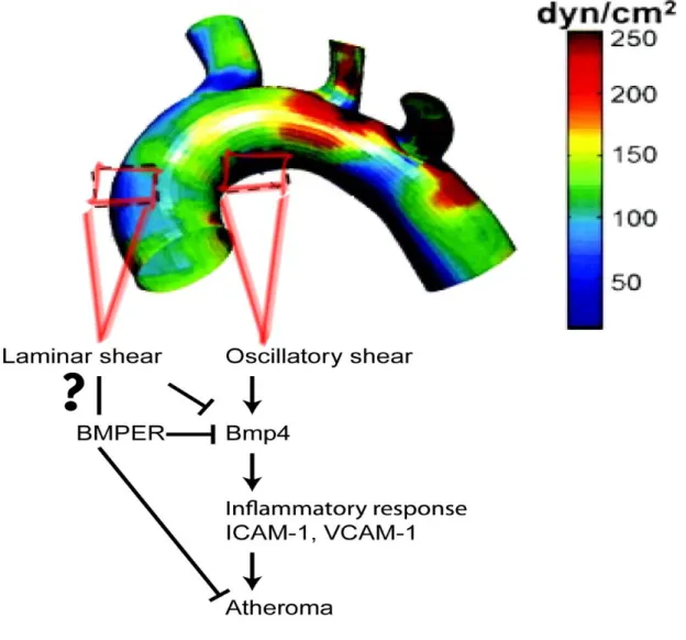
stress, resulting in low mean and oscillatory shear stress on the endothelial cells. In these lesion-prone regions, disturbed or oscillatory shear stress increases expression of bone morphogenetic proteins (BMPs) and their antagonists in the vascular endothelium.8,9 In turn,
BMPs activate an inflammatory response characterized by the expression of adhesion molecules, like intracellular adhesion molecule 1 (ICAM1) and vascular cell adhesion molecule 1 (VCAM1) on the endothelial surface.9 Despite this, animal studies examining the
atherosclerotic lesion formation when BMP activity is increased,10 whereas others conclude
that BMP plays a proinflammatory role.10,11 Although each of these studies support a central
role for BMP-mediated endothelial inflammation in atherosclerosis, some uncertainty remains about the precise contribution of BMP signaling to endothelial inflammation and atherosclerosis. BMPs belong to the transforming growth factor-β superfamily and play important roles in cellular processes, such as bone formation, proliferation, differentiation, motility, vasculogenesis, and angiogenesis (reviewed by Moreno-Miralles12). More
specifically, BMP4, together with BMP2 and BMP6, demonstrate important roles in endothelial differentiation, migration, and angiogenesis.13–16 Previously we identified
BMPER, a novel extracellular modulator of BMP signaling, which is required for hematopoietic and vascular development and hypoxia–induced retinal
neovascularization.13,14,17 We have also demonstrated that BMPER regulates BMP4 activity
in a dose-dependent manner.13 Recent reports show that BMPER is induced by
inflammatory-regulatory stimuli, such as oscillatory shear stress and mevastatin, and inhibits tumor necrosis factor-α–induced endothelial inflammation,18,19 suggesting that BMPER acts
in an anti-inflammatory capacity in endothelial cells by inhibiting BMP activity. This led us to question whether BMPER may also inhibit the endothelial inflammation and subsequent pathology associated with atherosclerosis. In this study, we used the apolipoprotein E– deficient (ApoE−/−) mouse atherosclerotic model to study the effects of BMPER
haploinsufficiency on the development of atherosclerosis. We used BMPER+/− mice instead of BMPER−/− mice because BMPER−/− mice die at birth.13 BMPER+/−;ApoE−/− mice fed a high-fat (HF) diet displayed an exacerbated inflammatory vascular response compared with ApoE−/− mice with the wild-type BMPER gene (BMPER+/+/ApoE−/−). Mechanistically, we demonstrate that BMPER exerts protective effects by inhibiting BMP activity. These data demonstrate for the first time that BMPER is a novel player in the development of
cells. Taken together, it suggests that BMPER is a novel protective regulator of vascular inflammation and vascular diseases, such as atherosclerosis.
2.2
RESULTS
2.2.1 BMPEREXPRESSION PROTECTS AGAINST ATHEROSCLEROTIC LESION FORMATION AND CALCIFICATION
Accumulated evidence suggests that BMPER protects endothelial cells from
inflammation by inhibiting BMP activity.18,19 Therefore, we used the ApoE−/− mouse model, in which a HF diet leads to accelerated atherosclerotic lesion formation and arterial
calcification, to analyze the in vivo effect of reduced BMPER expression on vascular inflammation. We crossed wild-type or ApoE−/− mice with mice that had either 1 or 2 functional BMPER alleles, resulting in 4 genotypes of experimental mice:
BMPER+/+/ApoE+/+, BMPER+/−/ApoE+/+, BMPER+/+/ApoE−/−, BMPER+/−/ApoE−/−. These mice were then fed either a standard chow or HF diet for 20 weeks, and the formation of atherosclerotic plaques in the aorta and aortic sinus regions was evaluated by Oil Red O staining. ApoE−/− mice that were haploinsufficient for BMPER expression
(BMPER+/−/ApoE−/−) responded to the HF diet with enhanced plaque formation compared with BMPER+/+/ApoE−/− mice (28.35 ± 2.23% versus 17.96 ± 3.00%, P=0.016), as measured by en face staining of aortic lesions (including both thoracic and abdominal aorta, Figure 2.1 A and 2.1B). In addition, cross-sectional analysis of aortic sinus lesions revealed that the plaques formed in the BMPER+/−/ApoE−/− mice were larger compared with lesions in BMPER+/+/ApoE−/− mice (0.38 ± 0.02% versus 0.29 ± 0.03%, P=0.018), demonstrating a protective effect of BMPER expression on the degree of plaque growth (Figure 2.1C and 2.1D). When lesion calcification was compared between genotypes, BMPER+/−/ApoE−/− mice again showed an exacerbated response, with a 120% increase over baseline levels of
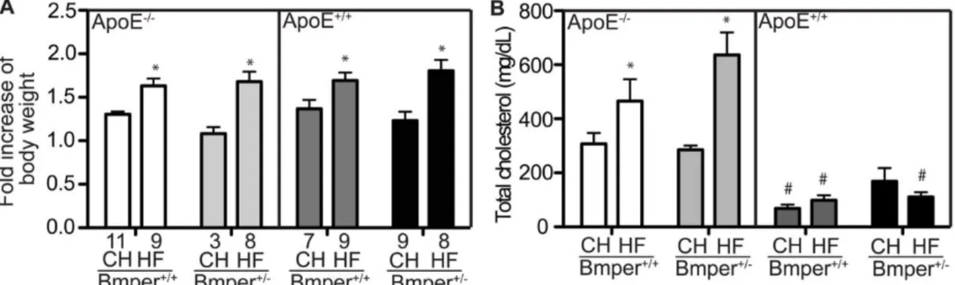
0.25% in BMPER+/−/ApoE−/− mice versus 0.37 ± 0.22% in BMPER+/+/ApoE−/− mice (Figure 2.1E and 2.1F). BMPER+/−/ApoE−/− mice fed the HF diet showed no differences in body weight or serum cholesterol levels compared with BMPER+/+/ApoE−/− mice (Figure 2.6 A and 2.6 B), indicating that diet induced increases in weight and lipid levels are not
responsible for the more robust atherosclerotic phenotype in the BMPER+/-/ApoE−/− mice. Together, these results indicate that BMPER plays a protective role in plaque formation and arterial calcification in an in vivo model of atherosclerosis.
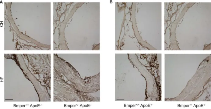
2.2.2 BMPEREXPRESSION INHIBITS AORTIC INFLAMMATION
Several reports have demonstrated a central role for the BMP signaling pathway in promoting endothelial inflammatory responses.8,9,18–20 Therefore, we sought to
determine whether the protective influence of BMPER on the development of atherosclerotic lesions is attributable to changes in vascular inflammation. To test this, we measured the degree of macrophage infiltration (a common phenotype observed with the onset of
expression of these proteins in response to vascular inflammation induced by HF diet. We observed a robust increase in BMPER (Figure 2.8 A) and BMP4 (Figure 2.8 B) levels in mice fed a HF diet compared with those fed a control diet. This increase in BMPER and BMP4 correlated with the increased expression of ICAM1/VCAM1 and CD68 signals (Figure 2.2), further supporting the notion that the modulation of BMP signaling by BMPER plays an important role in the inflammatory responses induced by HF diet. Collectively, these data demonstrate that BMPER haploinsufficiency leads to phenotypic changes correlative with an increased chronic, vascular inflammatory response and likely contributes to the aggravated atherosclerotic lesion formation observed in the BMPER+/−/ApoE−/− mice. 2.2.3 BMPERINHIBITS SHEAR STRESS–DEPENDENT INDUCTION OF INFLAMMATORY
ADHESION MOLECULES IN THE ENDOTHELIUM
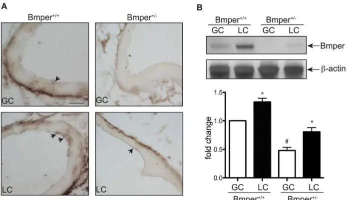
Aortic lesions develop predominantly in regions that are exposed to low or disturbed fluid shear stress, such as the LC of the aorta. Even under normal hemodynamic conditions, previous studies demonstrate higher endothelial inflammation in the LC compared with other regions of the aorta, such as the GC,3,4 possibly because of the increased expression of BMPs
in the vascular endothelium of these regions.8 This difference in lesion formation in the GC
observations, we used this inherent difference in BMPER expression between the LC and GC to analyze the effect of reduced BMPER expression on downstream mediators and effectors of BMP activation in these regions of mouse aortas. To simplify our experimental setup, we only examined mice on the ApoE+/+ background. Endothelial cells located in the LC region
of aortas of BMPER+/+ mice displayed abundant Smad1, 5, and 8 activation (as detected by
phosphorylated Smad1, 5, and 8 signals) compared with endothelial cells in the GC region (Figure 2.3A), consistent with previous reports. In contrast, BMPER+/− mice exhibited an enhanced increase in Smad activation in the LC region in comparison with BMPER+/+ mice;
but in addition, BMPER+/− mice had clearly detectable Smad activation in the GC region of the aortas, consistent with our in vivo data described above indicating that BMPER functions as an anti-inflammatory mediator (Figure 2.3B). Expression patterns of both ICAM1 and VCAM1 paralleled that of Smad activation (Figure 2.3C–2.3F), demonstrating that BMPER inhibits the endothelial response to oscillatory shear stress–mediated induction of endothelial inflammatory adhesion molecules, and in the context of atherosclerosis, may directly inhibit the endothelial inflammatory response.
2.2.4 BMPERINHIBITS BMP4-INDUCED INFLAMMATORY GENE EXPRESSION IN ENDOTHELIAL CELLS AND PREVENTS FLUID SHEAR STRESS–INDUCED INFLAMMATORY RESPONSES
demonstrating that BMPER directly antagonizes BMP4-mediated inflammatory signaling. Next, we examined the ability of endogenous BMP4 and BMPER to affect the expression of inflammatory markers. Given the robust inflammation seen in the normally quiescent GC region of the aorta in BMPER+/−/ApoE−/− mice, we hypothesized that reducing endogenous BMP4 or BMPER expression in endothelial cells would affect the inflammatory signature of the cells even in the absence of exogenous mediators. As expected, BMP4-targeted siRNA reduced expression of ICAM1 and VCAM1 in transfected cells, whereas siRNA-mediated reduction of endogenous BMPER expression resulted in a significant increase in these same inflammatory markers (Figure 2.4D and 2.4E). Collectively, our data demonstrate that the anti-inflammatory function of BMPER in endothelial cells is mediated, at least in part, through antagonizing BMP activity.
Given the effect of BMPER gene dosage on endothelial inflammation in vivo (Figure 2.3) and the in vitro effects of a reduction of BMPER expression on endothelial inflammation in the absence of any stimuli (Figure 2.4), we hypothesized that the inflammatory response to shear stress in the aorta may be mediated by changes in the levels of BMPER expression. To test this hypothesis directly, we subjected HUVECs to conditions that mimic the shear stress conditions in the LC and GC aortic regions using either oscillatory stress (±5 dyne cm−2) or laminar shear stress (20 dyne cm−2) for 8 hours, respectively. Consistent with our in vivo data, endothelial cells subjected to oscillatory shear had a larger inflammatory response (a 2.33-fold and 4.56-fold increase in ICAM1 and VCAM1 expression, respectively) compared with cells subjected to laminar shear, respectively (Figure 2.5A). Interestingly, we also observed increases in BMP4 and BMPER expression in oscillatory shear conditions
expression with control siRNA-treated cells. In control siRNA-treated cells, we detected higher ICAM1 and VCAM1 expression after oscillatory shear was compared with laminar shear (Figure2. 5C–2.5E), with similar patterns of BMP4 and BMPER expression as what was seen in untreated cells exposed to both modes of shear stress (Figure 2.5A). In BMPER siRNA-treated cells, we observed an increased inflammatory response in oscillatory shear conditions compared with control siRNA–treated cells (Figure 2.5C–2.5E), consistent with the increased inflammation we observed in the LC regions of BMPER+/− aortas (Figure 2.3). Additionally, even in the low inflammatory environment (laminar shear stress), reducing BMPER expression resulted in 34% and 68% higher expression of ICAM1 and VCAM1, respectively, compared with control siRNA– treated cells (Figure 2.5C–2.5E), paralleling our observations seen in the GC regions of BMPER+/− aortas (Figure 2.3). Given that laminar shear stress promotes endothelial survival and integrity by activating endothelial nitric oxide synthase (eNOS) signaling4 and that BMPER regulates eNOS protein expression and activity
under static conditions,19 we tested whether eNOS is regulated by BMPER under laminar or
2.3
DISCUSSION
In this study, we have identified a previously unrecognized, protective role for BMPER in the setting of atherosclerosis. ApoE−/− mice, which develop an atherosclerotic phenotype when fed a HF diet, displayed significantly worsened symptoms when they carried only 1 phenocopy of the BMPER gene (BMPER+/−;ApoE−/− mice). Not only did the decrease in BMPER levels in these mice cause a dramatic increase in lesion size, it also resulted in increased arterial calcification and a heightened induction of endothelial
inflammatory adhesion molecules in the aorta in regions subjected to oscillatory and laminar shear stress. Similar results were found in cultured cell studies, solidifying the notion that BMPER is a critical regulator of vascular inflammation and broadening our understanding of the role that BMPER plays in the myriad events that result from the BMP signaling pathway.
Previously, our studies revealed the essential roles of BMP and BMPER signaling in endothelial cell differentiation, migration, and angiogenesis.13,14,16,17,21,22 With the results
from the present study, we can now add vascular inflammation to the growing list of BMP signaling events that are regulated by BMPER. BMPER+/− mice demonstrated increased Smad activation and expression of the inflammatory markers ICAM1 and VCAM1 in the endothelium layer of the LC of the aorta, a region known to be predisposed to atherogenic activity attributable to the BMP-mediated vascular inflammation brought about by oscillatory shear stress effects on endothelial cells (Figure 2.3). In addition, decreased levels of BMPER in BMPER+/−/ApoE−/− mice led to an increase in the number of macrophages that were recruited and that migrated to the inflamed, atherogenic regions of the aortas (Figure 2.2A and 2.2B), supporting the notion that BMPER acts as a protective regulator of vascular inflammation. It is worth noting, however, that BMPER may also play a role in maintaining general vascular health in addition to its role in inflammatory responses. Our analysis of aortas taken from ApoE+/+ mice that were haploinsufficient for BMPER (BMPER+/−/ApoE+/+
regular diet (Figure 2.1A and 2.1B). This suggests that BMPER may be important in maintaining vascular health even under basal conditions, a theory that will require further experiments to determine.
As compelling as these in vivo results are, however, it is important to remember that the decrease in BMPER expression in the BMPER+/− mouse is not limited to endothelial cells. Because atherosclerosis is a pathological condition that results from the dysfunction of multiple cell types and involves different cellular events, it is not possible, from these in vivo studies, to localize the protective effect of BMPER to endothelial cells alone. Indeed,
published reports demonstrate that BMPs enhance smooth muscle cell migration and induce proinflammatory factors, such as inducible nitric oxide synthase and tumor necrosis factor in macrophages.23,24 The inhibition of BMP activity by specific inhibitors and antagonist
decreases vascular calcification, suggesting important roles of BMP in vascular calcification, as well as early vascular injury. Therefore, it is entirely possible that BMPER, a secreted extracellular BMP modulator, may also be able to influence BMP activity not only in endothelial cells but in additional cell types such as smooth muscle cells and macrophages. Therefore, the contribution of BMPER to protection against atherogenic processes and vascular inflammation will need further investigation.
As mentioned above, a number of published reports have detailed the role of
BMPER/BMP signaling in various aspects of endothelial cell function.13,14,16,17,21,22 However,
the ability of BMPER to inhibit endothelial inflammatory responses has not been investigated directly. To examine this aspect of endothelial BMP signaling, we cultured HUVECs and subjected them to fluid shear stress to induce an inflammatory response similar to what occurs within atherogenic areas of the aorta. We found that BMPER inhibits the
inflammatory effect on endothelial cells exposed to laminar shear stress. Laminar shear stress promotes endothelial survival and integrity by activating mitogen activated protein kinases, such as extracellular-signal regulated kinase/big mitogen-activated protein kinase 1 and endothelial NOS signaling.4 Our data demonstrated that BMPER is also involved in
protecting endothelial cells exposed to laminar shear stress, partly, because of increases in the activity of eNOS and decreases ICAM1 and VCAM1 expression (Figure 2.5C and 2.5F) that have recently been attributed to BMPER activity in endothelial cells.19 The exact
molecular mechanism through which BMPER modulates eNOS activity and other signaling pathways of laminar shear stress need further investigation. BMPER has been identified as a critical regulator of BMP signaling activity, important in both vascular development and in hypoxia-induced retinal neovascularization.13,14 Previously, we reported a gradient effect of
BMPER’s ability to influence BMP signaling, whereby superstoichiometric concentrations of BMPER compared with BMP inhibit BMP signaling and substoichiometric concentrations of BMPER compared with BMP activate BMP signaling.13 In this study, we observed 61% and
15% higher BMP4 and BMPER expression in oscillatory shear stress conditions, resulting in a significantly lower BMPER to BMP4 ratio compared with laminar shear stress conditions, an expression pattern consistent with an anti-inflammatory role for BMPER (Figure 2.5A and 2.5B). This observation further suggests that the fine-tuning of BMP activity by BMPER is essential for modulating BMP-mediated cellular functions. Collectively, the data presented in this report demonstrate that the regulation of BMP activity by BMPER is essential for the maintenance of normal vascular homeostasis (Figure 2.11) and its disruption increases the risk of inflammatory vascular diseases, such as atherosclerosis.
2.4
MATERIALS AND METHODS
2.4.1 ANIMALS AND DIETS
ApoE-/- mice on C57BL/6J background were kindly provided by Dr. Maeda (UNC,
genetic background1, were crossed with ApoE-/- mice to generate BMPER+/-/ApoE-/- mice.
We used the BMPER+/- mice instead of BMPER-/- mice because BMPER-/- mice die at birth.
All adult mice were fed with the standard chow or a high-fat/highcholesterol diet (Western diet) (Harlan Laboratories, Indianapolis, IN) for 20 weeks. Body weight of mice was monitored before and after they were fed with different diets. Blood serum was obtained every four weeks. All mouse experimental procedures were performed according to the National Institutes of Health Guide for the Care and Use of Laboratory Animals and approved by the Institutional Committee for the Use of Animals in Research. 2.4.2 LIPID ANALYSIS
Mice were fasted for 18 hours before blood sampling. Less than 200 μL of blood was collected through submandibular bleeding using a lancet. The total cholesterol level was measured enzymatically with a commercially available kit (Infinity kits, Thermo Scientific, Waltham, MA).
2.4.3 LESION QUANTIFICATION
previously described protocol. Twenty sections were stained with eosin and Oil Red O. The slides were imaged by light microscopy, and the atherosclerotic lesion area located in aortic sinus area was quantified with ImageJ and averaged over a 280 μm region.
2.4.4 CALCIFICATION QUANTIFICATION
Deposited calcium in the aorta was detected by staining with von Kossa. The 5 μm cryosections of aortic sinus were prepared as described above and subjected to the von Kossa staining procedure. The calcification area from each section was quantified as a percentage of the total vessel cross-sectional area using ImageJ software.
2.4.5 ELISAMEASUREMENTS
Blood samples were drawn from mice after consuming the HF diet or standard chow for 4 weeks. Soluble VCAM and soluble ICAM were measured in plasma in triplicate using an ELISA method (R&D Systems Minneapolis, MN).
2.4.6 REAGENTS
Recombinant human BMP4 and BMPER protein and antibodies recognizing BMPER and BMP4 were obtained from R&D Systems. VCAM1 antibodies were obtained from Santa Cruz Biotechnology (Santa Cruz, CA) for Western blotting and Chemicon for
immunofluorescence (Millipore, Billerica, MA). The ICAM1 antibody was purchased from Cell Signaling Technology (Danvers, MA) and was used for Western blotting experiments. An additional ICAM1 antibody (purchased from Chemicon) was used for
immunofluorescence experiments. The phosphorylated Smad1, 5, and 8 antibodies were purchased from Cell Signaling Technology and used for both Western blotting and immunofluorescence experiments.
2.4.7 CELL CULTURE AND SIRNATRANSFECTION
hydrocortisone, bovine brain extract, epidermal growth factor, and 2% fetal calf serum. The cells from passages 4–8 were used for experiments. The stealth small interfering RNA (siRNA) duplexes were obtained from Invitrogen (Grand Island, NY). The siRNAs against mouse BMPER are a mixture of the duplexes of
5ʹGAAUUUCAGCCAGAAGGAAGCAAAU-3ʹ and
5ʹ-GGAGAGAUGUGGUCCUCUAUCAAUU-3ʹ. The siRNA against mouse BMP4 is a duplex of 5ʹ-GCAUGUCAGGAUUAGCCGAUCGUUA-3ʹ. The control siRNA is the Stealth RNAi negative control duplex (Cat. No. 12935-300) and was purchased from Invitrogen. The siRNAs were transfected into HUVECs according to the manufacturer’s recommended protocol for Nucleofection (Lonza, Walkersville, MD; the HUVEC protocol). Briefly, for each sample, 2×10⁵ HUVECs were transfected with 300 pmol siRNA. The
experiments with BMP4 or BMPER siRNA-transfected HUVECs were performed 1 day or 4 days later, respectively. The siRNAs resulted in more than 70 percent knockdown of the protein levels of BMP4 and BMPER.
2.4.8 SHEAR STRESS ASSAYS
HUVECs were post-confluent for 48 hours before the performance of fluid shear stress experiment to decrease the background signal. Laminar shear stress assay was described previously. Briefly, confluent cells in a 10-cm dish were exposed to shear stress using the cone- and plate-flow chamber system for 8 hours at 20 dyne/cm2 for laminar shear
stress or ± 5 dyne/cm2 for oscillatory shear stress experiments. 2.4.9 IMMUNOBLOTTING
Cells were harvested in lysis buffer (1% Triton X-100, 50 mmol/L Tris (pH 7.4), 150 mmol/L NaCl, 1 mmol/L Na3VO4, and 0.1% protease inhibitor mixture; Sigma) and
2.4.10 IMMUNOFLUORESCENCE
The aortic arch segments were dissected out and gently cleaned of the adventitia. The aortic fragments located at the greater curvature (GC) and lesser curvature (LC) were
separated and fixed in 3.7% formaldehyde for 10 minutes at room temperature. The aortic fragments were sequentially treated with 70% ethanol for 30 minutes and 5% hydrogen peroxide in methanol. Then, the segments were washed with water for 5 minutes. For the phosphorylated Smad1, 5, and 8 antibody, the samples were soaked in boiling citric acid buffer (10 mmol/L; pH, 6.0) for 9 minutes to expose the antigens. Next, the aortic fragments or 5 μm cryosections of the aortic root were blocked with 5% heat-inactivated goat serum for 1 hour and then incubated overnight with primary antibodies against ICAM1, VCAM1, CD31, or CD68 diluted in the blocking solution. After 3 washes in tris buffered saline, cells were incubated in the dark with a second antibody conjugated with Alexa Fluor 488 or 568 (Molecular Probes, Eugene, OR) in blocking solution for 90 minutes at 37°C. After 3 washes in tris buffered saline, the fragments were counterstained with DAPI for phosphorylated Smad1, 5, and 8 staining. The en face images of the endothelial layer and the cross-sectional images of the aortic root were visualized by confocal laser scanning microscopy.
2.4.11 IMMUNOHISTOCHEMISTRY
The aortic arch segments were dissected out and gently cleaned of the adventitia. The aortic fragments located at the greater curvature (GC) and lesser curvature (LC) were
separated and fixed in 3.7% formaldehyde for 10 minutes at room temperature. After
incubating tissue in a sucrose gradient, aortic samples were embedded in OCT compound and submitted for frozen sectioning. For the anti-BMPER and BMP4 antibody, the samples were soaked in boiling citric acid buffer (10 mmol/L, pH 6.0) for 10 minutes to expose the
antigens. Next, the 5 μm cryosections of the aortic root or aortic arch were blocked with 5% heat inactivated rabbit serum for 1 hour and then incubated overnight with primary
washes in TBS, samples were incubated in a second antibody. For BMPER staining, we utilized amplification processes including the serial incubation with ABC (Vector Labs, Burlingame, CA, USA) and tyramide signal amplification reagent (Waltham, MA, USA) in blocking solution for 30 minutes for each at 37 °C. After 3 washes in TBS, the sections stained with BMPER and BMP4 antibodies were developed with DAB. The images were recorded using the bright field microscopy with 10x and 20x objective lens.
2.4.12 STATISTICAL ANALYSIS
Figure 2-1. BMPER haploinsufficiency leads to aggravated atherosclerotic plaque formation.
percentage of the total area of the aorta. *P<0.05, compared with mice with the same genotype but fed the control diet. #P<0.05, compared with mice fed with the same diet but with the ApoE−/− genotype. The numbers below each column are the number of mice used in the experiments. (C) Representative images of Oil Red O–stained sections of aortic sinus regions from mice of the designated genotype and food groups. Scale bar, 0.2 mm. (D) The lesions in the aortic sinus region were quantified as the percentage of the total lumenal area of aortas. *P<0.05, compared with mice with the same genotype but fed the control diet, n≥4. There were no detectable lesions formed in the aortic sinus regions of ApoE+/+ mice. (E) Representative images of calcification in the aortas and
Figure 2-2. BMPER haploinsufficiency increases macrophage infiltration and intimal inflammation in ApoE−/− mice.
Figure 2-3. BMPER haploinsufficiency results in increased BMP activity and expression of inflammatory adhesion molecules in the ApoE+/+ endothelium of the greater curvature (GC) and lesser curvature (LC) of the aortic arch.
(A) En face staining of the GC and LC in the aortic arch of BMPER+/- and BMPER+/- mice was
performed using an antibody specific for phosphorylated Smad (pSmad) 1, 5, and 8 (green). Nuclei were counterstained with DAPI (blue). Arrows indicate pSmad positive nuclei. Scale bar, 10 μm. (B) pSmad positive nuclei were quantified as a percentage of all nuclei per field. *P<0.05 compared with pSmad positive nuclei (%) in the GC region of the same mice; #P<0.05 compared with pSmad positive nuclei (%) in the same region of wild-type littermates, n=3. (C–F) En face staining was performed for intracellular adhesion molecule 1 (ICAM1) (green in C) and vascular cell adhesion molecule 1 (VCAM1) (green in E) and the endothelial marker CD31 (red in C and E). Scale bar, 10 μm. (D and F) the average intensity of ICAM1 (D) and VCAM1 (F) was measured as fold change over the level of intensity in the GC of wild-type mice. *P<0.05 compared with the relative
Figure 2-4. BMPER inhibits BMP4– induced ICAM1 and VCAM1 expression.
A) Human umbilical vein endothelial cells (HUVECs) were subjected to oscillatory shear stress (OS, ± 5 dyn/cm2) or laminar shear stress (LS, 20 dyn/cm2) or remained static in the incubator for 8 hours.
Cell lysates were used for immun0-blotting to detect the expression of BMPER and BMP4 and the induction of inflammatory adhesion molecules ICAM1 and VCAM1. (B) The band intensity of BMP4 and BMPER (A) was quantified with ImageJ and is presented as the ratio of BMP4/BMPER protein level compared with the LS condition. *P<0.05 compared with the cells exposed to laminar shear stress, n=4. C, HUVECs were transfected with BMPER or control (small interfering RNAs) siRNAs. Four days later, cells were exposed to OS (± 5 dyn/cm2), LS (20 dyn/cm2), or static
with the cells exposed to LS, n=3. (F) HUVECs were transfected with BMPER or control siRNAs. Four days later, cells were exposed to OS (± 5 dyn/cm2), LS (20 dyn/cm2), or static conditions for 8
Figure 2-6. The changes in body weight and total cholesterol level of BMPER/ApoE mice after 20 weeks on standard chow (CH) or high fat diet (HF).
Figure 2-7. sICAM1 and sVCAM1 plasma levels increased in BMPER+/- mice after 4 weeks consuming the high fat diet.
Figure 2-9. 9 BMPER haploinsufficiency leads to aggravated atherosclerotic plaque formation in aortic arch area.
Mice were fed a high fat diet (HF) or standard chow (CH) for twenty weeks. The aortas were dissected out and stained with Oil Red O. The lesions on the surface of each greater and lesser curvature of aorta arch were quantified as a percentage of the total area of GC and LC. *, P<0.002, compared to that lesions located in GC of the same mouse. #, P<0.05, compared to BMPER+/+ mice
Figure 2-10. BMPER protein level was increased in the LC compared to GC.
(A) The regions of GC and LC located in aortic arch of mouse aortas were processed for staining with BMPER antibody. Scale bar, 200 μm. The arrows represent the positive staining of BMPER protein. Scale bar: 200 μm. (B) The vessel lysates obtained from the GC and LC region of mice were
CHAPTER 3: LRP1-DEPENDENT BMPER SIGNALING REGULATES
LPS-INDUCED ACUTE VASCULAR INFLAMMATION
3.1
INTRODUCTION
Sepsis is a common cause of significant morbidity and mortality worldwide. It is the tenth leading cause of overall death in the United States, and the incidence of sepsis is 750,000 and costs $17 billion dollars annually creating a major healthcare burden.25–27
Despite being one of the oldest clinical syndromes in medicine, there is no specific or targeted therapy and the number of patients has continued to increase by 9% each year over the past two decades.25 Treatment has been constrained to the useage of antibiotics and
supportive care. This underscores the fact that fundamental gaps remain in our knowledge of the pathiophysiological mechanisms that drive sepsis. Injury and dysfunction of multiple organs are a clinical manifestation of sepsis, with the lung most commonly involved. Sepsis often leads to ALI or its more severe form, ARDS whose distinguishing features are
impairment of pulmonary vascular integrity and endothelial dysfunction. The bacterial endotoxin, lipopolysaccharide (LPS), is one of the leading contributing factors to ALI in sepsis.
eventually result in increased vascular permeability, allowing increased flux of proteins, fluid and immune cells across vessels into tissues. However, the underlying mechanisms
mediating endothelial activation in response to LPS remain largely unknown.
Toll-like receptor 4 (TLR4) has been recognized as the major receptor for LPS. Ligation of LPS to TLR4 activates NFκB and nuclear factor of activated T cells (NFAT) pathways. In human umbilical vein endothelial cells (HUVECs), TLR4, MyD88 and Mal/TIRAP adaptor proteins are required for the activation of NFκB and production of interleukin-6 (IL-6).28 Animal studies with inactive NFκB signaling indicate that, in response
to LPS-induced endotoxemia, NFκB activation acts as a quick adaptive response, which provides an important survival signal and maintains a normal but dynamic endothelial barrier function.29 On the other hand, LPS activates transcriptional factor NFAT in endothelial cells,
which results in increased expression of pro-inflammatory cytokines such as IFN-γ and TNFα.30 The activation of NFAT is mediated by reactive oxygen species (ROS)-driven Ca2+
signaling pathway and TLR4 may be required for ROS generation.30 In activated immune
cells such as dendritic cells, CD14 is another receptor of LPS leading to NFAT activation, likely through Src-family kinase and phospholipase Cγ2, and Ca2+/calcineurin signaling.31
In endothelial cells, CD14 has also been suggested to function together with TLR4 to mediate LPS-induced E-selectin and IL-6 production.32 Although the exact roles of TLR4
and CD14 in LPS-mediated NFAT activation and endothelial inflammatory responses remain to be further clarified, NFAT activation is recognized as a crucial transcription factor
controlling the expression of pro-inflammatory cytokines.
Endothelial integrity and vascular homeostasis are tightly regulated by multiple signaling pathways such as bone morphogenetic protein (BMP) signaling.33 BMP-binding
endothelial regulator (BMPER), an extracellular regulator of BMPs,13,21 has been identified
as an important regulator of vascular inflammation and atherosclerosis.18,19,34 Knockdown of
BMPER by its specific siRNA or its deficiency in BMPER+/- mice potentiates TNFα
is mediated by blocking BMP activity, which likely explains the atheroprotective function of BMPER.34 However, it is not clear whether BMPER regulates inflammatory responses in
endothelial cells in an acute setting, such as LPS challenge. We have recently demonstrated that LDLR-related protein (LRP) 1, a member of the LDL receptor family, binds with BMPER at the cell membrane, the complex is endocytosed and has been shown to regulate angiogenesis during zebrafish vein development.35 The mature form of LRP1 is a
heterodimer composed of a 515-kDa α chain (LRP1α), possessing four extracellular ligand binding domains, and an 85-kDa membrane-anchored cytoplasmic β chain (LRP1β), which remains non-covalently associated with α chain. LRP1 is an endocytic receptor for multiple signaling pathways and mediates their signals through its β chain, which interacts with many scaffolding proteins.36–42 In addition, processed forms of LRP1β can also translocate into
nucleus and regulate the enzyme activity of PARP143 and expression of PPAR target genes
by acting as a PPARγ co-activator.44 Although LRP1 is required for the endocytosis of
BMPER signaling complex in endothelial cells, it remains elusive whether the coupling of BMPER to LRP1 may also initiate their own signaling events.
BMPER null mice die at birth.13 In this study, we used BMPER haploinsufficient
mice to study the effect of reduced BMPER expression on LPS-induced endothelial inflammation. Surprisingly, we observed that BMPER+/- mice exhibit reduced vascular
inflammatory responses upon LPS treatment as shown by several parameters, including endothelial permeability, pulmonary edema, survival rate and production of
3.2
RESULTS
3.2.1 LPS-INDUCED LUNG INJURY AND MORTALITY IS REDUCED IN BMPER+/-MICE
BMPER has been well characterized in the setting of chronic inflammation and atherosclerosis and established as a protective mediator of these processes by inhibiting BMP activity in the endothelium.18,19,34 However, it is unknown how BMPER regulates vascular
responses during acute inflammation. We used a LPS-induced model of endotoxemia and BMPER+/- mice (BMPER-/- mice die at birth.13) to investigate the in vivo role of BMPER in
acute inflammation of the endothelium. We first challenged BMPER+/- mice and their
littermate controls (BMPER+/+ mice) with a lethal dose of LPS (15 mg/kg i.v.). We were
surprised to discover all BMPER+/+ mice died within 2 days while the majority of BMPER
+/-mice recovered with a 7-day survival rate at approximately 62.5 percent (Figure 3.1A). Next we evaluated histopathological changes in the lungs of BMPER+/- and BMPER+/+ mice 12
hours after a sub-lethal dose of LPS (10 mg/kg i.v.). We observed an increased number of inflammatory cells in the interstitum and alveolar space, proteinacous debris in the alveolar space, interalveolar septal thickening and interstitial edema in BMPER+/+ mice (Figure 3.1B),
while BMPER+/- mice exhibited much lower instances of these indices of lung injury and had
a much reduced lung injury score (Figure 3.1C). These data suggest that BMPER haploinsufficiency protects against endotoxemia-induced pulmonary injury. 3.2.2 LPS-INDUCED ENDOTHELIAL PERMEABILITY IS LESS SEVERE IN BMPER
HAPLOINSUFFICIENT MICE
To extend our in vivo analysis, we next compared the several lung injury-associated parameters of endothelial dysfunction, pulmonary edema and BALF protein concentration.
When BMPER+/+ control mice were challenged with a sub-lethal dose of LPS (10 mg/kg
i.v.), they rapidly developed symptoms consistent with sepsis (e.g., lethargy, ocular
discharge; data not shown), while BMPER haploinsufficient mice (BMPER+/-) demonstrated
less severe symptoms. Extravasation of Evans blue dye (EBD) from circulation into the lung
elevated capillary permeability. We observed that extravasated EBD content in the lung
tissue of BMPER+/- mice was significantly less than that from BMPER+/+ mice (Figure
3.2A,B) as well as total protein collected from broncioalveolar lavage fluid (BALF).
Similarly the lung wet/dry weight ratio, a measure of pulmonary edema, and the total protein
concentration in the BALF of BMPER+/- mice, following LPS injection was significantly
smaller than that of BMPER+/+ mice (Figure 3.2C, D). Taken together, these data suggest that
BMPER haploinsufficiency protects against pathological endothelial pulmonary
permeability.
3.2.3 LPS-INDUCED PRO-INFLAMMATORY CYTOKINE PRODUCTION AND LEUKOCYTE MIGRATION ARE ATTENUATED IN BMPER+/- MICE
Endotoxemia-induced lung inflammation is associated with increases in the
production of cytokines, both systemically and in the alveolar compartment, and leukocyte infiltration into target tissues. Therefore, we evaluated the concentration of LPS-induced pro-inflammatory cytokines in the serum of BMPER+/+ and BMPER+/- mice. IFNγ, IL-6 and
TNF-α were induced in BMPER+/+ mice by LPS, while BMPER+/- mice had a significantly
lower response (Figure 3.3A-C). To further assess the influence of BMPER
haploinsufficiency on pulmonary inflammation, we determined in the BALF several
parameters of lung inflammation, including cell infiltration, myeloperoxidase (MPO) activity and the levels of IFNγ, IL-6 and TNF-α. In response to LPS, both BMPER+/+ and BMPER
+/-mice had increased leukocyte infiltration in the BALF, but the cell count in BALF of BMPER+/- mice was significantly fewer than those in BMPER+/+ mice (Figure 3.4A).
Similarly, myeloperoxidase (MPO) activity was also dramatically lower in BMPER+/- mice,
compared to BMPER+/+ mice (Figure 3.4B) suggesting that BMPER haploinsufficiency