Coagulation Abnormalities of Sickle Cell Disease: Relationship
with Clinical Outcomes and the Effect of Disease Modifying
Therapies
Denis Noubouossie, MD, PhD, Nigel S. Key, MBChB, and Kenneth I. Ataga, MBBS Division of Hematology/Oncology, University of North Carolina at Chapel Hill, USA
Abstract
Sickle cell disease (SCD) is a hypercoagulable state. Patients exhibit increased platelet activation, high plasma levels of markers of thrombin generation, depletion of natural anticoagulant proteins, abnormal activation of the fibrinolytic system, and increased tissue factor expression, even in the non-crisis “steady state.” Furthermore, SCD is characterized by an increased risk of thrombotic complications. The pathogenesis of coagulation activation in SCD appears to be multi-factorial, with contributions from ischemia-reperfusion injury and inflammation, hemolysis and nitric oxide deficiency, and increased sickle RBC phosphatidylserine expression. Recent studies in animal models suggest that activation of coagulation may contribute to the pathogenesis of SCD, but the data on the contribution of coagulation and platelet activation to SCD-related complications in humans are limited. Clinical trials of new generations of anticoagulants and antiplatelet agents, using a variety of clinical endpoints are warranted.
Keywords
Sickle cell disease; Coagulation activation; Platelet activation; Hemolysis; Inflammation; Complications
1. Introduction
Sickle cell disease (SCD) refers to a group of genetic disorders defined by the presence of sickle hemoglobin (HbS), chronic hemolysis and multi-organ morbidity. More than 300 000 children were born with sickle cell anemia (SCA), the homozygous form of SCD, in 2010 (1) and it is predicted that more than 400 000 children will be born annually by 2050 (2). Comprehensive care in resource-rich countries, including newborn screening, infection prophylaxis with penicillin, and hydroxyurea therapy, has improved the survival as well as
Corresponding Author: Kenneth I. Ataga, MBBS, Division of Hematology/Oncology, University of North Carolina at Chapel Hill, Physicians’ Office Bldg., 3rd Floor, CB# 7305, 170 Manning Drive, Chapel Hill, NC 27599-7305, USA, Tel.: +1-919 843-7708, Fax: +1-919 966-6735, [email protected].
Conflict of interest
The authors declare no conflict of interest associated to this article.
Publisher's Disclaimer: This is a PDF file of an unedited manuscript that has been accepted for publication. As a service to our
customers we are providing this early version of the manuscript. The manuscript will undergo copyediting, typesetting, and review of
HHS Public Access
Author manuscript
Blood Rev
. Author manuscript; available in PMC 2017 July 01.Published in final edited form as:
Blood Rev. 2016 July ; 30(4): 245–256. doi:10.1016/j.blre.2015.12.003.
A
uthor Man
uscr
ipt
A
uthor Man
uscr
ipt
A
uthor Man
uscr
ipt
A
uthor Man
uscr
the quality of life of individuals with SCD (3). In addition to its well-known hemolytic and vaso-occlusive complications, SCD is characterized by a variety of thrombotic
complications, including ischemic stroke (4). Furthermore, multiple recent studies show that patients with SCD have an increased risk of venous thromboembolism (5–8). The high prevalence of thrombotic complications, combined with the well documented hemostatic alterations in the direction of a procoagulant phenotype shows that SCD can be considered to be a true hypercoagulable state (9–13). In an attempt to improve our understanding of the role of hypercoagulability in the pathogenesis of SCD, many groups have addressed the link between coagulation activation and various clinical manifestations of the disease. Using data from animal models and patients, the current review provides an update on coagulation abnormalities in SCD, their relationship with selected clinical complications, the effect of current disease-modifying treatments, and summarizes the published studies of
anticoagulants and anti-platelet agents.
2. Hemostatic alterations of SCD
2.1. In vivo thrombin and fibrin generation
Chronic activation of coagulation is commonly observed in patients with SCD at ‘steady-state’ compared to healthy control subjects with normal hemoglobin. This is evidenced by increased plasma levels of in vivo markers of thrombin and fibrin generation, including thrombin-antithrombin complexes (TAT), prothrombin fragment 1.2 (F1.2), fibrinopeptide A, D-dimers and plasmin-antiplasmin complexes (PAP) (14–21). There are conflicting reports regarding further increases in coagulation activation markers during painful crises as compared with the non-crisis, ‘steady-state” (14–21). There are also conflicting reports on the association between markers of coagulation activation and the frequency of painful crisis. A significant correlation was reported between D-dimer levels measured during the non-crisis state and the frequency of pain crises the following year (22). In addition, plasma D-dimer level was inversely correlated with the time to the next pain episode (22). However, no associations were found between both plasma TAT and D-dimer levels obtained at steady state and the frequency of acute pain crises in other studies of adults and children with SCD (23,24). The reason for these conflicting data is uncertain, but may be related to the difficulty in accurately defining the steady state in patients with SCD.
2.2. Ex vivo thrombin generation assays and thromboelastography
The capacity to generate thrombin reflects the balanced effect of all components of the coagulation cascade (both pro- and anticoagulant) and correlates with the bleeding or thrombotic phenotype (25,26). Thrombin generation assays (TGA) reliably assess an individual’s rate and potential to generate thrombin ex vivo in plasma and possibly in whole blood, following a calibrated trigger of coagulation (27,28). Although multiple studies are published (29–32), the results of ex vivo TGA in SCD patients at “steady state” compared with age-matched controls or with patients during acute painful episodes are inconsistent. This inconsistency may be due to heterogeneity in the genotypes and treatments of enrolled subjects, lack of race-matched controls in some studies, variability in the timing of blood collection, sample preparation and/or the analytical conditions of the assays (Table 1). Differences in these parameters have been shown to result in large inter-center variability of
A
uthor Man
uscr
ipt
A
uthor Man
uscr
ipt
A
uthor Man
uscr
ipt
A
uthor Man
uscr
results (33). Using a model of whole blood thrombin generation, higher maximum levels of
αTAT were generated in adults with HbSS at steady state than in race-matched controls,
irrespective of the intrinsic or extrinsic pathway of coagulation activation, in line with the increased peak of thrombin generation in platelet-poor plasma (PPP) (34).
Thromboelastography, another tool to assess global coagulation, measures the viscoelastic changes of a clotting sample from initiation to the formation of a stable clot. Whole blood is the common sample type used for thromboelastographic assessments. It is believed that the outcome reflects the effect of both plasma and cellular blood components including
platelets, white blood cells and red blood cells (RBC) that are altered in SCD. Children with HbSS and HbSC had higher angle, higher maximum amplitude and higher coagulation index values (a computed parameter designed by the manufacturer which measures the global coagulability of the sample) at “steady state” compared to race-matched controls (35). The reaction time was reduced in HbSS patients in the “steady state” compared with controls. In HbSS patients, maximum amplitude and coagulation index increased further during painful episodes (35). Finally, the reaction time of the thrombogram was positively correlated with protein C and protein S levels, alpha angle correlated with platelet count, and the maximal amplitude and coagulation index correlated with D-dimer levels (35).
2.3. Tissue factor and contact system activation
Tissue factor (TF), the physiological trigger of coagulation, is normally separated from contact with plasma proteins by an intact layer of endothelial cells, thus preventing coagulation activation. In patients with HbSS and compound heterozygous forms of SCD, increased levels of circulating TF are expressed by endothelial cells, monocytes and microparticles derived from these cells (20,36–38). As SCD is associated with endothelial injury, it is also likely that sub-endothelial TF is exposed to circulating blood at sites of vascular injury. No difference was observed in whole blood TF procoagulant activity between HbSS and HbSC patients in one study (20). However, a smaller study reported a significantly increased percentage of TF-positive monocytes and whole blood TF activity in HbSS compared with HbSC, although no difference was seen in TF-positive microparticles (36). The number of TF-positive monocytes, TF-positive circulating endothelial cells and TF-positive microparticles derived from these cells appear to increase during painful episodes compared to the non-crisis, “steady state” (37,38), although no difference in whole blood TF procoagulant activity was observed between these two clinical states (20).
Multiple studies show associations of markers of hemolysis with whole blood TF procoagulant activity, TF-positive monocytes, as well as plasma markers of thrombin and fibrin generation in patients with SCD (24,36,39,40). Heme, an inflammatory mediator and a product of intravascular hemolysis, induces functional TF expression in cultured human umbilical vein endothelial cells and human lung microvascular endothelial cells
independently of IL-1α and TNFα (41). Heme has also been reported to increase TF
expression on human blood mononuclear cells via toll-like receptor 4 (42) and on mouse leukocytes, although not on mouse lung endothelial cells (43). Increasing the bioavailability of nitric oxide (NO) either by breathing NO, addition of arginine, an NO precursor, to the diet or by breeding the animals to overexpress endothelial NO synthase led to significant
A
uthor Man
uscr
ipt
A
uthor Man
uscr
ipt
A
uthor Man
uscr
ipt
A
uthor Man
uscr
reduction in endothelial TF expression in two mouse models of SCD, thus demonstrating a role for NO in endothelial TF regulation and coagulation activation in SCD (44). At steady state, endothelial TF expression in the pulmonary veins is increased in sickle mice with severe disease phenotypes (BERK and S+S-Antilles mice), but it is similar in mild phenotypes (NY1DD and SAD mice) and non-sickle, control mice (45,46). Transient hypoxia-induced stress in sickle mice with mild disease phenotypes leads to up-regulation of endothelial cell expression of TF in the pulmonary veins (45,46), indicating a role for ischemia-reperfusion injury in TF expression in SCD. Increased TF expression in the
pulmonary veins following hypoxia-reoxygenation is primarily dependent upon NFκB
activation in monocytes (47). Recent data from animal models suggest that in addition to initiating coagulation, TF may trigger other biological pathways, including inflammation and vascular injury. Inhibition of TF with a blocking antibody effectively prevented the accelerated thrombus formation observed in mice that express hemoglobin S in a light/dye-induced model of cerebral microvascular thrombosis (48). Since increased TF expression was not detected in the cerebral vasculature of these mice, it is likely that the TF responsible for the effect was expressed on circulating hematopoietic cells. Antibody-mediated blockade of TF in another mouse model of SCD significantly reduced plasma levels of TAT,
interleukin-6 (IL-6), soluble vascular cell adhesion molecule-1 (VCAM-1), and serum amyloid protein, as well as neutrophil infiltration in the lung evaluated by the measurement of myeloperoxidase activity (49). In addition, specific deletion of the TF gene in endothelial cells reduced plasma level of IL-6 without affecting the plasma level of TAT, suggesting that endothelial TF plays a role in inflammation but not in coagulation activation in this mouse model (49).
In patients with SCD, markers of in vivo thrombin and fibrin generation, including plasma TAT, F1.2 and D-dimer, show only moderate or no correlation with whole blood TF procoagulant activity, total TF-positive microparticles, and TF-positive microparticles derived from monocytes or endothelial cells (20,36,38). While this may reflect the contribution of endothelial and sub-endothelial TF at sites of vascular injury that is not measured in blood, it may also reflect a contribution of activation of the intrinsic pathway to in vivo thrombin generation. Indeed, plasma levels of contact system proteins, including factor XII, prekallikrein and high molecular weight kininogen have been shown to be decreased in patients with SCD at “steady state” compared with control subjects, with further decreases during acute painful episodes (50–52). Autoactivation of contact system proenzymes is known to occur on negatively charged surfaces. Potential candidates for contact system activation in vivo include polyphosphates, nucleic acids, misfolded proteins, heparan sulfate, sulfatides, collagen and phosphatidylserine (53,54). Polyphosphates may be released by activated platelets (55) and an increased number of circulating microparticles are described in SCD (see below). In addition, increased levels of cell-free DNA and
nucleosomes released by activated neutrophils, and possibly other cells, have been detected in the plasma of SCD patients at “steady state,” with accentuated levels during acute painful episodes and acute chest syndrome (56–58). Kininogen deficient mice transplanted with bone marrow from Townes sickle mice show lower levels of plasma TAT compared to
normal kininogen littermates (59). In addition, in a model of TNFα-induced vaso-occlusive
crisis in Townes sickle mice, elevation of plasma levels of TAT strongly correlates with
A
uthor Man
uscr
ipt
A
uthor Man
uscr
ipt
A
uthor Man
uscr
ipt
A
uthor Man
uscr
elevation of plasma levels of cell-free DNA (59). Together, these data suggest a potential role for the contact system in the hypercoagulability of SCD.
2.4. Platelet activation, red blood cells and microparticles
Platelets are activated during the “steady state,” with further activation during acute painful episodes, as evidenced by increased levels of soluble markers of platelet activation including platelet-derived soluble CD40 ligand (60,61), platelet factor 3 (62), platelet factor 4
(22,63,64), beta-thromboglobulin (22,63,64) and thrombospondin-1 (65); decreased platelet content of thrombospondin-1 (65) and CD40 ligand (60,65); increased expression of surface markers of activation including P-selectin (22,66,67), CD63 (66), activated glycoprotein IIb/ IIIa (22,66) and phosphatidylserine (19,22); increased numbers of platelet-platelet (63), platelet-erythrocyte (68), and platelet-leukocyte aggregates (61); and increased numbers of platelet-derived microparticles. Functional assays show enhanced platelet aggregation in adult patients at “steady state” compared with control subjects, (63,69) while aggregation is reduced in children (70,71). As an increase in platelet aggregation is observed in
splenectomized non-SCD adults (69), it is hypothesized that the enhanced platelet aggregation in adults with SCD is due to the high number of circulating young and hyperactive platelets secondary to autosplenectomy (72). Platelet procoagulant activity is significantly increased in patients during acute pain episodes compared to the non-crisis state, and is significantly correlated with the number of acute pain episodes during the following year (22). In addition, a trend towards a higher level of soluble CD40 ligand was reported in patients with more frequent pain episodes (<3 episodes vs. ≥3 episodes in the previous year, p = 0.058), although the difference was not statistically significant (24). Platelet activation assessed by the activated fibrinogen receptor, glycoprotein IIb/IIIa, is correlated with echocardiography-derived tricuspid regurgitant jet velocity and laboratory markers of hemolysis (73). Furthermore, administration of sildenafil, a phosphodiesterase-5 inhibitor that potentiates NO-dependent signaling, has been shown to decrease platelet activation.
Loss of normal membrane phospholipid asymmetry, with resultant increased expression of phosphatidylserine (PS) at the surface of the outer cell membrane, is present in a
subpopulation of red blood cells (RBC) in SCD patients (74). Abnormal PS exposure functions as a recognition signal for cell removal during apoptosis of nucleated cells (75) and during aging of RBCs (76,77). Since patients with SCD have reduced or absent spleen function (72), the removal of senescent RBCs from the circulation is impaired leading to the presence of a high percentage of circulating PS-positive RBCs (19,78,79). PS provides a negatively charged surface which serves as a docking site for tenase and prothrombinase complexes involved in coagulation pathways (80). PS-positive RBCs in normal individuals support thrombin generation in PPP (81), suggesting a role in the hypercoagulability of SCD. The number of PS-positive sickle RBC, but not PS-positive platelets, is significantly correlated with plasma F1.2, D-dimer, and PAP complexes (19,82). However, PS expressed on sickle RBCs has also been shown to provide a catalytic surface for factor Va inhibition by activated protein C in vitro, indicating a possible role of PS-positive RBCs in
downregulation of thrombin generation in patients with SCD (83).
A
uthor Man
uscr
ipt
A
uthor Man
uscr
ipt
A
uthor Man
uscr
ipt
A
uthor Man
uscr
Exposure of PS is a hallmark of microparticles, which are submicron vesicles released by various cells during activation or apoptosis, and is used for their enumeration in flow cytometry analysis by quantifying binding by annexin V or lactadherin labeled with a fluorescent molecule. The phospholipid-dependent procoagulant activity of microparticles has also been measured using functional assays based on their ability to support the assembly of the prothrombinase complex (84,85). Compared to individuals with normal hemoglobin, patients with SCD have a higher total concentration of circulating
microparticles at “steady state” assessed by flow cytometry (38,78,86) and higher
procoagulant activity assessed by functional assays (87,88). These microparticles are derived from various cells, including RBCs (38,78,86), platelets (38,78,86), monocytes (38) and endothelial cells (38). The total concentration of microparticles is reported to be correlated with plasma F1.2 (38), D-dimers (38,86) and TAT (38) levels in adult patients with HbSS,
HbSβ-thalassemia and HbSC, although no correlations were found in one study of adult
HbSS patients (78). RBC- and platelet-derived microparticles can also trigger thrombin generation in a factor XII-dependent manner (89), possibly by the binding and autoactivation of contact system enzymes on PS (90). There are inconsistent data regarding further
increases of the concentration of total microparticles during acute painful episodes (38,88) (86,87). A history of at least 3 painful episodes in the previous year was associated with a higher “steady state” plasma concentration of monocyte-derived microparticles in a group of
adults with HbSS and HbSβ-thalassemia (91), while another study of adult HbSS patients
reported associations of painful episodes in the previous 2 years with lower concentrations of erythrocyte-derived microparticles, but higher total and platelet-derived microparticles (92).
2.5. Factor VIII, ADAMTS 13 and von Willebrand factor
The plasma level of coagulation factor VIII (FVIII) is elevated in patients with SCD at “steady state” and during acute pain episodes compared with non-SCD controls (14,23,93– 96). In one report, FVIII activity correlated with plasma levels of D-dimer, suggesting a contribution of elevated FVIII activity to coagulation activation in SCD (94). FVIII also strongly correlates with von Willebrand factor antigen (vWF:Ag) and markers of hemolysis, but not with high-sensitivity C-reactive protein, suggesting a role for hemolysis in the elevation of plasma levels of FVIII in SCD (94). Reduced ADAMTS13 activity has been reported in patients with SCD at “steady state” (97,98). Other studies have reported similar ADAMTS13 activity in SCD patients and controls, but reduced ADAMTS13 activity/ vWF:Ag ratio in patients at “steady state,” with further reduction during pain crisis (99,100). In vitro studies demonstrate that cell-free hemoglobin released during intravascular
hemolysis can bind the A2 domain of the von Willebrand molecule and prevent its cleavage by ADAMTS13 (97). In addition, high plasma levels of thrombospondin-1 have been observed in some SCD patients with undetectable levels of ADAMTS13 activity, suggesting an inhibitory effect of thrombospondin-1 on enzyme activity (98). In patients with SCD, reduced ADAMTS13 activity may account, at least in part, for the increased circulating levels of vWF, especially the ultra-large multimer forms (100,101), and subsequent elevation of plasma level of FVIII. However, in one study, plasma vWF level was not significantly different in SCD patients with and without undetectable ADAMTS13 activity, suggesting that ADAMTS13 activity is not the sole regulatory determinant of vWF levels in SCD (98).
A
uthor Man
uscr
ipt
A
uthor Man
uscr
ipt
A
uthor Man
uscr
ipt
A
uthor Man
uscr
2.6. Natural anticoagulant proteins
Reduced plasma levels of physiologic anticoagulants is commonly observed in patients with SCD. A moderate decrease in plasma levels of protein C and protein S is consistently observed during the “steady state,” with perhaps further decreases occurring during acute pain episodes (14–17,21,93,102,103). Among vitamin K-dependent proteins, protein S has the highest affinity for membranes exposing phosphatidylserine (104,105). Calcium-dependent binding of protein S at the surface of RBC microparticles and irreversibly sickled
RBCs (106) prevents the binding of protein S to β2-glycoprotein-1, thus enhancing its
inactivation by C4b-binding protein (107). The binding of protein S to β2-glycoprotein 1 is
also inhibited by antiphospholipid antibodies (108). Another potential reason for the low levels of physiologic anticoagulants in SCD is chronic consumption due to ongoing
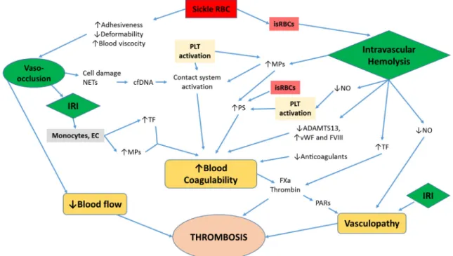
coagulation activation. In vivo, the protein C/protein S anticoagulant pathway is activated by the binding of thrombin and protein C to thrombomodulin and endothelial protein C-receptor (EPCR), their respective C-receptors expressed on endothelial cells. The pattern of expression of these transmembrane proteins in the various vascular beds in patients with SCD is unknown. Using a light/dye thrombosis model, enhanced thrombus formation in cerebral arterioles and venules was demonstrated in mice expressing hemoglobin S (48). These mice expressed lower levels of EPCR on the endothelium of cerebral arterioles and venules than wild type mice, and genetic intervention to increase EPCR in these vessels abrogated the enhanced thrombus formation in the brain (48). Furthermore, the capacity to generate thrombin ex vivo was significantly increased in children with SCD at steady state compared with age-matched controls only when the protein C/protein S anticoagulant pathway was activated by addition of exogenous thrombomodulin(32). Similarly, a higher peak thrombin generation was observed in adult HbSS patients than in age- and race-matched controls only when thrombomodulin or activated protein C was added to their plasma (34). Together, these findings indicate the relevance of the impaired protein C/protein S anticoagulant pathway in the hypercoagulability of SCD. While there are conflicting reports on the plasma levels of antithrombin (93,109), one study reported normal plasma levels of tissue factor pathway inhibitor (TFPI) antigen in SCD patients at steady state and during painful episodes (20). Plasma level of heparin cofactor II, a physiologic serine protease inhibitor, has been reported to be lower in HbSS and HbSC patients during both “steady state” and acute painful episodes compared to healthy controls (110). A summary of coagulation abnormalities and their potential contributions to hypercoagulability and thrombosis in SCD is shown in Figure 1.
3. Vasculopathy of SCD
Vasculopathy is a term that has been used to describe the progressive remodeling of the arterial vasculature, leading to impaired blood flow. The pathogenesis of vasculopathy in SCD is not fully elucidated. Several distinct concepts, histologic (111–114), radiologic (115–119) and mechanistic (120,121), all using the term ‘sickle vasculopathy’ have been described in the literature. Mechanistically, the term has been used to describe a generalized form of endothelial dysfunction with likely contributions from genetic factors (122,123), intravascular hemolysis, endothelial injury, vascular inflammation and chronic activation of coagulation ultimately leading to tissue hypoperfusion and damage (120,121). Intravascular
A
uthor Man
uscr
ipt
A
uthor Man
uscr
ipt
A
uthor Man
uscr
ipt
A
uthor Man
uscr
hemolysis is thought to account for a third of the total hemolysis occurring in patients with SCD (124). The resultant cell-free hemoglobin consumes nitric oxide (NO) to generate
methemoglobin and NO3− (125,126). Arginase, an enzyme also released from RBCs during
intravascular hemolysis, metabolizes L-arginine, the substrate for NO production by the enzyme NO synthase (125). Consumption and reduced production lead to impaired NO bioavailability which is associated with platelet activation and vascular endothelial dysfunction (125).
Recent data from in vitro studies and animal models support a role of chronic activation of coagulation in the development of vascular inflammation in SCD. Thrombin and other serine proteases of the clotting system have coagulation-independent activities that are mediated via binding to protease-activated receptors (PARs) (127). For instance, thrombin promotes fibrocyte proliferation in vitro, and blockade of TF pathway prevents intimal hyperplasia in a mouse model of vascular injury (128). As discussed previously, the TF pathway not only serves as a trigger for coagulation activation, but also promotes inflammation and vascular injury in sickle mice (49). Further analyses of the effect of downstream coagulation
proteases in this model show that TF, thrombin and factor Xa have differential contributions to vascular injury and inflammation in these mice. Factor Xa contributes to systemic inflammation (IL-6) through PAR-2 expressed on non-hematopoietic cells, while thrombin contributes to neutrophil infiltration in the lungs independently of PAR-1 expressed on non-hematopoietic cells (129). Consistent with the cross-talk of coagulation and inflammation, both plasma TAT and D-dimer levels have been reported to be correlated with soluble VCAM-1 in patients with SCD (24).
4. Thrombosis-Related Complications of SCD and the Link with
Hemostatic Alterations
4.1. Venous Thromboembolism (VTE)
Until recently, venous thromboembolism (VTE) has been overlooked as a cardiovascular complication of SCD. Two retrospective studies based on data from administrative databases of hospital discharge records addressed the risk of VTE in the SCD population (5,6). A significantly higher discharge diagnosis of pulmonary embolism (PE) was reported in African-Americans with SCD younger than 40 years than in African Americans without SCD (0.44% vs 0.12%), although a similar rate of deep venous thrombosis (DVT) was observed in both groups (5). Similarly, a higher incidence of inpatient PE was reported in patients with SCD than in the non-SCD population in the US state of Pennsylvania, although the prevalence of PE among SCD patients ≤50 years of age did not differ from that of non-SCD patients of similar age. In this study, non-SCD patients admitted with PE were older, had longer lengths of hospitalization, greater severity of illness and higher inpatient mortality than SCD admissions without PE (6). In a single center retrospective study of 404 SCD patients (7), 25% had a history of VTE, 31% of which were catheter-related. Of the patients with non-catheter-related VTE who had complete records for all provoking factors, 42% had no identifiable risk factors for VTE (7). Multiple studies based on administrative databases (110,130–135) and one small prospective, controlled, cohort study (136) have reported an increased risk of VTE in women with SCD during pregnancy and the puerperium as
A
uthor Man
uscr
ipt
A
uthor Man
uscr
ipt
A
uthor Man
uscr
ipt
A
uthor Man
uscr
compared with those without SCD (137). Analysis of VTE incidence and mortality risk in the cohort of patients enrolled in the Cooperative Study of Sickle Cell Disease (CSSCD) (8) supports the notion that SCD is associated with an increased risk of VTE (8). In addition, patients with VTE had a higher risk of death than those without VTE (5,7,8). The risk of
VTE was higher in patients with HbSS/Sβ°-thalassemia genotypes than in those with HbSC/
Sβ+-thalassemia, while co-inheritance of α-thalassemia was protective (8). Overall, PE
appears to be a more frequent manifestation of VTE than DVT in SCD (5,6,8).
4.2. Stroke and Silent Cerebral Infarct (SCI)
Stroke is a major cause of morbidity and mortality in patients with SCD (138), with cumulative risks of 11% and 24% for first event at 20 and 45 years old, respectively, in HbSS patients reported in the CSSCD (4). Both ischemic and hemorrhagic strokes are observed in SCD. A high incidence of ischemic stroke is observed in the first decade and after the third decade of life, while the incidence of hemorrhagic stroke peaks within the third decade in patients with HbSS (4). Clinical risk factors for ischemic stroke include prior transient ischemic attack, low steady-state hemoglobin level, systolic blood pressure, acute chest syndrome within two weeks of the stroke event as well as the rate per year of acute chest syndrome (4). Nocturnal hypoxemia is also recognized as a risk factor for acute
neurological events (139). Some genetic factors such as the co-inheritance of α-thalassemia
(140) and the nonsynonymous SNP VCAM1 G1238C (141) may be protective, while others
such as SNPs in the tumor growth factor-β and P-selectin genes identified using
genome-wise association studies have been associated with an increased risk of overt stroke in patients with SCA (142). The best predictor of stroke risk to date is an elevated transcranial Doppler (TCD) velocity, which is the qualifying criterion for primary stroke prevention by regular blood transfusion (115). However, the presence of thrombosis in the large and small cerebral arteries commonly described at autopsy of SCD patients with ischemic stroke suggests the participation of coagulation abnormalities to the pathogenesis of this devastating complication (111,112). Multiple studies have evaluated the association of various parameters of coagulation and cerebrovascular disease in SCD patients (21,23,24,30,143–146) (Table 2). All of these studies had cross-sectional designs and included only small numbers of patients with cerebrovascular disease. The conflicting results of these studies may also be due to the heterogeneity of patient genotypes, treatments and the criteria used to define cerebrovascular disease across the studies. Consequently, more studies are required to better understand the contribution of coagulation abnormalities to the pathogenesis of stroke of SCD.
The use of magnetic resonance imaging (MRI) has allowed the recognition of infarct-like lesions of the brain in the setting of a normal neurologic examination or the absence of an abnormality on neurological examination that can be explained by the location of this lesion (147). Silent cerebral infarcts (SCI) are detected in all SCD genotypes and are found in up to 37% of HbSS children before the age of 14 (147). Although referred to as “silent” infarcts, SCI is a morbid condition associated with neurocognitive impairment, poor academic performance, neurologic soft signs, and increased risk for subsequent overt stroke as compared with SCD children with normal MRI findings (147–149). The pathogenesis of SCI is unknown, and no autopsy study has specifically described the histopathological
A
uthor Man
uscr
ipt
A
uthor Man
uscr
ipt
A
uthor Man
uscr
ipt
A
uthor Man
uscr
lesions corresponding to the bright spots observed on MRI. Acute demyelination, sinus venous thrombosis and small artery and arteriole disease are suggested to account for the MRI lesions of SCI (147). One study reported lower steady state plasma levels of tissue-plasminogen activator (tPA) and ADAMTS13 in SCD children with SCI compared with those without SCI, although plasma levels of TAT, F1.2 and D-dimers were similar in both groups (23).
4.3. Acute Chest Syndrome and Pulmonary Hypertension
Acute chest syndrome is defined as the presence of a new pulmonary infiltrate on chest x-ray, associated with a variety of respiratory signs and symptoms, including chest pain, fever, dyspnea or cough in a patient with SCD (150). It is a common cause of hospitalization of patients with SCD, second only to acute pain episodes, and is a leading cause of death (151). The causes of acute chest syndrome were extensively evaluated by the National Acute Chest Syndrome Study Group, and include infection, fat embolism and possibly pulmonary infarction (150). In this multicenter study, it was presumed that pulmonary infarction was the cause of acute chest syndrome in 16% of episodes with complete study data, but in which no specific etiology was otherwise identified (150). While autopsy studies have shown
microscopic organized thrombi in the lungs of SCD patients (152), the contribution of this finding to the pathogenesis of acute chest syndrome is uncertain. Pulmonary thrombosis was detected, using computerized tomography imaging techniques, in 17% of patients during episodes of acute chest syndrome in a single center study (153). However, it is uncertain if these pulmonary thrombi were present before or occurred following the development of acute chest syndrome. Steady state levels of TAT and D-dimer were not significantly different in adult and pediatric patients with histories of acute chest syndrome compared to patients with no previous episodes (23,24). Similarly, a series of coagulation parameters, including plasma levels of FVIII, vWF, F1.2, TAT, D-dimer, ADAMTS13 antigen, plasminogen activator inhibitor and tPA, did not correlate with the rate of acute chest syndrome in children with SCD (23).
Pulmonary vasculopathic complications, such as echocardiography-derived elevation in tricuspid regurgitant jet velocity (TRV) and pulmonary hypertension, are increasingly recognized in adult patients with SCD. Although the prevalence of elevated TRV is high in SCD (154,155), a right heart catheterization is always required to confirm the diagnosis of pulmonary hypertension. Pulmonary hypertension is defined as a resting mean pulmonary arterial pressure (mPAP) ≥ 25 mm Hg by right heart catheterization (RHC) (156). Autopsy studies have reported the presence of in situ thrombi in the pulmonary vasculature of SCD patients with pulmonary hypertension, suggesting a role for hypercoagulability in this complication (113,114). There are no studies evaluating the relationship between markers of coagulation activation and RHC-confirmed pulmonary hypertension in SCD. No significant associations were observed between plasma markers of coagulation activation (TAT, D-dimer) and TRV in SCD (24,39,40). One pediatric study reported negative correlations between plasma levels of tPA and TRV (23). There are conflicting reports on the association of platelet activation with TRV, with one study showing a correlation between activated GPIIb/IIIa receptor with TRV (73), while another showed no association between soluble CD40 ligand and TRV (40). Furthermore, higher levels of both platelet- and
erythrocyte-A
uthor Man
uscr
ipt
A
uthor Man
uscr
ipt
A
uthor Man
uscr
ipt
A
uthor Man
uscr
derived microparticles have been reported in patients with histories of ACS and elevated TRV compared to those without either of these complications (143), although no significant differences were seen in the plasma concentrations of total-, endothelial cell derived-, and TF-positive microparticles in another study (91).
4.4. Other Complications
Avascular necrosis is a chronic complication which occurs in up to 50% of HbSS subjects by age 35 (157). In patients without SCD, avascular necrosis has been reported to be associated with thrombophilia, including elevated factor VIII activity, heterozygosity for factor V Leiden, and elevated plasma levels of TAT, F1.2, PAI-1, and platelet- and endothelial cell-derived microparticles (158,159). No association was observed between avascular necrosis and plasma levels of TAT, D-dimer and microparticle-associated TF in 2 cross-sectional studies of adult SCD patients (24)(160), although a higher total number of microparticles was observed in patients with avascular necrosis than in those without this complication (160). “Steady state” plasma levels of markers of in vivo thrombin and fibrin generation were not associated with histories of leg ulcers, retinopathy or priapism in adult SCD patients (24), nor with histories of splenic sequestration, hemolytic or aplastic crises in children (23). The number of platelet-derived microparticles was reported to be significantly higher in adult patients with albuminuria compared to those without, but no differences were observed in total microparticles or other circulating cell-derived microparticles (91).
5. Effect of Disease Modifying Treatments, Anticoagulants and
Anti-Platelet Agents on the Hypercoagulable State of SCD
5.1. Hydroxyurea
Hydroxyurea is approved by the US Food and Drug Administration specifically for treating SCD. It has been shown to reduce the frequency of acute painful episodes, dactylitis, acute chest syndrome, hospitalizations, and the need for blood transfusions in children and adults with sickle cell anemia (161,162). Observational studies have reported a reduction of TCD velocity (163–165), rate of first stroke (166) and the rate of stroke recurrence (167–170) in SCD patients treated with hydroxyurea. The Stroke With Transfusions Changing to Hydroxyurea (SWiTCH) trial, was a randomized, non-inferiority trial comparing
transfusions and iron chelation to hydroxyurea and therapeutic phlebotomy for children with sickle cell anemia, stroke, and iron overload, with a composite primary endpoint allowing an increased stroke risk but requiring superiority for removing iron (171). Although there were 7 strokes in the hydroxyurea/phlebotomy arm and none in the transfusions/chelation arm, within the non-inferiority stroke margin, the study was stopped after interim analysis revealed equivalent liver iron content, indicating futility for the composite primary endpoint. More recently, the randomized Transcranial Doppler With Transfusions Changing to Hydroxyurea (TWiTCH) study was stopped prematurely by the Data Monitoring Committee after hydroxyurea was found to be non-inferior to chronic RBC transfusions in lowering TCD velocities in children with SCD who were at high risk for stroke (172).
Several studies have evaluated the effect of hydroxyurea on coagulation parameters in patients with SCD. There are conflicting reports on the effect of hydroxyurea on total,
A
uthor Man
uscr
ipt
A
uthor Man
uscr
ipt
A
uthor Man
uscr
ipt
A
uthor Man
uscr
specific blood cell-derived and TF-bearing microparticles (31,78,91,173). A reduction in plasma D-dimer level in patients treated with hydroxyurea has been reported (78,174), with these studies reporting conflicting effects of hydroxyurea on circulating TAT complexes. Adult patients treated with hydroxyurea also manifest a longer lag time, slower rate and reduced peak of ex vivo thrombin generation in TGA compared with untreated patients (31). One study of pediatric patients with SCD reported a negative correlation between ETP and peak ex vivo thrombin generation normalized for age and duration of hydroxyurea treatment, suggesting a time-dependent effect of hydroxyurea on overall coagulation potential in children with SCD (146). However, all of these have been cross-sectional studies in which the steady state plasma levels of coagulation parameters were compared in SCD patients based on treatment with hydroxyurea, often with no consideration of patient dosage, adherence or duration of treatment. Finally, treatment of a small group of children with SCD
and β-thalassemia intermedia was reported to lower the plasma level of FVIII and protein C
after approximately 6 months of hydroxyurea therapy (175).
5.2. Chronic Blood Transfusion
Chronic blood transfusion is effective for the primary (176) and secondary (171) prevention of stroke as well as for reducing the risk of recurrent cerebral infarcts (177) in children with SCD. In addition, chronic transfusion therapy decreases the frequency of acute pain episodes and acute chest syndrome (178). A significant reduction in the concentration of erythrocyte-derived microparticles was reported in one study, although the concentrations of platelet-derived microparticles and total annexin V-positive microparticles remained similar before
and following RBC exchange transfusion (179). In children with HbSS and HbSβ0
-thalassemia receiving regular blood transfusion to keep HbS ≤20%, plasma levels of coagulation factor X and factor XI, total protein S, heparin cofactor II, F1.2 and TAT remained higher than in race- and age-matched controls, suggesting ongoing coagulation activation despite chronic transfusion (21).
5.3. Anticoagulant Therapy
Downregulation of coagulation with anticoagulant drugs has been used to assess the contribution of hemostatic alterations to the pathogenesis of SCD. However, the majority of the published studies are small, poorly controlled, and have focused mainly on the frequency of painful episodes as the primary endpoint (Table 3). Although low intensity
anticoagulation with the vitamin K antagonist, acenocoumarol, has been reported to normalize circulating markers of in vivo thrombin generation in patients with SCD
(180,181), no reduction was observed in the frequency of pain episodes (181). A small study of 4 patients with severe SCD reported a reduction of the number of days of hospitalization per year and number of days spent in the emergency department during periods when patients received prophylactic doses of unfractionated heparin compared to periods when they were off treatment (182). More recently, a randomized, double-blind,
placebo-controlled study of the low molecular weight heparin, tinzaparin, in 253 patients with HbSS showed a significant reduction in the number of days with the most severe pain scores, the overall duration of painful crisis, and the duration of hospitalization in the treatment group compared with placebo (183). However, it is uncertain whether the beneficial effects were
A
uthor Man
uscr
ipt
A
uthor Man
uscr
ipt
A
uthor Man
uscr
ipt
A
uthor Man
uscr
due to the anticoagulant property of tinzaparin or its anti-inflammatory and P-selectin blocking effects (184,185).
Studies in animal models of SCD have suggested a link between coagulation activation and vascular inflammation (186). Blockade of factor Xa or thrombin using specific direct inhibitors, rivaroxaban and dabigatran, respectively, significantly reduced TAT and local tissue inflammation, with decreased levels of myeloperoxidase and the number of neutrophils in the lungs of sickle mice (129). Furthermore, treatment of sickle mice with rivaroxaban resulted in decreased IL-6, suggesting an effect on systemic inflammation. Genetic reduction of prothrombin level to below 10% activity in sickle mice also resulted in lower plasma levels of steady state D-dimer, IL-6, soluble VCAM-1, as well as white blood cell and platelet counts despite similar RBC profiles compared with control mice (187) indicative of decreased coagulation activation, systemic inflammation and vascular injury. Interestingly, sickle mice with reduced prothrombin level experienced no significant bleeding and had decreased mortality and less damage to organs, including the lung, kidney, heart and liver (187). Together, these animal studies provide a proof of concept that
diminution of coagulation activation in SCD may indeed decrease end-organ damage. Based on the observed effects in sickle mice, a study of the factor Xa inhibitor, rivaroxaban, is ongoing in patients with SCD to assess its pharmacodynamic effects and safety (www.clinicaltrials.gov. identifier NCT02072668).
5.4. Anti-platelet Agents
There have been multiple studies of antiplatelet agents in SCD (Table 4), although most of these trials did not correlate the in vivo effect of the drugs on platelet activation with clinical endpoints. In a randomized, double-blind, placebo-controlled study, treatment with
ticlopidine resulted in a reduction in the frequency, duration, and severity of acute pain episodes in patients with SCD compared with placebo following 6 months of therapy (188).
Eptifibatide, a specific and reversible synthetic peptide inhibitor of the αIIbβ3 receptor, was
shown to inhibit platelet aggregation, and decrease soluble CD40L levels as well as plasma levels of inflammatory mediators in a phase 1 study of adults with HbSS (189). Although eptifibatide was not associated with major bleeding or thrombocytopenia, it did not improve times to discharge, crisis resolution or the total opioid use in a pilot, randomized, double-blind and placebo-controlled trial of adults with SCD admitted for acute pain episodes (190). However, this study was not adequately powered to assess clinical outcomes. Treatment with prasugrel, a third-generation platelet P2Y12 ADP antagonist, in a multicenter, phase 2 randomized, double-blind study resulted in reduced markers of platelet activation, with no hemorrhagic events requiring medical intervention in adults with SCD (191). Although efficacy was not a primary end-point of this study, the treatment group showed a non-significant trend towards reduction in the rate and intensity of pain. More recently, a phase 3, multinational study evaluating the efficacy of prasugrel in 341 children with sickle cell
anemia (HbSS and HbSβ0 thalassemia) showed no significant difference in the rate of
vaso-occlusive crisis (a composite of painful crisis and acute chest syndrome) among those who received prasugrel compared with placebo (192). However, subgroup analyses showed that the effect of prasugrel was greatest in the group of patients between the ages of 12 and 17 years and in patients not receiving hydroxyurea (192).
A
uthor Man
uscr
ipt
A
uthor Man
uscr
ipt
A
uthor Man
uscr
ipt
A
uthor Man
uscr
6. Conclusion
SCD is a hypercoagulable state characterized by chronic activation of coagulation in vivo and increased risk of both arterial and venous thrombosis. There is increasing evidence that the activation of coagulation in SCD is not just a secondary event, but may contribute to disease pathogenesis. Although treatment with hydroxyurea and chronic blood transfusion have important clinical benefits, patients continue to experience clinical complications, including thrombotic complications. Defining the contribution of the hypercoagulable state to disease pathogenesis requires further studies using transgenic animal models. Despite the disappointing results of the phase 3 trial of prasugrel in children, other well controlled clinical studies of new anticoagulants and antiplatelet agents, using a variety of clinical endpoints will help to further define the contribution of coagulation and platelet activation to the pathophysiology of SCD and its complications.
Acknowledgments
Funding source
This work was supported by NIH grant U01HL117659. Support for this work was also provided by an award from the North Carolina State Sickle Cell Program.
References
1. Piel FB, Patil AP, Howes RE, Nyangiri OA, Gething PW, Dewi M, et al. Global epidemiology of sickle haemoglobin in neonates: a contemporary geostatistical model-based map and population estimates. Lancet. 2013; 381:142–51. [PubMed: 23103089]
2. Piel FB, Hay SI, Gupta S, Weatherall DJ, Williams TN. Global burden of sickle cell anaemia in children under five, 2010–2050: modelling based on demographics, excess mortality, and interventions. PLoS Med. 2013; 10:e1001484. [PubMed: 23874164]
3. Chakravorty S, Williams TN. Sickle cell disease: a neglected chronic disease of increasing global health importance. Arch Dis Child. 2015; 100:48–53. [PubMed: 25239949]
4. Ohene-Frempong K, Weiner SJ, Sleeper LA, Miller ST, Embury S, Moohr JW, et al.
Cerebrovascular accidents in sickle cell disease: rates and risk factors. Blood. 1998; 91:288–94. [PubMed: 9414296]
5. Stein PD, Beemath A, Meyers FA, Skaf E, Olson RE. Deep venous thrombosis and pulmonary embolism in hospitalized patients with sickle cell disease. Am J Med. 2006; 119:897.e7–11. [PubMed: 17000225]
6. Novelli EM, Huynh C, Gladwin MT, Moore CG, Ragni MV. Pulmonary embolism in sickle cell disease: a case-control study. J Thromb Haemost JTH. 2012; 10:760–6. [PubMed: 22417249] 7. Naik RP, Streiff MB, Haywood C, Nelson JA, Lanzkron S. Venous thromboembolism in adults with
sickle cell disease: a serious and under-recognized complication. Am J Med. 2013; 126:443–9. [PubMed: 23582935]
8. Naik RP, Streiff MB, Haywood C, Segal JB, Lanzkron S. Venous thromboembolism incidence in the Cooperative Study of Sickle Cell Disease. J Thromb Haemost JTH. 2014; 12:2010–6. [PubMed: 25280124]
9. Ataga KI, Key NS. Hypercoagulability in sickle cell disease: new approaches to an old problem. Hematology Am Soc Hematol Educ Program. 2007:91–6. [PubMed: 18024615]
10. De Franceschi L, Cappellini MD, Olivieri O. Thrombosis and sickle cell disease. Semin Thromb Hemost. 2011; 37:226–36. [PubMed: 21455857]
11. Rahimi Z, Parsian A. Sickle cell disease and venous thromboembolism. Mediterr J Hematol Infect Dis. 2011; 3:e2011024. [PubMed: 21713075]
A
uthor Man
uscr
ipt
A
uthor Man
uscr
ipt
A
uthor Man
uscr
ipt
A
uthor Man
uscr
12. Lim MY, Ataga KI, Key NS. Hemostatic abnormalities in sickle cell disease. Curr Opin Hematol. 2013; 20:472–7. [PubMed: 23817169]
13. Pakbaz Z, Wun T. Role of the hemostatic system on sickle cell disease pathophysiology and potential therapeutics. Hematol Oncol Clin North Am. 2014; 28:355–74. [PubMed: 24589271] 14. Nsiri B, Gritli N, Bayoudh F, Messaoud T, Fattoum S, Machghoul S. Abnormalities of coagulation
and fibrinolysis in homozygous sickle cell disease. Hematol Cell Ther. 1996; 38:279–84. [PubMed: 8974793]
15. Westerman MP, Green D, Gilman-Sachs A, Beaman K, Freels S, Boggio L, et al. Antiphospholipid antibodies, proteins C and S, and coagulation changes in sickle cell disease. J Lab Clin Med. 1999; 134:352–62. [PubMed: 10521081]
16. Hagger D, Wolff S, Owen J, Samson D. Changes in coagulation and fibrinolysis in patients with sickle cell disease compared with healthy black controls. Blood Coagul Fibrinolysis Int J Haemost Thromb. 1995; 6:93–9.
17. Peters M, Plaat BE, ten Cate H, Wolters HJ, Weening RS, Brandjes DP. Enhanced thrombin generation in children with sickle cell disease. Thromb Haemost. 1994; 71:169–72. [PubMed: 8191393]
18. Kurantsin-Mills J, Ofosu FA, Safa TK, Siegel RS, Lessin LS. Plasma factor VII and thrombin-antithrombin III levels indicate increased tissue factor activity in sickle cell patients. Br J Haematol. 1992; 81:539–44. [PubMed: 1390242]
19. Setty BN, Rao AK, Stuart MJ. Thrombophilia in sickle cell disease: the red cell connection. Blood. 2001; 98:3228–33. [PubMed: 11719358]
20. Key NS, Slungaard A, Dandelet L, Nelson SC, Moertel C, Styles LA, et al. Whole blood tissue factor procoagulant activity is elevated in patients with sickle cell disease. Blood. 1998; 91:4216– 23. [PubMed: 9596669]
21. Liesner R, Mackie I, Cookson J, McDonald S, Chitolie A, Donohoe S, et al. Prothrombotic changes in children with sickle cell disease: relationships to cerebrovascular disease and transfusion. Br J Haematol. 1998; 103:1037–44. [PubMed: 9886316]
22. Tomer A, Harker LA, Kasey S, Eckman JR. Thrombogenesis in sickle cell disease. J Lab Clin Med. 2001; 137:398–407. [PubMed: 11385360]
23. Colombatti R, De Bon E, Bertomoro A, Casonato A, Pontara E, Omenetto E, et al. Coagulation Activation in Children with Sickle Cell Disease Is Associated with Cerebral Small Vessel Vasculopathy. PLoS ONE. 2013; 8:e78801. [PubMed: 24205317]
24. Ataga KI, Brittain JE, Desai P, May R, Jones S, Delaney J, et al. Association of coagulation activation with clinical complications in sickle cell disease. PloS One. 2012; 7:e29786. [PubMed: 22253781]
25. Hemker HC, Giesen P, AlDieri R, Regnault V, de Smed E, Wagenvoord R, et al. The calibrated automated thrombogram (CAT): a universal routine test for hyper- and hypocoagulability. Pathophysiol Haemost Thromb. 2002; 32:249–53. [PubMed: 13679651]
26. Hemker HC, Al Dieri R, De Smedt E, Béguin S. Thrombin generation, a function test of the haemostatic-thrombotic system. Thromb Haemost. 2006; 96:553–61. [PubMed: 17080210] 27. Hemker HC, Giesen P, Al Dieri R, Regnault V, de Smedt E, Wagenvoord R, et al. Calibrated
automated thrombin generation measurement in clotting plasma. Pathophysiol Haemost Thromb. 2003; 33:4–15. [PubMed: 12853707]
28. Ninivaggi M, Apitz-Castro R, Dargaud Y, de Laat B, Hemker HC, Lindhout T. Whole-blood thrombin generation monitored with a calibrated automated thrombogram-based assay. Clin Chem. 2012; 58:1252–9. [PubMed: 22665918]
29. Amin C, Adam S, Mooberry MJ, Kutlar A, Kutlar F, Esserman D, et al. Coagulation activation in sickle cell trait: an exploratory study. Br J Haematol. 2015; 171:638–46. [PubMed: 26511074] 30. Shah N, Thornburg C, Telen MJ, Ortel TL. Characterization of the hypercoagulable state in
patients with sickle cell disease. Thromb Res. 2012; 130:e241–5. [PubMed: 22959127]
31. Gerotziafas GT, Van Dreden P, Chaari M, Galea V, Khaterchi A, Lionnet F, et al. The acceleration of the propagation phase of thrombin generation in patients with steady-state sickle cell disease is associated with circulating erythrocyte-derived microparticles. Thromb Haemost. 2012; 107:1044– 52. [PubMed: 22535498]
A
uthor Man
uscr
ipt
A
uthor Man
uscr
ipt
A
uthor Man
uscr
ipt
A
uthor Man
uscr
32. Noubouossie DF, Lê PQ, Corazza F, Debaugnies F, Rozen L, Ferster A, et al. Thrombin generation reveals high procoagulant potential in the plasma of sickle cell disease children. Am J Hematol. 2012; 87:145–9. [PubMed: 22052675]
33. Dargaud Y, Luddington R, Gray E, Negrier C, Lecompte T, Petros S, et al. Effect of standardization and normalization on imprecision of calibrated automated thrombography: an international multicentre study. Br J Haematol. 2007; 139:303–9. [PubMed: 17897307]
34. Whelihan MF, Lim MY, Walton BL, Wolberg AS, Cai J, Ataga KI, et al. Hypercoagulability in Sickle Cell Disease: The Importance of the Cellular Component of Blood. Blood. 2014; 124:4060– 4060.
35. Yee DL, Edwards RM, Mueller BU, Teruya J. Thromboelastographic and hemostatic
characteristics in pediatric patients with sickle cell disease. Arch Pathol Lab Med. 2005; 129:760– 5. [PubMed: 15913424]
36. Setty BNY, Key NS, Rao AK, Gayen-Betal S, Krishnan S, Dampier CD, et al. Tissue factor-positive monocytes in children with sickle cell disease: correlation with biomarkers of haemolysis. Br J Haematol. 2012; 157:370–80. [PubMed: 22360627]
37. Solovey A, Gui L, Key NS, Hebbel RP. Tissue factor expression by endothelial cells in sickle cell anemia. J Clin Invest. 1998; 101:1899–904. [PubMed: 9576754]
38. Shet AS, Aras O, Gupta K, Hass MJ, Rausch DJ, Saba N, et al. Sickle blood contains tissue factor-positive microparticles derived from endothelial cells and monocytes. Blood. 2003; 102:2678–83. [PubMed: 12805058]
39. van Beers EJ, Spronk HMH, Ten Cate H, Duits AJ, Brandjes DPM, van Esser JWJ, et al. No association of the hypercoagulable state with sickle cell disease related pulmonary hypertension. Haematologica. 2008; 93:e42–4. [PubMed: 18450728]
40. Ataga KI, Moore CG, Hillery CA, Jones S, Whinna HC, Strayhorn D, et al. Coagulation activation and inflammation in sickle cell disease-associated pulmonary hypertension. Haematologica. 2008; 93:20–6. [PubMed: 18166781]
41. Setty BNY, Betal SG, Zhang J, Stuart MJ. Heme induces endothelial tissue factor expression: potential role in hemostatic activation in patients with hemolytic anemia. J Thromb Haemost. 2008; 6:2202–9. [PubMed: 18983524]
42. Rehani T, Mathson K, Belcher JD, Vercellotti GM. Heme Potently Stimulates Tissue Factor Expression By Peripheral Blood Monocytes: A Novel Mechanism For Thrombosis In Intravascular Hemolytic Diseases. Blood. 2013; 122:2215–2215.
43. Sparkenbaugh EM, Chantrathammachart P, Wang S, Jonas W, Kirchhofer D, Gailani D, et al. Excess of heme induces tissue factor-dependent activation of coagulation in mice. Haematologica. 2015; 100:308–14. [PubMed: 25596265]
44. Solovey A, Kollander R, Milbauer LC, Abdulla F, Chen Y, Kelm RJ, et al. Endothelial nitric oxide synthase and nitric oxide regulate endothelial tissue factor expression in vivo in the sickle transgenic mouse. Am J Hematol. 2010; 85:41–5. [PubMed: 20029945]
45. Solovey A, Kollander R, Shet A, Milbauer LC, Choong S, Panoskaltsis-Mortari A, et al.
Endothelial cell expression of tissue factor in sickle mice is augmented by hypoxia/reoxygenation and inhibited by lovastatin. Blood. 2004; 104:840–6. [PubMed: 15073034]
46. Aufradet E, DeSouza G, Bourgeaux V, Bessaad A, Campion Y, Canet-Soulas E, et al. Hypoxia/ reoxygenation stress increases markers of vaso-occlusive crisis in sickle SAD mice. Clin Hemorheol Microcirc. 2013; 54:297–312. [PubMed: 23696418]
47. Kollander R, Solovey A, Milbauer LC, Abdulla F, Kelm RJ, Hebbel RP. Nuclear factor-kappa B (NFkappaB) component p50 in blood mononuclear cells regulates endothelial tissue factor expression in sickle transgenic mice: implications for the coagulopathy of sickle cell disease. Transl Res J Lab Clin Med. 2010; 155:170–7.
48. Gavins FNE, Russell J, Senchenkova EL, De Almeida Paula L, Damazo AS, Esmon CT, et al. Mechanisms of enhanced thrombus formation in cerebral microvessels of mice expressing hemoglobin-S. Blood. 2011; 117:4125–33. [PubMed: 21304105]
49. Chantrathammachart P, Mackman N, Sparkenbaugh E, Wang J-G, Parise LV, Kirchhofer D, et al. Tissue factor promotes activation of coagulation and inflammation in a mouse model of sickle cell disease. Blood. 2012; 120:636–46. [PubMed: 22661702]
A
uthor Man
uscr
ipt
A
uthor Man
uscr
ipt
A
uthor Man
uscr
ipt
A
uthor Man
uscr
50. Gordon EM, Klein BL, Berman BW, Strandjord SE, Simon JE, Coccia PF. Reduction of contact factors in sickle cell disease. J Pediatr. 1985; 106:427–30. [PubMed: 3844465]
51. Verma PS, Adams RG, Miller RL. Reduced plasma kininogen concentration during sickle cell crisis. Res Commun Chem Pathol Pharmacol. 1983; 41:313–22. [PubMed: 6635322]
52. Miller RL, Verma PS, Adams RG. Studies of the Kallikrein-Kinin System in Patients with Sickle Cell Anemia. J Natl Med Assoc. 1983; 75:551–6. [PubMed: 6603519]
53. Smith SA, Travers RJ, Morrissey JH. How it all starts: Initiation of the clotting cascade. Crit Rev Biochem Mol Biol. 2015; 28:1–11.
54. Wu Y. Contact pathway of coagulation and inflammation. Thromb J. 2015; 13:17. [PubMed: 25949215]
55. Morrissey JH, Choi SH, Smith SA. Polyphosphate: an ancient molecule that links platelets, coagulation, and inflammation. Blood. 2012; 119:5972–9. [PubMed: 22517894]
56. Al-Humood S, Zueriq R, Al-Faris L, Marouf R, Al-Mulla F. Circulating cell-free DNA in sickle cell disease: is it a potentially useful biomarker? Arch Pathol Lab Med. 2014; 138:678–83. [PubMed: 24786126]
57. Vasavda N, Ulug P, Kondaveeti S, Ramasamy K, Sugai T, Cheung G, et al. Circulating DNA: a potential marker of sickle cell crisis. Br J Haematol. 2007; 139:331–6. [PubMed: 17897311] 58. Schimmel M, Nur E, Biemond BJ, van Mierlo GJ, Solati S, Brandjes DP, et al. Nucleosomes and
neutrophil activation in sickle cell disease painful crisis. Haematologica. 2013; 98:1797–803. [PubMed: 23911704]
59. Sparkenbaugh E, Key NS, Chandarajoti K, Gruber A, Mackman N, McCrae K, et al. Kininogen deficiency attenuates thrombin generation in a mouse model of sickle cell disease (ISTH Abstract). J Thromb Haemost. 2015; 13:S2, 17. [PubMed: 26149024]
60. Lee SP, Ataga KI, Orringer EP, Phillips DR, Parise LV. Biologically active CD40 ligand is elevated in sickle cell anemia: potential role for platelet-mediated inflammation. Arterioscler Thromb Vasc Biol. 2006; 26:1626–31. [PubMed: 16601237]
61. Jakubowski JA, Zhou C, Jurcevic S, Winters KJ, Lachno DR, Frelinger AL, et al. A phase 1 study of prasugrel in patients with sickle cell disease: effects on biomarkers of platelet activation and coagulation. Thromb Res. 2014; 133:190–5. [PubMed: 24368019]
62. Famodu AA, Oduwa D. Platelet count and platelet factor 3 (PF-3) availability in sickle cell disease. Br J Biomed Sci. 1995; 52:323–4. [PubMed: 8555788]
63. Westwick J, Watson-Williams EJ, Krishnamurthi S, Marks G, Ellis V, Scully MF, et al. Platelet activation during steady state sickle cell disease. J Med. 1983; 14:17–36. [PubMed: 6224876] 63. Papadimitriou CA, Travlou A, Kalos A, Douratsos D, Lali P. Study of platelet function in patients
with sickle cell anemia during steady state and vaso-occlusive crisis. Acta Haematol. 1993; 89:180–3. [PubMed: 8212998]
64. Browne PV, Mosher DF, Steinberg MH, Hebbel RP. Disturbance of plasma and platelet thrombospondin levels in sickle cell disease. Am J Hematol. 1996; 51:296–301. [PubMed: 8602630]
65. Wun T, Paglieroni T, Rangaswami A, Franklin PH, Welborn J, Cheung A, et al. Platelet activation in patients with sickle cell disease. Br J Haematol. 1998; 100:741–9. [PubMed: 9531343] 66. Proença-Ferreira R, Franco-Penteado CF, Traina F, Saad STO, Costa FF, Conran N. Increased
adhesive properties of platelets in sickle cell disease: roles for alphaIIb beta3-mediated ligand binding, diminished cAMP signalling and increased phosphodiesterase 3A activity. Br J Haematol. 2010; 149:280–8. [PubMed: 20136824]
67. Westwick J, Watson-Williams EJ, Krishnamurthi S, Marks G, Ellis V, Scully MF, et al. Platelet activation during steady state sickle cell disease. J Med. 1983; 14:17–36. [PubMed: 6224876] 68. Wun T, Paglieroni T, Tablin F, Welborn J, Nelson K, Cheung A. Platelet activation and
platelet-erythrocyte aggregates in patients with sickle cell anemia. J Lab Clin Med. 1997; 129:507–16. [PubMed: 9142047]
69. Kenny MW, George AJ, Stuart J. Platelet hyperactivity in sickle-cell disease: a consequence of hyposplenism. J Clin Pathol. 1980; 33:622–5. [PubMed: 7430367]
70. Mehta P, Mehta J. Abnormalities of platelet aggregation in sickle cell disease. J Pediatr. 1980; 96:209–13. [PubMed: 7351581]
A
uthor Man
uscr
ipt
A
uthor Man
uscr
ipt
A
uthor Man
uscr
ipt
A
uthor Man
uscr
71. Stuart MJ, Stockman JA, Oski FA. Abnormalities of platelet aggregation in the vaso-occlusive crisis of sickle-cell anemia. J Pediatr. 1974; 85:629–32. [PubMed: 4423856]
72. Brousse V, Buffet P, Rees D. The spleen and sickle cell disease: the sick(led) spleen. Br J Haematol. 2014; 166:165–76. [PubMed: 24862308]
73. Villagra J, Shiva S, Hunter LA, Machado RF, Gladwin MT, Kato GJ. Platelet activation in patients with sickle disease, hemolysis-associated pulmonary hypertension, and nitric oxide scavenging by cell-free hemoglobin. Blood. 2007; 110:2166–72. [PubMed: 17536019]
74. Wood BL, Gibson DF, Tait JF. Increased erythrocyte phosphatidylserine exposure in sickle cell disease: flow-cytometric measurement and clinical associations. Blood. 1996; 88:1873–80. [PubMed: 8781447]
75. Fadok VA, Voelker DR, Campbell PA, Cohen JJ, Bratton DL, Henson PM. Exposure of phosphatidylserine on the surface of apoptotic lymphocytes triggers specific recognition and removal by macrophages. J Immunol Baltim Md 1950. 1992; 148:2207–16.
76. McEvoy L, Williamson P, Schlegel RA. Membrane phospholipid asymmetry as a determinant of erythrocyte recognition by macrophages. Proc Natl Acad Sci U S A. 1986; 83:3311–5. [PubMed: 3458184]
77. Schroit AJ, Madsen JW, Tanaka Y. In vivo recognition and clearance of red blood cells containing phosphatidylserine in their plasma membranes. J Biol Chem. 1985; 260:5131–8. [PubMed: 3988747]
78. Westerman M, Pizzey A, Hirschman J, Cerino M, Weil-Weiner Y, Ramotar P, et al. Microvesicles in haemoglobinopathies offer insights into mechanisms of hypercoagulability, haemolysis and the effects of therapy. Br J Haematol. 2008; 142:126–35. [PubMed: 18422994]
79. Whelihan MF, Mooberry MJ, Zachary V, Bradford RL, Ataga KI, Mann KG, et al. The contribution of red blood cells to thrombin generation in sickle cell disease: meizothrombin generation on sickled red blood cells. J Thromb Haemost JTH. 2013; 11:2187–9. [PubMed: 24119168]
80. Zwaal RF, Schroit AJ. Pathophysiologic implications of membrane phospholipid asymmetry in blood cells. Blood. 1997; 89:1121–32. [PubMed: 9028933]
81. Whelihan MF, Zachary V, Orfeo T, Mann KG. Prothrombin activation in blood coagulation: the erythrocyte contribution to thrombin generation. Blood. 2012; 120:3837–45. [PubMed: 22968460] 82. Setty BN, Kulkarni S, Rao AK, Stuart MJ. Fetal hemoglobin in sickle cell disease: relationship to
erythrocyte phosphatidylserine exposure and coagulation activation. Blood. 2000; 96:1119–24. [PubMed: 10910931]
83. Bezeaud A, Venisse L, Helley D, Trichet C, Girot R, Guillin M-C. Red blood cells from patients with homozygous sickle cell disease provide a catalytic surface for factor Va inactivation by activated protein C. Br J Haematol. 2002; 117:409–13. [PubMed: 11972526]
84. Connor DE, Exner T, Ma DDF, Joseph JE. Detection of the procoagulant activity of microparticle-associated phosphatidylserine using XACT. Blood Coagul Fibrinolysis Int J Haemost Thromb. 2009; 20:558–64.
85. Marco A, Brocal C, Marco P. Measurement of procoagulant activity of microparticles in plasma: feasibility of new functional assays. Thromb Res. 2014; 134:1363–4. [PubMed: 25282540] 86. van Beers EJ, Schaap MCL, Berckmans RJ, Nieuwland R, Sturk A, van Doormaal FF, et al.
Circulating erythrocyte-derived microparticles are associated with coagulation activation in sickle cell disease. Haematologica. 2009; 94:1513–9. [PubMed: 19815831]
87. Noubouossie DCF, Lê P-Q, Rozen L, Debaugnies F, Ferster A, Demulder A. Evaluation of the procoagulant activity of endogenous phospholipids in the platelet-free plasma of children with sickle cell disease using functional assays. Thromb Res. 2012; 130:259–64. [PubMed: 22079446] 88. van Tits LJ, van Heerde WL, Landburg PP, Boderie MJ, Muskiet FaJ, Jacobs N, et al. Plasma
annexin A5 and microparticle phosphatidylserine levels are elevated in sickle cell disease and increase further during painful crisis. Biochem Biophys Res Commun. 2009; 390:161–4. [PubMed: 19799864]
89. Van Der Meijden PEJ, Van Schilfgaarde M, Van Oerle R, Renné T, Ten Cate H, Spronk HMH. Platelet- and erythrocyte-derived microparticles trigger thrombin generation via factor XIIa: