American Journal of Epidemiology
Published by Oxford University Press on behalf of the Johns Hopkins Bloomberg School of Public Health 2017. This work is written by (a) US Government employee(s) and is in the public domain in the US.
Vol. 186, No. 12 DOI: 10.1093/aje/kwx206 Advance Access publication: June 16, 2017
Original Contribution
Epidemiologic Risk Factors for In Situ and Invasive Breast Cancers Among
Postmenopausal Women in the National Institutes of Health-AARP Diet and
Health Study
Maeve Mullooly*, Zeina G. Khodr, Cher M. Dallal, Sarah J. Nyante, Mark E. Sherman, Roni Falk, Linda M. Liao, Jeffrey Love, Louise A. Brinton, and Gretchen L. Gierach
*Correspondence to Dr. Maeve Mullooly, Division of Cancer Epidemiology and Genetics and Division of Cancer Prevention, National Cancer Institute, 9609 Medical Center Drive, Bethesda, MD 20892 (e-mail: [email protected]).
Initially submitted September 29, 2016; accepted for publication March 9, 2017.
Comparing risk factor associations between invasive breast cancers and possible precursors may further our under-standing of factors related to initiation versus progression. Accordingly, among 190,325 postmenopausal participants in the National Institutes of Health-AARP Diet and Health Study (1995–2011), we compared the association between risk factors and incident ductal carcinoma in situ (DCIS;n=1,453) with that of risk factors and invasive ductal carcino-mas (n=7,525); in addition, we compared the association between risk factors and lobular carcinoma in situ (LCIS;
n=186) with that of risk factors and invasive lobular carcinomas (n=1,191). Hazard ratios and 95% confidence inter-vals were estimated from multivariable Cox proportional hazards regression models. We used case-only multivariable logistic regression to test for heterogeneity in associations. Younger age at menopause was associated with a higher risk of DCIS but lower risks of LCIS and invasive ductal carcinomas (Pfor heterogeneity<0.01). Prior breast biopsy was more strongly associated with the risk of LCIS than the risk of DCIS (Pfor heterogeneity=0.04). Increased risks associated with use of menopausal hormone therapy were stronger for LCIS than DCIS (Pfor heterogeneity=0.03) and invasive lobular carcinomas (Pfor heterogeneity<0.01). Associations were similar for race, age at menarche, age atfirst birth, family history, alcohol consumption, and smoking status, which suggests that most risk factor asso-ciations are similar for in situ and invasive cancers and may influence early stages of tumorigenesis. The differential associations observed for various factors may provide important clues for understanding the etiology of certain breast cancers.
breast cancer; DCIS; histology; LCIS; precursors; risk factors
Abbreviations: BMI, body mass index; CI, confidence interval; DCIS, ductal carcinoma in situ; ER, estrogen receptor; HR, hazard ratio; IDC, invasive ductal carcinoma; ILC, invasive lobular carcinoma; LCIS, lobular carcinoma in situ; MHT, menopausal hormone therapy; NIH, National Institutes of Health.
Most invasive breast carcinomas arise from well-characterized precursors, such as ductal carcinoma in situ (DCIS) and lobular carcinoma in situ (LCIS). Statistics from the Surveillance, Epide-miology, and End Results Program show that between 2004 and 2008, DCIS accounted for 83.8% of all breast carcinomas in situ, with LCIS accounting for 11.4% (1). Invasive ductal carci-noma (IDC) and invasive lobular carcicarci-noma (ILC) accounted for 70.6% and 8.3% of all invasive breast cancers, respectively (1).
Multiple studies suggest that DCIS is a nonobligate pre-cursor lesion and that most invasive breast tumors arise and
develop from this precursor lesion (2,3). In contrast, LCIS is thought to be a more general marker of the risk of invasive breast cancer, because many of the cancers that occur after a diagnosis of LCIS are of ductal histological type (2). Results from molecular studies that suggested a clonal link of LCIS with ILC have renewed interest in LCIS as a nonobligate precur-sor lesion, as well as a risk indicator (4).
Comparative analyses of the associations of risk factors for breast cancer with in situ and with invasive carcinomas have not been definitively established. Studies of etiological heterogeneity
in which researchers compare risk factors for in situ versus those for invasive breast cancer, particularly with additional differenti-ation by ductal versus lobular histological type, are limited (5–7). Although breast cancer risk factors are likely to be important for tumor initiation, risk factors such as menopausal hormone therapy (MHT) (8) may also contribute to tumor progression and may show differential associations with invasive breast cancer compared with DCIS and LCIS (9). Identifying breast cancer risk factor differences and similarities between and within in situ and invasive carcinomas may help to further our under-standing of disease etiology and progression.
The National Institutes of Health (NIH)-AARP Diet and Health Study is a large prospective cohort study that facilitates the analysis of risk relationships with specific tumor types, including in situ versus invasive carcinomas. We previously re-ported differences in risk factor associations by invasive histo-logical types of breast carcinoma in this cohort (10,11). In the present study, we aimed to expand thesefindings. First, we com-pared risk factor associations with DCIS to those with LCIS. We then compared risk factor associations for DCIS and LCIS with those observed for invasive breast tumors.
METHODS
Study population
We conducted this study using participants from the NIH-AARP Diet and Health Study, a longitudinal cohort established in 1995–1996 as previously described (12). Briefly, self-administered, baseline questionnaires regarding health and nutri-tion were mailed to 3.5 million members of the AARP (formerly, the American Association of Retired Persons) who resided in 6 states (California, Florida, Louisiana, New Jersey, North Caroli-na, and Pennsylvania) and 2 metropolitan areas (Atlanta, Geor-gia; and Detroit, Michigan). These regions were chosen for their large AARP populations and cancer registries with case ascer-tainment rates of 90% or more. Of this population, 566,398 satisfactorily completed and returned the questionnaire (12). We excluded participants with questionnaires completed by proxy (n=15,760), men (n=325,171), women who had a self-reported personal history of cancer other than nonmelanoma skin cancer (n=23,998), premenopausal women (n=9,418), women who reported extreme values for caloric intake (defined as>2 interquartile ranges above the 75th percentile or below the 25th percentile of log-transformed intake;n=1,703), and women who had no follow-up time (n=23). Thefinal analytic population included 190,325 postmenopausal women who were cancer-free at baseline and who were followed up through December 31, 2011. A second questionnaire was administered to enrolled participants between 1996 and 1997, and additional risk factor data were collected (n=120,780). The NIH-AARP Diet and Health Study was approved by the Special Studies Institu-tional Review Board of the US NaInstitu-tional Cancer Institute. All participants gave informed consent by virtue of completing and returning the questionnaire.
Risk factors for breast cancer
All data on risk factors were self-reported at baseline except a history of mammography, which was self-reported in the
sec-ond questionnaire (i.e., at least 1 mammogram in the past 3 years; no or yes). Risk factors included age at study entry (<55, 55–59, 60–64, 65–69, or≥70 years), race (white or nonwhite), age at menarche (≤12, 13–14, or≥15 years), age atfirst birth (<20, 20–24, 25–29, or≥30 years or nulliparous), and type of menopause (natural, which was further stratified into catego-ries of<45, 45–49, 50–54, or≥55 years; surgical, which was defined as cessation of menstrual cycle because of a surgical procedure; and medical or unknown, which was defined as cessation of menstrual cycle because of medical reasons or information not captured in the questionnaire). Use of MHT was defined as a combination of use of any MHT formula-tion and duraformula-tion of use (never; former; current,<5 years; cur-rent, 5–9 years; or current,≥10 years). Additional risk factors that were investigated included a family history of breast can-cer in a first-degree female relative (no or yes), past breast biopsy (no or yes), alcohol consumption during the 12 months before completion of the questionnaire (0, 0.01–10.00, 10.01–20.00, or≥20.01 g/day; estimated from the amount and frequency consumed, as previously described) (13), smoking status (never, former, or current), body mass index (BMI (calcu-lated as weight (kg)/height (m)2)<18.5, 18.5–24.9, 25–29.9, 30.0–34.9, or≥35.0) (14), educational level (no college vs. some college or more), and frequency of vigorous physical activity (never or rarely, 1–3 times/month, 1–2 times/week, 3–4 times/week, or≥5 times/week).
Ascertainment of breast cancer
Incident cases of in situ and invasive breast carcinomas were identified through probabilistic linkage to cancer registries in the 8 states in the cohort, as well as 3 in nearby states to which participants tended to move (Arizona, Nevada, and Texas). In a previous validation study of this cohort, Michaud et al. (15) esti-mated that 90% of all incident cancers were reported in states in which participants resided at baseline. Behavior and histology codes from theInternational Classification of Diseases for Oncology, Third Edition, were used to define invasive (behav-ior code 3), ductal (histology code 8500/3 or 8523/3), and lob-ular (histology code 8520/3 or 8524/3) histological types (16). To identify in situ disease that corresponded with the invasive codes included in this analysis, we defined DCIS and LCIS using the same histology codes and with the behavior code 2.
Statistical analysis
We estimated hazard ratios and 95% confidence intervals using Cox proportional hazards regression models with age as the time metric. Women were observed from the date on which the 1995–1996 study questionnaire was returned until the earliest of the following events occurred: movement out of the defined catchment area, death, diagnosis, or the end of follow-up (December 31, 2011). We used the Wald test to test for a linear trend between risk factors for breast cancer and risk of breast cancer. In multivariable models in which we evaluated histology-specific risk, women were censored at the date of diagnosis of other histological subtypes of breast cancer. Case-only logistic regression was used to test for statis-tical heterogeneity in the associations of risk factors with DCIS versus those with LCIS; associations with DCIS versus
Am J Epidemiol. 2017;186(12):1329–1340
those with IDC; and association with LCIS versus ILC. All multivariable models included adjustments for established risk factors for breast cancer that were determined in previous stud-ies of postmenopausal breast cancer (10). Participants with miss-ing data on covariates were retained in a designated“unknown” category in multivariable models. We repeated sensitivity analy-ses in a subset of women who completed the second question-naire and who had a mammogram within 3 years of it (n=
120,780). In additional sensitivity analyses, we examined whether the relationship between BMI and risk of the histologic subtypes varied by MHT use. We conducted analyses using SAS, version 9.3 (SAS Institute, Inc., Cary, North Carolina). Statistical tests were 2-sided.Pvalues≤0.05 were considered statistically significant.
RESULTS
Characteristics of study participants
A total of 1,453 DCIS, 186 LCIS, 7,525 IDC, and 1,191 ILC were diagnosed among these 190,325 postmenopausal women during an average of 13.3 (standard deviation, 4.3) years of follow-up. The distributions of breast cancer risk factors and patient characteristics stratified by cancer histological subtype are presented in Web Table 1 (available athttps://academic. oup.com/aje).
Participant characteristics and risks of DCIS and LCIS
Relationships of participant characteristics with risks of DCIS and LCIS are shown in Table1. We observed significant het-erogeneity in risk factor associations of DCIS and of LCIS with age at menopause, MHT use, and a history of breast biopsy (Table1). Specifically, compared with women whose age at natural menopause was 50–54 years, those who were younger natural menopause (<45 years) had a higher risk of DCIS (haz-ard ratio (HR)=1.30; 95% confidence interval (CI): 1.06, 1.61) but not LCIS (HR=0.71; 95% CI: 0.37, 1.34;Pfor het-erogeneity<0.01). Furthermore, the higher risk associated with MHT use was significantly stronger for LCIS than for DCIS (Pfor heterogeneity=0.03). Current MHT use was asso-ciated with at least 2.5-fold higher risk of LCIS, irrespective of duration of use (for<5 years of use, HR=2.59, 95% CI: 1.65, 4.05; for 5–9 years of use, HR=3.63, 95% CI: 2.36, 5.59; and for≥10 years of use, HR=2.71, 95% CI: 1.70, 4.30). In con-trast, the hazard ratios for the association between current MHT use and DCIS were 1.44 (95% CI: 1.22, 1.71), 1.76 (95% CI: 1.50, 2.07), and 1.57 (95% CI: 1.35, 1.82) for<5, 5–9, and≥10 years of use, respectively. Women who had a history of a previ-ous breast biopsy were more likely to be diagnosed with LCIS (HR=2.21; 95% CI: 1.65, 2.96) than with DCIS (HR=1.54, 95% CI: 1.38, 1.72;Pfor heterogeneity=0.04) (Table1). Dif-ferences between risk factor associations for DCIS and LCIS were not observed for age at entry, race, age at menarche, age atfirst birth, a family history of breast cancer, or alcohol or smoking (for DCIS vs. LCIS,Pfor heterogeneity>0.05 for each risk factor). Furthermore, differences were not observed for educational level and frequency of vigorous physical activ-ity (data not shown).
Participant characteristics and risk of in situ versus invasive breast cancers
Next, we examined the relationships of participant character-istics with risks of in situ breast cancers and invasive breast can-cer. Similar patterns of association for the risks of DCIS and IDC were observed for most risk factors, including age, race, age at menarche, age atfirst birth, MHT use, a family history of breast cancer, and histories of alcohol and smoking (Pfor heterogeneity >0.05) (Table1). Similar patterns were also observed for educa-tional level and frequency of vigorous physical activity (data not shown). Younger age at natural menopause was associated with a higher risk of DCIS, as noted before, but with a lower risk of IDC (Pfor heterogeneity<0.01). A slightly stronger association with a history of breast biopsy was observed for the risk of DCIS (HR=1.54, 95% CI: 1.38, 1.72) compared with the risk of IDC (HR=1.38, 95% CI: 1.31, 1.45;Pfor heterogeneity=0.05).
In a similar manner, when we compared risk relationships for LCIS and ILC, we observed few differences. Heterogene-ity in the risk of LCIS versus ILC was suggested for age at entry (Pfor heterogeneity<0.01). In addition, MHT use was more strongly associated with LCIS than with ILC (Pfor heterogeneity<0.01). Whereas risk of LCIS increased more than 2.5-fold with current MHT use, the hazard ratios for the association between ILC and with current MHT use were 1.38 (95% CI: 1.15, 1.66), 1.42 (95% CI: 1.18, 1.71), and 1.35 (95% CI: 1.14, 1.59) for<5, 5–9, and≥10 years of use, respectively (Table1).
Sensitivity analyses
As has been previously shown in this study population (17) and in other study populations (18,19), elevated BMI is associated with a higher risk of postmenopausal breast can-cer that is particularly strong among those who have never used MHT (17,20). Consistent with these studies, we also observed a significant interaction between BMI and MHT use with respect to risk (for current vs. noncurrent MHT users,
Pfor interaction<0.01). Therefore, we investigated the rela-tionships between BMI and the risks of in situ and invasive breast cancers stratified by MHT use (Table2). In general, risks of in situ and invasive ductal and lobular breast cancers were greatest among overweight and obese women compared with lean women (Table2), particularly among women who had never used MHT (for all histological types, P for trend <0.01). Among never users of MHT, the magnitude of association was strongest for LCIS (for BMI≥35 versus 18.5–24.9, HR= 3.08, 95% CI: 1.26, 7.54). The strength of this association was higher among former users of MHT, but no clear pat-tern of association was observed among current MHT users (Table2).
To attempt to account for potential bias as a result of screening practices that may have influenced risk factor relationships within this population, we conducted analyses among the 120,780 women who had a mammogram in the 3 years before adminis-tration of the 1996–1997 questionnaire (Table3). Overall, thefindings within this population were consistent with those in the full cohort, although thePvalues for heterogeneity were somewhat attenuated by the reduced sample size. It is notable
Am J Epidemiol. 2017;186(12):1329–1340
Table 1. Associations of Participant Characteristics With Risks of In Situ and Invasive Breast Cancers, by Histological Type, Among Postmenopausal Women (n=190,325), National Institutes of Health-AARP Diet and Health Study Cohort, 1995–2011a
Characteristic DCIS (n=1,453) LCIS (n=186) DCIS vs. LCIS Pfor Heterogeneity
IDC (n=7,525) DCIS vs. IDC Pfor Heterogeneity
ILC (n=1,191) LCIS vs. ILC Pfor Heterogeneity
HR 95% CI HR 95% CI HR 95% CI HR 95% CI
Age at entry, years 0.35 0.68 <0.01
<55 1.00 Referent 1.00 Referent 1.00 Referent 1.00 Referent
55–59 1.03 0.83, 1.27 0.86 0.53, 1.37 1.12 1.02, 1.23 0.92 0.72, 1.17
60–64 1.09 0.87, 1.37 1.06 0.61, 1.85 1.16 1.05, 1.28 1.05 0.81, 1.36
65–69 1.15 0.90, 1.47 1.04 0.54, 2.00 1.24 1.11, 1.39 1.24 0.95, 1.64
≥70 1.08 0.73, 1.60 1.67 0.60, 4.71 1.29 1.10, 1.53 1.07 0.70, 1.62
Pfor trend 0.24 0.54 <0.01 0.03
Race 0.83 0.06 0.35
White 1.00 Referent 1.00 Referent 1.00 Referent 1.00 Referent
Nonwhite 1.07 0.90, 1.28 1.03 0.62, 1.71 0.90 0.83, 0.97 0.79 0.64, 0.99
Age at menarche, years 0.36 0.24 0.57
≤12 1.00 Referent 1.00 Referent 1.00 Referent 1.00 Referent
13–14 1.06 0.95, 1.18 0.85 0.62, 1.16 0.98 0.93, 1.02 1.04 0.92, 1.18
≥15 0.83 0.68, 1.01 0.94 0.56, 1.58 0.91 0.84, 0.99 0.97 0.78, 1.19
Pfor trend 0.42 0.46 0.03 0.89
Age atfirst birth, years 0.80 0.06 0.62
<20 0.85 0.72, 1.00 0.91 0.57, 1.44 0.95 0.89, 1.02 0.78 0.64, 0.94
20–24 1.00 Referent 1.00 Referent 1.00 Referent 1.00 Referent
25–29 1.18 1.03, 1.37 1.40 0.95, 2.06 1.05 0.99, 1.12 1.38 1.19, 1.60
≥30 or nulliparous 1.39 1.22, 1.59 1.37 0.94, 2.00 1.21 1.14, 1.28 1.43 1.23, 1.65
Pfor trend <0.01 0.03 <0.01 <0.01
Age at menopause, years <0.01 <0.01 0.06
<45 1.30 1.06, 1.61 0.71 0.37, 1.34 0.77 0.69, 0.85 0.77 0.60, 0.99
45–49 1.00 0.84, 1.18 0.61 0.39, 0.96 0.93 0.87, 1.00 0.83 0.70, 0.99
50–54 1.00 Referent 1.00 Referent 1.00 Referent 1.00 Referent
≥55 1.23 0.99, 1.53 0.70 0.37, 1.33 1.20 1.10, 1.32 1.19 0.96, 1.48
Surgical 0.93 0.81, 1.08 0.41 0.28, 0.59 0.74 0.70, 0.79 0.72 0.62, 0.84
Unknown 1.71 1.31, 2.22 1.04 0.53, 2.05 0.91 0.79, 1.05 0.68 0.46, 1.02
Pfor trend 0.63 <0.01 <0.01 <0.01
Table continues
Am
J
Epidemiol.
2017;186(12):1329
–
1340
1332
Mullooly
et
Table 1. Continued
Characteristic DCIS (n=1,453) LCIS (n=186) DCIS vs. LCIS Pfor Heterogeneity
IDC (n=7,525) DCIS vs. IDC Pfor Heterogeneity
ILC (n=1,191) LCIS vs. ILC Pfor Heterogeneity
HR 95% CI HR 95% CI HR 95% CI HR 95% CI
MHT use 0.03 0.13 <0.01
Never 1.00 Referent 1.00 Referent 1.00 Referent 1.00 Referent
Former 1.14 0.93, 1.39 1.60 0.88, 2.92 1.05 0.96, 1.14 0.85 0.67, 1.07
Current (<5 years) 1.44 1.22, 1.71 2.59 1.65, 4.05 1.22 1.13, 1.32 1.38 1.15, 1.66
Current (5–9 years) 1.76 1.50, 2.07 3.63 2.36, 5.59 1.41 1.31, 1.52 1.42 1.18, 1.71
Current (≥10 years) 1.57 1.35, 1.82 2.71 1.70, 4.30 1.35 1.26, 1.44 1.35 1.14, 1.59
Pfor trend <0.01 <0.01 <0.01 <0.01
Family history of breast cancer 0.34 0.19 0.92
No 1.00 Referent 1.00 Referent 1.00 Referent 1.00 Referent
Yes 1.63 1.42, 1.86 1.15 0.76, 1.75 1.47 1.38, 1.56 1.28 1.09, 1.50
Breast biopsy 0.04 0.05 0.08
No 1.00 Referent 1.00 Referent 1.00 Referent 1.00 Referent
Yes 1.54 1.38, 1.72 2.21 1.65, 2.96 1.38 1.31, 1.45 1.60 1.42, 1.80
Alcohol consumption, g/day 0.46 0.39 0.60
0 1.00 Referent 1.00 Referent 1.00 Referent 1.00 Referent
0.01–10.00 1.05 0.93, 1.19 1.27 0.88, 1.83 1.03 0.97, 1.08 1.08 0.94, 1.24
10.01–20.00 1.02 0.84, 1.25 1.55 0.92, 2.63 1.11 1.02, 1.21 1.13 0.91, 1.40
≥20.01 1.08 0.87, 1.34 1.11 0.57, 2.14 1.26 1.15, 1.38 1.43 1.15, 1.79
Pfor trend 0.50 0.33 <0.01 <0.01
Smoking status 0.78 0.52 0.38
Never 1.00 Referent 1.00 Referent 1.00 Referent 1.00 Referent
Former 1.03 0.92, 1.16 0.89 0.64, 1.23 1.11 1.05, 1.17 1.05 0.93, 1.20
Current 1.02 0.87, 1.20 0.84 0.52, 1.35 1.13 1.05, 1.21 1.26 1.07, 1.50
Pfor trend 0.62 0.41 <0.01 0.01
Abbreviations: CI, confidence interval; DCIS, ductal carcinoma in situ; HR, hazard ratio; IDC, invasive ductal carcinoma; ILC, invasive lobular carcinoma; LCIS, lobular carcinoma in situ; MHT, menopausal hormone therapy.
aAll analyses were adjusted for age at entry, race, body mass index, smoking status, alcohol consumption, age at menarche, age atfirst birth, age at menopause, MHT use and duration of
use, past breast biopsy, family history of breast cancer in afirst-degree female relative, educational level, and frequency of vigorous physical activity.
Am
J
Epidemiol.
2017;186(12):1329
–
1340
Risk
Factors
for
Postmenopausal
Breast
Cancer
Table 2. Associations of Body Mass Index With Risks of In Situ and Invasive Breast Cancers, by Menopausal Hormone Therapy Use and Histological Type, Among Postmenopausal Women, National Institutes of Health-AARP Diet and Health Study Cohort, 1995–2011a
MHT Use Category and BMIb
Total
DCIS LCIS
DCIS vs. LCIS Pfor Heterogeneity
IDC
DCIS vs. IDC Pfor Heterogeneity
ILC
LCIS vs. ILC Pfor Heterogeneity
No. HR 95% CI No. HR 95% CI No. HR 95% CI No. HR 95% CI
Overallc 190,325 1,453 186 0.28 7,525 0.65 1,191 0.45
<18.5 0.78 0.46, 1.32 –d 0.80 0.63, 1.01 0.58 0.30, 1.12
18.5–24.9 1.00 Referent 1.00 Referent 1.00 Referent 1.00 Referent
25.0–29.9 1.07 0.95, 1.21 0.85 0.60, 1.22 1.12 1.06, 1.18 1.08 0.94, 1.23
30.0–34.9 1.09 0.93, 1.29 1.41 0.92, 2.15 1.24 1.16, 1.33 1.12 0.93, 1.34
≥35.0 1.25 1.02, 1.54 1.20 0.67, 2.16 1.44 1.32, 1.57 1.25 0.99, 1.57
Pfor trend 0.02 0.12 <0.01 0.02
Never users 87,271 530 47 0.21 3,145 0.97 503 0.42
<18.5 0.74 0.27, 1.99 –d 0.95 0.66, 1.35 0.68 0.25, 1.84
18.5–24.9 1.00 Referent 1.00 Referent 1.00 Referent 1.00 Referent
25.0–29.9 1.26 1.02, 1.56 1.01 0.45, 2.23 1.27 1.16, 1.39 1.25 1.00, 1.55
30.0–34.9 1.37 1.06, 1.77 2.00 0.87, 4.65 1.41 1.27, 1.57 1.26 0.96, 1.66
≥35.0 1.48 1.10, 2.00 3.08 1.26, 7.54 1.62 1.43, 1.83 1.64 1.21, 2.23
Pfor trend <0.01 <0.01 <0.01 <0.01
Former users 17,690 121 14 0.28 646 0.67 83 0.27
<18.5 –d 0.44 0.14, 1.38 1.05 0.14, 7.74
18.5–24.9 1.00 Referent 1.00 Referent 1.00 Referent 1.00 Referent
25.0–29.9 1.40 0.89, 2.22 1.44 0.41, 5.12 0.99 0.82, 1.19 1.13 0.66, 1.94
30.0–34.9 1.91 1.13, 3.23 1.82 0.41, 8.01 1.18 0.94, 1.48 1.62 0.86, 3.08
≥35.0 1.83 0.93, 3.62 1.50 1.13, 1.99 1.78 0.75, 4.21
Pfor trend 0.01 0.92 <0.01 0.06
Current users 84,820 798 125 0.31 3,723 0.17 604 0.58
<18.5 0.87 0.47, 1.64 –d 0.73 0.53, 1.02 0.47 0.17, 1.26
18.5–24.9 1.00 Referent 1.00 Referent 1.00 Referent 1.00 Referent
25.0–29.9 0.96 0.82, 1.13 0.79 0.51, 1.21 1.05 0.97, 1.13 0.98 0.81, 1.18
30.0–34.9 0.83 0.65, 1.07 1.27 0.75, 2.15 1.12 1.01, 1.24 0.96 0.73, 1.26
≥35.0 0.99 0.71, 1.38 0.70 0.28, 1.76 1.24 1.08, 1.44 0.76 0.49, 1.18
Pfor trend 0.40 0.97 <0.01 0.57
Abbreviations: BMI, body mass index; CI, confidence interval; DCIS, ductal carcinoma in situ; HR, hazard ratio; IDC, invasive ductal carcinoma; ILC, invasive lobular carcinoma; LCIS, lobular carcinoma in situ; MHT, menopausal hormone therapy.
aAll analyses were adjusted for age at entry, race, smoking status, alcohol consumption, age at menarche, age atfirst birth, age at menopause, past breast biopsy, family history of breast
cancer in afirst-degree female relative, educational level, and frequency of vigorous physical activity. bBMI was calculated as weight (kg)/height (m)2.
cAdditionally adjusted for MHT use and duration.
dThere were an insufficient number of cases to estimate the HR.
Am
J
Epidemiol.
2017;186(12):1329
–
1340
1334
Mullooly
et
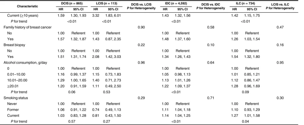
Table 3. Associations of Participant Characteristics With Risks of In Situ and Invasive Breast Cancers, by Histological Type, Among Postmenopausal Women Who Have Had a Mammogram (n=120,780)a, National Institutes of Health-AARP Diet and Health Study Cohort, 1995–2011b
Characteristic DCIS (n=865) LCIS (n=113) DCIS vs. LCIS Pfor Heterogeneity
IDC (n=4,592) DCIS vs. IDC Pfor Heterogeneity
ILC (n=734) LCIS vs. ILC Pfor Heterogeneity
HR 95% CI HR 95% CI HR 95% CI HR 95% CI
Age at entry, years 0.72 0.22 0.94
<55 1.00 Referent 1.00 Referent 1.00 Referent 1.00 Referent
55–59 0.97 0.72, 1.32 1.44 0.68, 3.06 1.09 0.96, 1.25 1.20 0.85, 1.70
60–64 1.28 0.92, 1.76 1.57 0.68, 3.65 1.14 0.99, 1.31 1.36 0.93, 1.97
≥65 1.26 0.89, 1.78 1.44 0.57, 3.65 1.24 1.06, 1.44 1.52 1.02, 2.27
Pfor trend 0.07 0.58 <0.01 0.03
Race 0.41 0.29 0.90
White 1.00 Referent 1.00 Referent 1.00 Referent 1.00 Referent
Nonwhite 1.08 0.85, 1.38 0.83 0.38, 1.80 0.94 0.84, 1.05 0.86 0.64, 1.14
Age at menarche, years 0.55 0.24 0.98
≤12 1.00 Referent 1.00 Referent 1.00 Referent 1.00 Referent
13–14 1.11 0.97, 1.28 1.02 0.69, 1.51 0.99 0.94, 1.06 1.02 0.86, 1.17
≥15 0.83 0.63, 1.08 1.04 0.53, 2.05 0.89 0.80, 0.99 0.96 0.73, 1.25
Pfor trend 0.87 0.91 0.12 0.84
Age atfirst birth, years 0.29 0.04 0.32
<20 0.79 0.63, 0.98 1.15 0.63, 2.09 0.97 0.89, 1.06 0.76 0.59, 0.98
20–24 1.00 Referent 1.00 Referent 1.00 Referent 1.00 Referent
25–29 1.21 1.01, 1.45 1.75 1.07, 2.86 1.07 0.98, 1.16 1.42 1.17, 1.71
≥30 or nulliparous 1.44 1.21, 1.71 1.74 1.08, 2.82 1.24 1.15, 1.34 1.32 1.09, 1.60
Pfor trend <0.01 0.03 <0.01 <0.01
Age at menopause, years 0.03 <0.01 0.36
<45 1.51 1.15, 1.97 0.77 0.35, 1.73 0.81 0.71, 0.93 0.74 0.53, 1.04
45–49 1.08 0.87, 1.34 0.62 0.35, 1.12 0.92 0.84, 1.00 0.97 0.78, 1.21
50–54 1.00 Referent 1.00 Referent 1.00 Referent 1.00 Referent
≥55 1.33 1.00, 1.75 0.59 0.25, 1.39 1.22 1.09, 1.36 1.34 1.03, 1.75
Surgical 1.01 0.84, 1.21 0.41 0.25, 0.67 0.71 0.66, 0.77 0.75 0.62, 0.91
Unknown 1.55 1.07, 2.23 0.78 0.30, 1.99 0.93 0.77, 1.12 0.60 0.34, 1.03
Pfor trend 0.28 0.03 <0.01 0.08
MHT use 0.06 0.23 0.01
Never 1.00 Referent 1.00 Referent 1.00 Referent 1.00 Referent
Former 1.17 0.90, 1.52 1.70 0.76, 3.79 1.08 0.97, 1.21 0.95 0.71, 1.28
Current (<5 years) 1.56 1.26, 1.95 2.72 1.47, 5.02 1.23 1.12, 1.35 1.42 1.13, 1.80
Current (5–9 years) 1.89 1.53, 2.32 4.62 2.66, 8.05 1.49 1.36, 1.64 1.50 1.19, 1.89
Table continues
Am
J
Epidemiol.
2017;186(12):1329
–
1340
Risk
Factors
for
Postmenopausal
Breast
Cancer
Table 3. Continued
Characteristic DCIS (n=865) LCIS (n=113) DCIS vs. LCIS Pfor Heterogeneity
IDC (n=4,592) DCIS vs. IDC Pfor Heterogeneity
ILC (n=734) LCIS vs. ILC Pfor Heterogeneity
HR 95% CI HR 95% CI HR 95% CI HR 95% CI
Current (≥10 years) 1.59 1.30, 1.93 3.32 1.83, 6.01 1.43 1.32, 1.56 1.42 1.15, 1.75
Pfor trend <0.01 <0.01 <0.01 <0.01
Family history of breast cancer 0.90 0.58 0.47
No 1.00 Referent 1.00 Referent 1.00 Referent 1.00 Referent
Yes 1.57 1.32, 1.87 1.43 0.87, 2.35 1.48 1.37, 1.60 1.26 1.03, 1.54
Breast biopsy 0.22 0.10 0.16
No 1.00 Referent 1.00 Referent 1.00 Referent 1.00 Referent
Yes 1.51 1.31, 1.74 2.08 1.42, 3.03 1.34 1.26, 1.43 1.54 1.32, 1.80
Alcohol consumption, g/day 0.96 0.64 0.95
0 1.00 Referent 1.00 Referent 1.00 Referent 1.00 Referent
0.01–10.00 1.16 0.99, 1.37 1.15 0.73, 1.83 1.05 0.98, 1.13 1.01 0.85, 1.21
10.01–20.00 1.29 1.00, 1.65 1.40 0.71, 2.73 1.13 1.01, 1.26 1.12 0.86, 1.47
≥20.01 1.20 0.91, 1.59 1.11 0.49, 2.50 1.22 1.09, 1.37 1.28 0.96, 1.69
Pfor trend 0.06 0.53 <0.01 0.09
Smoking status 0.29 0.71 0.30
Never 1.00 Referent 1.00 Referent 1.00 Referent 1.00 Referent
Former 1.06 0.91, 1.22 0.74 0.49, 1.13 1.11 1.04, 1.18 1.10 0.93, 1.29
Current 1.03 0.83, 1.28 0.81 0.43, 1.50 1.14 1.04, 1.25 1.27 1.01, 1.58
Pfor trend 0.57 0.27 <0.01 0.04
Abbreviations: BMI, body mass index; CI, confidence interval; DCIS, ductal carcinoma in situ; HR, hazard ratio; IDC, invasive ductal carcinoma; ILC, invasive lobular carcinoma; LCIS, lobular carcinoma in situ; MHT, menopausal hormone therapy.
aAll analyses were adjusted for age at entry, race, body mass index, smoking status, alcohol consumption, age at menarche, age atfirst birth, age at menopause, MHT use and duration of
use, past breast biopsy, family history of breast cancer in afirst-degree female relative, educational level, and frequency of vigorous physical activity.
bThe women had a mammogram within the 3 years before completion of the second study follow-up questionnaire. Follow-up for this analysis involved mammographic screening data
cap-tured on the second study questionnaire, and follow-up for this analysis began on the date on which that questionnaire was returned.
Am
J
Epidemiol.
2017;186(12):1329
–
1340
1336
Mullooly
et
that among this population, early age at natural menopause was still associated with a higher risk of DCIS but not LCIS (for DCIS, HR=1.51, 95% CI: 1.15, 1.97;Pfor heteroge-neity=0.03) or IDC (HR=0.81, 95% CI: 0.71, 0.93;Pfor heterogeneity<0.01). In addition, current MHT use was still most strongly associated with a higher risk of LCIS than of ILC (Pfor heterogeneity=0.01).
DISCUSSION
Within the NIH-AARP Diet and Health Study, we identified many similarities in and a few differences between associations of breast cancer risk factors with incident in situ and invasive breast cancers among postmenopausal women. Although asso-ciations were similar for race, age at menarche, age atfirst birth, a family history of breast cancer, and lifestyle factors (BMI, alcohol use, cigarette smoking status, and level of physical activity), we observed significant heterogeneity in risk associated with age at natural menopause, MHT use, and a history of breast biopsy. In particular, higher risks associated with MHT use and previous breast biopsy were stronger for LCIS than for ILC and DCIS, respectively. Thesefindings sug-gest that most risk factors for breast cancer act at the precursor stage, but there may be a few with differential associations for in situ versus invasive breast cancers.
There have been few longitudinal studies in which investi-gators have examined associations between risk factors and LCIS. Ourfindings, which demonstrated similar risk patterns in the associations of race, age at menarche, age atfirst birth, family history, alcohol use, and smoking status with both DCIS and LCIS, suggest that these noninvasive lesions may share at least some common etiology. Given that cases of LCIS and low-grade DCIS are nearly uniformly estrogen receptor (ER)–positive, comparisons might have been tighter had grade information been available. Ourfindings also suggest some possible eti-ological differences: Differences in patterns of association were observed for age at natural menopause and risk of DCIS versus LCIS. Ourfindings are consistent with those from a pre-vious analysis by Claus et al. (21) but were not in agreement with results from a case-control study that showed a lower risk of DCIS associated with younger age at menopause (22). Among women who experienced menopause at a younger age, we observed a significantly higher risk of DCIS but not LCIS. One possible biological hypothesis for thisfinding may be accounted for by the relationship between decreased mammographic breast density and menopause (23). Higher mammographic breast den-sity is associated with lower mammographic sensitivity (24). A possible implication of younger menopausal age is that mammo-graphic density will have also declined at a younger age, which potentially renders DCIS more visible on a mammogram during screening. Because LCIS are less frequently detected with mam-mography, it is not necessarily surprising that this association was not observed for LCIS. This is supported by thefindings in our sensitivity analysis, which showed even higher hazard ratios for the association of DCIS with a younger age at menopause compared with the association with LCIS when the population was restricted to women who reported a previous mammogram in the 3 years prior to the second study follow-up question-naire. However, because measurements of mammographic
breast density were not available for analysis among this study population, we cannot directly investigate this hypothesis. Because multiple statistical tests were carried out, we also acknowledge that thisfinding may be spurious.
In addition, we found that current MHT use was more strongly associated with the risk of LCIS than with the risk of DCIS,findings that are of a similar magnitude to a previ-ous analysis within the prospective cohort from the Million Women Study (MWS), although a larger number of LCIS cases were included in this present analysis than in the previous report (5). Further within the Million Women Study, Reeves et al. (5) showed strong relationships between MHT use and increased risks of both DCIS (n=1,443) and LCIS (n=86). In addition, in our prospective analysis we showed a stronger association of previous benign breast biopsy with LCIS risk than with DCIS, which is in agreement with results from 3 population-based case-control studies (6,7,21). The observed associations between a history of benign breast biopsy and an elevated risk of in situ lesions are complex and likely due to the influence of multiple factors. For example, thesefindings may be driven by biological mechanisms relating benign lesions to the risk of subsequent development of in situ lesions. Further-more, these associations may be the result of higher surveil-lance among women who would have had a prior biopsy than among women who did not.
DCIS is widely accepted as a nonobligate precursor of IDC, and both are thought to share common etiologies, as reviewed by Virnig et al. (25). In a second analysis conducted with the prospective cohort from the Million Women Study, Reeves et al. (26) compared risk factors for DCIS and IDC among that cohort of 1.1 million postmenopausal women from the United Kingdom, which included 3,715 women with DCIS and 21,137 with IDC. Similar to ourfindings, significant heteroge-neity was not observed between the risks of DCIS and IDC for most breast cancer risk factors, including age at menarche, age atfirst birth, MHT use, and a family history of breast cancer (26). However, in that prior study, investigators observed stron-ger associations of higher BMI and alcohol consumption with an increased risk of IDC than with DCIS (26). Although we observed somewhat stronger positive associations of BMI and alcohol consumption with the risk of IDC versus the risk of DCIS, the test for statistical heterogeneity was null in our study population.
The role of LCIS as a precursor lesion of ILC is less well understood (27). It is a less common histological type, it is often not recognized radiologically, and its classification is often surrounded by inconsistency (28). However, multiple studies have shown that LCIS is associated with an increased risk of invasive breast cancer (29–32). The most notable dif-ferences between LCIS and ILC risks in our analyses were observed for MHT use, with significantly stronger associa-tions with the risk of LCIS than with ILC,findings that were also observed in our sensitivity analysis restricted to women who had undergone a mammogram. Several lines of evidence support a hormonal influence on the development of LCIS. First, incidence trends for LCIS in the United States declined sharply between 2001 and 2004 (27,33), which is likely the result of decreased use of MHT after the publication of results from the Women’s Health Initiative (8). In addition,findings from the National Surgical Adjuvant Breast and Bowel Project
Am J Epidemiol. 2017;186(12):1329–1340
P-1 chemoprevention trial highlighted the potential for the selective ER modulator tamoxifen to reduce the development of LCIS (34), and the American Society for Clinical Oncology guidelines include recommendations for antiestrogen therapy as chemoprevention for future breast cancer risk among women with LCIS (35).
A strength of the present study was the large number of cases and extended follow-up period (through 2011). Although the NIH-AARP Diet and Health Study is population-based, it was geographically restricted. Most participants were older, with the largest proportion of women 65 years of age or older, which may limit the generalizability of thesefindings among younger postmenopausal women. In addition, compared with the other histological types assessed, the number of LCIS cases was somewhat limited. Despite this limitation, our study highlighted higher risks associated with current MHT use, particularly for LCIS. Because prior work has demonstrated differential asso-ciations of breast cancer risk by MHT formulation (e.g., estro-gen alone vs. combined estroestro-gen plus progestin) (5,17,36), future efforts in larger diverse populations are needed to exam-ine these relationships in order to further understand the risks of in situ and invasive breast cancers associated with specific MHT formulations. Furthermore, although the majority of breast cancers diagnosed in older women are ER-positive, we were unable to directly examine associations of risk factors specifically for pathologically confirmed ER-positive breast cancers. ER status was not systematically reported in Florida, Texas, and Pennsylvania registries. In addition, ER classifi ca-tion of in situ breast cancer was not routinely recorded in regis-tries until recently, with ER status for DCIS in the data captured by the Surveillance, Epidemiology, and End Results Program only reaching 72% completeness in 2005 (37,38). Further-more, within our analysis, a large number of statistical tests for heterogeneity were carried out; therefore, we cannot eliminate the possibility thatfindings may be the result of chance.
In conclusion, we found that associations of many breast cancer risk factors with in situ and invasive carcinomas were similar, which suggest that most risk factors act during the early stages of breast tumorigenesis. However, certain associations differed between in situ and invasive breast cancers, which may suggest that some factors act differentially during the in situ stage or that there are possibly unaccounted for effects, such as detectability by mammography. These similarities and differ-ences are important in order to increase our understanding of the etiology of histological tumor types and may help to inform prevention strategies. Future ongoing work will determine the importance of these risk factors among women who are diag-nosed with an in situ lesion and subsequently develop an inva-sive breast cancer at a later time point. Additional studies that include distinct molecular subtypes are required to expand these findings to further understand the role of MHT use, menopausal age, and benign breast disease in breast carcinogenesis.
ACKNOWLEDGMENTS
Author affiliations: Division of Cancer Epidemiology and Genetics, National Cancer Institute, Bethesda, Maryland (Maeve Mullooly, Zeina G. Khodr, Roni Falk, Linda
M. Liao, Louise A. Brinton, Gretchen L. Gierach); Cancer Prevention Fellowship Program, Division of Cancer Prevention, National Cancer Institute, Bethesda, Maryland (Maeve Mullooly); Department of Epidemiology and Biostatistics, School of Public Health, University of Maryland, College Park, College Park, Maryland (Cher M. Dallal); Department of Radiology, University of North Carolina at Chapel Hill, Chapel Hill, North Carolina (Sarah J. Nyante); Division of Cancer Prevention, National Cancer Institute, Bethesda, Maryland (Mark E. Sherman); and Strategic Issues Research, AARP Research Center, Washington, DC (Jeffrey Love).
This research was supported by the Intramural Research Program of the National Cancer Institute, National Institutes of Health. M.M. was supported by the Cancer Prevention Fellowship Program of the National Cancer Institute, National Institutes of Health.
We thank Sigurd Hermansen and Kerry Grace Morrissey from Westat, Inc. (Rockville, Maryland) for study outcomes ascertainment and management and Leslie Carroll at Information Management Services, Inc. (Rockville, Maryland) for data support and analysis.
Cancer incidence data from the Atlanta, Georgia, metropolitan area were collected by the Georgia Center for Cancer Statistics, Department of Epidemiology, Rollins School of Public Health, Emory University, Atlanta, Georgia. Cancer incidence data from California were collected by the California Cancer Registry, California Department of Public Health’s Cancer Surveillance and Research Branch, Sacremento, California. Cancer incidence data from the Detroit metropolitan area were collected by the Michigan Cancer Surveillance Program, Community Health Administration, Detroit, Michigan. The Florida cancer incidence data used in this report were collected by the Florida Cancer Data System (Miami, Florida) under contract with the Florida Department of Health, Tallahassee, Florida. Cancer incidence data from Louisiana were collected by the Louisiana Tumor Registry, Louisiana State University Health Sciences Center School of Public Health, New Orleans, Louisiana. Cancer incidence data from New Jersey were collected by the New Jersey State Cancer Registry, The Rutgers Cancer Institute of New Jersey, New Brunswick, New Jersey. Cancer incidence data from North Carolina were collected by the North Carolina Central Cancer Registry, Raleigh, North Carolina. Cancer incidence data from Pennsylvania were supplied by the Division of Health Statistics and Research, Pennsylvania Department of Health, Harrisburg, Pennsylvania. Cancer incidence data from Arizona were collected by the Arizona Cancer Registry, Division of Public Health Services, Arizona Department of Health Services, Phoenix, Arizona. Cancer incidence data from Texas were collected by the Texas Cancer Registry, Cancer Epidemiology and Surveillance Branch, Texas Department of State Health Services, Austin, Texas. Cancer incidence data from Nevada were collected by the Nevada Central Cancer Registry, Division of Public and Behavioral Health, State of Nevada Department of Health and Human Services, Carson City, Nevada.
The views expressed herein are solely those of the authors and do not necessarily reflect those of the Florida Cancer
Am J Epidemiol. 2017;186(12):1329–1340
Data System or the Florida Department of Health. The Pennsylvania Department of Health specifically disclaims responsibility for any analyses, interpretations, or conclusions.
Conflict of interest: none declared.
REFERENCES
1. Howlader N, Noone AM, Krapcho M, et al., eds.SEER Cancer Statistics Review, 1975–2008. Bethesda, MD: National Cancer Institute; 2011.http://seer.cancer.gov/csr/1975_2008/. Accessed July 1, 2016.
2. Morrow M, Schnitt SJ, Norton L. Current management of lesions associated with an increased risk of breast cancer.Nat Rev Clin Oncol. 2015;12(4):227–238.
3. Sherman ME, Mies C, Gierach GL. Opportunities for molecular epidemiological research on ductal carcinoma in-situ and breast carcinogenesis: interdisciplinary approaches.
Breast Dis. 2014;34(3):105–116.
4. Venkitaraman R. Lobular neoplasia of the breast.Breast J. 2010;16(5):519–528.
5. Reeves GK, Beral V, Green J, et al. Hormonal therapy for menopause and breast-cancer risk by histological type: a cohort study and meta-analysis.Lancet Oncol. 2006;7(11):910–918. 6. Trentham-Dietz A, Newcomb PA, Storer BE, et al. Risk factors
for carcinoma in situ of the breast.Cancer Epidemiol Biomarkers Prev. 2000;9(7):697–703.
7. Weiss HA, Brinton LA, Brogan D, et al. Epidemiology of in situ and invasive breast cancer in women aged under 45.Br J Cancer. 1996;73(10):1298–1305.
8. Ravdin PM, Cronin KA, Howlader N, et al. The decrease in breast-cancer incidence in 2003 in the United States.N Engl J Med. 2007;356(16):1670–1674.
9. Kerlikowske K, Barclay J, Grady D, et al. Comparison of risk factors for ductal carcinoma in situ and invasive breast cancer.
J Natl Cancer Inst. 1997;89(1):76–82.
10. Nyante SJ, Dallal CM, Gierach GL, et al. Risk factors for specific histopathological types of postmenopausal breast cancer in the NIH-AARP Diet and Health Study.Am J Epidemiol. 2013;178(3):359–371.
11. Nyante SJ, Gierach GL, Dallal CM, et al. Cigarette smoking and postmenopausal breast cancer risk in a prospective cohort.
Br J Cancer. 2014;110(9):2339–2347.
12. Schatzkin A, Subar AF, Thompson FE, et al. Design and serendipity in establishing a large cohort with wide dietary intake distributions: the National Institutes of Health-American Association of Retired Persons Diet and Health Study.Am J Epidemiol. 2001;154(12):1119–1125.
13. Lew JQ, Freedman ND, Leitzmann MF, et al. Alcohol and risk of breast cancer by histologic type and hormone receptor status in postmenopausal women: the NIH-AARP Diet and Health Study.Am J Epidemiol. 2009;170(3):308–317.
14. Kuczmarski RJ, Flegal KM. Criteria for definition of
overweight in transition: background and recommendations for the United States.Am J Clin Nutr. 2000;72(5):1074–1081. 15. Michaud DS, Midthune D, Hermansen S, et al. Comparison of
cancer registry case ascertainment with SEER estimates and self-reporting in a subset of the NIH-AARP Diet and Health Study.J Registry Manag. 2005;32(2):70–75.
16. Fritz A, Percy C, Jack A, et al.International Classification of Diseases for Oncology (ICD-O-3). 3rd ed. Geneva,
Switzerland: World Health Organization; 2000.
17. Brinton LA, Richesson D, Leitzmann MF, et al. Menopausal hormone therapy and breast cancer risk in the NIH-AARP Diet and Health Study Cohort.Cancer Epidemiol Biomarkers Prev. 2008;17(11):3150–3160.
18. Kabat GC, Xue X, Kamensky V, et al. Risk of breast, endometrial, colorectal, and renal cancers in postmenopausal women in association with a body shape index and other anthropometric measures.Cancer Causes Control. 2015;26(2): 219–229.
19. Munsell MF, Sprague BL, Berry DA, et al. Body mass index and breast cancer risk according to postmenopausal estrogen-progestin use and hormone receptor status.Epidemiol Rev. 2014;36:114–136.
20. Morimoto LM, White E, Chen Z, et al. Obesity, body size, and risk of postmenopausal breast cancer: the Women’s Health Initiative (United States).Cancer Causes Control. 2002;13(8): 741–751.
21. Claus EB, Stowe M, Carter D. Breast carcinoma in situ: risk factors and screening patterns.J Natl Cancer Inst. 2001; 93(23):1811–1817.
22. Phillips LS, Millikan RC, Schroeder JC, et al. Reproductive and hormonal risk factors for ductal carcinoma in situ of the breast.Cancer Epidemiol Biomarkers Prev. 2009;18(5): 1507–1514.
23. Boyd N, Martin L, Stone J, et al. A longitudinal study of the effects of menopause on mammographic features.Cancer Epidemiol Biomarkers Prev. 2002;11(10 pt 1):1048–1053. 24. Boyd NF, Guo H, Martin LJ, et al. Mammographic density and
the risk and detection of breast cancer.N Engl J Med. 2007; 356(3):227–236.
25. Virnig BA, Wang SY, Shamilyan T, et al. Ductal carcinoma in situ: risk factors and impact of screening.J Natl Cancer Inst Monogr. 2010;2010(41):113–116.
26. Reeves GK, Pirie K, Green J, et al. Comparison of the effects of genetic and environmental risk factors on in situ and invasive ductal breast cancer.Int J Cancer. 2012;131(4):930–937. 27. Ward EM, DeSantis CE, Lin CC, et al. Cancer statistics: breast
cancer in situ.CA Cancer J Clin. 2015;65(6):481–495. 28. Anderson BO, Calhoun KE, Rosen EL. Evolving concepts in
the management of lobular neoplasia.J Natl Compr Canc Netw. 2006;4(5):511–522.
29. Cutuli B, De Lafontan B, Kirova Y, et al. Lobular carcinoma in situ (LCIS) of the breast: is long-term outcome similar to ductal carcinoma in situ (DCIS)? Analysis of 200 cases.Radiat Oncol. 2015;10:110.
30. King TA, Pilewskie M, Muhsen S, et al. Lobular carcinoma in situ: a 29-year longitudinal experience evaluating
clinicopathologic features and breast cancer risk.J Clin Oncol. 2015;33(33):3945–3952.
31. Oppong BA, King TA. Recommendations for women with lobular carcinoma in situ (LCIS).Oncology (Williston Park). 2011;25(11):1051–1056, 1058.
32. Portschy PR, Marmor S, Nzara R, et al. Trends in incidence and management of lobular carcinoma in situ: a population-based analysis.Ann Surg Oncol. 2013;20(10):3240–3246. 33. Eheman CR, Shaw KM, Ryerson AB, et al. The changing
incidence of in situ and invasive ductal and lobular breast carcinomas: United States, 1999–2004.Cancer Epidemiol Biomarkers Prev. 2009;18(6):1763–1769.
34. Fisher B, Costantino JP, Wickerham DL, et al. Tamoxifen for prevention of breast cancer: report of the National Surgical Adjuvant Breast and Bowel Project P-1 Study.J Natl Cancer Inst. 1998;90(18):1371–1388.
35. Visvanathan K, Hurley P, Bantug E, et al. Use of
pharmacologic interventions for breast cancer risk reduction:
Am J Epidemiol. 2017;186(12):1329–1340
American Society of Clinical Oncology clinical practice guideline.J Clin Oncol. 2013;31(23):2942–2962. 36. Rossouw JE, Anderson GL, Prentice RL, et al. Risks and
benefits of estrogen plus progestin in healthy postmenopausal women: principal results from the Women’s Health Initiative randomized controlled trial.JAMA. 2002;288(3):321–333.
37. Morrow M. Refining the use of endocrine therapy for ductal carcinoma in situ.J Clin Oncol. 2012;30(12): 1249–1251.
38. Zujewski JA, Harlan LC, Morrell DM, et al. Ductal carcinoma in situ: trends in treatment over time in the US.Breast Cancer Res Treat. 2011;127(1):251–257.
Am J Epidemiol. 2017;186(12):1329–1340