M A J O R A R T I C L E
Evaluation of Serologic and Antigenic
Relationships Between Middle Eastern
Respiratory Syndrome Coronavirus and Other
Coronaviruses to Develop Vaccine Platforms for
the Rapid Response to Emerging Coronaviruses
Sudhakar Agnihothram,1,2,aRobin Gopal,4,aBoyd L. Yount Jr,1,2Eric F. Donaldson,1,2Vineet D. Menachery,1,2
Rachel L. Graham,1,2Trevor D. Scobey,1,2Lisa E. Gralinski,1,2Mark R. Denison,3Maria Zambon,4and Ralph S. Baric1,2
1
Department of Epidemiology and2Department of Microbiology and Immunology, University of North Carolina, Chapel Hill;3Departments of Pediatrics and Microbiology and Immunology, Vanderbilt University, Nashville, Tennessee; and4Viral Zoonosis Unit, Public Health of England, London, United Kingdom
Background. Middle East respiratory syndrome coronavirus (MERS-CoV) emerged in 2012, causing severe acute respiratory disease and pneumonia, with 44% mortality among 136 cases to date. Design of vaccines to limit the virus spread or diagnostic tests to track newly emerging strains requires knowledge of antigenic and serologic re-lationships between MERS-CoV and other CoVs.
Methods. Using synthetic genomics and Venezuelan equine encephalitis virus replicons (VRPs) expressing spike and nucleocapsid proteins from MERS-CoV and other human and bat CoVs, we characterize the antigenic re-sponses (using Western blot and enzyme-linked immunosorbent assay) and serologic rere-sponses (using neutraliza-tion assays) against 2 MERS-CoV isolates in comparison with those of other human and bat CoVs.
Results. Serologic and neutralization responses against the spike glycoprotein were primarily strain specific, with a very low level of cross-reactivity within or across subgroups. CoV N proteins within but not across subgroups share cross-reactive epitopes with MERS-CoV isolates. Our findings were validated using a convalescent-phase serum specimen from a patient infected with MERS-CoV (NA 01) and human antiserum against SARS-CoV, human CoV NL63, and human CoV OC43.
Conclusions. Vaccine design for emerging CoVs should involve chimeric spike protein containing neutralizing epitopes from multiple virus strains across subgroups to reduce immune pathology, and a diagnostic platform should include a panel of nucleocapsid and spike proteins from phylogenetically distinct CoVs.
Keywords. MERS-CoV Vaccine Design; Diagnostics; Serology; Synthetic Genomics.
Novel approaches are needed to respond rapidly to new emerging diseases, especially early in the epidemic, when prompt public health interventions can limit mortality and epidemic spread. Coronaviruses (CoVs) constitute a
group of phylogenetically diverse enveloped viruses that have the largest plus-strand RNA genomes and replicate efficiently in most mammals [1,2]. Human CoV (HCoV-229E, -OC43, -NL63, and -HKU 1) infections typically result in mild-to-severe upper and lower respiratory tract disease [3,4]. SARS-CoV emerged in 2002–2003, causing acute respiratory distress syndrome with 10% mortality overall and up to 50% mortality among aged individuals [5]. Most recently, Middle East respiratory syndrome CoV (MERS-CoV) emerged in the Middle East in April 2012, manifesting as severe pneumonia, acute respiratory distress syndrome, and acute renal failure. The virus is still circulating and has caused 136 human infections with 58 deaths (mortality rate, approximately 44%) [6,7].
Received 16 August 2013; accepted 11 October 2013; electronically published 18 November 2013.
a
S. A. and R. G. contributed equally to this work.
Presented in part: MERS-CoV study group meeting June 20-22, 2013, Cairo, Egypt. Correspondence: Ralph S. Baric, PhD, Department of Microbiology and Immunol-ogy, 3304 Michael Hooker Research Bldg, Campus Box 7435, Chapel Hill, NC 27599-7435 ([email protected]).
The Journal of Infectious Diseases 2014;209:995–1006
© The Author 2013. Published by Oxford University Press on behalf of the Infectious Diseases Society of America. All rights reserved. For Permissions, please e-mail: [email protected].
Phylogenetic analysis groups CoVs into 4 genera—
Alphacoronavirus, Betacoronavirus, Gammacoronavirus, and
Deltacoronavirus—and for many mammalian CoVs, bats are considered reservoirs [6,8,9]. SARS-CoV is closely related to bat CoV (BtCoV) HKU 3 [1,10–12], whereas MERS-CoV is closely related to PipistrellusBtCoV HKU 5 andTylonycteris BtCoV HKU 4 [9]. However, the serologic and antigenic rela-tionship between strains is unclear. Given the vast number of genetically distinct CoVs, well-defined serologic and virologic reagents are needed to rapidly track MERS-CoV and other CoV infections in natural populations and to optimize vaccine and therapeutic designs early in an outbreak setting, especially within and between phylogenetic subgroups.
The spike (S) and nucleocapsid (N) proteins are major im-munogenic components of CoVs and are produced in abun-dant quantities during infection. The S protein is the principle determinant of protective immunity and cross-species trans-mission in CoV [11]. Antibodies against S protein protect from homologous and heterologous SARS-CoV challenge in vivo [13], whereas N protein–specific immune responses may offer limited protection especially against low-dose challenge [13]. Therefore, antibodies against S and N protein have diagnostic and therapeutic potential [14,15].
In this article, we use alphavirus replicon vaccine vectors to express a panel of recombinant S and N proteins from distantly related alphacoronaviruses and betacoronaviruses, including MERS-CoV and other subgroup 2c CoVs. Using mouse poly-clonal antisera and recombinant proteins, we compare the cross-reactivity and neutralization titers of these antisera between distantly related human and bat CoVs. Our results in-dicate that the S glycoprotein but not the N protein is the major determinant of the neutralizing antibody response to MERS-CoV; that the N proteins of CoVs only cross-react within but not between subgroups; that little if any cross-neutralization or cross-reactivity exists between the S proteins of CoVs within subgroup 2c or any other subgroup; and that cross-neutraliza-tion and cross-reactive patterns were validated with the conva-lescent-phase serum sample from a patient infected with MERS-CoV Hu/England-N1/2012 and a donor panel of human antisera against 3 different HCoVs. Our approach pro-vides critical reagents, antisera, and recombinant virus vaccines that allow for rapid diagnosis of and intervention against MERS-CoV and other zoonotic CoVs that emerge in the future.
MATERIALS AND METHODS
Viruses, Cells, and Plaque Assays
MERS-CoV Hu/England-N1/2012 and MERS-CoV Hu/SA-N1/2012 were cultured on Vero 81 cells and grown in Opti-Mem (Gibco, Carlsbad, CA) with 5% fetal clone serum (Hyclone, South Logan, UT) and gentamicin/kanamycin
(Gibco). Viral growth assays in Vero and Calu-3 cells were per-formed as previously described [16].
Generation of Polyclonal Mouse Antisera, Neutralization Assays, and Western and Northern Blot Analysis
Genes encoding the indicated S and N proteins were synthe-sized from Bio Basic (Ontario, Canada) and packaged into Venezuelan equine encephalitis virus replicon particles (VRPs). Following vaccination, mouse polyclonal sera were generated from BALB/c mice, and neutralization assays involving MERS-CoV strains and SARS-MERS-CoV were as described previously [17]. For Western blots, VRP or virus-infected cell lysates and con-trols were prepared as described before in detail [8], and these blots were probed using the indicated mouse polyclonal sera. Vero cells inoculated with MERS-CoV isolates were harvested 12 hours after infection by means of Trizol reagent (Invitrogen) and were used to perform Northern blots [18].
Enzyme-Linked Immunosorbent Assay (ELISA) and Blocking Assay
An ELISA using indicated virus-infected cell lysates or antigens expressed from VRPs was performed as described previously [19], and the reactivity of mouse or human serum was deter-mined using a chemiluminescent substrate. Blocking ELISA was performed by sequentially reacting plate-bound MERS-CoV lysate antigen with convalescent-phase serum obtained from a patient with MERS, followed by mouse polyclonal serum raised against VRP-packaged MERS-CoV N or S pro-teins. Blocking was expressed as the percentage reduction in the reactivity of the mouse serum alone.
RESULTS
Molecular Characterization of MERS-CoV Hu/England-N1/2012
MERS-CoV Hu/England-N1/2012 (MERS Eng 1) was isolated from a 49-year-old patient with severe respiratory illness and was transferred to London for treatment [7]. Twenty-nine mu-tations in MERS Eng 1 at the amino acid level were identified and compared to the published sequence of MERS-CoV Hu/ SA-N1/2012 (MERS SA 1; GenBank JX869059.2; Supplementa-ry Figure S2).To identify whether these mutations altered virus growth, we analyzed the replication kinetics of the 2 isolates in Vero cells and a continuous epithelial cell line, Calu-3 (Figure 1A and 1B). Although the replication kinetics were slightly different between 2 isolates in Vero cells, peak viral titers were equivalent. In contrast, virus growth was markedly distinct in human Calu-3 cells and could have represented dif-ferences in strain-specific in vitro adaptation phenotypes or re-sulted from functional differences in the sensitivity to innate immune responses [20].
previous reports predicted 10 open reading frames in MERS SA 1 and MERS Eng 1 [9]. Northern blot analysis identified 8 sub-genomic mRNAs after infection in both viruses (Figure 1C). The observed nested set of subgenomic mRNA expression is consistent with observations for other CoVs [2,9]. The open reading frames encoded by each mRNA in these isolates are de-tailed inSupplementary Table 1.
Serologic Relationships Among MERS-CoV Strains
VRPs function as efficient expression and vaccine platforms for a variety of antigens [13,21]. We generated VRPs expressing MERS SA 1 S and N proteins and then immunized mice. N protein–specific antiserum recognized a discrete 50-kDa band at the predicted molecular weight in lysates from Vero cells (Supplementary Figure 1A) and Calu-3 cells (Figure1D) infect-ed with VRP-N or with the 2 different MERS-CoV isolates. For
the most part, similar expression patterns were evident between VRPs and viruses; however, the N protein of MERS Eng 1 had a slightly lower molecular weight, which was consistent with amino acid deletions at positions S391 and I392 ( Supplementa-ry Figure 2).
Mouse anti-S serum identified an approximately 180-kDa S protein in VRP-S–or MERS-CoV–infected Vero cells ( Supple-mentary Figure 1A). The observed molecular weights were con-sistent with the sizes of the S proteins of other CoVs [2,22]. We noted similar results in Calu-3 cells (Figure1D), and interest-ingly, an increased amount of a higher-molecular-weight form of S protein (glycosylated dimer) was noted in MERS Eng 1 in both cell lines. Antiserum against MERS SA 1 S and N proteins also recognized MERS Eng 1 in ELISAs (data not shown), and N and S proteins recognized the MERS-CoV Jordan isolate (Supplementary Figure 1B).
Cross-neutralization Patterns Across Strains
Plaque reduction neutralization tests (PRNT50) indicated
com-plete neutralization of both MERS-CoV isolates (PRNT50titer,
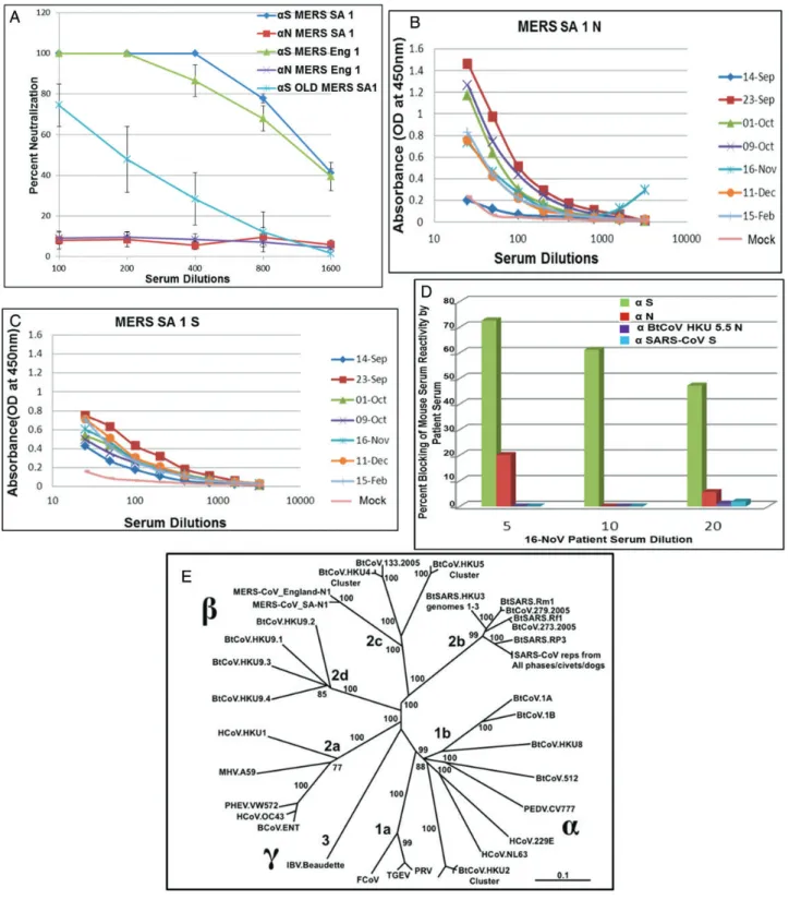
approximately 1:1400 for each; Figure2A) and the MERS-CoV Jordan isolate (Supplementary Figure1C) by VRP-S antiserum, whereas no neutralization was observed with N antiserum. Similarfindings have been reported with SARS-CoV, as well as with other known human and animal CoVs [13,23]. Interesting-ly, serum from aged mice vaccinated with VRP S showed a 6-fold reduction in PRNT50titers (approximately 1:200), indicating that
immunosenescence attenuates vaccine responses to MERS-CoV antigens, as was noted with SARS-CoV vaccines [13,17]. Using serum from NA 01 patient, ELISA demonstrated high reactivity of the patient’s serum to N and S antigens of MERS SA 1 ex-pressed from VRPs (Figure 2B and 2C). Titers of antibody against N protein in patient serum peaked 3–5 weeks after onset of illness (which occurred on 3 September 2012) and waned thereafter, but the antibodies were still detected up to 5 months after illness onset. Titers of antibody against S protein were con-sistent from 3 weeks to 5 weeks after illness onset, after which they remained detectable. Most importantly, patient serum col-lected on 16 November 2012 (which contained high titers against S protein) outcompeted the binding of mouse S antiserum to intact virus in a blocking assay (Figure2D). These data suggest that different/overlapping epitopes are recognized by human and mouse antisera following virus or VRP-S infection.
Cross-reactive and Cross-neutralizing Antibody Responses Within and Across Alphacoronaviruses and Betacoronaviruses
MERS SA 1 and MERS Eng 1 are closely related to BtCoV HKU 5 and BtCoV HKU 4 (Figure2E) [6,9,24]. To evaluate antigenic relationships with the subgroup 2c betacoronaviruses, VRPs ex-pressing S and N proteins of BtCoV HKU 4.2 and BtCoV HKU 5.5 were inoculated into mice. Antisera against both HKU 4.2 and 5.5 N proteins recognized the N proteins of both MERS-CoV isolates, whereas MERS SA 1 N antisera also detected the VRP-expressed HKU 4.2 and 5.5 N proteins, as revealed by Western blot (Figure 3A and3B). We obtained similar results using ELISA and immunofluorescence assays (data not shown). In contrast, there was little if any observable cross-reactivity ob-served between MERS SA 1 S antisera with the VRP-expressed S proteins of HKU 4.2 and HKU 5.5, whereas antisera to HKU 5.5 S protein but not HKU 4.2 S protein recognized the S proteins of both MERS-CoV isolates (Figure3Cand3D). We also measured serologic relationships using ELISA, which captures cross-reactiv-ity to conformational epitopes, and confirmed these antigenic re-lationships (Figure6C). Consistent with results of serologic tests, antisera against HKU 4.2 and HKU 5.5 S proteins did not cross-neutralize the MERS-CoV isolates. These data indicate that the N protein but not the S glycoprotein are antigenically conserved within the subgroup 2c betacoronaviruses evaluated in this panel.
We then extended our analysis to the highly pathogenic SARS-CoV and related subgroup 2b betacoronaviruses. Poly-clonal mouse sera to SARS-CoV or MERS SA 1 N or S proteins exhibited no cross-reactivity to the reciprocal strains (Figure4A
and4B). We observed very low levels of cross-neutralization of MERS SA 1 by mouse antisera to SARS-CoV, using very high but not low concentrations of serum (Figure4C), afinding that is consistent with a recent report [24]. Interestingly, ELISA results also showed very minimal cross-reactivity of the NA 01 patient sera obtained on 23 September 2012 to SARS-CoV S antigen (Figure4D). Consistent with this observation, binding of mouse SARS-CoV S antiserum to SARS-CoV was not inhibited by NA01 patient sera in blocking assays (Figure2D), indicating the absence of antibodies to SARS-CoV in the patient serum.
Consonant with these findings, no cross-reactivity was ob-served with antisera against the VRP-expressed N or S glyco-proteins of BtCoV HKU 3 and 279 and the MERS-CoV isolates (Figure5A–D). Furthermore, no cross-neutralization of MERS-CoV isolates by HKU 3S antiserum was observed, although this serum has previously been shown to neutralize a synthetically resurrected HKU 3 variant encoding the SARS S glycoprotein receptor binding domain [23]. Interestingly, we observed very low levels of cross-neutralization of SARS-CoV by BtCoV 279 S antiserum (Figure4C).
To further elucidate the antigenic relationships between the S glycoproteins of alphacoronaviruses and the MERS-CoV iso-lates, we expressed and generated mouse antisera to BtCoV 1A and BtCoV HKU 2 (group 1b alphacoronaviruses), using the VRP platforms. Despite efficient recombinant S glycoprotein expression (Figure7Aand7B), none of the recombinant S gly-coproteins were recognized by MERS SA 1 S antisera. Antisera against BtCoV1A and HKU 2 S glycoproteins had little if any cross-reactivity with and did not neutralize MERS-CoV (Figure7A–C).
Antigenic Relationships Among the HCoVs
We next analyzed the antigenic relationships between VRP-derived mouse serum with the following representative HCoVs from each subgroup, using ELISA (Figure 6C): MERS Eng 1, from subgroup 2c; SARS-CoV, from subgroup 2b; HCoV-NL63, from subgroup 1b; and HCoV-OC43, from subgroup 2a. MERS Eng 1 was recognized by antisera targeting the N but not the S glycoprotein of viruses within the subgroup 2c betacoronaviruses. Likewise, SARS-CoV was only recognized by antisera to N but not S glycoproteins of viruses with the subgroup 2b betacoronavi-ruses. None of the antiserum screened reacted with HCoV-NL63 (subgroup 1b) or HCoV-OC43 (subgroup 2a). Although BtCoV HKU 2 is genetically close to HCoV-NL63, we did not observe any cross-reactivity within the S glycoprotein.
with our previousfindings, human serum to SARS-CoV recog-nized BtCoV HKU 3N, BtCoV 279N, and SARS-CoV N (sub-group 2b) but did not recognize N proteins from other subgroups (Figure6B). Similarly, there was no cross-reactivity of the human antisera from HCoV-NL63 (subgroup 1b) and HCoV-OC43 (subgroup 2a) infections with any of the viral an-tigens within the panel. Serum collected from the patient in-fected with MERS-CoV NA01 showed cross-reactive binding only to BtCoV HKU 5.5 N (subgroup 2c), and little if any cross-reactivity was noted outside the subgroup (Figure 6A),
apart from very low, transient cross-detection of BtCoV 279 N and from cross-reactivity to SARS-CoV S and N recombinant proteins on a single day (23 September 2012).
DISCUSSION
Emerging respiratory CoVs offer a considerable threat to the health of global populations and the economy. Platforms for generating well-characterized molecular reagents and recombi-nant viruses are needed to detect and control the emergence of
new strains, especially early in an outbreak, before the develop-ment of type-specific serologic reagents and therapeutics. Here, we characterized the genome organization, subgenomic mRNA expression, and protein expression patterns of 2 isolates of MERS-CoV. Using alphavirus replicon particles and synthetic gene design, we assembled a panel of recombinant proteins from and donor antisera against phylogenetically distant alpha-coronaviruses and betaalpha-coronaviruses to evaluate the antigenic relationships between strains and to inform vaccine design. MERS-CoV is a highly pathogenic respiratory CoV of humans, causing acute respiratory distress syndrome, with mortality rates approaching 44%. CoV primer sets were not successful in diagnosing the etiology of the Jordan outbreak in April 2012, demonstrating a critical need for paneled reagent sets of recom-binant proteins and sera that allow for serologic evaluations of cases, contact cases, and asymptomatic infections, using Western blot or ELISA-based techniques [25]. This article is also thefirst report that describes the serologic characterization of MERS-CoV and CoV reagents. An advantage of the VRP platform is that it can also function as a vaccine vector,
affording the rapid production of candidate vaccines against newly emerged strains [23]. Using SARS-CoV and MERS-CoV as models, we clearly demonstrated that S protein–based re-combinant vaccines elicit robust neutralization responses in young and aged rodent models. Because VRP-S vectors against SARS-CoV protected young and aged animals [17], we have de-veloped a recombinant S vectored vaccine that could likely prove successful in preventing heterologous MERS-CoV infec-tion in aged individuals, but this remains to be tested.
MERS Eng 1 replicated to lower titers than SA1 in Calu-3 cells. As the 2 viruses have different passage histories in vitro, tissue culture adaptive mutations may account for these differ-ences, as reported with many SARS-CoV isolates [26]. Alterna-tively, 29 amino acid differences have been described, most of which reside in the replicase polyprotein (Supplementary Figure 2) and may affect replication efficiency. In addition, the S glycoprotein of MERS Eng 1 differs from that of MERS SA 1 by 2 amino acids, L506F and Q1020H (Supplementary Figure 2), which may account for the increased amount of the higher-molecular-weight form of S protein in MERS Eng 1
(Figure1DandSupplementary Figure 1A). Recent studies indi-cate that TMPRSS2 likely plays important roles in viral entry by enhancing fusogenic potential through proteolytic processing of MERS-CoV S glycoprotein [22]. In addition, we identified a unique mutation, T1015N, in the MERS SA 1 isolate but not in the MERS Eng 1 isolate and showed that this mutation is re-sponsible for increased in vitrofitness and for plaque morphol-ogy [20]. It is possible that the presence or absence of 1 or more of the S glycoprotein mutations in MERS Eng 1 may result in the slower growth phenotype in Calu3 cells.
Alphavirus VRPs have considerable potential as recombinant virus vaccine platforms in the absence of preexisting immunity [27–30]. We demonstrate efficient expression of several CoV S and N structural proteins both in vitro and in vivo, resulting in robust serologic responses in vaccinated mice. Antiserum to VRP-S glycoprotein but not to VRP-N protein neutralized both isolates of MERS-CoV. Furthermore, we and others have dem-onstrated that vaccine-induced immunopathology observed after challenge is minimized in VRP-S protein–based vaccines, partly because of the T-helper type 1–biased immune response
and high neutralization titers elicited by VRP vectors [13,31]. Importantly, vaccination of aged mice demonstrated that im-munosenescence contributes to a reduction in the magnitude of the antibody response to MERS-CoV S glycoprotein, an im-portant point to be considered in candidate vaccine designs. To date, wild-type and VRP 3526–coated VRP-S vaccines repre-sent one of the few vaccine platforms that functioned well in aged animals, in addition to recombinant subunit–based vac-cines and poxvirus-vectored vacvac-cines [21, 32–34]. In SARS-CoV pathogenesis, increased age-related susceptibility is linked to increased prostaglandin D2 expression; it remains uncertain whether increased prostaglandin D2 levels have contributed to reduced vaccine performance, as well [35]. Because we have not observed MERS-CoV replication in immunocompetent and immunocompromised mice, these vectors must be tested di-rectly in primates [36]. The safety of the VRP platform has been demonstrated in high-risk human populations and
immunosenescent nonhuman primates [27,28,37,38], and we believe that these vectors will be efficacious in healthcare workers and target populations infected with MERS-CoV.
Our results indicate the presence of strongly cross-reactive epitopes in the N protein within a particular subgroup but not between subgroups. Under identical conditions, little cross-re-activity or conservation of cross-neutralizing epitopes was ob-served between S proteins within and across subgroups. Similar studies showing strong conservation of cross-reactive epitopes between N proteins, but to a lesser extent between S proteins of the subgroup 2a CoVs, has been reported [39,40]. Importantly, the pattern of serologic and antigenic relationship observed using the mouse antisera was recapitulated using the human antiserum to 4 different CoVs. Neutralization assays demon-strated little if any conservation of cross-neutralizing epitopes between S glycoproteins of CoVs within and across subgroups. In particular, the absence of cross-neutralization of MERS-CoV
isolates by antiserum to HKU 4 or HKU 5 S glycoprotein and of SARS-CoV by antiserum to the HKU 3 or BtCoV 279 S gly-coprotein suggests very limited conservation or, possibly, the deliberate masking of conserved cross-neutralizing sites within a subgroup. Although speculative, these cross-neutralization re-lationships suggest that at least 3 antigenically distinct CoVs could emerge from zoonotic viruses circulating within sub-group 1a/b, 2b, and 2c reservoirs and then simultaneously cir-culate in humans. These findings are evidence that vaccine design for any new emerging CoV should either focus on the development of chimeric S glycoproteins containing neutraliz-ing epitopes from multiple strains within or across subgroups or on the development of new paradigms in structure-guided antigen design that improve the presentation of broadly neu-tralizing epitopes. Regions of S glycoprotein are interchange-able between CoVs within and across subgroups, rendering viable recombinant viruses [23]. Inclusion of N protein in such chimeric vaccines may broaden the protective response,
although this remains to be tested using lethal challenge viruses. Such a vaccine might provide robust protection against several homologous and heterologous viruses within or across genoclusters.
After the SARS-CoV epidemic and in stark contrast to the situation with emerging influenza viruses such as influenza A (H7N9), the research and biomedical communities failed to develop broadly applicable biopreparedness platforms for rapid response against future emerging CoV threats. Because CoVs have demonstrated an accelerating pattern of zoonotic emer-gence since the 1980s [25,41], our data indicate that an appro-priate diagnostic platform should include a large panel of phylogenetically distinct CoV S and N structural proteins, which must be validated using larger panels of antisera against other HCoVs in the general population. While molecular-based platforms like polymerase chain reaction and deep se-quencing offer clear advantages in early detection of active infections, public health response platforms would be
strengthened by the availability of recombinant proteins and subgroup- and type-specific antisera that can track subclinical infections, determine the prevalence of infection in popula-tions, and identify hospital-acquired infections. A recent report identified subclinical cases of MERS-CoV infection through reverse-transcription polymerase chain reaction, and the screen using the panel of recombinant proteins described here would have provided more-specific information about the presence of other CoVs in these cases [42]. The VRP platform we describe not only yields high-level expression of key recombinant pro-teins across the alphacoronaviruses and betacoronaviruses, it also provides thefirst candidate vaccine vectors with the poten-tial to augment the T-helper type 1–based immune responses to MERS-CoV infection and to reduce associated immune pa-thology. The VRP 3526–associated approach is also applicable to improving the public health response to and the control of future outbreaks of other highly pathogenic emerging infec-tious diseases due to CoV in human populations.
Supplementary Data
Supplementary materialsare available atThe Journal of Infectious Diseases online (http://jid.oxfordjournals.org/). Supplementary materials consist of data provided by the author that are published to benefit the reader. The posted materials are not copyedited. The contents of all supplementary data are the sole responsibility of the authors. Questions or messages regarding errors should be addressed to the author.
Notes
Acknowledgments. We thank the University of North Carolina–Chapel Hill genome analysis facility and Dr Mark Heise at Carolina Vaccine Insti-tute, for providing sequencing services and sharing laboratory space for animal experiments, respectively; Dr Michael Cooper at the Armed Forces Health Surveillance Center, Dr Emad Mohareb at Navy Medical Research Unit 3, and Dr Kanta Subbarao at the National Institute of Allergy and In-fectious Diseases, for providing us with MERS-CoV Hu/Jordan-N3/2012.
Financial support. This work was supported by the National Institutes of Allergy and Infectious Diseases, National Institute of Health (grants AI085524, AI057157, and U19 AI107810) and Public Health of England (formerly, Health Protection Agency, England).
Potential conflicts of interest. All authors: No reported conflicts. All authors have submitted the ICMJE Form for Disclosure of Potential Conflicts of Interest. Conflicts that the editors consider relevant to the content of the manuscript have been disclosed.
References
1. Graham RL, Baric RS. Recombination, reservoirs, and the modular spike: mechanisms of coronavirus cross-species transmission. J Virol 2010; 84:3134–46.
2. Masters PS. The molecular biology of coronaviruses. Adv Virus Res 2006; 66:193–292.
3. Pyrc K, Sims AC, Dijkman R, et al. Culturing the unculturable: human coronavirus HKU1 infects, replicates, and produces progeny virions in human ciliated airway epithelial cell cultures. J Virol 2010; 84: 11255–63.
4. van der Hoek L, Pyrc K, Jebbink MF, et al. Identification of a new human coronavirus. Nat Med2004; 10:368–73.
5. Rota PA, Oberste MS, Monroe SS, et al. Characterization of a novel co-ronavirus associated with severe acute respiratory syndrome. Science 2003; 300:1394–9.
6. Lau SK, Li KS, Tsang AK, et al. Genetic characterization of Betacoronavirus lineage C viruses in bats revealed marked sequence di-vergence in the spike protein of Pipistrellus bat coronavirus HKU5 in Japanese pipistrelle: implications on the origin of the novel Middle East respiratory syndrome coronavirus. J Virol2013; 87:8638–50.
7. Bermingham A, Chand MA, Brown CS, et al. Severe respiratory illness caused by a novel coronavirus, in a patient transferred to the United Kingdom from the Middle East, September 2012. Euro Surveill2012; 17:20290.
8. Huynh J, Li S, Yount B, et al. Evidence supporting a zoonotic origin of human coronavirus strain NL63. J Virol2012; 86:12816–25.
9. van Boheemen S, de Graaf M, Lauber C, et al. Genomic characteriza-tion of a newly discovered coronavirus associated with acute respiratory distress syndrome in humans. MBio2012; 3:e00473-12.
10. Müller MA, Raj VS, Muth D, et al. Human coronavirus EMC does not require the SARS-coronavirus receptor and maintains broad replicative capability in mammalian cell lines. MBio2012; 3:e00515-12.
11. Bolles M, Donaldson E, Baric R. SARS-CoV and emergent coronaviruses: viral determinants of interspecies transmission. Curr Opin Virol2011; 1:624–34.
12. Perlman S, Zhao J. Human coronavirus EMC is not the same as severe acute respiratory syndrome coronavirus. MBio2013; 4:e00002-13. 13. Deming D, Sheahan T, Heise M, et al. Vaccine efficacy in senescent
mice challenged with recombinant SARS-CoV bearing epidemic and zoonotic spike variants. PLoS Med2006; 3:e525.
14. Rockx B, Corti D, Donaldson E, et al. Structural basis for potent cross-neutralizing human monoclonal antibody protection against lethal human and zoonotic severe acute respiratory syndrome coronavirus challenge. J Virol2008; 82:3220–35.
15. Chan RW, Chan MC, Agnihothram S, et al. Tropism and innate immune responses of the novel human betacoronavirus lineage C virus in human ex vivo respiratory organ cultures. J Virol2013; 87:6604–14.
16. Josset L, Menachery VD, Gralinski LE, et al. Cell host response to infection with novel human coronavirus EMC predicts potential antivirals and im-portant differences with SARS coronavirus. MBio2013; 4:e00165-13. 17. Sheahan T, Whitemore A, Long K, et al. Successful vaccination strategies
that protect aged mice from lethal challenge from influenza virus and het-erologous severe acute respiratory syndrome coronavirus. J Virol2011; 85:217–30.
18. Donaldson EF, Sims AC, Graham RL, Denison MR, Baric RS. Murine hepatitis virus replicase protein nsp10 is a critical regulator of viral RNA synthesis. J Virol2007; 81:6356–68.
19. Höschler K, Gopal R, Andrews N, et al. Cross-neutralisation of anti-bodies elicited by an inactivated split-virion influenza A/Vietnam/ 1194/2004 (H5N1) vaccine in healthy adults against H5N1 clade 2 strains. Influenza Other Respi Viruses2007; 1:199–206.
20. Scobey T, Yount BL, Sims AC, et al. Reverse genetics with a full-length infectious cDNA of the Middle East respiratory syndrome coronavirus. Proc Natl Acad Sci U S A2013; 110:16157–62.
21. Sheahan T, Whitmore A, Long K, et al. Successful vaccination strategies that protect aged mice from lethal challenge from influenza virus and heterologous severe acute respiratory syndrome coronavirus. J Virol 2011; 85:217–30.
22. Gierer S, Bertram S, Kaup F, et al. The spike protein of the emerging be-tacoronavirus EMC uses a novel coronavirus receptor for entry, can be activated by TMPRSS2, and is targeted by neutralizing antibodies. J Virol2013; 87:5502–11.
23. Becker MM, Graham RL, Donaldson EF, et al. Synthetic recombinant bat SARS-like coronavirus is infectious in cultured cells and in mice. Proc Natl Acad Sci U S A2008; 105:19944–9.
25. Giménez LG, Rojas J, Rojas A, Mendoza J, Camacho AG. Development of an enzyme-linked immunosorbent assay-based test with a cocktail of nucleocapsid and spike proteins for detection of severe acute respirato-ry syndrome-associated coronavirus-specific antibody. Clin Vaccine Immunol2009; 16:241–5.
26. Vega VB, Ruan Y, Liu J, et al. Mutational dynamics of the SARS coro-navirus in cell culture and human populations isolated in 2003. BMC Infect Dis2004; 4:32.
27. Slovin SF, Kehoe M, Durso R, et al. A phase I dose escalation trial of vaccine replicon particles (VRP) expressing prostate-specific membrane antigen (PSMA) in subjects with prostate cancer. Vaccine 2013; 31:943–9.
28. Wecker M, Gilbert P, Russell N, et al. Phase I safety and immunogenici-ty evaluations of an alphavirus replicon HIV-1 subimmunogenici-type C gag vaccine in healthy HIV-1-uninfected adults. Clin Vaccine Immunol 2012; 19:1651–60.
29. Fillis CA, Calisher CH. Neutralizing antibody responses of humans and mice to vaccination with Venezuelan encephalitis (TC-83) virus. J Clin Microbiol1979; 10:544–9.
30. Tesh RB, Gajdusek DC, Garruto RM, Cross JH, Rosen L. The distribu-tion and prevalence of group A arbovirus neutralizing antibodies among human populations in Southeast Asia and the Pacific islands. Am J Trop Med Hyg1975; 24:664–75.
31. Tseng CT, Sbrana E, Iwata-Yoshikawa N, et al. Immunization with SARS coronavirus vaccines leads to pulmonary immunopathology on challenge with the SARS virus. PLoS One2012; 7:e35421.
32. Bolles M, Deming D, Long K, et al. A double-inactivated severe acute respiratory syndrome coronavirus vaccine provides incomplete protec-tion in mice and induces increased eosinophilic proinflammatory pul-monary response upon challenge. J Virol2011; 85:12201–15.
33. Ben-Yehuda A, Ehleiter D, Hu AR, Weksler ME. Recombinant vaccinia virus expressing the PR/8 influenza hemagglutinin gene overcomes the impaired immune response and increased susceptibility of old mice to influenza infection. J Infect Dis1993; 168:352–7.
34. Asanuma H, Hirokawa K, Uchiyama M, et al. Immune responses and protection in different strains of aged mice immunized intranasally with an adjuvant-combined influenza vaccine. Vaccine2001; 19:3981–9. 35. Zhao J, Legge K, Perlman S. Age-related increases in PGD(2)
expres-sion impair respiratory DC migration, resulting in diminished T cell re-sponses upon respiratory virus infection in mice. J Clin Invest2011; 121:4921–30.
36. Munster VJ, de Wit E, Feldmann H. Pneumonia from human coronavi-rus in a macaque model. N Engl J Med2013; 368:1560–2.
37. Fine DL, Roberts BA, Teehee ML, et al. Venezuelan equine encephalitis virus vaccine candidate (V3526) safety, immunogenicity and efficacy in horses. Vaccine2007; 25:1868–76.
38. Fine DL, Roberts BA, Terpening SJ, Mott J, Vasconcelos D, House RV. Neu-rovirulence evaluation of Venezuelan equine encephalitis (VEE) vaccine candidate V3526 in nonhuman primates. Vaccine2008; 26:3497–506. 39. Dea S, Verbeek AJ, Tijssen P. Antigenic and genomic relationships among
turkey and bovine enteric coronaviruses. J Virol1990; 64:3112–8. 40. Hogue BG, King B, Brian DA. Antigenic relationships among proteins
of bovine coronavirus, human respiratory coronavirus OC43, and mouse hepatitis coronavirus A59. J Virol1984; 51:384–8.
41. Cao Z, Liu L, Du L, et al. Potent and persistent antibody responses against the receptor-binding domain of SARS-CoV spike protein in recovered patients. Virol J2010; 7:299.