Transmission
of
SARS
and
MERS
coronaviruses
and
influenza
virus
in
healthcare
settings:
the
possible
role
of
dry
surface
contamination
q
J.A.
Otter
a,*,
C.
Donskey
b,
S.
Yezli
c,
S.
Douthwaite
d,
S.D.
Goldenberg
d,
D.J.
Weber
eaImperialCollegeHealthcareNHSTrust,London,UK
bClevelandVeteransAffairsMedicalCenter,Cleveland,OH,USA
cGlobalCentreforMassGatheringsMedicine,Riyadh,SaudiArabia
dCentreforClinicalInfectionandDiagnosticsResearch(CIDR),Guy’sandStThomasNHSFoundationTrust&King’sCollege
London,UK
eDivisionofInfectiousDiseases,UniversityofNorthCarolina,ChapelHill,NC,USA
A R T I C L E I N F O
Article history: Received 24 July 2015 Accepted 28 August 2015 Available online 3 October 2015
Keywords:
Healthcare-associated infection Influenza virus
MERS-CoV SARS-CoV
Surface contamination Transmission
S U M M A R Y
Viruses with pandemic potential including H1N1, H5N1, and H5N7 influenza viruses, and severe acute respiratory syndrome (SARS)/Middle East respiratory syndrome (MERS) coronaviruses (CoV) have emerged in recent years. SARS-CoV, MERS-CoV, and influenza virus can survive on surfaces for extended periods, sometimes up to months. Factors influencing the survival of these viruses on surfaces include: strain variation, titre, surface type, suspending medium, mode of deposition, temperature and relative hu-midity, and the method used to determine the viability of the virus. Environmental sampling has identified contamination in field-settings with SARS-CoV and influenza virus, although the frequent use of molecular detection methods may not necessarily represent the presence of viable virus. The importance of indirect contact transmission (involving contamination of inanimate surfaces) is uncertain compared with other transmission routes, principally direct contact transmission (independent of surface contamination), droplet, and airborne routes. However, influenza virus and SARS-CoV may be shed into the environment and be transferred from environmental surfaces to hands of patients and healthcare providers. Emerging data suggest that MERS-CoV also shares these properties. Once contaminated from the environment, hands can then initiate self-inoculation of mucous membranes of the nose, eyes or mouth. Mathematical and animal models, and intervention studies suggest that contact transmission is the most important route in some scenarios. Infection prevention and control implications include the need for hand hygiene and personal protective
qThisworkwaspresentedinpartattheInfectionPreventionSocietyConference,Glasgow,September29thtoOctober1st,2014.
* Correspondingauthor.Address:InfectionPreventionandControl,6thFloor,ClarenceWing,St.Mary’sHospital,PraedStreet,LondonW21NY, UK.Tel.: þ44(0)2033133271.
E-mailaddress:[email protected](J.A.Otter).
equipment to minimize self-contamination and to protect against inoculation of mucosalsurfaces and therespiratory tract,and enhanced surface cleaningand disin-fection in healthcare settings.
Introduction
A number of viruses with pandemic potential have emerged in recent years. The 2002 emergence of severe acute respira-tory syndrome coronavirus (SARS-CoV), 2009 pandemic of H1N1 influenza, continued circulation of influenza H5N1 and H5N7 strains, and the recent emergence of the Middle East respira-tory syndrome coronavirus (MERS-CoV) illustrate the current threat of these viruses.1e4
Despite fundamental differences in their structure and epidemiology, these pandemic viral threats share a number of important properties. They are zoonotic enveloped RNA res-piratory viruses that rarely transmit between humans in their native form, but could mutate to allow more efficient human-to-human transmission. This was illustrated by the 2002e2003 SARS pandemic and the 2009 H1N1 influenza pandemic.3,4 Frequent and accepted transmission routes are ‘droplet transmission’, where droplets (>5
m
m diameter, travelling<1 m) containing viable viruses make contact with the nose, mouth, eyes, or upper respiratory tract, and ‘airborne trans-mission’, where droplet nuclei (5
m
m diameter, which can travel >1 m) are inhaled by susceptible individuals (Figure 1).5e8 The role of ‘direct contact transmission’ (notinvolving contaminated surfaces) and ‘indirect contact trans-mission’ (involving contaminated surfaces) in the spread of these viruses with pandemic potential has been controversial (Figure 1).6e8However, several reviews and models have sug-gested that indirect contact transmission is the predominant transmission route for some respiratory viruses, including influenza, in some settings.7e9
Contaminated surfaces are an established route of trans-mission for important nosocomial pathogens including Clos-tridium difficile, meticillin-resistant Staphylococcus aureus (MRSA), vancomycin-resistant enterococci (VRE), Acineto-bacter baumanniiand norovirus, which share the capacity to survive on surfaces for extended periods.10e12There is a gen-eral perception that enveloped viruses, such as influenza and human coronaviruses including MERS-CoV and SARS-CoV, have a very limited capacity to survive on dry surfaces.13e15However, several studies suggest that SARS-CoV, MERS-CoV and influenza virus have the capacity to survive on dry surfaces for a suffi-cient duration to facilitate onward transmission.16e18SARS-CoV and surrogates, and influenza virus can also survive in envir-onmental reservoirs such as water, on foods, and in sewage for extended periods.19e25Here, we review the studies evaluating influenza and human coronavirus survival on dry surfaces, field
Infected
individual Susceptible individual
Droplets
>5 µm diameter, travel ≤1 m
Droplet nuclei
≤ 5 µm diameter, travel >1 m
* Transmission routes involving a combination of hand & surface = indirect contact.
investigations that have performed surface sampling for these viruses, and we consider the importance of contaminated surfaces in the transmission of these viruses.
Search strategy
PubMed searches without date or language restrictions were performed on November 22nd, 2014 using the following search terms: [coronavirus or influenza] survival surface OR fomite transmission OR surface contamination OR disinfection trans-mission. Studies evaluating contamination of any surface were included. A total of 254 articles were identified using these search terms (Appendix A). Articles were also identified by hand-searching of bibliographies and related articles on PubMed.
Survival on dry surfaces
Tables I and II summarize in-vitro studies evaluating the capacity of human coronaviruses (including SARS-CoV and MERS-CoV) and influenza to survive when inoculated on to dry surfaces. Important methodological differences include varia-tion in the choice of virus species and strain, method used to detect virus, deposition mode, titre and volume applied, sur-face substrate, suspending medium, temperate and relative humidity (RH), and drying time. These differences mean that direct comparison of reported survival times between studies is often not meaningful. In some of the reviewed studies, these factors have been experimental variables, allowing comment on the influence of the method used to detect virus, species and strain, titre, substrate, suspending medium, and temper-ature/RH on drying time (Tables I and II).
Notwithstanding differences in methodology, some common themes emerge. Survival times for SARS-CoV, MERS-CoV, and surrogates such as transmissible gastroenteritis virus (TGEV) are generally measured in days, weeks, or months.16,26,28e30,43 Survival times for influenza virus are generally shorter, often measured in hours rather than days.16,32e34 However, some studies have reported considerably longer survival times for influenza virus, measured in days rather than hours.35,36,39e42 This apparent conflict is most likely explained by experi-mental factors. The difference in survival capacity between influenza virus and that of SARS-CoV and MERS-CoV is best illustrated by van Doremalen et al. who tested both H1N1 influenza and MERS-CoV.16 Viable MERS-CoV was recovered after 48 h, with a half-life ranging from w0.5 to 1 h. By contrast, no viable H1N1 was recovered after 1 h under any of the conditions tested.
SARS-CoV and MERS-CoV appear to have an unusual capacity to survive on dry surfaces compared with other human coro-naviruses (229E, OC43, and NL63).17,28,27,31,44 SARS-CoV, like the non-enveloped adenovirus comparator, survived for more than six days when dried on to Petri dishes compared with human coronavirus HCoV-229E, which survived for less than 72 h.28 Although data are limited, it appears that MERS-CoV may survive on surfaces for longer than most human corona-viruses.16 Since other human coronaviruses do not share the unusual survival properties of SARS-CoV, TGEV and mouse hepatitis virus (MHV) are often used as surrogates.26,43,45
No study has tested more than one strain of SARS-CoV or MERS-CoV. However, some studies have tested more than one
strain of influenza, highlighting considerable strain varia-tion.18,35,39,42 Further work is necessary to evaluate the importance of strain variation in influenza and coronavirus survival.
There appears to be a ‘dose response’ in terms of survival, with more concentrated viral suspensions surviving longer than less concentrated suspensions.29,33,39For example, SARS-CoV survived on disposable gowns for 1 h at 104TCID
50/mL vs 2 days at 106TCID50/mL.29Similarly, H3N2 influenza survived on bank notes for 1 h at 1.1105TCID
50/mL vs 2 days at 8.9105TCID50/mL.39
Substantial variation in survival times is evident for coro-naviruses and influenza on different surface sub-strates.30,34,37,41 Coronaviruses and influenza both have the capacity to survive on a wide range of porous and non-porous materials, including metals, plastics (such as light switches, telephones, perspex, latex, rubber, and polystyrene), woven and non-woven fabrics (including cotton, polyester, handker-chiefs, and disposable tissues), paper (including magazine pages), wood, glass, stethoscopes, tissue, Formica, bank notes, tiles, eggs, feathers, and soft toys.16,27,31e34,39,41,43The properties of different surfaces are likely to influence survival times. For example, the survival of influenza dried on to copper surfaces was considerably shorter than on stainless steel.40
Several studies have evaluated the capacity for SARS-CoV (and the surrogate TGEV), and influenza virus to survive on materials widely used as personal protective equipment (PPE) such as gowns, gloves, and respirators.29,37,43 For example, TGEV survived on isolation gowns, nitrile and latex gloves, N95 respirators, and scrubs with a<102reduction for>4 h, and was detected on some items after 24 h.43 One study showed that H1N1 influenza virus dried on to various materials could be transferred to the hands of volunteers for at least 24 h following inoculation on some surfaces, with clear implications for the acquisition of viable viruses on the hands of healthcare personnel during the removal of PPE.42 A more recent study identified viable pandemic H1N1 influenza after six days on coupons made from N95 respirators.18
The suspending medium used to dry the viruses on to sur-faces is another important factor influencing survival times.18,28,39,46For example, adding mucus increased the sur-vival time of influenza dried on bank notes from hours to up to 17 days.39A related variable is the mode of deposition of the viruses. Most studies dried a small volume of a known con-centration of virus in a cell culture medium. However, several studies have evaluated the use of deposited virus from clinical specimens, which may be more representative of the clinical scenario and tends to result in shorter survival times.32,33,39
In all studies that tested varying temperature and RH, lower temperature and RH favoured the survival of both corona-viruses and influenza.16e18,26,35,36,38
Different methods have been applied to detect virusemost often cell culture assays but also RNA detection using poly-merase chain reaction (PCR) or indirect methods such as fluo-rescence or haemagglutinin assays.27,33,34,37,40Intact viral RNA appears to remain detectable on surfaces for longer than viruses that retain the ability to infect cells.32,33,42Since PCR assays only detect a small portion of RNA they cannot be used to replace culture-based methods in determining viability.
Table I
Survival of SARS-CoV, MERS-CoV, and surrogates on dry surfaces
Study Year Location Test virus Load
applied
Substrate(s) Suspending medium
Volume applied (mL)
Temperature (C)/RH (%)
Drying time (min) for time
0 sample
Results
van
Doremalen
et al.16
2013 USA MERS-CoV 105 Steel and plastic Cell culture
medium only
100 Variable 10 Viable virus detected after 48 h at 20C/40% RH. Less survival at 30C/80% RH (8 h) and 30C/30% RH (24 h). Half-life ranged fromw0.5 to 1 h.
Chanet al.17 2011 Hong
Kong
SARS-CoV 105 Plastic Cell culture
medium only
10 Variable Until
visibly dry
SARS-CoV survived for 5 days with <10-fold reduction in titre at room temperature and humidity, and was viable for>20 days. The virus was more stable at lower temperatures (28 vs 38C) and lower humidity (80e89% vs>95%). The reduction in viral titre was similar in suspension compared with virus dried on surfaces. Casanova
et al.26
2010 USA TGEV >103 Latex/nitrile gloves, N95 respirator, hospital scrubs, isolation gowns
Cell culture medium only
10 20/50 0 TGEV survived with<102
reduction on all items after 4 h and was detected on some items after 24 h
Casanova
et al.19
2009 USA TGEV, MHV 105 Stainless steel discs Cell culture medium only
10 Variable Until
visibly dry
Both TGEV and MHV could survive in excess of 28 days under some conditions, with lower temperature and relative humidity resulting in improved survival. TGEV and MHV did not differ significantly in their survival properties.
Muller
et al.27
2008 Germany HCoV-NL63, human
metapneumovirus Not specified
Latex gloves, thermometer caps, stethoscopes, plastic table
Cell culture medium only
Not specified
Ambient Not specified
Viable virus not detected after drying; viral RNA detectable for up to 7 days
Rabenau
et al.28
2005 Germany SARS-CoV, HCoV-229E, herpes simplex virus, adenovirus
106e107 Polystyrene Petri dish
Cell culture medium
20% fetal calf serum
500 Ambient Until
visibly dry
and volume of virus applied to surfaces will be influenced by the type and volume of respiratory secretion, as will the sus-pending medium. The temperature and RH of the hospital environment is likely to be controlled to comfortable levels, meaning that some of the extremes of temperature and rela-tive humidity testedin vitromay not be so relevant in the field.
Survival in aerosols
Respiratory virus symptoms such as sneezing and coughing result in the generation of virus-containing particles, in a size continuum from 1 to 500
m
m.47,48 Whereas the generation of small droplet nuclei has traditionally been associated with ‘aerosol-generating procedures’, several recent studies have identified aerosols (droplet nuclei,5m
m diameter) in the vi-cinity of patients infected with influenza who are not under-going recognized aerosol-generating procedures.49e51 Coronaviruses especially have the ability to survive for long periods in aerosols. For example, HCoV-229E aerosol remained infectious for six days at 20C and 50% RH.52 One study has evaluated the survival of MERS-CoV aerosols, finding a 7% reduction over 10 min (at 40% RH).16By contrast, H1N1 suffered a 95% reduction over the same time period, suggesting that influenza virus may be less robust as an aerosol than corona-viruses. However, other studies have shown extended survival times for influenza aerosols (surviving up to 36 h).53e55Environmental contamination in field settings
A number of studies have performed environmental sam-pling for influenza or SARS in field settings (Table III). No studies have yet been published evaluating MERS-CoV contamination in field settings.
The major limitation with field studies is the use of PCR to detect viral RNA, which is best seen as a marker of virus shedding rather than indicating the presence of viable virus on surfaces, which must be confirmed by the recovery of viruses able to infect cells. In a number of influenza virus studies, a considerably lower rate of detection was identified by viral culture than by PCR, and in one study no viable virus was detected by culture despite the detection of influenza virus RNA.56e58Similarly, regarding SARS, two studies have detected environmental reservoirs of SARS-CoV RNA by PCR, but no viable virus by culture.44,63
Three studies have evaluated influenza contamination of surfaces in healthcare settings. A UK study detected influenza virus RNA on two (0.5%) of 397 samples from surfaces around infected individuals, one of which grew viable influenza.57 More than half of the patients in the study were receiving antiviral medication, which may have reduced shedding. Influenza virus RNA was recovered from 38.5% of 13 environ-mental surfaces around hospitalized patients in Mexico.61 In one case, one out of five surfaces (a bed rail) was positive from a patient’s room 72 h after patient discharge and terminal cleaning. Pappaset al.sampled toys in the waiting room of a general paediatric practice, finding that only one out of 59 toys was contaminated with influenza RNA.59 However, a higher proportion of toys was contaminated with picornavirus RNA (19.2%), including four out of 15 after cleaning. The identifi-cation of viral RNA on surfaces after cleaning and disinfection may be a marker of ineffective cleaning and disinfection.
Lai et al. 29 2005 China SARS-CoV
Table II
Survival of influenza viruses on dry surfaces
Study Year Location Test virus Load applied Substrate (s) Suspending medium
Volume applied (mL)
Temp (C)/RH (%)
Drying time (min) for time
0 sample
Results
van
Doremalen
et al.16
2013 USA H1N1 (human
isolate)
105 Steel and plastic Cell culture medium only
100 Variable 10 No viable virus recovered
after 4 h. No difference between plastic and steel. Coulliette
et al.18
2013 USA H1N1 (pandemic strain)
104 Coupons from
N95 respirators
Cell culture medium/2% FBS/mucin
100 Variable 60 102TCID50per coupon
recovered from time 0 samples (after drying). Viable virus was recovered after 6 days with a 10-fold reduction. Viral survival was longer in FBS and mucin compared with cell culture medium. Lower absolute humidity favoured longer survival. Zuo
et al.32
2013 USA Avian influenza H9N9
Liquid spike (104e105)
Three non-woven fabrics
Cell culture medium only
20 Ambient 0 min; until visibly dry; 30 min after visibly dry
Viable virus survival for>1 h on each of the materials tested; survival times varied significantly by material. Survival on hydrophilic nylon lower than on hydrophobic materials. Choice of eluent did not significantly affect recovery. Virus recovery following deposition as an aerosol was considerably lower. Mukherjee
et al.33
2012 USA Field study of 20 influenza-infected individuals
Participants coughed or sneezed on hands then touched surfaces
Door handle, telephone, pillowcase, cotton handkerchief
n/a n/a Ambient n/a Virus RNA recovered
from three door handles and one telephone; no samples were tissue culture positive.
H1N1 (recovered from two participants)
Dilution series (10e105)
Formica, vinyl, stainless steel, cotton pillowcase, facial tissue
Cell culture medium only
20 Ambient 5 Viable virus detected by
culture after 1 h; virus RNA detectable after 1 h on some surfaces. Greatorex
et al.34
2011 UK H1N1 (PR8) 106 Common porous
and non-porous household materials
Cell culture medium plus 1% bovine serum albumin
10 17e21/23e24 0 (drying times ranged from 5 min to 7 h)
Viral RNA detected with minimal reduction on most surfaces over 24 h; viral infectivity falls away more rapidly, with infective virus at low titre detectable from most surfaces at 4 h but from only stainless steel at 9 h H1N1 (AH04):
recent clinical isolate
104 Cell culture
medium only
Semiquantitative fluorescence assay indicated the presence of virus at 4e24 h on hard surfaces but<4 h on porous surfaces. Dublineau
et al.35
2011 Paris H1N1 seasonal and pandemic strains
105e106 Watch glass Cell culture medium only
50 Variable 5e17 h Both viruses survived for>3 days under all conditions tested; pandemic H1N1 survived for>7 days at 35C and 2 months at 4C. Wood
et al.36
2010 USA H5N1 106 Glass and
galvanized steel
Cell culture medium only
100 4/variable 60 Influenza stable at low
temperature, regardless of humidity, with 13-day survival and reduction by factor of<1 on both substrates. Surface survival not tested at room temperature. Sakaguchi
et al.37
2010 Japan H1N1 104 Personal
protective equipment: rubber gloves, N95 mask, surgical mask, Tyvek gown, coated wood, steel
Cell culture medium only
500 25.2/55 0 The haemagglutinin titre
of the virus remained stable on all surfaces up to 24 h. The virus remained infective by TCID50on all materials up to 8 h, and on rubber for up to 24 h.
(continued on next page)
J.A.
Otter
et
al.
/
Journal
of
Hospital
Infection
92
(2016)
235
e
250
Table II(continued)
Study Year Location Test virus Load applied Substrate (s) Suspending medium
Volume applied (mL)
Temp (C)/RH (%)
Drying time (min) for time
0 sample
Results
McDevitt
et al.38
2010 USA H1N1 (PR8) 104e105 Stainless steel Purchased virus suspension
50 Variable Until visibly dry (w30 min)
Virus survival assessed at 15, 30 and 60 min at variable temperature 55e65C) and relative humidity (25e75%). Virus survived for>60 min with a 101.5reduction at the lowest temperature/humidity combination (55C/25%). Linear association between increasing humidity and logarithmic reduction. Thomas
et al.39
2008 Switzerland H3N2 (2 strains), H1N1 and influenza B
103e108 Bank notes Cell culture medium only
50 21e28
(avg. 22)/30e50
Dried under laminar airflow; time not specified
Survival varied by strain from 3 h to 3 days, depending on the virus tested.
Spiked pooled negative nasopharyngeal secretions
Bank notes Cell culture medium only
50 21e28
(avg. 22)/30e50
Dried under laminar airflow; time not specified
Higher inocula survived for longer on surfaces; the addition of respiratory mucus significantly increased survival, usually from hours to up to 17 days. Influenza-positive
nasopharyngeal secretions
Bank notes Cell culture medium only
50 21e28
(avg. 22)/30e50
Dried under laminar airflow; time not specified
Infective influenza recovered from 7/14 (50%) of notes after 24 h, 5/14 (36%) of notes after 48 h, and in one case, after 12 days.
Noyce
et al.40
2007 UK H1N1 106 Stainless steel
or copper
Cell culture medium only
20 20e24/50e60 Not specified 105viable virus recovered from stainless steel after 24 h vs 102viable virus on copper after 6 h
Tiwari
et al.41
2006 USA Avian influenza virus, avian metapneumovirus
104 Steel, wood, tile, tire, gumboot, feather, egg shell, egg tray, plastic, latex, cotton and polyester
Cell culture medium only
10 Ambient Until visibly dry (w30e40 min)
Several studies have evaluated influenza RNA or viable influenza in homes, day-care centres and elementary schools.58,60,62 The proportion of sites contaminated with influenza virus RNA varied from 3% to >50% in these studies, with evidence of seasonal variation in the study by Boone et al.62In Bangkok, households randomized to a handwashing intervention had a lower proportion of sites contaminated with influenza virus RNA than did control households (11.1% of 45 vs 24.4% of 45).58
Influenza RNA was detected on 15% of the 1862 environ-mental samples collected from bird markets in Indonesia, and almost half of the markets (47%) were contaminated at one or more site(s).56Viable influenza was cultured from 4.6% of 280 samples tested. Markets that slaughtered birds, as well as one particular province, were associated with contamination, whereas zoning of poultry activities and daily disposal of solid waste were protective.
Two studies have evaluated SARS-CoV contamination. A study of areas used to care for patients with SARS in Bangkok and Taipei found that 38.1% of 63 sites were contaminated with SARS-CoV RNA.44 Furthermore, 6.4% of 31 public areas were also contaminated with SARS-CoV RNA. A lower rate of contamination was identified at a Canadian hospital, where 3.5% of 85 surfaces in SARS units were contaminated with SARS-CoV RNA.63Viral culture did not detect viable SARS-CoV from any of the surfaces in these studies. A study of public surfaces in Jeddah Airport, Saudi Arabia, identified human coronavirus RNA from three (7.5%) of 40 surface samples. No viral culture was performed in the study.64
Importance of contaminated surfaces in
transmission
Direct and indirect contact transmission is an established transmission route for several respiratory and gastrointestinal viruses, including rhinovirus, respiratory syncytial virus, noro-virus, and rotavirus.7,47,65e67However, the importance of in-direct contact transmission (contact transmission involving contaminated surfaces;Figure 1) in the spread of respiratory viruses, including influenza, SARS-CoV and MERS-CoV, compared with other transmission routes is uncertain.6e8,68
For contaminated surfaces to play a role in transmission, a respiratory pathogen must be shed into the environment, have the capacity to survive on surfaces, transfer to hands or other equipment at a concentration above the infectious dose, and be able to initiate infection through contact with the eyes, nose or mouth.11
Human coronaviruses and influenza are shed in respiratory secretions.14,69 They can also survive in the gastrointestinal tract and have been associated with diarrhoea, which causes widespread environmental dissemination.14,69e74In the case of SARS-CoV, viral loads in nasopharyngeal (up to 106/mL) and stool (up to 109/g) specimens may be high.69Titres of influenza in nasopharyngeal specimens (generally ranging from 105 to 107, but can be up to 1011copies/mL) and stool specimens (up to 107/g) exhibit a similar range.57,74e76Emerging data suggest that MERS-CoV are shed in approximately equal quantities to SARS-CoV.77,78 By contrast with the high titre shed from the respiratory and gastrointestinal tracts, the infectious dose may be low. For example, the infectious dose for influenza can be
<1 TCID50, and<20 plaque-forming units for SARS-CoV. 13,79
Bean et
al.
42
1982
USA
H1N1
and
influenza Bclinical isolates
10
2 e
10
4
Steel,
plastic,
cotton
handkerchief,
paper
tissue,
magazine
page,
cotton
panamas
Cell
culture
medium
only
100
26
e
29/35
e
56
Up
to
1.5
h
Viruses
survived
for
48
e
72
h
on
non-porous
surfaces
(steel
and
plastic)
and
for
shorter
periods
on
porous
surfaces.
Influenza
A
survived
significantly
longer
than
influenza
B.
Viruses
dried
on
to
surfaces
could
be
transferred
to
hands
from
all
surfaces
for
15
min,
and
from
steel
for
24
h.
FBS,
feta
l
b
ovine
serum;
TCID,
ti
ssue
culture
infect
ious
dose;
avg.
,
averag
Table III
Field sampling for influenza and human coronaviruses including SARS-CoV environmental contamination
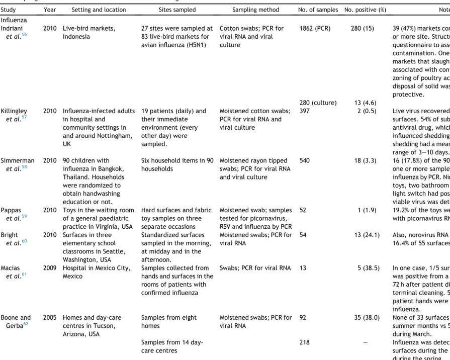
Study Year Setting and location Sites sampled Sampling method No. of samples No. positive (%) Notes
Influenza Indriani
et al.56
2010 Live-bird markets, Indonesia
27 sites were sampled at 83 live-bird markets for avian influenza (H5N1)
Cotton swabs; PCR for viral RNA and viral culture
1862 (PCR) 280 (15) 39 (47%) markets contaminated at one or more site. Structured
questionnaire to assess risk factors for contamination. One province and markets that slaughtered birds associated with contamination; zoning of poultry activities and daily disposal of solid waste were protective.
280 (culture) 13 (4.6) Killingley
et al.57
2010 Influenza-infected adults in hospital and
community settings in and around Nottingham, UK
19 patients (daily) and their immediate environment (every other day) were sampled.
Moistened cotton swabs; PCR for viral RNA and viral culture
397 2 (0.5) Live virus recovered from 1/2 positive surfaces. 54% of subjects took an antiviral drug, which may have influenced shedding. Duration of virus shedding had a mean of 6.2 days and a range of 3e10 days.
Simmerman
et al.58
2010 90 children with influenza in Bangkok, Thailand. Households were randomized to obtain handwashing education or not.
Six household items in 90 households
Moistened rayon tipped swabs; PCR for viral RNA and viral culture
540 18 (3.3) 16 (17.8%) of the 90 households had one or more samples positive for influenza by PCR. Nine TV remotes, six toys, two bathroom knobs and one light switch had positive results. No viable virus was detected by culture. Pappas
et al.59
2010 Toys in the waiting room of a general paediatric practice in Virginia, USA
Hard surfaces and fabric toy samples on three separate occasions
Moistened swab; samples tested for picornavirus, RSV and influenza by PCR
52 1 (1.9) 19.2% of the toys were contaminated
with picornavirus RNA.
Bright
et al.60
2010 Surfaces in three elementary school classrooms in Seattle, Washington, USA
Standardized surfaces sampled in the morning, at midday and in the afternoon.
Moistened swabs; PCR for viral RNA
54 13 (24.1) Also, norovirus RNA was found on 16.4% of 55 surfaces sampled.
Macias
et al.61
2009 Hospital in Mexico City, Mexico
Samples collected from hands and surfaces in the rooms of patients with confirmed influenza
Swabs; PCR for viral RNA 13 5 (38.5) In one case, 1/5 surfaces (a bed rail) was positive from a patient’s room 72 h after patient discharge and terminal cleaning. 5/6 samples from patient hands were positive for influenza.
Boone and Gerba62
2005 Homes and day-care centres in Tucson, Arizona, USA
Samples from eight homes
Moistened swabs; PCR for viral RNA
92 35 (38.0) None of 33 surfaces sampled during summer months vs 59% of 59 samples during March.
Samples from 14 day-care centres
218 e Influenza was detected on 23% of
SARS-CoV, MERS-CoV and influenza virus can survive on dry surfaces for extended periods, particularly when suspended in human secretions (Tables I and II), and may contaminate hand-touch sites in the field (Table III).
Viral and bacterial surface contamination can be trans-ferred to hands, and serial transfer to a number of surfaces from contaminated hands may occur.11,42,80e85 For example, Beanet al.calculated that an infectious dose of virus could be transmitted for at least 2 h and possibly up to 8 h from stainless steel surfaces to hands.42
In order for the virus to initiate indirect contact trans-mission, oral inoculation or contact with mucous membranes must occur to transfer sufficient viruses. Nasal inoculation is a frequent route for establishing influenza and SARS infection.86e90Whereas oral inoculation has not been reported for SARS, it may occur for influenza and other viruses.13,91,92
Thus, the steps necessary to facilitate indirect contact transmission of both SARS-CoV and influenza are established. Although data are more limited for MERS-CoV, it appears to have the key properties to facilitate indirect contact transmission.
Determining which route is most important is challenging, but it seems that direct contact, indirect contact, droplet and airborne transmission do occur with both SARS-CoV and influ-enza viruses on occasion.8,68Few data are available evaluating transmission routes for coronaviruses, but the relative impor-tance of the various routes for influenza virus has been evalu-ated through mathematical models, animal models, and intervention studies.9,93,94
Several mathematical models have been applied to SARS transmission, but none has considered an environmental route.93,95However, some influenza transmission models have evaluated the relative importance of airborne, droplet, and contact influenza transmission.9,96,97 Two of these models conclude that contact transmission of influenza is at least as important as airborne or droplet spread, whereas one study found that contact transmission was negligible compared with other routes.9,96,97However, it is important to note that the relative contribution of contact, droplet, and airborne trans-mission depends on a combination of viral factors (e.g. ca-pacity to survive on surfaces), host factors (e.g. frequency of hand contact with the nose) and environmental factors (e.g. size of enclosure and density of shedders). Varying these and other parameters will change the relative contribution of the various transmission routes.9
Several influenza transmission models have compared the importance of indirect contact transmission (involving surface contamination) with direct contact transmission (that occurs independently of surface contamination).98,99One model in-dicates that indirect transmission via contaminated surfaces generates touch frequency-dependent patterns whereas transmission via the air generates human density-dependent patterns.98 Another model compared the involvement of droplet-contaminated versus hand-contaminated surfaces.99 Droplet-contaminated surfaces were more likely to be involved in transmission than hand-contaminated surfaces (w10-fold difference), and large surfaces (such as table tops) had a higher transmission potential than small surfaces (such as door handles). A number of simplifying assumptions were made, which may be unsoundefor example, that people touch portions of the fomite homogeneously, and that pathogens on fomites are homogeneously distributed. Also, transportation of
contamination from one type of fomite to another via human hands was not modelled. Notwithstanding these limitations, the study provides some useful data on indirect contact transmission of influenza.
An alternative approach is the use of animal models. For example, a guinea-pig model evaluated the relative contribu-tion of airborne, droplet, and indirect contact transmission.94 Indirect contact transmission was evaluated by placing unin-fected animals in cages vacated by experimentally inunin-fected animals without changing bedding, food dishes, and water bottles. Animals were exposed to these cages for 24 h and tested for infection using nasal washings. Around a quarter of exposed guinea-pigs became infected, which was less efficient than transmission through airborne and droplet experiments (25e100% efficiency). Experimental contamination of surfaces in the cages was unable to establish infection. Another guinea-pig model showed that increasing the temperature to 30C blocked aerosol but not contact transmission of influenza.100 This provides further evidence that the relative importance of the various transmission routes is context dependent.
A small number of studies have demonstrated that in-terventions in field settings to improve surface or hand hygiene reduce influenza transmission, demonstrating the importance of contact transmission.63,101,102 For example, introducing regular cleaning using disinfectant wipes reduced the rate of respiratory and diarrhoeal disease in elementary schools.60
Implications for cleaning and disinfection, and
infection prevention and control in healthcare
settings
The likely contribution of droplet, direct and indirect con-tact, and to a lesser extent the airborne route in the trans-mission of influenza, SARS and MERS dictates that each route must be separately addressed by infection prevention and control interventions. The use of a surgical mask will protect the respiratory tract from droplets, an N95 (FFP3) respirator will protect the respiratory tract from droplet nuclei, and gloves, gowns and eye protection will prevent contact with mucous membranes and contamination of clothing or hands for subsequent nasal inoculation.103Emerging literature suggests that doffing PPE presents a challenging risk for the acquisition of important viruses on hands.104,105Thus, protocols should be in place for minimizing the risk of contamination of hands and clothing, and hand hygiene should be performed following removal of PPE.
The extended survival of influenza virus, SARS-CoV and MERS-CoV on surfaces (Tables I and II) and some evidence of contamination in field settings (Table III) argue for enhanced disinfection, particularly at the time of patient discharge.59,61 A range of hospital disinfectants are active against SARS-CoV and surrogates, and influenza, including alcohol, hypo-chlorites (bleach), quaternary ammonium compounds, and hydrogen peroxide, although inactivation is time and concen-tration dependent and will be influenced by other factors such as type of contaminated surface, specific product, and protein load.28,45,106,107However, in-vitro disinfectant effectiveness is a poor predictor for the elimination of contamination from surfaces if cleaning/disinfection is inadequate, which is often the case in hospitals.108,109 Thus, there may be a role for automated room disinfection (ARD) systems, such as hydrogen
peroxide vapour and ultraviolet (UV) light, when patients known to be infected with pandemic influenza or coronaviruses are discharged.45,108
There may be the potential for extended survival of an in-fectious viral aerosol in patients’ rooms following their discharge. Using MERS-CoV as an illustrative example, infec-tious aerosol above the infecinfec-tious dose could be present after the discharge of the patient for up to 26 h, assuming no air changes in the room and depending on the shed titre (Table IV). ARD systems address both contaminated air and surfaces, which may be important if infectious aerosol above the infec-tious dose remains following patient discharge.
Another consideration is the requirement for large quanti-ties of N95 (FFP3) respirators in the event of a pandemic of influenza or MERS/SARS. Stockpiles of N95 respirators required for a pandemic are large, and stock shortages were acknowl-edged during the 2009 N1H1 influenza pandemic.110 Both influenza virus and SARS-CoV surrogates have been shown to survive for extended periods on N95 respirator material.18,37,43 This survival represents a barrier to the reuse of N95 respira-tors. One approach is to disinfect the N95 respirarespira-tors. Several candidate technologies have been evaluated for the disinfec-tion of N95 respirators; UV light, hydrogen peroxide vapour, and ethylene oxide show most promise.111
Conclusion
We reviewed the capacity of viruses with pandemic poten-tial, influenza SARS-CoV and MERS-CoV, to survive on dry sur-faces. The experimental methods used to test survival are important, but it seems that surface survival of SARS/MERS-CoV is greater than that of influenza virus. Important factors that influence the survival of these viruses on surfaces include: strain variations, a ‘doseeresponse’ relationship between the titre applied and survival time, the surface substrate (including the ability to survive on materials used to make PPE), the suspending medium (with the addition of mucus increasing substantially the survival time of influenza), the mode of deposition, temperature and RH, and the method used to determine the presence of the virus (specifically culture versus the use of PCR to detect viral RNA). All three viruses are able to survive in an aerosol for a considerable length of time (>24 h), which may have important infection control implications.
Table IV
Calculating the time that an infectious aerosol shed by a patient infected with Middle East respiratory syndrome coronavirus could survive
Shed titre Time to reach 20 virus particles
1,000,000 26 h
100,000 20 h
10,000 15 h
1000 9 h
100 4 h
The calculation assumes an infectious dose equal to severe acute respiratory syndrome coronavirus (<20 plaque-forming units) and a decay rate of 7% over 10 min in a room with no air changes.13,16The
calculation used the following equation: P(t) ¼ P0e rt, where
P(t)¼the amount of some quantity at timet, P0¼initial amount at
Environmental sampling has been performed for influenza virus and human coronaviruses (including SARS-CoV) in a number of field settings. Most studies have used PCR to detect viral RNA, which may not necessarily represent the presence of viable virus, but should be seen as a marker of virus shedding. Some studies have demonstrated the presence of viable influ-enza virus on surfaces using cell culture. There is a wide range in terms of the frequency of sites contaminated with influenza virus or SARS-CoV RNA, ranging from<5% to>50%, including hand-touch sites.
The importance of indirect contact transmission is uncertain compared with other transmission routes, principally direct contact transmission, droplet, and airborne routes. Influenza virus, SARS-CoV and probably MERS-CoV are shed into the environment at concentrations far in excess of the infective dose, they can survive for extended periods on surfaces, and sampling has identified contamination of hospital surfaces. Contaminated surfaces could result in onward contamination of hands or equipment, which could then initiate inoculation through contact with the nose, eyes, or mouth. Thus, the steps required for indirect contact transmission are established. Mathematical modelling, animal models, and intervention trials suggest that contact transmission may be the most important route for influenza, but that this is context dependent.
The infection prevention and control implications of these findings include the need to wear appropriate PPE to account for contact, droplet and airborne routes, paying particular attention to the risk of contamination of hands and clothing during PPE removal. The potential for inadequate distribution and contact time during manual cleaning and disinfection, combined with the risk of extended survival of infectious aerosol, may argue for the use of ARD systems. These systems may also have a role in disinfection and reuse of N95/FFP3 respirators.
Viruses with pandemic potential including influenza, MERS-CoV, and SARS-CoV can survive for extended periods on dry surfaces, cause contamination in field settings and may require enhanced cleaning and disinfection to assure effective infec-tion preveninfec-tion and control.
Conflict of interest statement
J.A.O. is a consultant to Gama Healthcare. All other authors have no conflict to declare.
Funding sources None.
Appendix A. PubMed searches
coronavirus survival surfaces (June 11th, 2013: 9 studies) influenza survival surfaces (June 11th, 2013: 29 studies) coronavirus fomite transmission (June 20th, 2013: 8 studies) influenza virus fomite transmission (June 20th, 2013: 43 studies)
coronavirus surface contamination (June 20th, 2013: 4 studies) influenza virus surface contamination (June 20th, 2013: 14 studies)
disinfection influenza transmission (June 04th, 2014: 112 studies)
disinfection SARS transmission (June 04th, 2014: 35 studies) Updated May 21st, 2014
References
1.de Groot RJ, Baker SC, Baric RS,et al. Middle East Respiratory
Syndrome Coronavirus (MERS-CoV); Announcement of the
Coro-navirus Study Group.J Virol2013;87:7790e7792.
2.Zaki AM, van Boheemen S, Bestebroer TM, Osterhaus AD,
Fouchier RA. Isolation of a novel coronavirus from a man with
pneumonia in Saudi Arabia.N Engl J Med2012;367:1814e1820.
3.Fineberg HV. Pandemic preparedness and response e lessons
from the H1N1 influenza of 2009. N Engl J Med
2014;370:1335e1342.
4.Hayden FG. Respiratory viral threats. Curr Opin Infect Dis
2006;19:169e178.
5.World Health Organization. Annex C: Respiratory droplets. In:
Atkinson J, Chartier Y, Pessoa-Silva CL,et al., editors.Natural
ventilation for infection control in health-care settings. Geneva:
WHO; 2009.
6.Bridges CB, Kuehnert MJ, Hall CB. Transmission of influenza:
implications for control in health care settings.Clin Infect Dis
2003;37:1094e1101.
7.Boone SA, Gerba CP. Significance of fomites in the spread of
respiratory and enteric viral disease. Appl Environ Microbiol
2007;73:1687e1696.
8.Brankston G, Gitterman L, Hirji Z, Lemieux C, Gardam M.
Transmission of influenza A in human beings.Lancet Infect Dis
2007;7:257e265.
9. Spicknall IH, Koopman JS, Nicas M, Pujol JM, Li S, Eisenberg JN.
Informing optimal environmental influenza interventions: how the host, agent, and environment alter dominant routes of
transmission.PLoS Comput Biol2010;6:e1000969.
10.Otter JA, Yezli S, Salkeld JA, French GL. Evidence that
contam-inated surfaces contribute to the transmission of hospital path-ogens and an overview of strategies to address contaminated
surfaces in hospital settings. Am J Infect Control
2013;41:S6eS11.
11.Otter JA, Yezli S, French GL. The role played by contaminated
surfaces in the transmission of nosocomial pathogens. Infect
Control Hosp Epidemiol2011;32:687e699.
12.Weber DJ, Rutala WA, Miller MB, Huslage K, Sickbert-Bennett E.
Role of hospital surfaces in the transmission of emerging health
care-associated pathogens: norovirus, Clostridium difficile,
and Acinetobacter species. Am J Infect Control
2010;38:S25eS33.
13.Yezli S, Otter JA. Minimum infective dose of the major human
respiratory and enteric viruses transmitted through food and the
environment.Food Environ Microbiol2011;3:1e30.
14.Geller C, Varbanov M, Duval RE. Human coronaviruses: insights
into environmental resistance and its influence on the
devel-opment of new antiseptic strategies. Viruses 2012;4:
3044e3068.
15.Kramer A, Schwebke I, Kampf G. How long do nosocomial
path-ogens persist on inanimate surfaces? A systematic review.BMC
Infect Dis2006;6:130.
16.van Doremalen N, Bushmaker T, Munster VJ. Stability of Middle
East respiratory syndrome coronavirus (MERS-CoV) under
different environmental conditions. Euro Surveill2013;18. pii:
20590.
17.Chan KH, Peiris JS, Lam SY, Poon LL, Yuen KY, Seto WH. The
effects of temperature and relative humidity on the viability of
the SARS Coronavirus.Adv Virol2011;734690.
18.Coulliette AD, Perry KA, Edwards JR, Noble-Wang JA. Persistence
of the 2009 pandemic influenza A (H1N1) virus on N95 respirators.
Appl Environ Microbiol2013;79:2148e2155.
19.Casanova L, Rutala WA, Weber DJ, Sobsey MD. Survival of
sur-rogate coronaviruses in water.Water Res2009;43:1893e1898.
20.Mullis L, Saif LJ, Zhang Y, Zhang X, Azevedo MS. Stability of
bovine coronavirus on lettuce surfaces under household
21. Yepiz-Gomez MS, Gerba CP, Bright KR. Survival of respiratory viruses on fresh produce. Food Environ Virol 2013. http://
dx.doi.org/10.1007/s12560-013-9114-4.
22.Wang XW, Li J, Guo T,et al. Concentration and detection of SARS
coronavirus in sewage from Xiao Tang Shan Hospital and the
309th Hospital of the Chinese People’s Liberation Army.Water
Sci Technol2005;52:213e221.
23.Shigematsu S, Dublineau A, Sawoo O, et al. Influenza A virus
survival in water is influenced by the origin species of the host
cell.Influenza Other Respir Viruses2014;8:123e130.
24.Chmielewski R, Swayne DE. Avian influenza: public health and
food safety concerns. Annu Rev Food Sci Technol
2011;2:37e57.
25.Nazir J, Haumacher R, Ike A, Stumpf P, Bohm R, Marschang RE.
Long-term study on tenacity of avian influenza viruses in water (distilled water, normal saline, and surface water) at different
temperatures.Avian Dis2010;54:720e724.
26.Casanova LM, Jeon S, Rutala WA, Weber DJ, Sobsey MD. Effects of
air temperature and relative humidity on coronavirus survival on
surfaces.Appl Environ Microbiol2010;76:2712e2717.
27.Muller A, Tillmann RL, Muller A, Simon A, Schildgen O. Stability of
human metapneumovirus and human coronavirus NL63 on
medi-cal instruments and in the patient environment. J Hosp Infect
2008;69:406e408.
28.Rabenau HF, Cinatl J, Morgenstern B, Bauer G, Preiser W,
Doerr HW. Stability and inactivation of SARS coronavirus. Med
Microbiol Immunol2005;194:1e6.
29.Lai MY, Cheng PK, Lim WW. Survival of severe acute respiratory
syndrome coronavirus.Clin Infect Dis2005;41:e67e71.
30.Duan SM, Zhao XS, Wen RF,et al. Stability of SARS coronavirus
in human specimens and environment and its sensitivity
to heating and UV irradiation. Biomed Environ Sci
2003;16:246e255.
31.Sizun J, Yu MW, Talbot PJ. Survival of human coronaviruses 229E
and OC43 in suspension and after drying on surfaces: a possible
source of hospital-acquired infections. J Hosp Infect
2000;46:55e60.
32. Zuo Z, de Abin M, Chander Y, Kuehn TH, Goyal SM, Pui DY. Comparison of spike and aerosol challenge tests for the recovery of viable influenza virus from non-woven fabrics.Influenza Other Respi Viruses2013.http://dx.doi.org/10.1111/irv.12095.
33.Mukherjee DV, Cohen B, Bovino ME, Desai S, Whittier S,
Larson EL. Survival of influenza virus on hands and fomites in
community and laboratory settings. Am J Infect Control
2012;40:590e594.
34.Greatorex JS, Digard P, Curran MD,et al. Survival of influenza A
(H1N1) on materials found in households: implications for
infec-tion control.PLoS One2011;6:e27932.
35.Dublineau A, Batejat C, Pinon A, Burguiere AM, Leclercq I,
Manuguerra JC. Persistence of the 2009 pandemic influenza A
(H1N1) virus in water and on non-porous surface. PLoS One
2011;6:e28043.
36.Wood JP, Choi YW, Chappie DJ, Rogers JV, Kaye JZ.
Environ-mental persistence of a highly pathogenic avian influenza (H5N1)
virus.Environ Sci Technol2010;44:7515e7520.
37.Sakaguchi H, Wada K, Kajioka J,et al. Maintenance of influenza
virus infectivity on the surfaces of personal protective equipment
and clothing used in healthcare settings. Environ Health Prev
Med2010;15:344e349.
38.McDevitt J, Rudnick S, First M, Spengler J. Role of absolute
hu-midity in the inactivation of influenza viruses on stainless steel
surfaces at elevated temperatures. Appl Environ Microbiol
2010;76:3943e3947.
39.Thomas Y, Vogel G, Wunderli W,et al. Survival of influenza virus
on banknotes.Appl Environ Microbiol2008;74:3002e3007.
40.Noyce JO, Michels H, Keevil CW. Inactivation of influenza A virus
on copper versus stainless steel surfaces.Appl Environ Microbiol
2007;73:2748e2750.
41. Tiwari A, Patnayak DP, Chander Y, Parsad M, Goyal SM. Survival
of two avian respiratory viruses on porous and nonporous
sur-faces.Avian Dis2006;50:284e287.
42. Bean B, Moore BM, Sterner B, Peterson LR, Gerding DN,
Balfour Jr HH. Survival of influenza viruses on environmental
surfaces.J Infect Dis1982;146:47e51.
43. Casanova L, Rutala WA, Weber DJ, Sobsey MD. Coronavirus
sur-vival on healthcare personal protective equipment.Infect
Con-trol Hosp Epidemiol2010;31:560e561.
44. Dowell SF, Simmerman JM, Erdman DD,et al. Severe acute
res-piratory syndrome coronavirus on hospital surfaces.Clin Infect
Dis2004;39:652e657.
45. Goyal SM, Chander Y, Yezli S, Otter JA. Evaluating the virucidal
efficacy of hydrogen peroxide vapour. J Hosp Infect
2014;86:255e259.
46. Parker ER, Dunham WB, MacNeal WJ. Resistance of the
Mel-bourne strain of influenza virus to desiccation.J Lab Clin Med
1944;29:37e42.
47. Musher DM. How contagious are common respiratory tract
in-fections?N Engl J Med2003;348:1256e1266.
48. Gerone PJ, Couch RB, Keefer GV, Douglas RG, Derrenbacher EB,
Knight V. Assessment of experimental and natural viral aerosols.
Bacteriol Rev1966;30:576e588.
49. Tran K, Cimon K, Severn M, Pessoa-Silva CL, Conly J. Aerosol
generating procedures and risk of transmission of acute
respira-tory infections to healthcare workers: a systematic review.PLoS
One2012;7:e35797.
50. Bischoff WE, Swett K, Leng I, Peters TR. Exposure to influenza
virus aerosols during routine patient care. J Infect Dis
2013;207:1037e1046.
51. Thompson KA, Pappachan JV, Bennett AM, et al. Influenza
aerosols in UK hospitals during the H1N1 (2009) pandemicethe
risk of aerosol generation during medical procedures.PLoS One
2013;8:e56278.
52. Ijaz MK, Brunner AH, Sattar SA, Nair RC, Johnson-Lussenburg CM.
Survival characteristics of airborne human coronavirus 229E.
J Gen Virol1985;66(Pt 12):2743e2748.
53. Schaffer FL, Soergel ME, Straube DC. Survival of airborne
influ-enza virus: effects of propagating host, relative humidity, and
composition of spray fluids.Arch Virol1976;51:263e273.
54. Mitchell CA, Guerin LF. Influenza A of human, swine, equine and
avian origin: comparison of survival in aerosol form.Can J Comp
Med1972;36:9e11.
55. Tellier R. Review of aerosol transmission of influenza A virus.
Emerg Infect Dis2006;12:1657e1662.
56. Indriani R, Samaan G, Gultom A,et al. Environmental sampling
for avian influenza virus A (H5N1) in live-bird markets, Indonesia.
Emerg Infect Dis2010;16:1889e1895.
57. Killingley B, Greatorex J, Cauchemez S,et al. Virus shedding and
environmental deposition of novel A (H1N1) pandemic influenza
virus: interim findings.Health Technol Assess2010;14:237e354.
58. Simmerman JM, Suntarattiwong P, Levy J,et al. Influenza virus
contamination of common household surfaces during the 2009 influenza A (H1N1) pandemic in Bangkok, Thailand: implications
for contact transmission.Clin Infect Dis2010;51:1053e1061.
59. Pappas DE, Hendley JO, Schwartz RH. Respiratory viral RNA on
toys in pediatric office waiting rooms. Pediatr Infect Dis J
2010;29:102e104.
60. Bright KR, Boone SA, Gerba CP. Occurrence of bacteria and
vi-ruses on elementary classroom surfaces and the potential role of
classroom hygiene in the spread of infectious diseases.J Sch Nurs
2010;26:33e41.
61. Macias AE, de la Torre A, Moreno-Espinosa S, Leal PE, Bourlon MT,
Ruiz-Palacios GM. Controlling the novel A (H1N1) influenza virus:
don’t touch your face!J Hosp Infect2009;73:280e281.
62. Boone SA, Gerba CP. The occurrence of influenza A virus on
household and day care center fomites. J Infect
63. Booth TF, Kournikakis B, Bastien N,et al. Detection of airborne severe acute respiratory syndrome (SARS) coronavirus and
en-vironmental contamination in SARS outbreak units.J Infect Dis
2005;191:1472e1477.
64. Memish ZA, Almasri M, Assirri A,et al. Environmental sampling for
respiratory pathogens in Jeddah airport during the 2013 Hajj
season.Am J Infect Control2014;42:1266e1269.
65. Gwaltney Jr JM, Hendley JO. Transmission of experimental
rhinovirus infection by contaminated surfaces.Am J Epidemiol
1982;116:828e833.
66. Abad FX, Pinto RM, Bosch A. Survival of enteric viruses on
envir-onmental fomites.Appl Environ Microbiol1994;60:3704e3710.
67. Hall CB. Respiratory syncytial virus: its transmission in the
hos-pital environment.Yale J Biol Med1982;55:219e223.
68. Chan PK, Tang JW, Hui DS. SARS: clinical presentation,
trans-mission, pathogenesis and treatment options. Clin Sci (Lond)
2006;110:193e204.
69. Hung IF, Cheng VC, Wu AK,et al. Viral loads in clinical specimens
and SARS manifestations.Emerg Infect Dis2004;10:1550e1557.
70. Zhang XM, Herbst W, Kousoulas KG, Storz J. Biological and
gen-etic characterization of a hemagglutinating coronavirus isolated
from a diarrhoeic child.J Med Virol1994;44:152e161.
71. Vabret A, Dina J, Gouarin S, Petitjean J, Corbet S, Freymuth F.
Detection of the new human coronavirus HKU1: a report of 6
cases.Clin Infect Dis2006;42:634e639.
72. Peiris JS, Chu CM, Cheng VC,et al. Clinical progression and viral
load in a community outbreak of coronavirus-associated SARS
pneumonia: a prospective study.Lancet2003;361:1767e1772.
73. Pinsky BA, Mix S, Rowe J, Ikemoto S, Baron EJ. Long-term
shed-ding of influenza A virus in stool of immunocompromised child.
Emerg Infect Dis2010;16:1165e1167.
74. Chan MC, Lee N, Chan PK, Leung TF, Sung JJ. Fecal detection of
influenza A virus in patients with concurrent respiratory and
gastrointestinal symptoms.J Clin Virol2009;45:208e211.
75. Kaiser L, Fritz RS, Straus SE, Gubareva L, Hayden FG. Symptom
pathogenesis during acute influenza: interleukin-6 and other
cytokine responses.J Med Virol2001;64:262e268.
76. Hall CB, Douglas Jr RG, Geiman JM, Meagher MP. Viral shedding
patterns of children with influenza B infection. J Infect Dis
1979;140:610e613.
77. Drosten C, Seilmaier M, Corman VM, et al. Clinical features
and virological analysis of a case of Middle East respiratory
syndrome coronavirus infection. Lancet Infect Dis
2013;13:745e751.
78. Guery B, Poissy J, el Mansouf L,et al. Clinical features and viral
diagnosis of two cases of infection with Middle East Respiratory Syndrome coronavirus: a report of nosocomial transmission.
Lancet2013;381:2265e2272.
79. Watanabe T, Bartrand TA, Weir MH, Omura T, Haas CN.
Devel-opment of a doseeresponse model for SARS coronavirus. Risk
Anal2010;30:1129e1138.
80. Oelberg DG, Joyner SE, Jiang X, Laborde D, Islam MP,
Pickering LK. Detection of pathogen transmission in neonatal
nurseries using DNA markers as surrogate indicators.Pediatrics
2000;105:311e315.
81. Barker J, Vipond IB, Bloomfield SF. Effects of cleaning and
disinfection in reducing the spread of Norovirus contamination
via environmental surfaces.J Hosp Infect2004;58:42e49.
82. Guerrero DM, Nerandzic MM, Jury LA, Jinno S, Chang S,
Donskey CJ. Acquisition of spores on gloved hands after contact
with the skin of patients withClostridium difficileinfection and
with environmental surfaces in their rooms.Am J Infect Control
2012;40:556e558.
83. Rusin P, Maxwell S, Gerba C. Comparative surface-to-hand and
fingertip-to-mouth transfer efficiency of gram-positive bacteria,
gram-negative bacteria, and phage. J Appl Microbiol
2002;93:585e592.
84.Jiang X, Dai X, Goldblatt S,et al. Pathogen transmission in child
care settings studied by using a cauliflower virus DNA as a
sur-rogate marker.J Infect Dis1998;177:881e888.
85.Rheinbaben F, Schunemann S, Gross T, Wolff MH. Transmission of
viruses via contact in a household setting: experiments using
bacteriophage straight phiX174 as a model virus. J Hosp Infect
2000;46:61e66.
86.Nagata N, Iwata N, Hasegawa H, et al. Pathology and virus
dispersion in cynomolgus monkeys experimentally infected with severe acute respiratory syndrome coronavirus via different
inoculation routes.Int J Exp Pathol2007;88:403e414.
87.McAuliffe J, Vogel L, Roberts A,et al. Replication of SARS
coro-navirus administered into the respiratory tract of African Green,
rhesus and cynomolgus monkeys.Virology2004;330:8e15.
88.Henle W, Henle G, Stokes J, Maris EP. Experimental exposure of
human subjects to viruses of influenza. J Immunol
1946;52:145e165.
89.Frankova V. Inhalatory infection of mice with influenza A0/PR8
virus. I. The site of primary virus replication and its spread in the
respiratory tract.Acta Virol1975;19:29e34.
90.Qin C, Wang J, Wei Q,et al. An animal model of SARS produced by
infection of Macaca mulatta with SARS coronavirus. J Pathol
2005;206:251e259.
91.Quan FS, Compans RW, Kang SM. Oral vaccination with
inacti-vated influenza vaccine induces cross-protective immunity.
Vaccine2012;30:180e188.
92.VanDalen KK, Franklin AB, Mooers NL, Sullivan HJ, Shriner SA.
Shedding light on avian influenza H4N6 infection in mallards:
modes of transmission and implications for surveillance. PLoS
One2010;5:e12851.
93.van Kleef E, Robotham JV, Jit M, Deeny SR, Edmunds WJ.
Modelling the transmission of healthcare associated infections: a
systematic review.BMC Infect Dis2013;13:294.
94.Mubareka S, Lowen AC, Steel J, Coates AL, Garcia-Sastre A,
Palese P. Transmission of influenza virus via aerosols and fomites
in the guinea pig model.J Infect Dis2009;199:858e865.
95.Kwok KO, Leung GM, Lam WY, Riley S. Using models to identify
routes of nosocomial infection: a large hospital outbreak of SARS
in Hong Kong.Proc Biol Sci2007;274:611e617.
96.Atkinson MP, Wein LM. Quantifying the routes of transmission for
pandemic influenza.Bull Math Biol2008;70:820e867.
97.Nicas M, Jones RM. Relative contributions of four exposure
pathways to influenza infection risk. Risk Anal
2009;29:1292e1303.
98.Li S, Eisenberg JN, Spicknall IH, Koopman JS. Dynamics and
control of infections transmitted from person to person through
the environment.Am J Epidemiol2009;170:257e265.
99.Zhao J, Eisenberg JE, Spicknall IH, Li S, Koopman JS. Model
analysis of fomite mediated influenza transmission. PLoS One
2012;7:e51984.
100. Lowen AC, Steel J, Mubareka S, Palese P. High temperature (30
degrees C) blocks aerosol but not contact transmission of
influ-enza virus.J Virol2008;82:5650e5652.
101. Cowling BJ, Chan KH, Fang VJ,et al. Facemasks and hand
hy-giene to prevent influenza transmission in households: a cluster
randomized trial.Ann Intern Med2009;151:437e446.
102. Apisarnthanarak A, Apisarnthanarak P, Cheevakumjorn B,
Mundy LM. Intervention with an infection control bundle to reduce transmission of influenza-like illnesses in a Thai
pre-school.Infect Control Hosp Epidemiol2009;30:817e822.
103. Seto WH, Tsang D, Yung RW,et al. Effectiveness of precautions
against droplets and contact in prevention of nosocomial
trans-mission of severe acute respiratory syndrome (SARS). Lancet
2003;361:1519e1520.
104. Beam EL, Gibbs SG, Boulter KC, Beckerdite ME, Smith PW.
A method for evaluating health care workers’ personal protective
105. Johnson DW, Sullivan JN, Piquette CA, et al. Lessons learned: critical care management of patients with Ebola in the United
States.Crit Care Med2015;43:1157e1164.
106. Hulkower RL, Casanova LM, Rutala WA, Weber DJ, Sobsey MD.
Inactivation of surrogate coronaviruses on hard surfaces by
health care germicides.Am J Infect Control2011;39:401e407.
107. Jeong EK, Bae JE, Kim IS. Inactivation of influenza A virus H1N1
by disinfection process.Am J Infect Control2010;38:354e360.
108. Otter JA, Yezli S, Perl TM, Barbut F, French GL. Is there a role for
“no-touch” automated room disinfection systems in infection
prevention and control?J Hosp Infect2013;83:1e13.
109. Carling PC, Parry MM, Rupp ME, Po JL, Dick B, Von Beheren S.
Improving cleaning of the environment surrounding patients in 36
acute care hospitals. Infect Control Hosp Epidemiol
2008;29:1035e1041.
110. Anonymous. Interim guidance on infection control measures for
2009 H1N1 influenza in healthcare settings, including protection
of healthcare personnel.Miss RN2009;71:13e18.
111. Viscusi DJ, Bergman MS, Eimer BC, Shaffer RE. Evaluation of five
decontamination methods for filtering facepiece respirators.Ann