Identification and Validation of Small Molecules That Enhance
Recombinant Adeno-associated Virus Transduction following
High-Throughput Screens
Sarah C. Nicolson,a,bChengwen Li,a,cMatthew L. Hirsch,a,dVincent Setola,eR. Jude Samulskia,b Gene Therapy Center,a
Department of Pharmacology,b
Department of Pediatrics,c
and Department of Ophthalmology,d
University of North Carolina at Chapel Hill, Chapel Hill, North Carolina, USA; Department of Physiology and Pharmacology, West Virginia University, Morgantown, West Virginia, USAe
ABSTRACT
While the recent success of adeno-associated virus (AAV)-mediated gene therapy in clinical trials is promising, challenges still face the widespread applicability of recombinant AAV(rAAV). A major goal is to enhance the transduction efficiency of vectors in order to achieve therapeutic levels of gene expression at a vector dose that is below the immunological response threshold. In an attempt to identify novel compounds that enhance rAAV transduction, we performed two high-throughput screens compris-ing 2,396 compounds. We identified 13 compounds that were capable of enhanccompris-ing transduction, of which 12 demonstrated vec-tor-specific effects and 1 could also enhance vector-independent transgene expression. Many of these compounds had similar properties and could be categorized into five groups: epipodophyllotoxins (group 1), inducers of DNA damage (group 2), effec-tors of epigenetic modification (group 3), anthracyclines (group 4), and proteasome inhibieffec-tors (group 5). We optimized dosing for the identified compounds in several immortalized human cell lines as well as normal diploid cells. We found that the group 1 epipodophyllotoxins (teniposide and etoposide) consistently produced the greatest transduction enhancement. We also ex-plored transduction enhancement among single-stranded, self-complementary, and fragment vectors and found that the com-pounds could impact fragmented rAAV2 transduction to an even greater extent than single-stranded vectors.In vivoanalysis of rAAV2 and all of the clinically relevant compounds revealed that, consistent with ourin vitroresults, teniposide exhibited the greatest level of transduction enhancement. Finally, we explored the capability of teniposide to enhance transduction of frag-ment vectorsin vivousing an AAV8 capsid that is known to exhibit robust liver tropism. Consistent with ourin vitroresults, teniposide coadministration greatly enhanced fragmented rAAV8 transduction at 48 h and 8 days. This study provides a founda-tion based on the rAAV small-molecule screen methodology, which is ideally used for more-diverse libraries of compounds that can be tested for potentiating rAAV transduction.
IMPORTANCE
This study seeks to enhance the capability of adeno-associated viral vectors for therapeutic gene delivery applicable to the treat-ment of diverse diseases. To do this, a comprehensive panel of FDA-approved drugs were tested in human cells and in animal models to determine if they increased adeno-associated virus gene delivery. The results demonstrate that particular groups of drugs enhance adeno-associated virus gene delivery by unknown mechanisms. In particular, the enhancement of gene delivery was approximately 50 to 100 times better with than without teniposide, a compound that is also used as chemotherapy for can-cer. Collectively, these results highlight the potential for FDA-approved drug enhancement of adeno-associated virus gene ther-apy, which could result in safe and effective treatments for diverse acquired or genetic diseases.
A
deno-associated viral (AAV) vectors have emerged to be one of the most promising types of vectors for gene therapy. In-deed, recent and ongoing clinical trials have reported improve-ments in patients with hemophilia B (1), Parkinson’s disease (2, 3), Leber congenital amaurosis (4–7), and Canavan disease (8). Such clinical successes have led to the approval of recombinant AAV (rAAV)-mediated gene therapy in the European Union for the treatment of lipoprotein lipase deficiency (LPLD) (9). Enthu-siasm for using rAAV vectors stems from the unique properties of the virus itself. As naturally occurring AAV requires a helper virus such as adenovirus or herpes simplex virus in order to carry out a productive infection, AAV on its own is not known to cause dis-ease in humans. AAV vectors are comprised of transgenic DNA (⬍5 kb) with the only viral sequence being the 145-nucleotide inverted terminal repeats (ITRs). Furthermore, naturally occur-ring serotypes and engineered capsids have been shown to display diverse tissue tropism, as well as the ability to infect both dividingand nondividing cells (for a review, see reference 10). From a vector perspective, the use of AAV for gene therapy applications is limited only by the size of the vector, which consists of a transgene of approximately 4.7 kb (11). However, even the size limitations of rAAV vectors are being challenged with the development of trans-splicing and fragment vector (fAAV) technology; that is, transgene
Received9 December 2015 Accepted20 April 2016
Accepted manuscript posted online4 May 2016
CitationNicolson SC, Li C, Hirsch ML, Setola V, Samulski RJ. 2016. Identification and validation of small molecules that enhance recombinant adeno-associated virus transduction following high-throughput screens. J Virol 90:7019 –7031.
doi:10.1128/JVI.02953-15.
Editor:G. McFadden, University of Florida
Send correspondence to R. Jude Samulski, [email protected].
cassettes that rely on cellular recombination pathways to restore a full-length, large genome upon delivery by multiple rAAV vectors (12–19).
Despite its remarkable safety profile, gene therapy using AAV vectors has had limited success in applications requiring systemic delivery, namely, to nonimmunoprivileged sites (as opposed to direct injection into various tissues or to immunoprivileged re-gions). Results from clinical trials utilizing either rAAV2 or rAAV8 to deliver human factor IX to hemophilia B patients have suggested that capsid-specific cytotoxic T lymphocytes (CTLs) might have eliminated the majority of transduced cells, thus im-peding successful gene expression (1,20). In these dose escalation trials, therapeutic protein levels were achieved at the highest vec-tor doses administered; however, these levels were transient and their decline corresponded with a rise in liver transaminases, sug-gestive of transduced cell death. There was no rise in liver enzymes detected at the lower doses tested, suggesting that gene transfer to achieve protein levels that would be deemed therapeutic is limited by the amount of capsid that can be delivered to the host (21). It has thus been postulated that if transduction efficiency could be improved (including both gene transfer and expression), fewer vectors would be required to achieve a given level of gene expres-sion, and thus a clinically relevant therapeutic protein level could be obtained without eliciting a host immune response (22).
Several strategies have been employed in order to enhance transduction efficiency. Both rational mutagenesis and library-based approaches have been designed to create capsids that have increased transduction efficiency for particular tissue types as well as decreased tropism for nontarget organs such as the liver (23– 28). Barriers within the intracellular trafficking pathway have also been circumvented based on rationally engineered capsids (29, 30). Improvements to transgene design, including self-comple-mentary rAAV and tissue-specific promoters, ensure faster and more-robust onset of gene expression in target tissues (31–34). Finally, enhancing transduction efficiency through the use of pharmacological agents has been explored. Previous work has shown that topoisomerase inhibitors and anthracyclines can en-hance transduction bothin vitroandin vivo(35–42). Proteasome inhibitors, particularly the FDA-approved bortezomib (Velcade), have been shown to enhance transductionin vitroas well as in both small- and large-animal models (39,40,43). Collectively, these strategies have been important in reaching the level of trans-duction required for therapeutic benefit without eliciting a host immune response; however, efficient transduction in a wide vari-ety of clinical applications is still a major goal that is actively being pursued.
In this study, we performed a high-throughput, small-mole-cule screen with the purpose of identifying additional compounds that enhance rAAV2 transduction. Validation of this approach was confirmed by the transduction augmentation of the previ-ously identified topoisomerase II inhibitors, anthracyclines, and proteasome inhibitors in our small-molecule screen. Addition-ally, we identified several novel compounds and further char-acterized those with reported functions based on existing liter-ature. Collectively, these transduction-enhancing compounds could be grouped into 5 categories: epipodophyllotoxin topo-isomerase II inhibitors (group 1), DNA damage inducers (group 2), effectors of epigenetic modification (group 3), anthracyclines (group 4), and proteasome inhibitors (group 5). We also identi-fied a single agent that enhanced rAAV2 transduction through an
unrelated mechanism. The ability of these compounds to enhance transduction was confirmed in vitro using several human cell lines, including a normal diploid cell line, NHF-1. Some of these compounds were shown to be effective in enhancing self-comple-mentary rAAV2 (scAAV2) but to a lesser extent than single-stranded rAAV2. Interestingly, the compounds exerted the great-est degree of enhancement on fAAV2, rgreat-estoring transduction to levels equivalent to or surpassing that of single-stranded rAAV2. We further examined the activity of compounds identified in our screen that are currently approved for clinical usein vivo and found that, consistent with ourin vitroresults, one such com-pound—teniposide— exhibited the greatest level of transduction enhancement. Given the impact of teniposide on fAAV2, we ex-plored its capability to enhance transduction of fragment vectors
in vivousing an AAV8 capsid that is known to exhibit robust liver tropism. Consistent with ourin vitroresults using fAAV2, tenipo-side coadministration enhanced fragmented rAAV8 transduction both at 48 h and through the duration of the experiment (8 days). Our results demonstrate a simple, effective method of discovering compounds that enhance rAAV2 transduction. We anticipate that this approach can be applied to vectors derived from other sero-types and in other cell lines. Furthermore, our design provides a foundation to investigate the plethora of commercially available compound libraries that span the small-molecule and FDA-ap-proved drug milieu.
MATERIALS AND METHODS
Cell culture. HeLa cells, U87 cells, and normal human fibroblasts (NHF1s) were grown in Dulbecco’s modified Eagle medium that was supplemented with 10% heat-inactivated fetal calf serum, 100 U/ml pen-icillin, and 100 g/ml streptomycin (complete DMEM). HepG2 cells were grown in RPMI 1640 medium, supplemented as described above. All cell lines were maintained at 37°C and 5% CO2.
Virus production.Virus was produced in HEK-293 cells as previously described (44). Briefly, using polyethylenimine (PEI) Max (Polysciences), cells were triple transfected with a Rep and Cap plasmid (pXR2 or pXR8), an inverted terminal repeat-flanked transgene plasmid (single-stranded pTR-CBA-Luciferase, self-complementary pTR-CMV-gaussia-Lucifer-ase, or oversized pTR-CBA-LuciferpTR-CMV-gaussia-Lucifer-ase, where CBA is chicken-beta actin and CMV is cytomegalovirus), and the pXX6-80 helper plasmid. Between 48 and 72 h posttransfection, cells were harvested and virus was purified by cesium chloride gradient density centrifugation overnight at 55,000 rpm. Fractions that contained peak virus titers were dialyzed in dialysis buffer (1⫻phosphate-buffered saline [PBS], 0.5% sorbitol, 0.5 mM cal-cium chloride, and 1 mM magnesium chloride). Titers were calculated by quantitative PCR (qPCR) according to established procedures (16) by using a LightCycler 480 instrument with Sybr green PCR master mix. Conditions used for the reaction were as follows: 1 cycle at 95°C for 10 min; 45 cycles at 95°C for 10 s, 62°C for 10 s, and 72°C for 10 s; and 1 cycle at 95°C for 30 s, 65°C for 1 min, and 99°C for acquisition.
Compound screen, 384-well format.HeLa cells were plated at least 18 h prior to compound treatment and infection at a density of 8⫻103
cells/well. Compounds were prepared in complete DMEM so that delivery would yield a final concentration of 1M. Compounds were added di-rectly to wells. Two hours posttreatment, rAAV2-CBA-Luciferase was ad-ministered at a dose of 1,000 vector genomes (vg)/cell. Cells were har-vested 24 h postransduction by medium removal followed by incubation with passive lysis buffer (Promega) for 15 min. Luciferase activity was measured in accordance with the manufacturer’s instructions (Promega). Luciferase activity was measured either a PerkinElmer 1450 MicroBeta TriLux LSC and luminescence counter or a PerkinElmer 2450 MicroBeta2
fur-ther study. Cell viability was measured using the CellTiterGlo luminescent cell viability assay (Promega).
Secondary screen, 96-well format.HeLa cells were plated at least 18 h prior to compound treatment and infection at a density of 2⫻104cells/
well. Compounds were prepared in complete DMEM at a concentration of 10M. Medium was replaced with medium containing each com-pound. Two hours posttreatment, rAAV2-CBA-Luciferase was adminis-tered at a dose of 500 vg/cell. Cells were harvested 24 h postinfection via incubation with passive lysis buffer (Promega) for 15 min. Luciferase activity was measured in accordance with the manufacturer’s instructions (Promega). Luciferase activity was measured with a Wallac 1420 Victor3 plate reader. Compounds that enhanced transduction ⬎5-fold over DMSO treatment were considered hits for further study.
Transduction assays.Cells were plated at least 18 h prior to infection in 96-well plates at a density of 2⫻104cells/well. Compound treatment
was performed 2 h prior to infection. Compounds were either provided by the Drug Testing Program or were commercially available (teniposide from Sigma; nanaomycin from Apex Bio; daunorubicin and vorinostat from LC Laboratories). Cells were infected with purified rAAV2-CBA_Luc at 500 vector genomes/cell. Cells were harvested 24 h postinfec-tion, and luciferase activity was determined as described above. For scAAV, cells were treated as described above. At 24 h postinfection, 20l of medium was transferred to a black 96-well plate. The luciferase assay was performed using coelenterazine (Nanolight) as the reagent. Briefly, the coelenterazine was resuspended to 10 mg/ml in methanol. To make the working solution, the concentrated stock was dissolved in Tris-EDTA (TE) buffer containing NaCl (0.6 M) at a 1:200 dilution. The working solution was added to the wells at a 1:1 ratio of medium to coelenterazine, and luciferase activity was recorded.
Transfection assays.Two million HeLa cells were plated at least 18 h prior to transfection in a 10-cm plate. Cells were transfected with 1g pTR-CBA-Luc using PEI Max. Twenty-four hours posttransfection, cells were plated in a 96-well plate at a density of 2⫻104cells/well. At 48 h
posttransfection, cells were treated with the indicated drugs. At 72 h post-transfection, cells were harvested and luciferase activity was measured.
Animal studies.Housing and handling of BALB/c mice used in the study were carried out in compliance with National Institutes of Health guidelines and approved by the IACUC at the University of North Caro-lina Chapel Hill. All drugs and rAAV-Luc were coadministered through the intravenous route (tail vein) in a total volume of 200l (normalized
with l⫻PBS). For toxicity analysis, 24 h postadministration blood was collected and serum was assessed for blood urea nitrogen (BUN), aspar-tate aminotransferase (AST), alanine aminotransferase (ALT), and crea-tine kinase (CK). Bioluminescence of Luc expression was visualized by using a Xenogen IVIS Lumina imaging system (PerkinElmer) following intraperitoneal injection of luciferin substrate (120l; Nanolight). Image acquisition and analysis were carried out by using Living Image software.
RESULTS
Primary and secondary screens.The overall goal of our study was to identify and characterize small molecules that enhanced rAAV transductionin vitroand, if the small molecule was FDA approved at the time,in vivo. Two screens were performed to identify such compounds. The first was a stringent screen using a 1M final compound concentration to identify compounds capable of po-tentiating rAAV2 transduction at low concentrations. In each plate, column 1, row 1 contained no virus and no compounds, column 2, row 2 contained rAAV2 but no compounds. Column 24, row 4 contained 1M MG132 as a positive control. Column 23, row 3 contained rAAV2 with only DMSO (vehicle control). All compounds were administered in duplicate. Finally, each plate contained two sets of the given controls and compounds, whereby rows 1 to 8 (top, gray) were assayed for transduction activity and rows 9 to 16 (bottom, white) were assayed for viability using the CellTiterGlo system. As the vast majority of AAV biology has been discovered using AAV2 or rAAV2 in HeLa cells, our primary and secondary screens were performed using these parameters. Owing to its wide range in output and linear relationship to vector dose, the CBA-luciferase transgene, and thus luciferase activity, was chosen as the reporter for transduction efficiency. A secondary screen utilized a high (10M) final compound concentration in tandem with employing rAAV2 to HeLa cells at 500 vg/cell. The format of this screen utilized DMSO added to all wells in column 1, row 1 as a vehicle control, while two wells in column 12, row 2 contained MG132 as a positive control. Cell viability was not as-sessed at this stage. A combined list of the compounds that were pursued further is provided inTable 1.
TABLE 1Compounds characterized in this studya
Group no. Compound name NSC IDb Clinical use Group Known mechanism of action
1 Teniposide 122819 Chemotherapy Podophyllotoxin Topoisomerase II inhibition
Etoposide 141540 Chemotherapy
2 Bleomycin 125066 Chemotherapy, plantar warts Glycopeptide antibiotic DNA damage
Parthenin 85239 Sesquiterpene lactone
RH-1 697726 Phase I, solid malignancies Diaziridinylbenzoqui none
3 Vorinostat 701852 Chemotherapy HDAC inhibitor HDAC inhibition
Nanaomycin A 267461 DNMT3B inhibitor DNMT3B inhibition
4 Menogaril 269148 Anthracycline DNA intercalation, topoisomerase
II inhibition, polymerase inhibition, free radical damage to DNA
Pyrromycin 267229
Daunorubicin 82151 Chemotherapy
5 Bortezomib 681239 Chemotherapy Dipeptide Proteasome inhibition
Physalin B 287088 Physalin
Siomycin 285116 Thiazole antibiotic
Tetrocarcin A 333856 Microbial metabolite BCL-2 inhibitor
aCompounds that emerged as hits were selected based on known function in cells or clinical utility. b
In the primary screen, an initial “hit” was defined as a com-pound that enhanced transduction at least 2-fold, while in the secondary screen, an initial “hit” was defined as a compound that enhanced transduction at least 4-fold. From the hits found in both screens (a total of 72), we chose to further study compounds that had known functions based on existing literature. However, it is noteworthy to mention that several compounds with as-yet-un-known function were identified as hits, some enhancing transduc-tion 5- to 10-fold. These compounds warrant validatransduc-tion in further studies, but further characterization was deferred in order to bet-ter understand the compounds with reported mechanisms of ac-tion. Of these hits, compounds could be further categorized into groups based on their known cellular mechanisms of action ( Ta-ble 1). Group 1 compounds included the epipodophyllotoxins teniposide and etoposide, which have been shown to inhibit topo-isomerase II activity and cause DNA damage. Of these, etoposide has been previously identified as having rAAV2 transduction-po-tentiating activity, likely through mechanisms related to DNA damage repair proteins induced by etoposide-mediated inhibition of topoisomerase II (38). Group 2 compounds comprised other small molecules that are known to cause DNA damage, although not necessarily through topoisomerase II inhibition. These included the glycopeptide antibiotic bleomycin, which is known to cause DNA double-strand breaks (45), the sesquiterpene lactone parthenin, which has been shown to induce chromatid breaks (46), and the diaziridinylbenzoquinone RH1, a DNA cross-linker (47).
Group 3 compounds, nanaomycin A and vorinostat, are char-acterized by their ability to facilitate epigenetic modifications of DNA. While the quinine antibiotic nanaomycin A has been shown to generate oxygen free radicals and cause DNA damage (48,49), it has also recently been identified as an inhibitor of DNA meth-yltransferase 3B (DNMT3B) (50), thereby inhibiting epigenetic repression of gene expression. Vorinostat (suberanilohydroxamic acid [SAHA]) belongs to the class of histone deacetylase inhibi-tors, which function in cells to cause an accumulation of acety-lated histones and transcription factors, which in general increases gene expression (51,52). Group 4 compounds consisted of an-thracyclines, which are compounds that have been shown to exert a variety of effects, including inhibition of topoisomerase and proteasomal activity. These compounds included menogaril, pyr-romycin, and the chemotherapeutic daunorubicin. A related an-thracycline, doxorubicin, has been characterized for its enhance-ment of rAAV transduction (39,40); therefore, it is likely that these other anthracyclines potentiate rAAV transduction through similar mechanisms.
We also identified several nonanthracycline compounds that have been shown to have proteasome-inhibiting activity (group 5). These included bortezomib, physalin B, and siomycin. Bort-ezomib, an FDA-approved proteasome-inhibiting agent, has been explored for rAAV applications in a variety of cell lines and animal models and using different serotypes (43,53–56). Thus, the iden-tification of bortezomib within the screen inadvertently served as an internal control in validating our experimental setup. In addi-tion to its proteasome-inhibiting properties (57), physalin B has also been shown to have anti-inflammatory and other immuno-modulatory activity (58). The proteasome-inhibiting activity of siomycin has been correlated with its role as an inhibitor of the transcription factor forkhead box M1 (FOXM1) (59–61).
Finally, we identified one compound, tetrocarcin A, that could not be categorized into any of groups 1 to 4 but did show modest
levels of rAAV2 transduction enhancement. Tetrocarcin A has been shown to inhibit the antiapoptotic function of Bcl-2 (62) and induce endoplasmic reticulum (ER) stress (63), the latter condition of which we previously demonstrated to increase transduction (64).
In addition, our screen yielded several compounds that are known to interfere with microtubule dynamics, including colchi-cine, vincristine, and vinblastine, among others; however, these were excluded from further study as it has already been demon-strated that these microtubule inhibitors inhibit the cell cycle at low concentrations (65), which is likely the mechanism behind their transduction enhancement. In fact, at higher concentrations, these drugs have been shown to inhibit viral trafficking to the nucleus as well as transduction (66,67).
Dose optimization of compounds in HeLa cells.In order to further characterize the active compounds from our screen, we wanted to determine an optimal concentration that would result in the greatest enhancement of transduction without causing overt toxicity. To avoid potential side effects caused by DMSO, the final concentration of compounds was limited to 100M for high-dose applications. Based on these concentration boundaries, it is possible that compounds that both were well tolerated by cells and substantially enhanced transduction at higher concentrations (such as etoposide and bleomycin) may be capable of enhancing transduction at an even greater level than what was determined under our experimental conditions. For some compounds, the therapeutic window was quite small in that there was a very sharp decrease in cell viability as the concentration of the compound was increased. A notable example was menogaril, which at 5 M showed a⬎10-fold enhancement and approximately 100% viabil-ity but at 10M resulted in an approximately 50% reduction in viability, which corresponded to a reduction in transduction en-hancement as well (data not shown). The epipodophyllins tenipo-side and etopotenipo-side, as well as the DNA-damaging agent bleomy-cin, were the strongest augmenters in this assay, each increasing transduction over 20-fold. The proteasome inhibitors bortezomib and physalin B also showed enhancement over 10-fold.
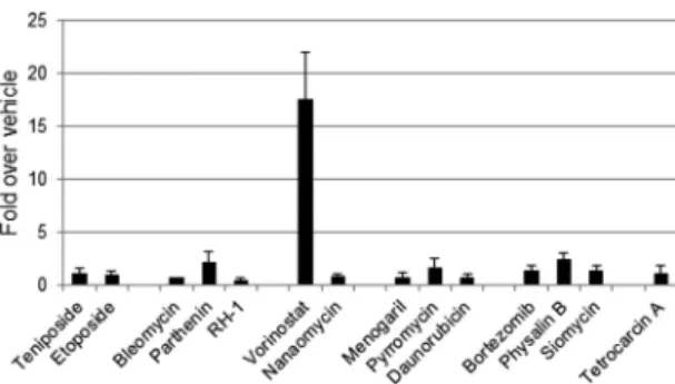
Effects of compounds on vector transduction versus general gene expression. Given the identified compounds’ impressive utility across multiple cell lines, it was important to investigate whether the observed increase in transgene expression was due to a mechanism related specifically to rAAV vectors. To this end, HeLa cells were transfected with pTR-CBA-Luc, the transgene plasmid used to generate the above-described single-stranded vec-tor constructs, and assayed for enhancement of luciferase activity following compound administration (Fig. 1). Most compounds elicited a negligible change in luciferase activity. For example, physalin B induced an approximately 2-fold increase in plasmid luciferase expression but a⬎10-fold increase in transduction, Some of the compounds even elicited a decrease in gene expres-sion when tested with the plasmid vector cassette. The only excep-tion was the histone deacetylase (HDAC) inhibitor vorinostat, which increased luciferase expression of the transfected plasmid to an extent similar to that seen with incoming vectors. It is noted that increased plasmid-borne transgene expression in this case does not simply imply that a similar mechanism of enhancement occurs with AAV vectors. However, in attempts to focus on drug enhancement of AAV vectors specifically, vorinostat was excluded from further analysis.
Characterization of compound activity in human cell lines.
transduction of rAAV2 in HeLa cells (Fig. 2A), we further ex-plored the utility of these compounds in different cell types to better understand their potential universal utility. The first such cell line examined was U87, a human glioblastoma cell line that is amenable to rAAV2 transduction (Fig. 2B). Overall, the fold en-hancement of each of the compounds was very similar to what was observed in HeLa cells, with a few exceptions. While teniposide enhanced transduction approximately 40-fold in HeLa cells, en-hancement was seen at 20-fold in U87 cells. Interestingly, while bortezomib enhanced transduction by approximately 15-fold in HeLa cells, enhancement was only approximately 3-fold in U87 cells. However, the level of enhancement obtained with by another
compound with proteasome-inhibiting activity, physalin B, re-mained similar in both cell lines (12-fold in HeLa cells and 10.5-fold in U87 cells). We next evaluated these compounds in HepG2 cells, a hepatocellular carcinoma cell line, as the liver is the site of transduction for systemically delivered rAAV2 (Fig. 2C). For treatment with pyrromycin, nanaomycin A, physalin B, bort-ezomib, and tetrocarcin A, transduction enhancement was ob-served at approximately the same magnitude as that seen with HeLa cells. Treatment with the epipodophyllotoxins, anthracy-clines, siomycin, and RH-1 resulted in enhancements 4- to 12-fold greater than that observed in HeLa cells. Notably, menogaril, which enhanced rAAV2 transduction in HeLa cells approximately 12-fold, enhanced transduction in HepG2 cells 138-fold. Finally, our compound collection was applied to NHF1 cells (a kind gift from William Kaufman), a diploid cell line derived from neonatal foreskin and immortalized by the expression of telomerase reverse transcriptase (hTERT) that displays contact inhibition and stationary growth once conflu-ent (68) (Fig. 2D). Again, treatment with the epipodophyllo-toxins and anthracyclines (especially menogaril) significantly enhanced transduction at magnitudes much greater than what was observed in HeLa cells. Treatment of NHF-1 cells with bleomycin also resulted in impressive transduction enhance-ment (approximately 100-fold). These results suggest that in general, the group 1 epipodophyllotoxins, causing DNA dam-age from topoisomerase II inhibition, consistently yielded the greatest enhancement in transduction. Proteasome inhibition, particularly by bortezomib, was more variable and cell type dependent. It is possible that the enhancing ability of these
FIG 1Effects of compounds on plasmid gene expression. HeLa cells were transfected with pCBA-Luc and treated with the compounds identified in our screen. Transduction was assessed 24 h after compound treatment. Of the hits, vorinostat appears to be the only compound that enhances plasmid gene ex-pression.
compounds depends on the abundance of their actionable tar-gets within cells. Thus, these results highlight the differences in response to pharmacological modulation in different cell types, which may influence the type of agent chosen to complement rAAV-mediated gene therapy in a given target tissue.
Characterization of transduction-specific compound activ-ity with vectors comprised of differentially formed transgenes.
Recent and emerging gene therapy applications have utilized transgenes that differ in form from the prototypical single-stranded AAV genome. For example, self-complementary vectors contain a transgene whereby a third inverted terminal repeat (ITR) in the middle of the transgene is mutated to eliminate the Rep nicking stem (33). This yields a double-stranded gene prod-uct, thus eliminating the rate-limiting step of second-strand syn-thesis and providing a faster, more robust onset of expression (33, 69). Other emerging vectors designed to deliver large transgenes are derived from the forced packaging of oversized genomes into the viral capsid (13, 15, 16, 18, 19, 70). It is thought that the packaged DNA becomes truncated at the 5=end once AAV has reached its packaging limit, but delivery of these transgene “frag-ments” still results in expression of the intact transgene through a reannealing process modulated by the homologous recombina-tion protein Rad51C (16). However, the transduction efficiency of fAAV is impaired compared to that of rAAV or scAAV.
We therefore wanted to test the capabilities of the screen-iden-tified compounds in enhancing the transduction of scAAV and fAAV in HeLa cells. For scAAV, a vector carrying a cytomegalovi-rus promoter-drivenGaussialuciferase transgene (CMV-gLuc) was utilized. For fAAV, a previously described vector carrying the
same promoter and transgene as the single-stranded vector (CBA-Luc), but with an additional 2.3-kb “stuffer sequence” inserted into an intronic region upstream of the luciferase (16), was uti-lized. A schematic of each of these vector transgenes is provided in Fig. 3A. We applied the same optimized concentration of each compound and tested their ability to enhance scAAV and fAAV transduction at the same vector dose. With the exception of daunorubicin and tetrocarcin A, the compounds still enhanced scAAV transduction, but the magnitude of the effect was signifi-cantly reduced (Fig. 3B). This suggests that these compounds may also be acting at the step of second-strand (ss) synthesis. Since we still observed enhancement of scAAV, albeit at a reduced capacity compared to ssAAV, it is likely that these compounds augment steps in transduction either before second-strand synthesis (such as subcellular trafficking, nuclear entry, or uncoating) or in estab-lishing the persistence of gene expression following second-strand synthesis. Impressively, application of the compounds in con-junction with fAAV2 enhanced transduction to a much greater extent than with rAAV2 (Fig. 3C). Notably, treatment with teni-poside, bortezomib, or bleomycin enhanced transduction over 250-fold, which was an 8- to 10-fold increase over the enhance-ment seen in rAAV2. This result is particularly interesting, as each of these drugs produces a different reported effect within cells. In untreated cells, fAAV2 transduction was approximately 21- to 28-fold lower than intact rAAV2, which is in agreement with previ-ously reported results comparing fAAV2 and intact rAAV2 at 5,000 vg/cell (16). Treatment with any of the compounds restored transduction of fAAV2 to levels (i) above untreated intact rAAV2 levels and (ii) within 2- to 10-fold of drug-treated intact rAAV2
levels, depending on which compound is used. Taken together, these results suggest that the identified compounds can enhance the transduction efficiency of vectors with different forms of transgenes and disproportionately benefit fAAV2 transduction in HeLa cells.
In vivoanalysis of FDA-approved hits.Owing to the appeal of repurposing FDA-approved drugs to augment rAAV-mediated gene transfer, we performed an in vivocomparison of rAAV2 transduction-potentiating capabilities of the hits identified in our screen that are already FDA approved. Therefore, teniposide, eto-poside, bleomycin, daunorubicin, and bortezomib were assessed. The doses selected forin vivostudy were chosen based on conver-sions from the FDA-approved dosing in humans to the murine equivalent based on body surface area and establishedKmfactors
(71). As teniposide and etoposide are recommended for use over a wide range of doses, they were administered in this study at a conservative dose of 20 mg/kg of body weight (human equivalent dose, (60 mg/m2). Each pharmaceutical was coadministered with 1⫻1011vg rAAV2-CBA-Luc through tail vein injection in
age-matched female BALB/c mice. Two vehicle cohorts were included in the study. We evaluated toxicity by measuring the levels of blood urea nitrogen (BUN), aspartate transaminase (AST), ala-nine transaminase (ALT), and creatine kinase (CK). These levels appeared within normal ranges and were comparable to those in the vehicle-treated mice. Average levels were similar for drug-treated and vehicle-drug-treated cohorts (data not shown).
Due to the impressive enhancement that we observedin vitroat 24 h postransduction, an early time point of gene expression was measured to determine whether the pharmaceuticals could also provide rapid, high-level gene expressionin vivo. At 48 h postad-ministration, all of the selected drugs enhanced transductionin vivo, but to various degrees (Fig. 4A). Consistent with what was seenin vitro, the epipodophyllotoxins as well as daunorubicin enhanced transduction to the greatest extent. Notably, cotreat-ment with teniposide enhanced transduction an average of nearly 100-fold (Fig. 4B). While teniposide and etoposide have the same mechanism of action in cells (i.e., inhibition of the religation of DNA ends through interference of the DNA-topoisomerase com-plex), teniposide was a more potent augmenter of transduction at this time point at the given dose. Upon assessment of transduction 8 days postadministration, enhanced transduction was noted among all of the drug-treated cohorts (Fig. 4A). Teniposide coad-ministration yielded the greatest enhancement in transduction at this time point, followed by bortezomib. Daunorubicin cotreat-ment also yielded impressive transduction enhancecotreat-ment at the 8 day time point. Bleomycin and etoposide cotreatment produced only modest enhancement of transduction efficiency. This result could be reflective of thein vivodose limitations in order to reflect clinical levels of each drug. In general, variability among treatment groups, including vehicle-treated mice, was noticed and could have been due to either an immune response against the luciferase transgene in some mice, as has been historically observed, or per-haps an effect of the vehicle (DMSO) onin vivotransduction.
Because coadministration of rAAV2 with teniposide showed the most-robust transduction enhancement, and because of the robust enhancement observed for fAAV2, we chose to test the ability of teni-poside to enhance fragment rAAVin vivoas well. Due to the limited systemic transduction capability of fAAVin vivo(16), we chose to test the transduction using an AAV8 capsid, as intact rAAV8 vectors have been shown to exhibit strong liver transduction (1,44,72). Due to the
difficulty in producing high-titer fAAV8, mice were administered only 5⫻1010vg. At 48 h postransduction, the teniposide-treated cohort exhibited 34-fold-higher levels of luciferase activity than the vehicle-treated cohort. Impressively, this enhancement was even greater at 8 days postransduction, with the teniposide-treated cohort exhibiting 86-fold-higher luciferase activity than the vehicle-treated cohort (Fig. 5B).
Taken together, these results confirmed our observationsin vitroand corroborated that the greatest transduction augmenta-tion arises from cotreatment with topoisomerase or proteasome inhibitors. Furthermore, teniposide was shown to exhibit a robust effect on fAAV8 transduction.
DISCUSSION
Recent gene therapy applications using rAAV have been met with both success and challenges that depend on the indication, route of administration, serotype, and vector dose utilized. Indeed, clin-ical efficacy for one indication, lipoprotein lipase deficiency, has been achieved to the extent that the first-ever gene therapy prod-uct, an rAAV1 vector carrying a lipoprotein lipase transgene, is now approved for use in the European Union (9). Results from clinical trials have exposed the current limitations of rAAV-medi-ated gene therapy, one of which is the lack of ability to achieve robust, long-term therapeutic gene expression without eliciting an immune response induced by high vector doses. Several strat-egies have been employed to combat this challenge, including cap-sid and transgene modification as well as pharmaceutical inter-vention; however, they have been met with varied success and may be limited to a particular serotype or tissue target. For example, the elimination of certain tyrosines on the capsids of AAV2 and AAV6 has allowed for enhanced transduction efficiency and lower rates of proteasomal degradation (29,30,73); however, it has been shown that this strategy cannot be applied to rAAV9 and attempts with rAAV8 have produced mixed results (74,75). Similarly, the proteasome inhibitor bortezomib has been shown to enhance transduction of both rAAV2 and rAAV8 vectors, resulting in in-creased transgene expression in both small- and large-animal models (43,54,55); however, a recent report has shown that this strategy cannot be applied for rAAV9-mediated therapy for car-diac failure (76). Thus, the need for a robust, widely applicable strategy to enhance transduction would be highly beneficial in the translation of rAAV-mediated gene therapy to a broad range of applications. In addition, compounds that enhance rAAV trans-duction may fall into classes that are tissue preferred; therefore, new compounds will always be of interest to explore.
The epipodophyllotoxin and anthracycline topoisomerase II inhibitors form complexes with DNA and topoisomerase II, an enzyme required for the prevention of DNA supercoiling in eu-karyotes. The topoisomerase II cycle includes (i) binding and cleavage of duplexed DNA, (ii) passage of a second strand of DNA through the complex, and (iii) religation of the broken DNA ends (79). Both classes of topoisomerase II inhibitors prevent the reli-gation step of this cycle, thus causing both single-stranded and double-stranded DNA damage. While the direct role of topoisom-erase II in the mechanism of rAAV transduction enhancement cannot be ruled out, previous research suggests that the DNA
damage response, an indirect result of topoisomerase II inhibi-tion, is likely the main contributor to the increase in transducinhibi-tion, either through increased second-strand synthesis or by a currently unknown mechanism (38,80,81). Indeed, this screen identified several topoisomerase II-independent DNA-damaging agents, such as bleomycin, parthenin, and RH1, as potentiators of rAAV2 transduction. In fact, bleomycin enhanced transduction to levels similar to those of teniposide and etoposide treatment in U87 cells (Fig. 1A). Several groups have shown that proteins involved in the DNA damage response, including both homologous recombina-tion and nonhomologous end joining, interact with incoming
ral and vector genomes and have important roles in genome pro-cessing. These processes are thought to include the conversion of the single-stranded genome into double-stranded DNA (69,82), opening of ITR hairpins (83), concatamerization and circulariza-tion (78, 84–86), and transgene expression (87). Notably, the Mre11/Rad50/Nbs1 (MRN) complex, important for double-strand break repair, recombination, and telomere maintenance, has been shown to bind to AAV ITRs, negatively affecting rAAV transduction, wild-type AAV (wtAAV) replication, and double-stranded rAAV DNA accumulation (80, 81). It is currently thought that cellular DNA damage, induced by radiation, small molecules, or other means, may serve as a “decoy,” recruiting the MRN complex to the sites of damage and away from single-stranded AAV DNA, thus allowing for double-strand conversion. Interestingly, Choi et al. observed that ATM, Mre11, and NBS1 are required for scAAV DNA circularizationin vitroand ATM and DNA-PK(CS) are required for scAAV DNA circularizationin vivo
(84). Taken together, these results suggest that DNA damage re-sponse proteins may have a dual function in rAAV DNA process-ing and the positive or negative effects may depend on the state of the DNA within the cell.
Proteasome inhibition is thought to be beneficial to rAAV transduction through a mechanism that differs from a DNA dam-age response. It is believed that inhibition of the proteasome facil-itates bulk flow of particles away from degradation pathways, thereby redirecting them to routes that favor transduction (i.e., nuclear translocation) (54,88,89). Bortezomib, the only currently
FDA-approved proteasome inhibitor, has been used in conjunc-tion with rAAV to enhance transducconjunc-tion in hemophilia A and hemophilia B models (43). Its effects seem to be thus far limited to these applications, as a recent study using rAAV9 to deliver SERCA2a to preserve cardiac function in a rat model demon-strated no additional benefit when bortezomib was coadminis-tered (76). Indeed, our results suggest that the effects of protea-some inhibition by bortezomib are cell type dependent, as bortezomib-mediated enhancement was not observed in U87 and NHF cell lines, and not as robust in HepG2 cells as in HeLa cells. Interestingly, two compounds that have been previously impli-cated in proteasome inhibition were identified, physalin B and siomycin, which outperformed bortezomib in U87, HepG2, and NHF cell lines. These compounds either may inhibit proteasome function through a mechanism that is different from that of bort-ezomib or may perform additional functions in cells that are ben-eficial to rAAV2 transduction.
The level of involvement of DNA damage response proteins and proteasome inhibition seems to differ between ssAAV2, scAAV2, and fAAV2. While the ssAAV2 and fAAV2 used in this study share identical promoters and luciferase genes, the scAAV2 cassette differed in promoter (CMV versus CBA) as well as trans-gene (Gaussiaversus firefly luciferase). Therefore, a direct com-parison can be made between ssAAV2 vectors and fAAV2 vectors, but considerations must be made when evaluating the perfor-mance of the scAAV2 vector. Initial conclusions seem to indicate that, for all of the compounds identified in our screen, transduc-tion enhancement seemed to be less pronounced for scAAV2. This finding is in agreement with the current theory of how DNA dam-age proteins may be inhibitory for ssAAV DNA, since scAAV DNA does not require second-strand synthesis and would therefore be unaffected by any proteins limiting this type of processing. Since it has been shown that Mre11 and ATM are required for scAAV genome circularization, it will be interesting to evaluate the long-term effects of these compounds on scAAV gene expression in the future. Interestingly, some enhancement of scAAV2 transduction was observed with the cotreatment with the epipodophyllotoxins and anthracyclines, suggesting that these compounds might en-hance transduction by mechanisms in addition to facilitating sec-ond-strand synthesis.
Perhaps the most striking observation was the dramatic in-crease in transduction seen for fAAV2 treated with the com-pounds identified in this screen. Notably, each compound boosted fAAV2 transduction to levels greater than in ssAAV2 treated with vehicle alone. Previous studies have shown that fAAV2 transduction relies on the annealing of sections of vector DNA that comprise the entire oversized transgene cassette (16). We previously noted that this process is dependent upon Rad51C, a single-stranded DNA binding protein involved in supporting homologous recombination during DNA double-strand break re-pair (16). Therefore, it is possible that in the case of fAAV2, cellu-lar DNA damage serves two purposes: (i) derepression of the sin-gle-stranded cassette by inhibitory proteins such as the MRN complex and (ii) recruitment of homologous recombination pro-teins such as Rad51C to facilitate the annealing of fragment vector DNA. Interestingly, treatment with bortezomib also resulted in high levels of fAAV2 enhancement. Treatment with proteasome inhibitors has been shown to increase the sheer volume of vector particles that reach the nucleus. It is possible that the deficit ob-served in unassisted fAAV2 transduction is simply a number
game; i.e., as more particles, and therefore presumably more un-coated genomes, transit to the nucleus, there is an increased chance for fragment strand reannealing. Alternatively, inhibition of the proteasome may also inhibit the degradation of proteins that might be essential for fAAV2 reannealing. Indeed, work by Bennett and Knight has shown that proteasome-mediated degra-dation of Rad51 occurs during DNA repair and this process is regulated in part by Rad51C (90). Additionally, the proteasome has been shown to be associated with double-strand breaks and has been suggested to play a role in degrading proteins upon com-pletion of DNA repair in yeast (91). Regardless of the mechanism, our results suggest that the deficit in fAAV2 transduction can be restored to ssAAV2 levels through pharmacological intervention, thus paving the way for future studies in pharmaco-gene therapy for large gene applications.
While the compounds that induced DNA damage seemed to enhance rAAV transduction to the greatest extent, caution is nec-essary before potentially moving forward with any of these drugs. Beyond concerns of direct unwanted side effects of the various drugs (for example, etoposide has⬎30 undesired consequences), indirect altered vector maintenance should also be thoroughly analyzed. Though the primary mechanism of DNA persistence is through concatamerization and circularization, random integra-tion has been demonstrated for rAAV (92–97). Indeed, a previous study has shown that integration of rAAV DNAin vitroincreased upon treatment with etoposide (38,98). While the epipodophyl-lotoxins have been shown to produce breaks in DNA “hot spots” (99–101), a thorough assessment of in vivo integration events would need to be carried out in order to gauge the risks of this kind of pharmaco-gene therapy. Finally, the effects of these agents on episomal expression should also be evaluated. It is tempting to envision a scenario in which inducing DNA damage through one of these agents could reactivate persistent-but-silenced rAAV epi-somes, thus eliminating the need for additional administration of vector if transgene expression falls below therapeutic levels.
ACKNOWLEDGMENTS
Animal studies were performed within the LCCC Animal Studies Core facility at the University of North Carolina at Chapel Hill. The LCCC Animal Studies Core is supported in part by an NCI Center Core support grant (CA16086) to the UNC Lineberger Comprehensive Cancer Center. This work was supported by the Northwest Genome Engineering Consortium, the Jain Foundation, and the NIH (RO1AI072176-06A1, RO1AR064369-01A1). Funding was also pro-vided in part through a departmental unrestricted grant from Research to Prevent Blindness, New York, NY.
Serum markers for toxicity were evaluated by the UNC Clinical Chem-istry Core facility.
We thank Shophia Shih for determining all the rAAV titers used here.
FUNDING INFORMATION
This work, including the efforts of Matthew L. Hirsch and R. Jude Sam-ulski, was funded by HHS | NIH | National Institute of Allergy and Infec-tious Diseases (NIAID) (RO1AI072176-06A1). This work, including the efforts of Matthew L. Hirsch, was funded by Research to Prevent Blindness (RPB). This work, including the efforts of Matthew L. Hirsch and R. Jude Samulski, was funded by HHS | NIH | National Institute of Arthritis and Musculoskeletal and Skin Diseases (NIAMS) (R01AR064369).
REFERENCES
1.Nathwani AC, Tuddenham EG, Rangarajan S, Rosales C, McIntosh J, Linch DC, Chowdary P, Riddell A, Pie AJ, Harrington C, O’Beirne J,
Smith K, Pasi J, Glader B, Rustagi P, Ng CY, Kay MA, Zhou J, Spence Y, Morton CL, Allay J, Coleman J, Sleep S, Cunningham JM, Srivas-tava D, Basner-Tschakarjan E, Mingozzi F, High KA, Gray JT, Reiss UM, Nienhuis AW, Davidoff AM.2011. Adenovirus-associated virus vector-mediated gene transfer in hemophilia B. N Engl J Med365:2357– 2365.http://dx.doi.org/10.1056/NEJMoa1108046.
2.Kaplitt MG, Feigin A, Tang C, Fitzsimons HL, Mattis P, Lawlor PA, Bland RJ, Young D, Strybing K, Eidelberg D, During MJ.2007. Safety and tolerability of gene therapy with an adeno-associated virus (AAV) borne GAD gene for Parkinson’s disease: an open label, phase I trial. Lancet369: 2097–2105.http://dx.doi.org/10.1016/S0140-6736(07)60982-9.
3.LeWitt PA, Rezai AR, Leehey MA, Ojemann SG, Flaherty AW, Eskan-dar EN, Kostyk SK, Thomas K, Sarkar A, Siddiqui MS, Tatter SB, Schwalb JM, Poston KL, Henderson JM, Kurlan RM, Richard IH, Van Meter L, Sapan CV, During MJ, Kaplitt MG, Feigin A.2011. AAV2-GAD gene therapy for advanced Parkinson’s disease: a double-blind, sham-surgery controlled, randomised trial. Lancet Neurol10:309 –319.
http://dx.doi.org/10.1016/S1474-4422(11)70039-4.
4.Bainbridge JW, Smith AJ, Barker SS, Robbie S, Henderson R, Balag-gan K, Viswanathan A, Holder GE, Stockman A, Tyler N, Petersen-Jones S, Bhattacharya SS, Thrasher AJ, Fitzke FW, Carter BJ, Rubin GS, Moore AT, Ali RR.2008. Effect of gene therapy on visual function in Leber’s congenital amaurosis. N Engl J Med358:2231–2239.http://dx .doi.org/10.1056/NEJMoa0802268.
5.Cideciyan AV, Aleman TS, Boye SL, Schwartz SB, Kaushal S, Roman AJ, Pang JJ, Sumaroka A, Windsor EA, Wilson JM, Flotte TR, Fish-man GA, Heon E, Stone EM, Byrne BJ, Jacobson SG, Hauswirth WW. 2008. Human gene therapy for RPE65 isomerase deficiency activates the retinoid cycle of vision but with slow rod kinetics. Proc Natl Acad Sci U S A105:15112–15117.http://dx.doi.org/10.1073/pnas.0807027105. 6.Maguire AM, High KA, Auricchio A, Wright JF, Pierce EA, Testa F,
Mingozzi F, Bennicelli JL, Ying GS, Rossi S, Fulton A, Marshall KA, Banfi S, Chung DC, Morgan JI, Hauck B, Zelenaia O, Zhu X, Raffini L, Coppieters F, De Baere E, Shindler KS, Volpe NJ, Surace EM, Acerra C, Lyubarsky A, Redmond TM, Stone E, Sun J, Mc-Donnell JW, Leroy BP, Simonelli F, Bennett J.2009. Age-dependent effects of RPE65 gene therapy for Leber’s congenital amaurosis: a phase 1 dose-escalation trial. Lancet 374:1597–1605. http://dx.doi .org/10.1016/S0140-6736(09)61836-5.
7.Maguire AM, Simonelli F, Pierce EA, Pugh EN, Jr, Mingozzi F, Bennicelli J, Banfi S, Marshall KA, Testa F, Surace EM, Rossi S, Lyubarsky A, Arruda VR, Konkle B, Stone E, Sun J, Jacobs J, Dell’Osso L, Hertle R, Ma J X, Redmond TM, Zhu X, Hauck B, Zelenaia O, Shindler KS, Maguire MG, Wright JF, Volpe NJ, McDonnell JW, Auricchio A, High KA, Bennett J.2008. Safety and efficacy of gene transfer for Leber’s congenital amaurosis. N Engl J Med358:2240 –2248.
http://dx.doi.org/10.1056/NEJMoa0802315.
8.Leone P, Shera D, McPhee SW, Francis JS, Kolodny EH, Bilaniuk LT, Wang DJ, Assadi M, Goldfarb O, Goldman HW, Freese A, Young D, During MJ, Samulski RJ, Janson CG.2012. Long-term follow-up after gene therapy for canavan disease. Sci Transl Med4:165ra163.http://dx .doi.org/10.1126/scitranslmed.3003454.
9.Kastelein JJ, Ross CJ, Hayden MR.2013. From mutation identification to therapy: discovery and origins of the first approved gene therapy in the Western world. Hum Gene Ther 24:472– 478.http://dx.doi.org/10.1089 /hum.2013.063.
10. Asokan A, Schaffer DV, Samulski RJ.2012. The AAV vector toolkit: poised at the clinical crossroads. Mol Ther20:699 –708.http://dx.doi.org /10.1038/mt.2011.287.
11. Grieger JC, Samulski RJ.2005. Packaging capacity of adeno-associated virus serotypes: impact of larger genomes on infectivity and postentry steps. J Virol 79:9933–9944. http://dx.doi.org/10.1128/JVI.79.15.9933 -9944.2005.
12. Allocca M, Doria M, Petrillo M, Colella P, Garcia-Hoyos M, Gibbs D, Kim SR, Maguire A, Rex TS, Di Vicino U, Cutillo L, Sparrow JR, Williams DS, Bennett J, Auricchio A.2008. Serotype-dependent pack-aging of large genes in adeno-associated viral vectors results in effective gene delivery in mice. J Clin Invest118:1955–1964.http://dx.doi.org/10 .1172/JCI34316.
13. Dong B, Nakai H, Xiao W.2010. Characterization of genome integrity for oversized recombinant AAV vector. Mol Ther18:87–92.http://dx.doi .org/10.1038/mt.2009.258.
ca-pacity with trans-splicing or overlapping vectors: a quantitative compar-ison. Mol Ther4:383–391.http://dx.doi.org/10.1006/mthe.2001.0456. 15. Grose WE, Clark KR, Griffin D, Malik V, Shontz KM, Montgomery
CL, Lewis S, Brown RH, Jr, Janssen PM, Mendell JR, Rodino-Klapac LR.2012. Homologous recombination mediates functional recovery of dysferlin deficiency following AAV5 gene transfer. PLoS One7:e39233.
http://dx.doi.org/10.1371/journal.pone.0039233.
16. Hirsch ML, Li C, Bellon I, Yin C, Chavala S, Pryadkina M, Richard I, Samulski RJ.2013. Oversized AAV transduction is mediated via a DNA-PKcs-independent, Rad51C-dependent repair pathway. Mol Ther21: 2205–2216.http://dx.doi.org/10.1038/mt.2013.184.
17. Kapranov P, Chen L, Dederich D, Dong B, He J, Steinmann KE, Moore AR, Thompson JF, Milos PM, Xiao W.2012. Native molecular state of adeno-associated viral vectors revealed by single-molecule se-quencing. Hum Gene Ther 23:46 –55. http://dx.doi.org/10.1089/hum .2011.160.
18. Lai Y, Yue Y, Duan D.2010. Evidence for the failure of adeno-associated virus serotype 5 to package a viral genome⬎or⫽8.2 kb. Mol Ther 18:75–79.http://dx.doi.org/10.1038/mt.2009.256.
19. Wu Z, Yang H, Colosi P.2010. Effect of genome size on AAV vector packaging. Mol Ther18:80 – 86.http://dx.doi.org/10.1038/mt.2009.255. 20. Manno CS, Pierce GF, Arruda VR, Glader B, Ragni M, Rasko JJ, Ozelo MC, Hoots K, Blatt P, Konkle B, Dake M, Kaye R, Razavi M, Zajko A, Zehnder J, Rustagi PK, Nakai H, Chew A, Leonard D, Wright JF, Lessard RR, Sommer JM, Tigges M, Sabatino D, Luk A, Jiang H, Mingozzi F, Couto L, Ertl HC, High KA, Kay MA.2006. Successful transduction of liver in hemophilia by AAV-Factor IX and limitations imposed by the host immune response. Nat Med12:342–347.http://dx .doi.org/10.1038/nm1358.
21. Mingozzi F, Maus MV, Hui DJ, Sabatino DE, Murphy SL, Rasko JE, Ragni MV, Manno CS, Sommer J, Jiang H, Pierce GF, Ertl HC, High KA.2007. CD8(⫹) T-cell responses to adeno-associated virus capsid in humans. Nat Med13:419 – 422.http://dx.doi.org/10.1038/nm1549. 22. Mingozzi F, High KA.2013. Immune responses to AAV vectors:
over-coming barriers to successful gene therapy. Blood122:23–36.http://dx .doi.org/10.1182/blood-2013-01-306647.
23. Asokan A, Conway JC, Phillips JL, Li C, Hegge J, Sinnott R, Yadav S, DiPrimio N, Nam HJ, Agbandje-McKenna M, McPhee S, Wolff J, Samulski RJ. 2010. Reengineering a receptor footprint of adeno-associated virus enables selective and systemic gene transfer to muscle. Nat Biotechnol28:79 – 82.http://dx.doi.org/10.1038/nbt.1599. 24. Dalkara D, Byrne LC, Klimczak RR, Visel M, Yin L, Merigan WH,
Flannery JG, Schaffer DV.2013. In vivo-directed evolution of a new adeno-associated virus for therapeutic outer retinal gene delivery from the vitreous. Sci Transl Med 5:189ra176. http://dx.doi.org/10.1126 /scitranslmed.3005708.
25. Excoffon KJ, Koerber JT, Dickey DD, Murtha M, Keshavjee S, Kaspar BK, Zabner J, Schaffer DV. 2009. Directed evolution of adeno-associated virus to an infectious respiratory virus. Proc Natl Acad Sci U S A106:3865–3870.http://dx.doi.org/10.1073/pnas.0813365106. 26. Gray SJ, Blake BL, Criswell HE, Nicolson SC, Samulski RJ, McCown
TJ, Li W.2010. Directed evolution of a novel adeno-associated virus (AAV) vector that crosses the seizure-compromised blood-brain barrier (BBB). Mol Ther18:570 –578.http://dx.doi.org/10.1038/mt.2009.292. 27. Maheshri N, Koerber JT, Kaspar BK, Schaffer DV. 2006. Directed
evolution of adeno-associated virus yields enhanced gene delivery vec-tors. Nat Biotechnol24:198 –204.http://dx.doi.org/10.1038/nbt1182. 28. Pulicherla N, Shen S, Yadav S, Debbink K, Govindasamy L,
Agbandje-McKenna M, Asokan A.2011. Engineering liver-detargeted AAV9 vec-tors for cardiac and musculoskeletal gene transfer. Mol Ther19:1070 – 1078.http://dx.doi.org/10.1038/mt.2011.22.
29. Zhong L, Li B, Jayandharan G, Mah CS, Govindasamy L, Agbandje-McKenna M, Herzog RW, Weigel-Van Aken KA, Hobbs JA, Zolo-tukhin S, Muzyczka N, Srivastava A.2008. Tyrosine-phosphorylation of AAV2 vectors and its consequences on viral intracellular trafficking and transgene expression. Virology381:194 –202.http://dx.doi.org/10 .1016/j.virol.2008.08.027.
30. Zhong L, Li B, Mah CS, Govindasamy L, Agbandje-McKenna M, Cooper M, Herzog RW, Zolotukhin I, Warrington KH, Jr, Weigel-Van Aken KA, Hobbs JA, Zolotukhin S, Muzyczka N, Srivastava A.2008. Next generation of adeno-associated virus 2 vectors: point mutations in tyrosines lead to high-efficiency transduction at lower doses. Proc Natl
Acad Sci U S A 105:7827–7832. http://dx.doi.org/10.1073/pnas .0802866105.
31. Chen SJ, Johnston J, Sandhu A, Bish LT, Hovhannisyan R, Jno-Charles O, Sweeney HL, Wilson JM.2013. Enhancing the utility of adeno-associated virus gene transfer through inducible tissue-specific expression. Hum Gene Ther Methods24:270 –278.http://dx.doi.org/10 .1089/hgtb.2012.129.
32. Johnson MC, Garland AL, Nicolson SC, Li C, Samulski RJ, Wang B, Tisch R.2013. beta-cell-specific IL-2 therapy increases islet Foxp3⫹Treg and suppresses type 1 diabetes in NOD mice. Diabetes62:3775–3784.
http://dx.doi.org/10.2337/db13-0669.
33. McCarty DM, Monahan PE, Samulski RJ.2001. Self-complementary recombinant adeno-associated virus (scAAV) vectors promote efficient transduction independently of DNA synthesis. Gene Ther8:1248 –1254.
http://dx.doi.org/10.1038/sj.gt.3301514.
34. Phillips MI, Tang Y, Schmidt-Ott K, Qian K, Kagiyama S. 2002. Vigilant vector: heart-specific promoter in an adeno-associated virus vector for cardioprotection. Hypertension39:651– 655.http://dx.doi.org /10.1161/hy0202.103472.
35. Halbert CL, Standaert TA, Aitken ML, Alexander IE, Russell DW, Miller AD.1997. Transduction by adeno-associated virus vectors in the rabbit airway: efficiency, persistence, and readministration. J Virol71: 5932–5941.
36. Hong SY, Lee MH, Kim KS, Jung HC, Roh JK, Hyung WJ, Noh SH, Choi SH.2004. Adeno-associated virus mediated endostatin gene ther-apy in combination with topoisomerase inhibitor effectively controls liver tumor in mouse model. World J Gastroenterol10:1191–1197. 37. Koeberl DD, Alexander IE, Halbert CL, Russell DW, Miller AD.1997.
Persistent expression of human clotting factor IX from mouse liver after intravenous injection of adeno-associated virus vectors. Proc Natl Acad Sci U S A94:1426 –1431.http://dx.doi.org/10.1073/pnas.94.4.1426. 38. Russell DW, Alexander IE, Miller AD.1995. DNA synthesis and
topo-isomerase inhibitors increase transduction by adeno-associated virus vectors. Proc Natl Acad Sci U S A92:5719 –5723.http://dx.doi.org/10 .1073/pnas.92.12.5719.
39. Yan Z, Zak R, Zhang Y, Ding W, Godwin S, Munson K, Peluso R, Engelhardt JF.2004. Distinct classes of proteasome-modulating agents cooperatively augment recombinant adeno-associated virus type 2 and type 5-mediated transduction from the apical surfaces of human airway epithelia. J Virol78:2863–2874.http://dx.doi.org/10.1128/JVI.78.6.2863 -2874.2004.
40. Zhang LN, Karp P, Gerard CJ, Pastor E, Laux D, Munson K, Yan Z, Liu X, Godwin S, Thomas CP, Zabner J, Shi H, Caldwell CW, Peluso R, Carter B, Engelhardt JF.2004. Dual therapeutic utility of proteasome modulating agents for pharmaco-gene therapy of the cystic fibrosis air-way. Mol Ther10:990 –1002.http://dx.doi.org/10.1016/j.ymthe.2004.08 .009.
41. Zhang S, Wu J, Wu X, Xu P, Tian Y, Yi M, Liu X, Dong X, Wolf F, Li C, Huang Q. 2012. Enhancement of rAAV2-mediated transgene expression in retina cells in vitro and in vivo by coadministration of low-dose chemotherapeutic drugs. Invest Ophthalmol Vis Sci53:2675– 2684.http://dx.doi.org/10.1167/iovs.11-8856.
42. Zhang T, Hu J, Ding W, Wang X.2009. Doxorubicin augments rAAV-2 transduction in rat neuronal cells. Neurochem Int55:521–528.http://dx .doi.org/10.1016/j.neuint.2009.05.005.
43. Monahan PE, Lothrop CD, Sun J, Hirsch ML, Kafri T, Kantor B, Sarkar R, Tillson DM, Elia JR, Samulski RJ.2010. Proteasome inhib-itors enhance gene delivery by AAV virus vectors expressing large ge-nomes in hemophilia mouse and dog models: a strategy for broad clinical application. Mol Ther18:1907–1916.http://dx.doi.org/10.1038/mt.2010 .170.
44. Nathwani AC, Gray JT, Ng CY, Zhou J, Spence Y, Waddington SN, Tuddenham EG, Kemball-Cook G, McIntosh J, Boon-Spijker M, Mertens K, Davidoff AM.2006. Self-complementary adeno-associated virus vectors containing a novel liver-specific human factor IX expres-sion cassette enable highly efficient transduction of murine and nonhu-man primate liver. Blood 107:2653–2661. http://dx.doi.org/10.1182 /blood-2005-10-4035.
45. Povirk LF, Han YH, Steighner RJ. 1989. Structure of bleomycin-induced DNA double-strand breaks: predominance of blunt ends and single-base 5=extensions. Biochemistry28:5808 –5814.http://dx.doi.org /10.1021/bi00440a016.
sesquiterpene lactone of Parthenium hysterophorus L. is a high toxicity clastogen. Mutat Res 514:19 –27. http://dx.doi.org/10.1016/S1383 -5718(01)00321-7.
47. Berardini MD, Souhami RL, Lee CS, Gibson NW, Butler J, Hartley JA. 1993. Two structurally related diaziridinylbenzoquinones preferentially cross-link DNA at different sites upon reduction with DT-diaphorase. Biochemistry32:3306 –3312.http://dx.doi.org/10.1021/bi00064a013. 48. Hayashi M, Unemoto T, Minami-Kakinuma S, Tanaka H, Omura S.
1982. The mode of action of nanaomycins D and A on a gram-negative marine bacterium Vibrio alginolyticus. J Antibiot (Tokyo)35:1078 – 1085.http://dx.doi.org/10.7164/antibiotics.35.1078.
49. Marumo H, Kitaura K, Morimoto M, Tanaka H, Omura S.1980. The mode of action of nanaomycin A in Gram-positive bacteria. J Antibiot (Tokyo)33:885– 890.http://dx.doi.org/10.7164/antibiotics.33.885. 50. Kuck D, Caulfield T, Lyko F, Medina-Franco JL.2010. Nanaomycin A
selectively inhibits DNMT3B and reactivates silenced tumor suppressor genes in human cancer cells. Mol Cancer Ther9:3015–3023.http://dx .doi.org/10.1158/1535-7163.MCT-10-0609.
51. Marks P, Rifkind RA, Richon VM, Breslow R, Miller T, Kelly WK. 2001. Histone deacetylases and cancer: causes and therapies. Nat Rev Cancer1:194 –202.http://dx.doi.org/10.1038/35106079.
52. Marks PA, Dokmanovic M.2005. Histone deacetylase inhibitors: dis-covery and development as anticancer agents. Expert Opin Investig Drugs14:1497–1511.http://dx.doi.org/10.1517/13543784.14.12.1497. 53. Finn JD, Hui D, Downey HD, Dunn D, Pien GC, Mingozzi F, Zhou S,
High KA.2010. Proteasome inhibitors decrease AAV2 capsid derived peptide epitope presentation on MHC class I following transduction. Mol Ther18:135–142.http://dx.doi.org/10.1038/mt.2009.257. 54. Mitchell AM, Samulski RJ.2013. Mechanistic insights into the
enhance-ment of adeno-associated virus transduction by proteasome inhibitors. J Virol87:13035–13041.http://dx.doi.org/10.1128/JVI.01826-13. 55. Nathwani AC, Cochrane M, McIntosh J, Ng CY, Zhou J, Gray JT,
Davidoff AM.2009. Enhancing transduction of the liver by adeno-associated viral vectors. Gene Ther16:60 – 69.http://dx.doi.org/10.1038 /gt.2008.137.
56. Paulk NK, Loza LM, Finegold MJ, Grompe M.2012. AAV-mediated gene targeting is significantly enhanced by transient inhibition of non-homologous end joining or the proteasome in vivo. Hum Gene Ther 23:658 – 665.http://dx.doi.org/10.1089/hum.2012.038.
57. Vandenberghe I, Creancier L, Vispe S, Annereau JP, Barret JM, Pouny I, Samson A, Aussagues Y, Massiot G, Ausseil F, Bailly C, Kruczynski A.2008. Physalin B, a novel inhibitor of the ubiquitin-proteasome path-way, triggers NOXA-associated apoptosis. Biochem Pharmacol76:453– 462.http://dx.doi.org/10.1016/j.bcp.2008.05.031.
58. Soares MB, Bellintani MC, Ribeiro IM, Tomassini TC, Ribeiro dos Santos R.2003. Inhibition of macrophage activation and lipopolysac-charide-induced death by seco-steroids purified from Physalis angulata L. Eur J Pharmacol 459:107–112. http://dx.doi.org/10.1016/S0014 -2999(02)02829-7.
59. Bhat UG, Halasi M, Gartel AL.2009. FoxM1 is a general target for proteasome inhibitors. PLoS One 4:e6593. http://dx.doi.org/10.1371 /journal.pone.0006593.
60. Bhat UG, Halasi M, Gartel AL.2009. Thiazole antibiotics target FoxM1 and induce apoptosis in human cancer cells. PLoS One4:e5592.http://dx .doi.org/10.1371/journal.pone.0005592.
61. Radhakrishnan SK, Bhat UG, Hughes DE, Wang IC, Costa RH, Gartel AL.2006. Identification of a chemical inhibitor of the oncogenic tran-scription factor forkhead box M1. Cancer Res66:9731–9735.http://dx .doi.org/10.1158/0008-5472.CAN-06-1576.
62. Nakashima T, Miura M, Hara M.2000. Tetrocarcin A inhibits mito-chondrial functions of Bcl-2 and suppresses its anti-apoptotic activity. Cancer Res60:1229 –1235.
63. Anether G, Tinhofer I, Senfter M, Greil R. 2003. Tetrocarcin-A– induced ER stress mediates apoptosis in B-CLL cells via a Bcl-2– independent pathway. Blood101:4561– 4568.http://dx.doi.org/10.1182 /blood-2002-08-2501.
64. Johnson JS, Gentzsch M, Zhang L, Ribeiro CM, Kantor B, Kafri T, Pickles RJ, Samulski RJ.2011. AAV exploits subcellular stress associated with inflammation, endoplasmic reticulum expansion, and misfolded proteins in models of cystic fibrosis. PLoS Pathog7:e1002053.http://dx .doi.org/10.1371/journal.ppat.1002053.
65. Jordan MA, Thrower D, Wilson L.1991. Mechanism of inhibition of cell proliferation by Vinca alkaloids. Cancer Res51:2212–2222.
66. Sanlioglu S, Benson PK, Yang J, Atkinson EM, Reynolds T, Engelhardt JF.2000. Endocytosis and nuclear trafficking of adeno-associated virus type 2 are controlled by rac1 and phosphatidylinositol-3 kinase activa-tion. J Virol 74:9184 –9196. http://dx.doi.org/10.1128/JVI.74.19.9184 -9196.2000.
67. Xiao PJ, Samulski RJ.2012. Cytoplasmic trafficking, endosomal escape, and perinuclear accumulation of adeno-associated virus type 2 particles are facilitated by microtubule network. J Virol86:10462–10473.http: //dx.doi.org/10.1128/JVI.00935-12.
68. Boyer JC, Kaufmann WK, Brylawski BP, Cordeiro-Stone M. 1990. Defective postreplication repair in xeroderma pigmentosum variant fi-broblasts. Cancer Res50:2593–2598.
69. Ferrari FK, Samulski T, Shenk T, Samulski RJ.1996. Second-strand synthesis is a rate-limiting step for efficient transduction by recombinant adeno-associated virus vectors. J Virol70:3227–3234.
70. Hirsch ML, Agbandje-McKenna M, Samulski RJ.2010. Little vector, big gene transduction: fragmented genome reassembly of adeno-associated virus. Mol Ther18:6 – 8.http://dx.doi.org/10.1038/mt.2009 .280.
71. Reagan-Shaw S, Nihal M, Ahmad N. 2008. Dose translation from animal to human studies revisited. FASEB J22:659 – 661.
72. Davidoff AM, Gray JT, Ng CY, Zhang Y, Zhou J, Spence Y, Bakar Y, Nathwani AC.2005. Comparison of the ability of adeno-associated viral vectors pseudotyped with serotype 2, 5, and 8 capsid proteins to mediate efficient transduction of the liver in murine and nonhuman primate models. Mol Ther11:875– 888.http://dx.doi.org/10.1016/j.ymthe.2004 .12.022.
73. Qiao C, Zhang W, Yuan Z, Shin JH, Li J, Jayandharan GR, Zhong L, Srivastava A, Xiao X, Duan D.2010. Adeno-associated virus serotype 6 capsid tyrosine-to-phenylalanine mutations improve gene transfer to skeletal muscle. Hum Gene Ther21:1343–1348.http://dx.doi.org/10 .1089/hum.2010.003.
74. Petrs-Silva H, Dinculescu A, Li Q, Min SH, Chiodo V, Pang JJ, Zhong L, Zolotukhin S, Srivastava A, Lewin AS, Hauswirth WW. 2009. High-efficiency transduction of the mouse retina by tyrosine-mutant AAV serotype vectors. Mol Ther17:463– 471.http://dx.doi.org/10.1038 /mt.2008.269.
75. Qiao C, Yuan Z, Li J, Tang R, Xiao X.2012. Single tyrosine mutation in AAV8 and AAV9 capsids is insufficient to enhance gene delivery to skeletal muscle and heart. Hum Gene Ther Methods23:29 –37.http://dx .doi.org/10.1089/hgtb.2011.229.
76. Chaanine AH, Nonnenmacher M, Kohlbrenner E, Jin D, Kovacic JC, Akar FG, Hajjar RJ, Weber T.2014. Effect of bortezomib on the efficacy of AAV9.SERCA2a treatment to preserve cardiac function in a rat pres-sure-overload model of heart failure. Gene Ther21:379 –386.http://dx .doi.org/10.1038/gt.2014.7.
77. Alexander IE, Russell DW, Miller AD.1994. DNA-damaging agents greatly increase the transduction of nondividing cells by adeno-associated virus vectors. J Virol68:8282– 8287.
78. Sanlioglu S, Benson P, Engelhardt JF. 2000. Loss of ATM function enhances recombinant adeno-associated virus transduction and integra-tion through pathways similar to UV irradiaintegra-tion. Virology268:68 –78.
http://dx.doi.org/10.1006/viro.1999.0137.
79. Vos SM, Tretter EM, Schmidt BH, Berger JM.2011. All tangled up: how cells direct, manage and exploit topoisomerase function. Nat Rev Mol Cell Biol12:827– 841.http://dx.doi.org/10.1038/nrm3228.
80. Cervelli T, Palacios JA, Zentilin L, Mano M, Schwartz RA, Weitzman MD, Giacca M.2008. Processing of recombinant AAV genomes occurs in specific nuclear structures that overlap with foci of DNA-damage-response proteins. J Cell Sci121:349 –357.http://dx.doi.org/10.1242/jcs .003632.
81. Schwartz RA, Palacios JA, Cassell GD, Adam S, Giacca M, Weitzman MD. 2007. The Mre11/Rad50/Nbs1 complex limits adeno-associated virus transduction and replication. J Virol81:12936 –12945.http://dx .doi.org/10.1128/JVI.01523-07.
82. Zentilin L, Marcello A, Giacca M.2001. Involvement of cellular double-stranded DNA break binding proteins in processing of the recombinant adeno-associated virus genome. J Virol75:12279 –12287.http://dx.doi .org/10.1128/JVI.75.24.12279-12287.2001.