INVESTIGATION OF THE METHYLATION STATUS OF FGF2 GENE IN PERIODONTAL TISSUES AS DETERMINANTS OF HEALING RESPONSE AFTER
IMPLANT SURGERY
Ping Seung Alice Wu
A thesis submitted to the faculty of the University of North Carolina at Chapel Hill in partial fulfillment of the requirements for the degree of Master of Science in the
Department of Periodontology, School of Dentistry.
Chapel Hill 2012
Approved by:
Adviser: Dr. Steven Offenbacher
Reader: Dr. Silvana Barros
ii ©2012
iii
ABSTRACT
PING SEUNG ALICE WU: INVESTIGATION OF THE METHYLATION STATUS OF FGF2 GENE IN PERIODONTAL TISSUES AS DETERMINANTS OF HEALING
RESPONSE AFTER IMPLANT SURGERY (Under the direction of Dr. Steven Offenbacher)
Periodontal wound healing is a complex process involving a series of interactions of growth factors, including FGF2. Previous studies suggested FGF2 gene may be down-regulated in diseased periodontal tissues. Aberrant gene expression in smokers and diabetics has also been reported. The aims of this study were to determine the association of FGF2 methylation level with wound healing after implant surgery and the association of smoking and diabetes with FGF2 methylation. Recruited subjects were distributed into control, smoking and diabetic groups. During the implant surgery, gingival tissue
iv
ACKNOWLEDGEMENTS
I would like to acknowledge the guidance and support of my committee members, laboratory colleagues, statisticians and members of General Oral Health Center.
Steven Offenbacher, Silvana Barros and Eric Everett David Barrow, Russ Levy, Shaoping Zhang and Ning Yu
John Preisser and Kevin Moss
ST Phillips, Luisito Mendoza, Kristi Laan, and Wendy Lamm
My sincere thanks are offered to Salvador Nares for all his help in making this research project possible.
v
TABLE OF CONTENTS
LIST OF TABLES ... vii
LIST OF FIGURES ... viii
Chapter 1. INTRODUCTION AND BACKGROUND ...1
Fibroblast Growth Factor -2 (FGF2) and Its Role in Periodontal Wound Healing ...1
DNA Methylation – An Overview ...3
Smoking and Its Effect on the Periodontium...5
DNA Methylation and Smoking ...8
Diabetes and Its Effect on the Periodontium ...10
DNA Methylation and Type II Diabetes ...15
2. AIMS...17
3. HYPOTHESES ...18
4. MATERIALS AND METHODS ...19
Study Population ...19
Gingival Tissue Biopsies, Gingival Crevicular Fluid (GCF) Collection and Implant Stability Quotient (ISQ) Measurements ...20
vi
DNA Isolation, Selective Digestion and Quantitative
Real-Time PCR ...21
Mediator Assays in Gingival Crevicular Fluid ...22
Statistical Analysis ...23
5. RESULTS ...24
Participant Recruitment Process ...24
Study Population Demographics ...26
Selected Implant Sites ...28
Differential DNA Methylation of FGF2 Gene in Smokers and Diabetic Participants ...30
Differential mRNA Expression of FGF2 Gene in Smokers and Diabetic Participants ...32
Relationship between DNA Methylation and mRNA Expression of FGF2 Gene ...34
Levels of FGF2 Protein detected in GCF during Wound Healing ...36
Implant Stability and WHI during Wound Healing ...36
Relationship between DNA Methylation of FGF-2 Gene and Implant Stability ...39
Relationship between DNA Methylation of FGF2 Gene and WHI ...42
6. DISCUSSION ...44
APPENDIX I ...56
vii
LIST OF TABLES
Table
Table 1. Proposed Mechanisms for the Negative Periodontal
Effects of Smoking ...7
Table 2. Participant Recruitment Process ...25
Table 3. Study Participant Demographics ...27
Table 4. Implant Sites of Study Participants ...29
viii
LIST OF FIGURES
Figure
Figure 1. The DNA Methylation Process. ...4 Figure 2. Possible Linkage between Diabetes and Periodontal
Disease Severity. ...14 Figure 3. Impact of Hyperglycemia on Bone...15 Figure 4. Methylation Level of FGF2 Gene in Smokers or
Diabetics in Comparison to Control Subjects. ...31 Figure 5. Messenger Level of FGF2 Gene in Smokers or Diabetics
in Comparison to Control Subjects. ...33 Figure 6. Group Means of Methylation Level and RNA Expression
of FGF2 Gene. ...35 Figure 7. Implant Stability of All Three Groups of Subjects after
Implant Placement. ...38 Figure 8. Levels of (a) Edema, (b) Erythema, (c) Pain, and (d)
Degree of Flap Closure Experienced by All Three Groups
of Subjects after Implant Placement.. ...39 Figure 9. Mean Methylation Level and Overall Implant Stability of
the Three Study Groups.. ...41 Figure 10. Mean Methylation Level and Overall (a) Edema Index,
(b) Erythema Index, (c) Pain Index, and (d) Flap
1
INTRODUCTION AND BACKGROUND
Fibroblast Growth Factor -2 (FGF2) and Its Role in Periodontal Wound Healing
Fibroblast growth factor (FGF) was first discovered in 1974 as a protein in bovine pituitary glands with the ability to induce proliferation of fibroblasts (Gospodarowicz 1974). In 1984, two FGF proteins with different isoelectric points were identified from bovine pituitary glands. While one was known as the acidic FGF (aFGF) or FGF1, the other became known as the basic FGF (bFGF) or FGF2 (Bohlen et al. 1984). Two years later, the nucleotide sequence of human FGF2 was isolated (Abraham et al. 1986).
FGF2 is a member of the heparin-binding growth factor family and is found in various body tissues. It induces endothelial cell proliferation and angiogenesis, thereby mediating smooth muscle cell growth and tissue repair. It stimulates hematopoiesis by enabling stem cell survival and differentiation of granulocytes. It also exerts a range of biologic effects on cells of the nervous system, the lung, the eye, the reproductive system, the skeleton and the skin (Bikfalvi et al. 1997, Lee et al. 2010).
2
matrix synthesis, and increases chemotaxis, proliferation and differentiation of
endothelial cells (Kaigler et al. 2006). FGF2 promotes angiogenesis, and exhibits potent mitogenic ability on mesenchymal cells within the periodontal ligament (Murakami et al. 1999). During the bone remodeling phase, FGF2 inhibits mineralized nodule formation in the periodontal ligament cells (Lee et al. 2010), and stimulates migration of mesenchymal progenitor cells which ultimately differentiate into osteoblasts and cementoblasts
(Kaigler et al. 2006, Murakami et al. 1999).
Animal (Murakami et al. 1999, Murakami et al. 2003, Takayama et al. 2001, Rossa et al. 2000, Sato et al. 2004) and human (Kitamura et al. 2008, Kitamura et al. 2011) studies have reported FGF2 to be effective in periodontal regeneration. In 1999, Murakami showed surgically treated 3-wall intrabony defects in beagle dogs and primates grafted with recombinant FGF2 were able to demonstrate significantly greater periodontal ligament, cementum and bone formation at 6 – 8 weeks. Epithelial down growth, ankylosis or root resorption were not observed in grafted sites (Murakami et al. 1999). In 2001, Takayama demonstrated a dose-dependent response with cementum and bone regeneration in primates using rhbFGF (Takayama et al. 2001). Two years later, Murakami again found that topical application of recombinant bFGF (rhbFGF) in surgically treated class II furcation defects in beagle dogs resulted in increase in formation of PDL, cementum and bone in 6 weeks (Murakami et al. 2003).
3
L (0.03% FGF2), Group M (0.1% FGF2) or Group H (0.3% FGF2). The group found significant difference in rate of alveolar bone growth between Group P and Group H at 36 weeks (Kitamura et al. 2008). Three years later, the group performed a subsequent larger clinical trial which involved 253 patients. In this study, 200µL of 0%, 0.2%, 0.3% or 0.4% FGF2 was administered to 2- or 3-walled bony defects at time of modified Widman flap surgery. All FGF2 sites demonstrated significant higher percentage of bone fill at 36 weeks. No clinical safety problems, including an abnormal increase in alveolar bone or ankylosis, were identified (Kitamura et al. 2011).
DNA Methylation – An Overview
The term “epigenetics” was first used by Conrad H. Waddington, a developmental biologist and evolutionist, in the middle of the twentieth century to describe changes in patterns of gene expression which do not involve changes in the DNA sequence (Barros & Offenbacher2009). It refers to events which chemically modify certain DNA regions, leading to alteration of the chromatin and ultimately silencing or activating a gene
(Gomez et al. 2009). Various mechanisms of epigenetic alterations were discovered in the past half century. Among them, DNA methylation is the most extensively studied
mechanism in mammals (Barros & Offenbacher2009).
DNA methylation is characterized by the addition of methyl group onto a cytosine residue within the cytosine-guanosine dinucleotide (CpG) regions (Barros &
4
important regulatory functions in normal cellular processes. During corneal wound healing, hyper-methylation of the maspin gene promoter causes down-regulation of maspin synthesis in corneal stromal cells, enabling these cells to undergo phenotypic changes to fibroblasts and myofibroblasts (Horswill et al. 2008). Similarly, while the conversion of hepatic stellate cells (HSC) into myofibroblasts is important for hepatic wound healing, such process can be blocked by treating HSC with DNA methylation inhibitor 5-aza-2’-deoxycytidine (Mann et al. 2007). In the periodontal ligament of aged individuals, the decreased expression of collagen α1(1) gene is associated with elevated levels of methylation at most CpG sites in the proximal and distal regions of the promoter (Ohi et al. 2006).
Figure 1. The DNA Methylation Process (adapted from Barros & Offenbacher 2009
and reproduced with permission).
5
(Wilson 2008). It was recognized that hemocysteine can inhibit arterial endothelial cell growth by increasing methylation of the FGF2 gene promoter region. This disrupts endothelial integrity and predisposes the individual to atherogenesis and angiostasis (Chang et al. 2008a). A persistent periodontal inflammation can cause DNA methylation, which inactivates suppressors of cytokine signaling and results in exaggerated cytokine production in the periodontium. This renders an individual susceptible to periodontal disease (Gomez et al. 2009). In our laboratory, we discovered that chronic periodontal disease is associated with increased DNA methylation of the prostaglandin-endoperoxide synthase-2 (PTGS2 or COX2) promoter, resulting in dampering of COX2 expression. The methylation level of the PTGS2 promoter from diseased gingival biopsies demonstrated a 5.06-fold increase when compared with non-diseased samples (p = 0.03) (Zhang et al. 2010).
Although DNA methylation has been identified as important components in various physiological and pathological processes, its role in periodontal wound healing has not yet been investigated.
Smoking and Its Effect on the Periodontium
According to the National Health Interview Survey performed by the U.S. Census Bureau in 2009, 21% of the US adult population was current smokers (Pleis 2010). In 2008, the Centers of Disease and Control estimated that cigarette smoking caused
6
factor for heart attack, stroke, chronic obstructive pulmonary disease, emphysema and cancer.
The adverse effects exerted by smoking on periodontal health have been demonstrated by compelling evidence. Locally, smoking causes a reduction in the gingival blood flow, thereby reducing the number of circulating leukocytes and the amount of oxygen reaching the gingiva (Giannopoulou et al. 1999). Systemically,
smoking reduces serum immunoglobulin G production (Johnson & Guthmiller 2007) and inhibits chemotactic and phagocytic activities of peripheral blood neutrophils
(Giannopoulou et al. 1999, Palmer et al. 2005). This in turn weakens the immune and inflammatory host responses.
7
Table 1. Proposed Mechanisms for the Negative Periodontal Effects of Smoking (adapted from Johnson & Guthmiller 2007 and reproduced with permission)
Proposed Mechanisms for the Negative Periodontal Effects of Smoking
- Decreased immunoglobulin G2 production
- Chronic reduction in blood flow and vascularity
- Increased prevalence of potential periodontal pathogens
- Shift in neutrophil function towards destructive activities
- Negative effects on cytokine and growth factor production
- Inhibition of fibroblast growth, attachment and collagen production
8
Smoking also has a negative impact on outcomes of periodontal therapy. A meta-analysis which evaluated the effect of smoking on non-surgical therapy found that probing depth reduction was significantly less in smokers compared with non-smokers (Labriola et al. 2005). A review which also included various forms of surgical therapies indicated that smokers exhibited only 50-75% as much improvement in probing depth and attachment level as non-smokers (Johnson & Guthmiller 2007). These sub-optimal therapeutic outcomes are likely attributable to the compromised healing response in smokers.
Smoking can increase the risk of failure in implant therapy. Based on a review article which has investigated studies of a variety of implant designs and surfaces, smokers were found to have at least twice the implant failure rate compared to non-smokers. The survival rates were summarized to be 80-100% and 93-98% for smokers and non-smokers respectively (Johnson & Guthmiller 2007). However, majority of these studies were retrospective in nature.
DNA Methylation and Smoking
cross-9
sectional studies were evaluated. The group found an odd ratio of 2.25 (CI = 1.81 – 2.80) of p16INK4α methylation in NSCLC patients with smoking habits compared to those without the habits. This positive association was even stronger in Asian population (Zhang et al. 2011). In a separate study, Murphy investigated the relationship between in utero exposure to cigarette smoking, DNA methylation and birth weight of infants in 418 pregnant women. Results showed that approximately 20% of smoking-related low birth weight in male infants was mediated by DNA methylation at the insulin-like growth factor 2 differentially methylated region (Murphy et al. 2011).
10 Diabetes and Its Effect on the Periodontium
Diabetes is a common metabolic disease that affects a significant proportion of the adult U.S. population. Because of disturbances in insulin production and/or decreased response of cells in other parts of the body to the insulin produced, hyperglycemia and abnormal metabolism of sugar, fat and protein are resulted (Southerland 2006). The condition is associated with an increased incidence of hypertension, obesity, stroke, heart failure, blindness, limb amputations, end-stage renal disease, birth complications and sexual dysfunction (Winer & Sowers 2004). In 2007, the American Diabetes Association reported the total estimated cost of diabetes to be $174 billion, and stated the medical expenditures of diabetic patients were on average 2.3 times higher than those without diabetes (American Diabetic Association 2008). The prevalence of diabetes has increased dramatically over the past forty years. It was projected that at least 21 million people (8.9% of U.S. population) will suffer from the condition in U.S. by 2025 (Winer & Sowers 2004).
11
growth factor (Nishimura 1996). Desta created surgical wounds on the palatal gingiva of type I and type II diabetic mice. The group noted the diabetic mice had less epithelial wound coverage, less new connective tissue formation and reduced fibroblast density five days after the surgery (Desta2010). These studies demonstrated the ability of diabetes to compromise periodontal wound healing.
Diabetes is a well-known risk factor for periodontal disease. In 1990, Nelson investigated in the rate of periodontal disease in 2273 Pima Indians with non-insulin dependent diabetes mellitus (NIDDM) and found it to be 2.6 times higher than those without (Nelson 1990). In 1991, Emrich also assessed the relationship between diabetes mellitus and oral health status in 1342 Pima Indians. Their results indicated subjects with type II diabetes had an odds ratio of 2.81 for attachment loss and an odds ratio of 3.43 for bone loss when compared to non-diabetic subjects (Emrich 1991). In 1998, Taylor
studied data obtained on the oral health status of 362 Gila River Indians and found the NIDDM subjects were at 4.23 times higher risk of alveolar bone loss (Taylor 1998).
12
periodontal disease as measured by the mean values of gingival index, probing depth and clinical attachment loss (Khader 2006). The relationship between diabetes mellitus and destructive periodontal disease was further strengthened when Chavarry performed a systematic review in 2009. Among the 49 cross-sectional studies included in the review, 27 detected more periodontal diseases in diabetic subjects compared with non-diabetic subjects. A significant association was detected in clinical attachment level and
periodontal pocket depth for type II diabetic subjects. The group concluded that type II diabetes should be considered as a risk factor of periodontitis (Chávarry 2009). Recently, Morita showed that the risk of developing periodontal disease was associated with HbA1c level. This again confirmed the effects diabetes can exert on the periodontium (Morita 2012).
Similar to smoking, diabetes can increase the risk of failure of implant therapy. De Morais performed an animal study in which dental implants were placed in the tibia of 40 adult rats. The group stated that diabetes impaired bone density around the implants but insulin could help maintain it (de Morais 2009). Javed and Romanos performed a systematic review of 18 studies selected between 1982 and 2009. They reported that implant osseointegration was negatively affected by poorly controlled diabetes but occurred successfully in patients with optimal glycemic control (Javed 2009).
13
degrade the newly formed collagen (Sorsa 1992). Sustained hyperglycemia has also been demonstrated to cause proteins to become glycated and form advanced glycation end products (AGEs). AGEs often form on collagen, and this increases collagen cross-linking and formation of collagen macro-molecules. These macro-molecules are resistant to enzyme degradation (Monnier 1996). The net effect of decreased collagen synthesis, increased MMP production and resultant AGE formation is the rapid degradation of newly formed collagen and the accumulation of collagen macro-molecules (Mealey 2006). This changes the normal homeostatic collagen turnover and alters the wound healing responses in the periodontium.
Abnormal growth and impaired regeneration of vessels have been demonstrated in the periodontium of diabetic subjects. AGEs have been shown to accumulate on the walls of blood vessels, thickening the walls and narrowing the lumens (Wautier 1998). This ultimately impairs tissue perfusion and wound healing. When AGEs form on collagen in bone, altered osteoblastic differentiation (McCarthy 2001) and diminished bone healing (Santana2003) can be observed.
While most culture studies demonstrated bacterial compositions in periodontally diseased sites in both diabetic and non-diabetic patients were similar, alterations in immune and inflammatory responses of the host were believed to be the major cause of increased extent and severity of periodontal diseases in diabetic patients (Mealey2006). Neutrophil adherence, chemotaxis and phagocytosis have been shown to be defective, resulting in decreased bacterial killing and increased periodontal destruction
(Manouchehr-Pour 1981, McMullen 1981). On the other hand, monocytes and
14
pro-inflammatory cytokines and mediators (Naguib2004). Hyperglycemia has been observed to inhibit osteoblast proliferation and collagen production, which lead to less bone formation and formation of weakened bone (Lu 2003, Lu 2003, Gooch 2000). Finally, AGEs have been demonstrated to activate a receptor called “receptor of AGEs” (RAGE) on various cells (Schmidt 1996, Schmidt 1999). The AGE-RAGE interaction on monocytes can increase cellular oxidant stress and activate a transcription factor called nuclear factor kappa B (Schmidt 1996). This results in increased production of pro-inflammatory cytokines, ultimately causing increased tissue destruction in the periodontium.
Figure 2. Possible Linkage between Diabetes and Periodontal Disease Severity
15
Figure 3. Impact of Hyperglycemia on Bone (adapted from Javed 2009 and
reproduced with permission).
DNA Methylation and Type II Diabetes
Currently, no investigations have been initiated on the impact of diabetes on the DNA methylation status of the periodontal tissues. There are only limited studies that have examined the relationship among epigenetic status in target tissues, insulin production and development of type II diabetes. One study reported that DNA
16
2008). In another study that examined the role of DNA methylation in the regulation of mouse and human insulin gene expression, the CpG sites in both mouse Ins2 and human INS promoters were found to be demethylated in healthy pancreatic beta cells. When these CpG regions were methylated, insulin promoter activity was suppressed by almost 90% (Kuroda et al. 2009). Reviews on epigenetics in diabetic kidney disease also highlighted the implications of DNA methylation in development of diabetes, in
fibroblast proliferation and fibrosis in injured kidneys, and in vascular complications of diabetic patients (Ling & Groop 2009, Reddy & Natarajan 2011).
Insulin resistance, a condition often seen in type II diabetes, may also be related to DNA methylation. Zhao recently performed a twin study that examined the association between global DNA methylation of Alu repeats in peripheral blood leukocytes and insulin resistance in 84 pairs of monozygotic twins. Results showed that methylation levels of all four CpG sites were individually associated with insulin resistance (p ≤
17
AIMS
The overall goal of this study is to identify alterations of methylation status of the FGF2 gene after implant surgeries in healthy individuals, smokers and diabetics. The levels of FGF2 present at baseline and following surgery will be followed in the GCF to relate it to wound healing as measured by implant integration using Osstell ISQ
instrument which is an ultrasonic measure of bone integration.
The specific aims include:
1. To determine whether smoking and diabetes are associated with differential methylation of FGF2 gene.
18
HYPOTHESES
19
MATERIALS AND METHODS
Study Population
20
calcium, antagonists, cyclosporin, warfarin) within 1 month of screening examination, had systemic conditions except smoking and diabetes that are known to affect the periodontal status, had active infectious diseases such as hepatitis A – E, HIV or tuberculosis, and were known to be pregnant or breastfeeding.
Gingival Tissue Biopsies, Gingival Crevicular Fluid (GCF) Collection and Implant
Stability Quotient (ISQ) Measurements
For each enrolled subject, a screening periodontal examination was performed within two weeks of the implant surgery. Plaque index (PI), gingival index (GI), probing depth (PD), percentage of bleeding on probing (BOP %) and clinical attachment level (CAL) were recorded for all teeth in the same quadrant of implant placement. Glycosylated hemoglobin (HbA1c) levels of the diabetic study subjects were also measured by a DCA 2000®+ Analyzer (Bayer Inc., Toronto, ON, Canada). On the day of surgery and prior to implant placement, 8 GCF samples were collected from the mesiolingual, mesiobuccal, distolingual and distobuccal surfaces of the two teeth that are in the closest proximity to the implant site. GCF samples were collected using filter paper strips (Pro Flow, Inc., Amityville, NY). During the implant surgery of each subject, a biopsy was obtained over the implant site using a 4mm punch biopsy blade or a scalpel blade. Gingival biopsy samples were immediately placed in RNAlater® (Applied Biosystems / Ambion Inc., Austin, TX) overnight at 4°C and stored at -80°C.
21
sites, and GI and PI were scored for all teeth in the same quadrant of implant placement during these three visits. Degree of osseointegration in terms of implant stability was measured using Osstell ISQ instrument (Osstell Inc., Linthicum, MD) at 4 weeks and 6 weeks after implant surgeries.
RNA Isolation and Quantitative Real-Time Reverse Transcription PCR
RNA was isolated from the collected gingival tissue samples using a TissueLyser LT (Qiagen, Valencia, CA) and a RNeasy Mini Kit (Qiagen, Valencia, CA). The RNA was quantified using the NanoDrop (Thermo Scientific, Wilmington, DE). For each sample, a volume of 600ng of RNA was used to generate complementary DNA (cDNA) through reverse transcription reactions using the SuperScript® VILOTM cDNA Synthesis Kit (InvitrogenTM, Life TechnologiesTM, Grand Island, NY). Quantitative real-time PCR (qRT-PCR) was performed with 3µL of synthesized cDNA, 22.5µL of Taqman®
Universal PCR Master Mix, 2.25µL of Taqman® Gene Expression Assay Mix for FGF2 gene and 17.25µL of ddH2O in a 7000 Sequence Detection System (ABI Prism, Applied Biosystems, Carlsbad, CA).
DNA Isolation, Selective Digestion and Quantitative Real-Time PCR
22
250ng DNA was aliquoted into four equal portions. Each portion was subjected to a different reaction: Mock digest (no digestion), methylation sensitive digest (digestion of unmethylated and partially methylated DNA only), methylation dependent digest
(digestion of methylated DNA only), and double digest (digestion of both methylated and unmethylated DNA). DNA digestion was allowed to take place at 37°C for 6 hours to overnight. qRT-PCR was performed after each digestion reaction using 18.75µL of RT2 SYBR GREEN ROX qPCR master mix, 1.5µL of PCR primer mix for FGF2 gene (Catalogue no. MePH06727-1A, SABiosciences, Frederick, MD), 7.5µL of digested DNA and 19.5µ L of ddH2O in a 7000 Sequence Detection System (ABI Prism, Applied
Biosystems, Carlsbad, CA). The relative fractions of methylated, intermediate methylated and unmethylated DNA were calculated using a standard ∆Ct method, normalizing the
amount of DNA in each digest against the total amount of DNA in the mock digest.
Mediator Assays in Gingival Crevicular Fluid
8 GCF samples were collected from the mesiolingual, mesiobuccal, distolingual and distobuccal surfaces of the two teeth that are in the closest proximity to the implant site. The samples of each subject were collected onto filter paper strips (Pro Flow, Inc., Amityville, NY), and the volume on each strip was determined using a Periotron 8000 (Harco Electronics Limited, Winnipeg, MB, Canada). Samples were wrapped in
23 Statistical Analysis
24
RESULTS
Participant Recruitment Process
25
Table 2. Participant Recruitment Process
Recruitment Process Number (%) Subjects
Screening 231 (100.0%)
Reason for Disqualification: 202 (87.4%)
Ex-smoker 73 (36.1%)
Age > Upper Limit 36 (17.8%)
Unable to Collect Tissue Sample 24 (11.9%)
Not interested 15 (7.4%)
Unable to Obtain Consent 9 (4.5%)
< 2 Teeth Remained in Same Quadrant 5 (2.5%)
Use of NSAIDs < 1 month 4 (2.0%)
Smoking < 5 pack-years 3 (1.5%)
Others 33 (16.3%)
26 Study Population Demographics
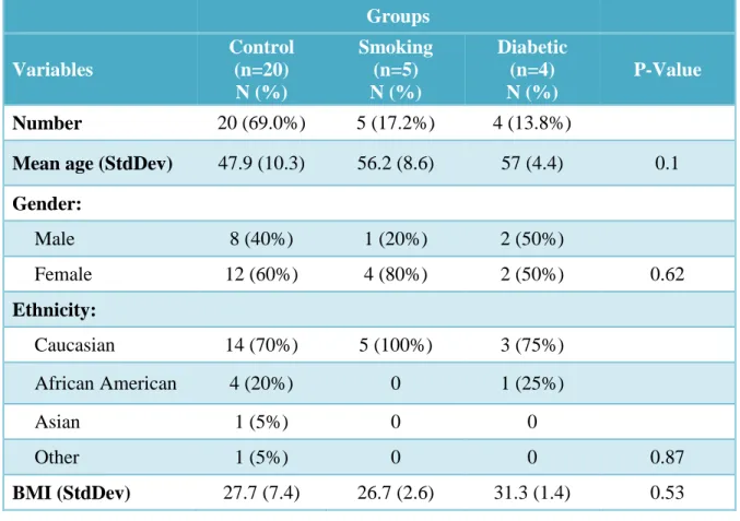
The 29 recruited participants included 20 control subjects, 5 smokers and 4 diabetics. The mean participant age varied from 47.9 to 56.2 years with a standard deviation ranged from 4.4 to 10.3 (Table 3). Majority of the participants were Caucasians, and more female participants were recruited in the control and smoking groups. BMI was recorded for all participants and ranged from 26.7 to 31.3 kg/m2. Statistical significance was not found with age, gender, race and BMI among the three study groups.
27
Table 3. Study Participant Demographics (Total n = 29)
Groups
Variables
Control (n=20) N (%)
Smoking (n=5) N (%)
Diabetic (n=4) N (%)
P-Value
Number 20 (69.0%) 5 (17.2%) 4 (13.8%)
Mean age (StdDev) 47.9 (10.3) 56.2 (8.6) 57 (4.4) 0.1
Gender:
Male 8 (40%) 1 (20%) 2 (50%)
Female 12 (60%) 4 (80%) 2 (50%) 0.62
Ethnicity:
Caucasian 14 (70%) 5 (100%) 3 (75%)
African American 4 (20%) 0 1 (25%)
Asian 1 (5%) 0 0
Other 1 (5%) 0 0 0.87
28 Selected Implant Sites
29
Table 4. Implant Sites of Study Participants (Total n = 29)
Groups
Implant sites Control (n=20) N (%)
Smoking (n=5) N (%)
Diabetic (n=4)
N (%) P-Value
Maxillary
molar 3 (15%) 1 (20%) 1 (25%)
Maxillary
premolar 5 (25%) 2 (40%) 1 (25%)
Maxillary
anterior 1 (5%) 1 (20%) 2 (50%)
Mandibular
molar 9 (45%) 0 0
Mandibular
premolar 2 (10%) 1 (20%) 0
Mandibular
30
Differential DNA Methylation of FGF2 Gene in Smokers and Diabetic Participants
A comparison among the control subjects, smokers and diabetic participants indicated increased methylation of FGF2 gene was present in the smoking and diabetic groups (Figure 4). The percentages of hyper-methylation in the gingival tissues of the control, smoking and diabetic groups were 6.1±6.0, 23.2±24.9 and 15.1±23.5
respectively. The p-values were 0.02 and 0.25 for the gingival samples from smokers and diabetics respectively as compared to the control samples. This indicated that the
31
Figure 4. Methylation Level of FGF2 Gene in Smokers or Diabetics in
Comparison to Control Subjects. Graph shows mean % methylation values with bars indicating standard errors.
0 5 10 15 20 25 30 35 40
Control Smoking Diabetic
%
o
f
H
y
p
er
m
et
h
y
la
ti
o
n
Group
Figure 4
32
Differential mRNA Expression of FGF2 Gene in Smokers and Diabetic Participants
33
Figure 5. Messenger Level of FGF2 Gene in Smokers or Diabetics in Comparison to Control Subjects. The fold change of all participants was computed against the mean expression level of the control group. The graph shows the adjusted mean fold change of each group with bars indicating standard errors.
0 0.2 0.4 0.6 0.8 1 1.2 1.4 1.6
Control Smoking Diabetic
F
o
ld
C
h
a
n
g
e
Group
Figure 5
34
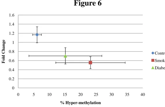
Relationship between DNA Methylation and mRNA Expression of FGF2 Gene
35
Figure 6. Group Means of Methylation Level and RNA Expression of FGF2 Gene. Horizontal error bars indicate standard errors of mean % methylation values whereas vertical error bars indicate standard errors of mean fold change values.
0 0.2 0.4 0.6 0.8 1 1.2 1.4 1.6
0 5 10 15 20 25 30 35 40
F
o
ld
C
h
a
n
g
e
% Hyper-methylation
Figure 6
Control
Smoking
36
Levels of FGF2 Protein detected in GCF during Wound Healing
The levels of FGF2 protein in GCF were below detection level (0.3pg/ml) of ELISA for all except one set of GCF samples that were collected at the 6-week time point. A literature search was performed on PubMed to identify studies that had evaluated FGF2 protein level in human GCF. One such study (Aykol et al. 2011) was identified, and it reported a shaking and incubation period of ≥ 18 hours instead of 30 minutes that was used previously on the GCF strips. The elution protocol was therefore modified, and ELISA was performed again on GCF samples collected at 0, 2 and 4 weeks from 2 subjects of each group. However, the levels of FGF2 proteins were still below detection for all time points.
Implant Stability and WHI during Wound Healing
The ISQ values at 4 and 6 weeks after implant surgery for all three study groups were illustrated in Figure 7. The values for the control, smoking and diabetic groups were respectively 62.3±11.5, 58.5±16.6 and 58.7±14.5 at 4 weeks and 68.4±7.9, 63.1±9.4 and 67.7±7.3 at 6 weeks. Statistical significant difference was not found between the two time points and among any groups. However, two general patterns can be observed: 1) ISQ values were higher for all three groups at 6 weeks than 4 weeks, indicating increased implant stability with time. 2) At both 4 and 6 weeks, ISQ values were the highest in control subjects and lowest in smokers. This result indicated that rate of wound healing was reduced in subjects who smoked or who had diabetes.
37
38
Figure 7. Implant Stability of All Three Groups of Subjects after Implant
Placement. Graph shows mean ISQ values with bars indicating standard errors.
45 50 55 60 65 70 75
4 Weeks 6 Weeks
IS
Q
V
a
lu
e
Weeks after Implant Surgery
Figure 7
Control
Smoking
Diabetic
39
Relationship between DNA Methylation of FGF-2 Gene and Implant Stability
Figure 8. Levels of (a) Edema, (b) Erythema, (c) Pain, and (d) Degree of Flap Closure Experienced by All Three Groups of Subjects after Implant Placement. Graphs show mean (a) edema index, (b) erythema index, (c) pain index and (d) flap closure index with bars indicating the corresponding standard errors.
0 0.2 0.4 0.6 0.8 1 1.2 1.4
2 Weeks 4 Weeks 6 Weeks
E ry th e m a I n d e x
Weeks after Implant Surgery
Figure 8(b)
Control Smoking Diabetic † * * 0 0.2 0.4 0.6 0.8 1 1.2 1.42 Weeks 4 Weeks 6 Weeks
F la p C lo su re I n d e x
Weeks after Implant Surgery
Figure 8(d)
Control Smoking Diabetic * 0 0.2 0.4 0.6 0.8 1 1.22 Weeks 4 Weeks 6 Weeks
E d e m a I n d e x
Weeks after Implant Surgery
Figure 8(a)
Control Smoking Diabetes † * * 0 0.1 0.2 0.3 0.4 0.5 0.62 Weeks 4 Weeks 6 Weeks
P a in I n d e x
Weeks after Implant Surgery
Figure 8(c)
Control Smoking Diabetic † † † ** p ≤ 0.05 when compared to control at 2 and 4 weeks
* p ≤ 0.05 when compared to control at 2 weeks † p ≤ 0.05 when compared to control at 6 weeks
* p ≤ 0.05 when compared to control at 2 weeks † p ≤ 0.05 when compared to diabetic at 6 weeks
40
Relationship between DNA Methylation of FGF2 Gene and Implant Stability
41
Figure 9. Mean Methylation Level and Overall Implant Stability of the Three Study Groups. Overall Implant stability is defined as the average ISQ value of all visits. Horizontal error bars indicate standard errors of mean % methylation values whereas vertical error bars indicate standard errors of overall ISQ
values. 54 56 58 60 62 64 66 68 70
0 5 10 15 20 25 30 35 40
42
Relationship between DNA Methylation of FGF2 Gene and WHI
A positive trend between methylation level of FGF2 gene and edema or erythema index was demonstrated for 2, 4 and 6 weeks after implant placement. In other words, the higher is the level of hyper-methylation, the higher are the edema and erythema levels. On the other hand, the association pattern between methylation level of FGF2 gene and pain or flap closure index was more inconsistent.
Except for edema index at 6 weeks and erythema index at 4 weeks, the
associations for the four indices at the three time points were not statistically significant (p > 0.05). The odds ratio and p-value were 1.08 and 0.03 respectively for edema index at 6 weeks and were 1.05 and 0.05 respectively for erythema index at 4 weeks. The mean methylation level and overall WHI indices of the three visits among all three study groups were illustrated in Figures 10(a)-(d).
43
Figure 10. Mean Methylation Level and Overall (a) Edema Index, (b) Erythema Index, (c) Pain Index, and (d) Flap Closure Index of the Three Study Groups. Overall edema, erythema, pain or flap closure index is defined as the average value of all visits of the corresponding index. Horizontal error bars indicate standard errors of mean % methylation values whereas vertical error bars indicate standard errors of overall (a) edema index, (b) erythema index, (c) pain index and (d) flap closure index.
0 0.2 0.4 0.6 0.8 1
0 20 40
O v er a ll E ry th em a I n d ex % Hyper-methylation
Figure 10(b)
Control Smoking Diabetic 0.4 0.5 0.6 0.7 0.8 0.9 10 20 40
O v er a ll F la p C lo su re I n d ex % Hyper-methylation
Figure 10(d)
Control Smoking Diabetic 0 0.1 0.2 0.3 0.4 0.5 0.6 0.7 0.80 20 40
O v er a ll E d em a I n d ex % Hyper-methylation
Figure 10(a)
Control Smoking Diabetic -0.05 0 0.05 0.1 0.15 0.2 0.250 20 40
44
DISCUSSION
In this study, hyper-methylation of FGF2 gene was consistently observed in the gingivae from smokers and diabetic subjects relative to the gingivae from healthy subjects. The level of mRNA expression of the FGF2 gene also tends to be down-regulated in gingivae from smokers and diabetics. Overall, differential methylation pattern of FGF2 gene among gingivae from healthy individuals, smokers and diabetics were associated with differing levels of FGF2 gene expression.
To date, no previous studies have investigated the effects of smoking and diabetes on methylation status of the FGF2 gene. Limited literature relating these two
environmental and systemic factors with DNA methylation of other genes could be identified. Regarding smoking, majority focused on bronchial or lung tissues in lung cancer patients (Belinsky et al. 2002, Soria et al. 2002, Divine et al. 2005, Liu et al. 2007, Han et al. 2009, Tekpli et al. 2011). Almost all of these studies demonstrated a positive correlation between cigarette smoking and abnormal promoter methylation patterns. In a meta-analysis performed by Zhang on the association between smoking and p16INK4α methylation in tumor cells from lung carcinoma patients, more than 2-fold increase of p16INK4α methylation in NSCLC patients with smoking habits could be noted. Zhang later demonstrated that the association was stronger in Asian population (Zhang et al. 2011). Lin demonstrated nicotine-derived nitrosamine ketone, a cigarette smoke carcinogen, was capable of influencing the expression of DNA methyltransferase 1 (DNMT1) and
45
lung tumor tissues (Lin et al. 2010). Jin also reported an increase in the risk of non-small cell lung cancer with hypermethylation of the p16 gene promoter by 5.16 folds in
smokers with CYP1A1 variants when compared to those without the variants (Jin et al. 2010). This finding was striking, as it implied the relationship between smoking and abnormal methylation pattern could be complicated by gene polymorphism. On the other hand, in the two studies performed by Oliveira (Oliveira et al. 2009, De Oliveira et al. 2011) regarding the impact of smoking on the DNA methylation status of IL-8, TLR2 and TLR4 gene promoters in the periodontal tissues, both failed to demonstrate a correlation between smoking and methylation of these gene promoters. The inconsistent results may be contributed by differences in the smoking dose. Smokers were defined as subjects who consumed at least 5 pack-years in the current study but 1.25 pack-years in Oliveira’s studies. Regarding diabetes, literature has also been identified. One study reported that DNA methylation in the promoter of transcriptional co-activator, PRARGC1A, was elevated in pancreatic islets from patients with type II diabetes (Ling et al. 2008).
Another study demonstrated methylation of the CpG sites in both mouse Ins2 and human INS promoters in pancreatic beta cells suppressed the insulin promoter activity by as much as 90% (Kuroda et al. 2009). A third study demonstrated the methylation levels of CpG sites in the Alu repeats of peripheral blood leukocytes were associated with insulin resistance (Zhao et al. 2011). In general, existing evidences were supportive to the finding of the current study, that smoking and diabetes are associated with hyper-methylation pattern of FGF2 gene.
46
severity of the systemic condition. The five smokers each had a smoking history of 30, 20, 20, 25 and 50 pack-years, indicating they were all heavy smokers. While the dose-response relationship exerted by smoking on periodontal health has been well
documented in literature (Bergstrom et al. 1991, Haffajee & Socransky2001,
Bergstrom2004, Machuca et al. 2000, Natto et al. 2005, Rosa et al. 2008), the degree of DNA hyper-methylation in the gingival tissues can be expected to be high. On the other hand, the four diabetic subjects all had HbA1C level of 7.0% or less, indicating that their diabetic conditions were controlled. Controlled diabetic patients have been shown to exhibit better periodontal status than those with poor glycemic control (Taylor 2001). Therefore, it is reasonable to observe that only differential methylation pattern of FGF2 gene in the gingivae of smokers reached statistical significance in this study.
FGF2 is an important protein in the development, repair and regeneration of various types of tissues in human body. DNA methylation of FGF2 gene by other factors has also been reported. Chang and his group(Chang et al. 2008b)demonstrated elevated plasma level of hemocysteine decreased proliferation of endothelial cell through methylation of FGF2 gene. Using cultured human coronary and bovine aortic endothelial cells, they found that S-adenosylhomocysteine (SAH), a metabolite of hemocysteine, could modulate the transcription control of CG-rich FGF2 promoter through a G-protein signaling pathway. The endothelial cells exhibited decreased DNA synthesis and delayed progression through the cell cycle at G1/S transition. These effects could be counteracted by exogenous FGF2 proteins. The group concluded hemocysteine contributed to
47
This observation might play a role in hyper-methylation of FGF2 gene in obesity (Campion et al. 2009). Li and his group (Li et al. 2008)investigated the mechanism by which methylated-CpG binding protein 1 (Mbd1), a transcriptional repressor of FGF2, regulated the expression of FGF2 in adult neural stem/progenitor cells (NSPCs). Using mouse brain-derived NSPCs, they demonstrated Mbd1 bound to the FGF2 promoter in adult NSPCs, and the FGF2 promoter became hypomethylated in the absence of functional Mbd1. FGF2 was identified to be essential in maintaining NSPCs in their multi-potent state. A deficiency in Mbd1 reduced repression of FGF2 and promoted increased neural differentiation of NSPCs. Knockdown of Mbd1 or over-expression of FGF2 resulted in inhibition of differentiation of NSPCs. The group concluded a reduction of DNA methylation of FGF2 gene in NSPC is necessary for neural commitment to occur.
48
thus the specific locus of CpG methylation within the promoter could not be obtained for further evaluation.
FGF2 can upregulate the expression of many genes and can initiate various downstream reactions, but the exact mechanisms by which the FGF2 gene is regulated remain unclear. At present time there have been no studies investigating how smoking can affect RNA expression of FGF2 gene during wound healing. Only one study could be identified to support the inhibitory effect that smoking has on the activity of FGF2 protein. Pauwels and his group (Pauwels et al. 2010) evaluated whether cigarette smoke triggered pulmonary and systemic pentraxin 3 (PTX3) expression in vivo in a murine model. PTX3, a soluble pattern recognition receptor, is produced in response to pro-inflammatory cytokines IL-1β and TNF-α and mediates angiogenesis by influencing FGF2 activity. They detected elevated level of PTX3 and reduced mRNA expression of FGF2 at 4 weeks and 24 weeks after exposure to cigarette smoke. This could possibly be related to binding of FGF2 protein by the N-terminal domain of PTX3, thereby inhibiting the proangiogenic activity of FGF2.
49
identified in patients with type 2 diabetes, induced endothelial cell apoptosis by
suppressing FGF2 expression. FGF2 expression exerted most of its effects by activating the phosphatidylinositol 3-kinase (PI3K)-Akt pathway. It activated PI3K, which then activated Akt. The phorsphorylated Akt activated endothelial nitric oxide synthase, which is essential for survival of endothelial cell. Lu suggested FGF2 was auto-regulated by Akt activation through a positive feedback loop. They isolated L5 from type 2 diabetic
patients, and found L5 caused reduction in Akt phosphorylation and decreased transcription of FGF2 gene. The group concluded L5 could potentially impair FGF2 dependent re-endothelialization and could play a role in diabetes-associated wound impairment and atherosclerosis.
In this study, an increase in implant stability from 4 weeks to 6 weeks was
demonstrated in all three groups of subjects. The highest and lowest degrees of stability were consistently demonstrated in the control group and the smoking group respectively. Differential methylation pattern of FGF2 gene among gingivae from healthy subjects, smokers and diabetics was inversely associated with implant stability at both 4 and 6 weeks.
50
et al. 1995) discovered that over-expressing FGF2 in mice induced abnormal long bone formation, and Montero (Montero et al. 2000) later noted bone formation was inhibited by deactivating FGF2.
Using human calvaria cell cultures, Debiais and his group (Debiais et al. 1998) discovered the effect of FGF2 was dependent on the stage of osteoblast maturation. In immature cells, FGF2 increased cell growth, decreased alkaline phosphatase (ALP) activity, reduced osteocalcin synthesis and lowered matrix mineralization whereas in mature cells, it increased osteocalcin production and matrix mineralization. The group later found that FGF2 increased expression and function of N-cadherin, a transmembrane glycoprotein which plays a role in osteoblast differentiation, in human calvaria
osteoblasts (Debiais et al. 2001).
Using Fgf2-null mice, Fei and his group (Fei et al. 2011) demonstrated a role of FGF2 in osteoblastogenesis through Wnt signaling. Wnts are a family of 19 glycoproteins secreted in mammals, and were well demonstrated to positively regulate osteoblast
differentiation and bone formation (Krishnan et al. 2006). Wnt10b had been shown to stimulate osteogenic transcription factor Runx2 (Bennett et al. 2005, Bennett et al. 2007). Wnt ligands had also been shown to bind to β-catenin, enabling the complex to
51
The effects of FGF2 can be enhanced with bone morphogenetic protein 2 (BMP2). Exposure of rat marrow-derived mesenchymal stem cells with FGF2 and BMP2 induced expression of osteoclacin mRNA and formation of bone-like nodules earlier than either factor alone (Hanada et al. 1997). Supplementing mesenchymal stromal cell culture sequentially with FGF2 before BMP2 resulted in high ALP activity, elevated bone-specific osteocalcin expression and abundant bone matrix formation than adding either alone (Maegawa et al. 2007). In adult rabbits that underwent intertransverse process fusions at L4-L5 implanted with mesenchymal stem cells, only the group with both BMP2 and FGF2 addition demonstrated bone in-growth at 6 weeks, and this group had a higher 6-week fusion rate than the other groups with only BMP2 or FGF2 (Minamide et al. 2007). Recently, FGF2 has been demonstrated to reduce expression of BMP receptor 1A on murine osteoprecursor cells, thereby influencing osteoblast differentiation
(Park2011). In addition to BMP2, a review written by Marie in 2003 also provided evidence to support interaction of FGF2 with other biological factors, such as cAMP and TGF-β to enhance osteogenesis (Marie2003).
The ability of FGF2 to regulate bone remodeling is supported by studies that
52
hand, the direct mechanisms involved FGF2 binding to FGF receptor type 1, a receptor tyrosine kinase, on osteoclasts to induce bone resorption (Kawaguchi et al. 2000, Chikazu et al. 2001) and on osteoclast precursors to prevent their differentiation (Chikazu et al. 2001).
No statistical significance was identified between DNA methylation of FGF2 gene and implant stability among any groups and at any time points. This can partly be
53
was also not possible to stratify the study population according to these factors because of the limited sample size.
The wound healing index (WHI) developed to evaluate degree of soft tissue healing in this study was built upon several similar indices published in literature. The Loe and Silness Gingival Index (Loe and Silness 1963) is a scale of 0-3 used to evaluate gingival inflammation instead, but it contains several characteristics of gingival presentation, such as edema and redness, that are also important aspects to evaluate during wound healing. The wound healing index developed by Wang (Huang et al. 2005a, Huang et al. 2005b) is a scale of 1-3 that jointly evaluated degree of gingival edema, erythema, suppuration, patient discomfort or flap dehiscence. These elements are essential when assessing wound healing, but the index does not allow evaluation of each separately. This problem was overcome by the early healing index (EHI) developed by Hurzeler (Wachtel et al. 2003) and soft tissue swelling and gingiva color indices developed by Weijden
(Hagenaars et al. 2004). EHI is a scale of 1-5 that assesses the degree of flap closure while the swelling and color indices are scales of 0-2 that each assesses the degree of swelling and redness respectively after surgery. Therefore, combining these indices resulted in the WHI used in the current study.
This newly developed WHI is not without limitations. The main problem lied in the evaluation of pain and flap closure. The pain index in WHI was based on patient’s
54
the incision area, which is affected by the incision design and surgical technique. These limitations can be reflected in the inconsistent WHI results among the three groups and the three time points.
Several other limitations can be identified in the study. 1) The principle one is the small sample sizes in the smoking and diabetic groups. The target number of recruitment was 20 for the control group, 12 for the smoking group and 12 for the diabetic group. However, the control group was the only category that met the goal. With only 4-5 subjects in each of the two test groups, the study was under-powered. 2) Another important one is the cross-sectional design used to evaluate impact of smoking and diabetes on DNA methylation. Causality therefore cannot be implied. 3) Smoking history and dosage were self-reported, and HbA1c was not evaluated in the other two
55
In conclusion, this study identified important association patterns of differential methylation of FGF2 gene with smoking, diabetes and wound healing, and it
demonstrated hyper-methylation level of FGF2 gene coincided with down-regulation of its mRNA expression. For the first time, systemic and environmental factors were shown to have epigenetic effects on periodontal tissues, ultimately affecting their healing response. The findings from this study imply a potential role of the epigenetic
56
APPENDIX I
SUPPLEMENTARY MATERIALS AND METHODS
Institutional Review Board, University of North Carolina at Chapel Hill IRB Study Number 10-1184
1. Clinical Study Design
This is a longitudinal case-control study of 6-8 weeks in duration. Clinical parameters and biological samples were collected from healthy subjects, smokers and diabetics during the course of implant therapy. The study was performed in a single masked or blinded manner. All laboratory assessments were performed without knowledge of the subjects’ type of treatment and health condition.
2. Human Subjects
57
within 3 months prior to screening. Screening and follow-up examinations were
performed in the General Oral Health Center in the School of Dentistry. Implant surgery was performed in the Graduate Periodontology clinic.
The inclusion criteria of study participation included: (1) males or females between the age of 18 and 70 years (inclusive), (2) must be able and willing to follow study procedures and instructions, (3) must have read, understood and signed an informed consent form, (4) must be in good general health, (5) must have one or more implant placements as their future treatment needs, (6) must fit into one of the study groups as listed previously, and (7) must have probing depth ≤ 4 mm or 5mm without bleeding on probing for all teeth at the same quadrant of implant placement as determined by two calibrated examiners .
Individuals were not enrolled into the study if they presented with the following exclusion criteria: (1) chronic disease with oral manifestations (e.g. Crohn’s disease, lupus erythematosus, Behcet’s disease), (2) gross oral pathology, (3) use of either antibiotics or NSAIDs within 2 weeks prior to screening examination, (4) chronic treatment (i.e. two weeks or more) with any medication known to affect periodontal status (e.g. phenytoin, calcium, antagonists, cyclosporin, warfarin) within 1 month prior to screening examination, (5) systemic conditions, except smoking and diabetes, that were known to affect the periodontal status, (6) active infectious diseases such as hepatitis A-E, HIV or tuberculosis, and (7) known to be pregnant or breastfeeding.
58
be withdrawn. Infection around an implant would be reported as an adverse event and subject would be followed until it was resolved.
3. Clinical Study Method
The following procedures were performed on study participants during a total of 5 visits within a study period of 6-8 weeks.
Visit 1 (≤ 2 weeks before implant surgery):
• Study personnel obtained a signed written informed consent from the subject. • Study personnel obtained medical history and demographics related to the patient. • Study personnel performed HbA1C test on all subjects in the diabetic group (if a
test result within past 3 months was not available). A drop of blood was obtained by finger prick and was loaded into a DCA 2000 HbA1C reagent cartridge, which was then processed immediately by a DCA 2000®+ Analyzer (Bayer Inc., Toronto, ON, Canada) for HbA1C level.
• Patient vital signs (blood pressure and pulse), height and weight were recorded. • A dental examiner performed a general oral examination for presence of any
abnormalities.
• A dental examiner scored all teeth in the same quadrant of implant placement for plaque index, gingival index, probing depth, percent bleeding on probing and clinical attachment level.
Visit 2 (Implant surgery):
59
• A dental examiner performed a general oral examination for presence of any abnormalities.
• The dental examiner collected 8 GCF samples from the mesiolingual, mesiobuccal, distolingual and distobuccal surfaces of the two teeth that were in the closest proximity to the implant site. Samples were collected using filter paper strips (Pro Flow, Inc., Amityville, NY).
• The dental examiner collected 1 biopsy at the site of implant placement for quantitative PCR, DNA methylation sequence analysis and differential methylation analysis. The biopsy sample was obtained with a 4mm punch biopsy blade or a scalpel blade. It was removed in the same way as how discarded tissues will be removed. It was removed at the beginning of the surgery as the clinician exposed the underlying bone at the implant site, or before suturing when the clinician reduced redundant tissues to achieve better flap closure.
Visits 3-5 (2 weeks ± 2 days, 4 weeks ± 2 days, 6 weeks ± 2 days after surgery): • Study personnel updated medical history and obtained vital signs.
• A dental examiner performed a general oral examination for presence of any abnormalities.
• A dental examiner scored all teeth in the same quadrant of implant placement for plaque index and gingival index.
• The dental examiner recorded wound healing indices (WHI) as indicators of degree of soft tissue healing for the implant site.
60
• The dental examiner collected 8 GCF samples in the same areas and using the same method as indicated previously.
Table 5. Schedule of Procedures by Visit
Week
Visit 1 -2 to -1
Visit 2 0
Visit 3 2
Visit 4 4
Visit 5 6
Procedure:
Informed Consent X
Medical/Dental History X X X X X
Demographics X
Vital Signs X X X X X
HbA1C Test (for diabetic group
only) X
Oral Examination X X X X X
PI, GI X X X X
PD, BOP, CAL X
WHI X X X
Implant Stability Measurement X X
Biopsy Sample X
GCF Sample X X X X
Saliva Sample X X X X
Adverse Experience X X X X X
4. Clinical Periodontal Assessments
61
parameters were measured using a manual University of North Carolina (UNC-15) periodontal probe. These parameters were measured at six periodontal sites per tooth (i.e., mesiobuccal, buccal, distobuccal, mesiolingual, lingual, and distolingual) and at all teeth in the same quadrant where implant placement is desired (including the third molar). Silness and Löe Plaque Index (PI) (SILNESS & LOE 1964):
PI was defined as the relative amount of supragingival plaque and was recorded on an ordinal scale of 0-3.
0- No plaque in the gingival area.
1- A film of plaque adhering to the free gingival margin and the adjacent tooth. The plaque may be recognized only by running a probe across the tooth surface
2- Moderate accumulation of soft deposits within the gingival pocket and on the gingival margin and/or adjacent tooth surface, which can be seen by the naked eye.
3- Abundance of soft matter within the gingival pocket and/or on the gingival margin and the adjacent tooth surface.
Löe and Silness Gingival Index (GI) (LOE & SILNESS 1963):
GI was defined as the degree of gingival inflammation and was recorded on an ordinal scale of 0-3.
0- Normal gingiva.
62
2- Moderate inflammation (redness, edema, glazing); bleeding on palpation (i.e., sulcular sweep).
4- Severe inflammation (i.e., marked redness, edema); ulceration, tendency to spontaneous bleeding.
Probing depth (PD):
PD was defined as the linear distance from the gingival margin (GM) to base of the pocket. If a PD reading fell between two millimeter readings, the rule was to round down and the lower of the two readings was recorded.
Bleeding on Probing (BOP):
BOP was defined as the presence or absence of bleeding to manual probing and was recorded as a dichotomous variable.
0- No bleeding within 10 seconds after probing. 1- Bleeding within 10 seconds after probing. Clinical attachment level (CAL):
CAL was defined as the linear distance from the cemento-enamel junction (CEJ) to base of the pocket. If a CAL reading fell between two millimeter readings, the rule was to round down and the lower of the two readings was recorded.
Wound healing indices (WHI):
WHI was defined as the degree of soft tissue healing and consisted of four indices. Each index was recorded on an ordinal scale of 0-3.
Degree of Edema
63 2- Moderate gingival edema. 3- Severe gingival edema.
Degree of Erythema
0- No erythema. 1- Mild erythema. 2- Moderate erythema. 3- Severe erythema.
Degree of Pain on Palpation
0- No pain. 1- Mild pain. 2- Moderate pain. 3- Severe pain.
Degree of Flap Closure
0- No fibrin line or folding line in the incision area. 1- Fine fibrin line or folding line in the incision area. 2- Fibrin clot in the incision area.
3- Partial or complete necrosis of flap. Implant Stability:
64
65
REFERENCES
(U.S.) C. f. D. C. (2009) State-specific smoking-attributable mortality and years of potential life lost--United States, 2000-2004. MMWR.Morbidity and mortality weekly report 58, 29-33.
Abraham J. A., Whang J. L., Tumolo A., Mergia A., Friedman J., Gospodarowicz D. & Fiddes J. C. (1986) Human basic fibroblast growth factor: nucleotide sequence and genomic organization. The EMBO journal 5, 2523-2528.
Aksoy U., Eratalay K. & Tözüm T. (2009) The possible association among bone density values, resonance frequency measurements, tactile sense, and histomorphometric
evaluations of dental implant osteotomy sites: A preliminary study. Implant dentistry 18, 316-325.
Association A. (2008) Economic costs of diabetes in the U.S. In 2007. Diabetes care 31, 596-615.
Aykol G., Baser U., Maden I., Kazak Z., Onan U., Tanrikulu-Kucuk S., Ademoglu E., Issever H. & Yalcin F. (2011) The Effect of Low-Level Laser Therapy as an Adjunct to Non-Surgical Periodontal Treatment. Journal of periodontology 82, 481.
Barros S. P. & Offenbacher S. (2009) Epigenetics: connecting environment and genotype to phenotype and disease. Journal of dental research 88, 400-408. doi:
10.1177/0022034509335868
Belinsky S. A., Palmisano W. A., Gilliland F. D., Crooks L. A., Divine K. K., Winters S. A., Grimes M. J., Harms H. J., Tellez C. S., Smith T. M., Moots P. P., Lechner J. F., Stidley C. A. & Crowell R. E. (2002) Aberrant promoter methylation in bronchial
epithelium and sputum from current and former smokers. Cancer research 62, 2370-2377. Bennett C. N., Ouyang H., Ma Y. L., Zeng Q., Gerin I., Sousa K. M., Lane T. F.,
Krishnan V., Hankenson K. D. & MacDougald O. A. (2007) Wnt10b increases postnatal bone formation by enhancing osteoblast differentiation. Journal of bone and mineral research : the official journal of the American Society for Bone and Mineral Research 22, 1924-1932. doi: 10.1359/jbmr.070810
Bennett C. N., Longo K. A., Wright W. S., Suva L. J., Lane T. F., Hankenson K. D. & MacDougald O. A. (2005) Regulation of osteoblastogenesis and bone mass by Wnt10b. Proceedings of the National Academy of Sciences of the United States of America 102, 3324-3329. doi: 10.1073/pnas.0408742102
66
Bergstrom J., Eliasson S. & Preber H. (1991) Cigarette smoking and periodontal bone loss. Journal of periodontology 62, 242-246.
Bikfalvi A., Klein S., Pintucci G. & Rifkin D. B. (1997) Biological roles of fibroblast growth factor-2. Endocrine reviews 18, 26-45.
Bohlen P., Baird A., Esch F., Ling N. & Gospodarowicz D. (1984) Isolation and partial molecular characterization of pituitary fibroblast growth factor. Proceedings of the National Academy of Sciences of the United States of America 81, 5364-5368. Campion J., Milagro F. I. & Martinez J. A. (2009) Individuality and epigenetics in obesity. Obesity reviews : an official journal of the International Association for the Study of Obesity 10, 383-392. doi: 10.1111/j.1467-789X.2009.00595.x
Chang P. Y., Lu S. C., Lee C. M., Chen Y. J., Dugan T. A., Huang W. H., Chang S. F., Liao W. S., Chen C. H. & Lee Y. T. (2008a) Homocysteine inhibits arterial endothelial cell growth through transcriptional downregulation of fibroblast growth factor-2 involving G protein and DNA methylation. Circulation research 102, 933-941. doi: 10.1161/CIRCRESAHA.108.171082
Chang P. Y., Lu S. C., Lee C. M., Chen Y. J., Dugan T. A., Huang W. H., Chang S. F., Liao W. S., Chen C. H. & Lee Y. T. (2008b) Homocysteine inhibits arterial endothelial cell growth through transcriptional downregulation of fibroblast growth factor-2
involving G protein and DNA methylation. Circulation research 102, 933-941. doi: 10.1161/CIRCRESAHA.108.171082
Chávarry N. G. M. (2009) The relationship between diabetes mellitus and destructive periodontal disease: a meta-analysis. Oral health & preventive dentistry 7, 107-27. Chikazu D., Katagiri M., Ogasawara T., Ogata N., Shimoaka T., Takato T., Nakamura K. & Kawaguchi H. (2001) Regulation of osteoclast differentiation by fibroblast growth factor 2: stimulation of receptor activator of nuclear factor kappaB ligand/osteoclast differentiation factor expression in osteoblasts and inhibition of macrophage colony-stimulating factor function in osteoclast precursors. Journal of bone and mineral research : the official journal of the American Society for Bone and Mineral Research 16, 2074-2081. doi: 10.1359/jbmr.2001.16.11.2074
Coffin J. D., Florkiewicz R. Z., Neumann J., Mort-Hopkins T., Dorn G. W.,2nd,
Lightfoot P., German R., Howles P. N., Kier A. & O'Toole B. A. (1995) Abnormal bone growth and selective translational regulation in basic fibroblast growth factor (FGF-2) transgenic mice. Molecular biology of the cell 6, 1861-1873.