Received 22 Mar 2016
|
Accepted 30 Nov 2016
|
Published 6 Feb 2017
Identification of BPIFA1/SPLUNC1 as an
epithelium-derived smooth muscle relaxing factor
Tongde Wu
1
, Julianne Huang
1,2
, Patrick J. Moore
1
, Michael S. Little
2
, William G. Walton
2
, Robert C. Fellner
1
,
Neil E. Alexis
3
, Y. Peter Di
4
, Matthew R. Redinbo
2
, Stephen L. Tilley
1,3,
* & Robert Tarran
1,5,
*
Asthma is a chronic airway disease characterized by inflammation, mucus hypersecretion and
abnormal airway smooth muscle (ASM) contraction. Bacterial permeability family member
A1, BPIFA1, is a secreted innate defence protein. Here we show that BPIFA1 levels are reduced
in sputum samples from asthmatic patients and that BPIFA1 is secreted basolaterally from
healthy, but not asthmatic human bronchial epithelial cultures (HBECs), where it suppresses
ASM contractility by binding to and inhibiting the Ca
2þinflux channel Orai1. We have
localized this effect to a specific, C-terminal
a
-helical region of BPIFA1. Furthermore, tracheas
from
Bpifa1
/mice are hypercontractile, and this phenotype is reversed by the addition of
recombinant BPIFA1. Our data suggest that BPIFA1 deficiency in asthmatic airways promotes
Orai1 hyperactivity, increased ASM contraction and airway hyperresponsiveness. Strategies
that target Orai1 or the BPIFA1 deficiency in asthma may lead to novel therapies to treat this
disease.
DOI: 10.1038/ncomms14118
OPEN
1Cystic Fibrosis Center/Marsico Lung Institute, Marsico Hall, 125 Mason Farm Road, University of North Carolina at Chapel Hill, Chapel Hill, North Carolina
27599-7248, USA.2Department of Chemistry, Genome Science Building, 250 Bell Tower Road, University of North Carolina at Chapel Hill, Chapel Hill, North Carolina 27599-7248, USA.3Center for Environmental Medicine, Asthma, and Lung Biology, US EPA Human Studies Facility, 104 Mason Farm Road, University of North Carolina at Chapel Hill, Chapel Hill, North Carolina 27599-7248, USA.4Department of Environmental and Occupational Health,
University of Pittsburgh, 331 Bridgeside Point Building, Pittsburgh, Pennsylvania 15260, USA.5Department of Cell Biology & Physiology, 5200 Medical
A
sthma affects
B
334 million people worldwide, yet little
is known regarding the underlying aetiology of the
exaggerated airway smooth muscle (ASM) contraction
that leads to airway hyperresponsiveness (AHR). More recently,
research has focused on the role of airway epithelia in
driving asthma. In ASM, the degree of contraction is
directly proportional to the cytosolic Ca
2þlevels
1,2. As such,
ASM Ca
2þhomeostasis is tightly regulated and often involves
store-operated Ca
2þentry (SOCE), a process whereby stromal
interacting
molecule
1
(STIM1)
relocates/aggregates
at
the sarcoplasmic reticulum (SR)-plasma membrane junction
where it activates Orai1 to allow Ca
2þinflux, leading to
contraction
3,4. SOCE is defective in ASM from asthma
patients and in murine asthma models, which show increased
Orai1 activity
5,6. For over 30 years, researchers have predicted
that an epithelium-derived smooth muscle relaxing factor
(EDSMRF) exists in normal subjects where it regulates
ASM Ca
2þlevels, and that EDSMRF is deranged in asthmatics,
leading to an exaggerated Ca
2þresponse to agonists such
as methacholine (Mch). The existence of an EDSMRF was
first proposed when researchers demonstrated that trachea
denuded of epithelia induced AHR and that placing epithelia
from another animal in the same organ bath reduced
contractility
7. Candidate molecules for EDSMRF have included
NO, arachidonic acid metabolites, and cytokines, but all
have been ultimately rejected since they do not meet the
necessary criteria of (i) being secreted directly into the media
and (ii) being modulated by inflammatory mediators
8. Bacterial
permeability family member A1 (BPIFA1), also known as
short palate and nasal epithelial clone 1 (SPLUNC1), secreted
protein from the upper respiratory tract, LUNg-specific protein-X
and nasopharyngeal carcinoma-related protein, is a
multi-functional protein that is secreted by airway epithelia and
has both antimicrobial activity and regulates ion channels
9.
BPIFA1 production has previously been shown to be inhibited
by Th2-type cytokines that are upregulated in asthma, such as
IL-13, and in allergic mouse models, suggesting that it plays a role
in the allergic response to allergens
10. However, its effects on
ASM have not previously been described. We observed that
BPIFA1 protein levels were diminished in sputa from asthma
patients. Surprisingly, we found that BPIFA1 was secreted
basolaterally from normal human bronchial epithelial cultures
(HBECs)
and
that
basolateral
BPIFA1
secretions
were
significantly reduced in asthma-derived HBECs. On the basis
of these data, we tested the hypothesis that BPIFA1 was
the EDSMRF.
In support of this hypothesis, we found that serosal media
from normal but not asthmatic HBECs lowered cytosolic
ASM Ca
2þlevels and prevented ASM contractions. We also
found that Bpifa1-deficient (
Bpifa1
/) mice exhibited
sponta-neous AHR and that excised tracheas from
Bpifa1
þ/þbut
not
Bpifa1
/mice were capable of reducing ASM contraction.
Furthermore,
recombinant BPIFA1
blocked
Ca
2þinflux
and bound specifically to the plasma membrane Ca
2þchannel
Orai1, leading to Orai1 internalization. Thus, we propose
that BPIFA1 (SPLUNC1) is the EDSMRF and it functions
by internalizing Orai1.
Results
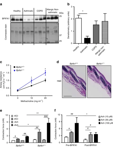
BPIFA1 levels inversely correlate with asthma and AHR
.
To investigate the role of BPIFA1 in asthma pathogenesis,
we measured sputum BPIFA1 levels in healthy donors, asthmatic
patients,
chronic
obstructive
pulmonary
disease
patients
and atopic individuals without asthma, the latter two cohorts
serving as disease controls. Immunoblot analyses indicated
significantly decreased BPIFA1 protein levels in asthmatic
patients’ samples compared with the other donors (Fig. 1a,b,
Supplementary Table 1). To determine whether a decrease
in BPIFA1 levels was associated with abnormal ASM activity,
we next tested whether
Bpifa1
/mice exhibit AHR.
These mice showed a significant increase in airway resistance
following Mch challenge compared with their
Bpifa1
þ/þlitter-mate controls (Fig. 1c), indicating an inverse correlation between
Bpifa1 expression and ASM contraction. Haematoxylin and
eosin (H&E) staining and quantification of smooth muscle
mass of tracheas from these mice showed no obvious difference in
ASM morphology between
Bpifa1
þ/þand
Bpifa1
/animals
(Fig. 1d and Supplementary Fig. 1), suggesting that the increase
in airway resistance in
Bpifa1
/mice was not due to
ASM hypertrophy. To further study the effect of Bpifa1 on
ASM, we excised tracheas from these mice and mounted
them on wire myographs to measure contractility
ex vivo
.
Tracheal rings from
Bpifa1
/mice showed significant
hyper-contractility
compared
with
wild-type
controls
following
exposure to acetylcholine (Ach) or KCl (Fig. 1e). Since this effect
was seen with both Ach, which stimulates Muscarinic/Ach
receptors and KCl, which depolarizes the plasma membrane to
induce Ca
2þinflux
11,12, we concluded that this effect was
not due to abnormal receptor function. Furthermore,
pretrea-tment with recombinant BPIFA1 protein at a physiologically
relevant dose
13(10
mM) for 1 h suppressed contraction (Fig. 1f),
suggesting that BPIFA1 is an EDSMRF.
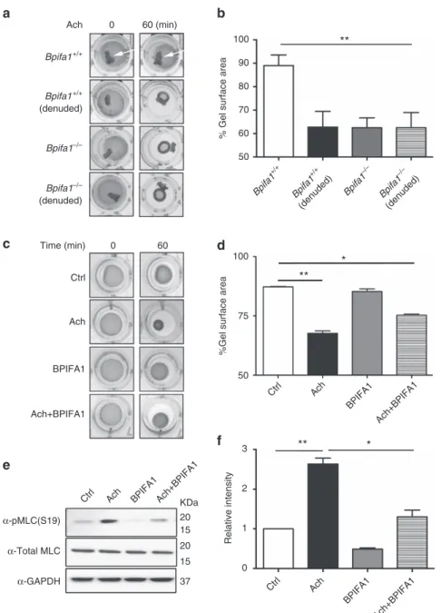
BPIFA1
regulates
ASM
contraction
in
vitro. Due
to
the potential importance of this observation, we further
investi-gated the ASM–BPIFA1 interaction in ASM cells (ASMCs)
cultured in collagen matrices. We observed that ASMC
contrac-tion was significantly reduced in the presence of
Bpifa1
þ/þtracheas with intact epithelium. In contrast, denuded tracheas
from both genotypes and whole
Bpifa1
/tracheas showed
no inhibitory effect on ASMC contraction (Fig. 2a,b). Similarly,
pre-incubation with recombinant BPIFA1 for 1 h reduced ASMC
contraction (Fig. 2c,d), suggesting that epithelium-derived
BPIFA1 is responsible for modulating ASM contractility.
In ASMC, contraction is regulated by cross-bridge formation
between phosphorylated myosin light chain (MLC) and
actin. Ach enhances MLC phosphorylation in a Ca
2þ-dependent
manner and therefore enhances contraction
14. MLC
phos-phorylation was increased by Ach, whereas pre-treatment with
BPIFA1 decreased both basal and induced MLC phosphorylation
(Fig. 2e,f and Supplementary Fig. 2).
BPIFA1 inhibits store-operated Ca
2þentry in ASM
. Since
BPIFA1 downregulated MLC phosphorylation, we next
investi-gated whether BPIFA1 regulated ASMC Ca
2þsignalling. When
ASMCs were exposed to serosal media from healthy HBECs,
or to recombinant BPIFA1, the thapsigargin (TG)-induced
Ca
2þincrease was significantly reduced in a dose-dependent
manner (Fig. 3c,d and Supplementary Fig. 5a–c). After
TG exposure, Ca
2þenters the cytoplasm from multiple sources
including the endoplasmic reticulum (ER) and the extracellular
milieu
15. In the absence of extracellular Ca
2þ, TG triggered
ER Ca
2þrelease and only moderately raised cytoplasmic Ca
2þ,
whereas reintroduction of Ca
2þextracellular Ca
2þinduced
SOCE
and
further
elevated
cytosolic
Ca
2þ(Fig.
3e).
Pre-treatment with BPIFA1 had no effect on ER Ca
2þrelease,
but significantly suppressed SOCE (Fig. 3e and Supplementary
Fig. 5d). Since SOCE supports intracellular Ca
2þoscillations
16,
we also examined the role of BPIFA1 in regulating this
phenomenon. Pre-treatment with BPIFA1 for 1 h significantly
attenuated Mch-induced Ca
2þoscillations (Fig. 3f,g), indicating
that BPIFA1 inhibits the Ca
2þentry required for oscillations.
To map out the structural region within BPIFA1 that
was responsible for these effects, we used a series of BPIFA1
mutants/peptides
and
tested
their
ability
to
suppress
Ca
2þsignalling. Inhibition was not different for mouse
and human BPIFA1 (Fig. 3h). BPIFA1 utilizes its N-terminal
‘S18’ region to regulate epithelial Na
þchannel (ENaC)
plasma membrane density
17,18. The S18 peptide
17did not
inhibit SOCE, suggesting that the action of BPIFA1 was
Healthy Asthmatic COPD Allergic Non-asthmatic
BPIFA1
Coomassie stain
a
KDa 25
25 37
Densitometry (arbitrary units)
3
2
0 1
Healthy
Asthmatic
COPD
Allergic non
-asthmatic
*
b
Airway resistance (cm H
2
O s
–1
ml
–1
)
0.0 0.5 1.0 1.5 2.0
Bpifa1+/+ Bpifa1–/–
0 10 20
Methacholine (mg ml–1)
*
*
c
Bpifa1+/+ Bpifa1–/–H&E
d
Bpifa1+/+ 0
3 6 9 12
Bpifa1–/–
Contraction force (mN)
–KCl +KCl –Ach
+Ach *
**
# ##
## ###
e
Contraction force (mN)
0 3 6 9 12
Ach (10 μM) Ach (30 μM) Ach (100 μM)
# #
#
## *
Pre-BPIFA1 Post-BPIFA1
f
Figure 1 | BPIFA1 is diminished in asthmatic airways and is associated with airway hyperresponsiveness (AHR) in mice.Induced sputum was collected from healthy normal controls, asthmatic and chronic obstructive pulmonary disease (COPD) patients, and allergic non-asthmatics. (a) Representative immunoblots of BPIFA1 (upper) and coomassie loading control (lower). (b) Mean densitometry of BPIFA1 normalized to total protein. (n¼6 subjects per group). (c) Evaluation of peripheral airway resistance by flexiVent after methacholine challenge inBpifa1/ andBpifa1þ/þ littermate controls.
(d) Representative haematoxylin and eosin (H&E) staining of tracheas fromBpifa1/ andBpifa1þ/þmice (n¼3 mice/genotype). Scale bar is 200mm. (e) Tracheal rings (n¼6 mice per genotype) were extracted fromBpifa1þ/þ andBpifa1/ mice and mounted onto wire myographs. Contraction was measured under both resting and agonist (KCl and Ach)-induced conditions. (f) Contractile force was measured pre- and post-bilateral BPIFA1 addition to the bath 1 h before agonist addition (alln¼6). Data inb,c,eandfare mean±s.e.m. The data were analysed using one-way ANOVA followed by Tukey
ENaC-independent. BPIFA1 lacking its N-terminal S18 region
(
D44BPIFA1) or deletion of alpha helix 4 (
Da4BPIFA1) still
inhibited Ca
2þsignalling. However, deletion of alpha helix
6 (
Da6BPIFA1) significantly abolished its ability to inhibit
Ca
2þsignalling (Fig. 3h), indicating that the C-terminal
a6 helix is required to inhibit SOCE.
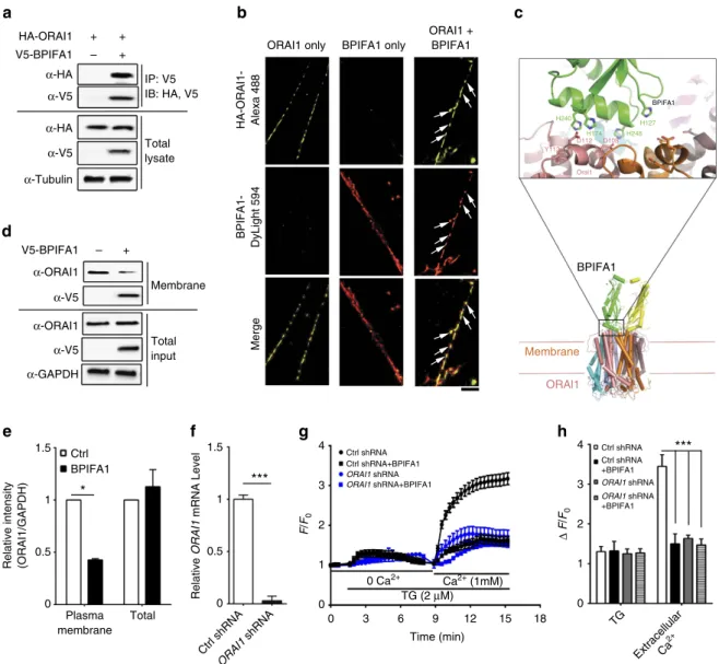
BPIFA1 binds to and internalizes Orai1
. Since Orai1
mediates SOCE and both are hyperactive in murine asthma
models where BPIFA1 is diminished
5,19, we tested whether
these proteins interact. BPIFA1 and Orai1, but not an alternate
Ca
2þchannel (transient receptor potential cation channel 3,
TRPC3), could be co-immunoprecipitated (Fig. 4a and
Supple-mentary
Fig.
6).
Using
ground
state
depletion
(GSD)
super resolution microscopy to give a resolution of 30 nm
2per
pixel, BPIFA1 and Orai1 were found to co-localize in ASMC
plasma membranes after 1 h of co-incubation (Fig. 4b).
The structures of Orai1 and BPIFA1 have been solved
9,20and
our docking model suggested that the histidines in BPIFA1’s
a6 helix, fit into the highly conserved negatively charged
regions of Orai1 (Fig. 4c). Surface biotinylation/immunoblot
and confocal microscopy demonstrated that plasma membrane
Orai1 levels decreased by
B
50% after 4 h of BPIFA1 exposure
a
Ach 0 60 (min)
Bpifa1+/+
Bpifa1–/– Bpifa1+/+
(denuded)
Bpifa1–/– (denuded)
0
Ctrl
Ach
BPIFA1
Ach+BPIFA1
Time (min) 60
c
% Gel surface area
Bpifa1 +/+
Bpifa1 +/+
(denuded) Bpifa1
–/–
Bpifa1 –/–
(denuded) 50
60 70 80 90 100
b
**
d
Ctrl Ach
BPIFA1 Ach+BPIFA1 50
75 100
%Gel surface area
**
*
Ctrl Ach BPIFA1 Ach+BPIFA1KDa
α-pMLC(S19)
α-Total MLC
α-GAPDH
20 15
20
15
37
e
Ctrl Ach
**
*
BPIFA1
Ach+BPIFA1
Relative intensity
0 1 2 3
f
Figure 2 | BPIFA1 decreases airway smooth muscle (ASM) contractility by suppressing myosin light chain (MLC) phosphorylation.Human ASMCs were grown in type I collagen matrices in 24-well plates. Gel contractions were measured as indicated over 60 min. (a) Human ASMCs were co-cultured with whole and denuded tracheal rings fromBpifa1þ/þ orBpifa1/ mice for 48 h before contraction was induced with Ach. White arrows show the location of excised tracheas on the cultures. (c) ASMCs were pretreated with BPIFA1 (10mM) or vehicle for 1 h and gel contraction was then measured±Ach (100mM). Representative images of gel contraction are shown. (b,d) Summary of data fromaandc, respectively, expressed as the % decrease in gel surface area at 60 min (alln¼3 per group). (e) Representative immunoblots probed for total and phosphorylated MLC. (f) Mean densitometry taken frome(n¼3). Data inb,dandfare mean±s.e.m. The data were analysed using one-way ANOVA followed by Tukey
c
4 Healthy_1
Healthy_2 Healthy_3
Asthmatic_1 Asthmatic_2 Asthmatic_3
0
0 3 6 9 12
Time (min)
F
/
F0
TG (2 μM) 1
2 3
1.0 1.5 2.0
Δ
F
/
F0
2.5
10–11 10–10 10–9 10–8 10–7 10–6 10–5 BPIFA1 (M)
10–4
d
e
Time (min)
F
/
F0
BPIFA1
0 3 6 9 12
4
3
2
1
0
0 Ca2+ Ca2+ (1 mM) TG (2 μM) Ctrl
h
1.0 1.5
**
2.0
Δ
F
/
F0
2.5
Ctrl
Human BPIFA1Mouse Bpifa1
Δ44BPIFA1Δα4BPIFA1Δα6BPIFA1
S18
f
F
/
F0
0.0 0.5 1.0 1.5 2.0
0 500 1,000 1,500 2,000 Time (s)
0 5 10 15 20 25 (min) Ctrl
BPIFA1
MeCh (5 μM)
g
0 5 10 15 20 25 30
Time (min) Ctrl
BPIFA1 4
3
2
1
0
Spikes per 5 min
*
* *
* * α-BPIFA1
α-BPIFA1
α-BPIFA1
α-GAPDH
HealthyAsthmaticKDa
25
25
25
37
Basolateral
media
Apical lavage
Cell lysate
a
KDa
25
25
25
37
b
α-BPIFA1
α-BPIFA1
α-BPIFA1
α-GAPDH
Ctrl IL-13
Basolateral
media
Apical lavage
Cell lysate
Absolute BPIFA1 intensity
Healthy
** **
*
Asthmatic
0 10,000 20,000 30,000 40,000
Absolute BPIFA1 intensity
0
Basolateral
media Apical lavage
Cell
Cell
lysate
Basolateral
media Apical lavage lysate
10,000 20,000 30,000
40,000 Ctrl IL-13
*** *** ***
Figure 3 | BPIFA1 from normal but not asthmatic HBECs is secreted basolaterally and blocks Ca2þ influx in ASMCs.(a) Representative immunoblots (left panel) and densitometry (right panel) showing BPIFA1 from healthy and asthmatic HBECs (n¼6 per group). (b) Representative immunoblots (left panel) and densitometry (right panel) showing BPIFA1 levels in vehicle and IL-13 exposed HBECs (n¼3 per group). (c) Mean changes in fura-2 fluorescence in ASMC with time, as an indicator of cytosolic Ca2þ. Serosal media from normal and asthmatic HBECs were co-incubated with human ASMCs for 1 h. (d) BPIFA1 inhibits TG-induced cytosolic Ca2þincreases in a dose-dependent manner. ASMCs were incubated with BPIFA1 and changes in the fura-2 emission ratio over time were recorded.DF/F0represents the average peak fluorescent intensity changes of three independent experiments. (e) Mean change in the fura-2 emission ratio over time following TG and the addition of extracellular Ca2þ. (f) Typical Ca2þoscillations in
(Fig. 4d,e). To verify that Orai1 was BPIFA1’s target,
we knocked down endogenous
ORAI1
in ASMCs by shRNA,
as
confirmed
by
both
qPCR
(Fig.
4f)
and
immuno-blot (Supplementary Figs 7a and 8a).
ORAI1
knockdown
decreased BPIFA1 binding to ASMC plasma membranes
(Supplementary Fig. 7) and the ability of BPIFA1 to inhibit
Ca
2þinflux and ASMC contrition
in vitro
were also lost
(Fig. 4g,h, Supplementary Figs 9 and 10). Additionally,
phos-phorylation of myosin light chain phosphatase (MYPT1)
at threonine 583, which was reportedly induced by Ach in
mice tracheas
21, was downregulated by BPIFA1
(Suppleme-ntary Fig. 8). Taken together, our data suggest that BPIFA1
binds to and inhibits Orai1 to block Ca
2þinflux in
ASMC, resulting in a decrease in MLC phosphorylation and
ASM contractility.
Discussion
Our
previous
studies,
and
those
conducted
elsewhere,
have focused on BPIFA1’s secretion into the lung lumen
18,22,23.
For example, BPIFA1 negatively regulates Na
þabsorption across
airway epithelia by binding extracellularly to
b-ENaC
18,24.
We note that BPIFA1’s S18 region, which inhibits ENaC,
is located N-terminally
12, while the
a6 helix, which
antago-nizes Orai1, lies close to BPIFA1’s C terminus. Thus, these
domains are in distinct regions of BPIFA1, which may support
BPIFA1
BPIFA1
c
α-HA V5-BPIFA1 HA-ORAI1
–
IP: V5 IB: HA, V5
Total lysate + + +
α-V5
α-HA
α-V5
α-Tubulin
a
α-V5 V5-BPIFA1 –
Membrane
Total input +
α-ORAI1
α-V5
α-ORAI1
α-GAPDH
d
e
Relative intensity (ORAI1/GAPDH)
0 0.5
Plasma membrane
Total 1
1.5
BPIFA1 Ctrl
*
f
0 0.5 1 1.5
Relative
ORAI1
mRNA Level
Ctrl shRNA
ORAI1
shRNA
***
b
Merge
BPIFA1 only ORAI1 only
ORAI1 + BPIFA1
HA-ORAI1- Alexa 488
BPIFA1-DyLight 594
0 3 6 9 12 15 18 Time (min)
Ctrl shRNA Ctrl shRNA+BPIFA1 ORAI1 shRNA ORAI1 shRNA+BPIFA1
0 1 2 3 4
F
/
F0
Ca2+ (1mM) 0 Ca2+
TG (2 μM)
g
h
Ctrl shRNA Ctrl shRNA +BPIFA1 ORAI1 shRNA
ORAI1 shRNA +BPIFA1
***
0 1 2 3 4
Δ
F
/
F0
TG
ExtracellularCa
2+
H240 H174
H127 H248
D108 D112 Y113
Orai1
ORAl1
Membrane
their differing functions. Chu and colleagues demonstrated that
Bpifa1
/mice
have
a
more
severe
phenotype
than
WT mice when sensitized by ovalbumin, including
eosino-philic inflammation
25. These authors hypothesized that apically
secreted BPIFA1 normally ‘mopped up’ excess bacterial
lipopolysaccharide and that after allergen exposure, reduced
BPIFA1 levels increased lipopolysaccharide, leading to an
exaggerated allergen response. However, they did not factor
basolaterally secreted BPIFA1 into their model. It is likely
therefore, that the ovalbumin-induced decrease in BPIFA1
levels also contributed to AHR via the dysregulation of
BPIFA1–Orai1 interactions. We found that BPIFA1 levels were
significantly diminished in sputum derived from asthma patients
(Fig.
1a,b)
and
that
a
similar
decrease
in
BPIFA1
levels was observed in apical secretions from both asthmatic
and IL-13-exposed HBECs (Fig. 3a,b). Perhaps surprisingly, we
demonstrated that BPIFA1 was also basolaterally secreted
from airway epithelia, and that basolateral BPIFA1 secretions
were greatly diminished in asthmatic and IL-13 exposed
HBECs (Fig. 2a,b). Mirroring these decreases, we found that
asthma
and
IL-13
downregulated
BPIFA1
mRNA
and intracellular protein levels (Fig. 2a,b and Supplementary
Fig 3). Why asthmatic HBECs continued to display reduced
BPIFA1 levels remains to be determined. They may have
a ‘memory’ of the host inflammation and/or exhibit as yet
unknown genetic variations that predispose them to reduced
BPIFA1 levels.
BPIFA1 binds extracellularly to Orai1, leading to Orai1’s
internalization,
inhibition
of
SOCE
and
a
decrease
in
ASM contraction (Figs 2 and 4), a process that is deranged in
asthmatic airways (Figs 1 and 3). Consistent with these data,
we have also discovered a hitherto unnoticed phenotype of
the
Bpifa1
/mice, that is, they exhibit spontaneous
AHR that is reversed in excised tracheas by the addition
of recombinant BPIFA1 (Fig. 1c–f). The inhibitory effect
of BPIFA1 on ASM contraction also requires an intact of
airway epithelia. Indeed, denuding tracheas from
Bpifa1
þ/þmice abolished their ability to suppress ASM contraction
in vitro
, an effect that was absent from whole or denuded
Bpifa1
/tracheas (Fig. 2a,b). Taken together, these data
strongly suggest that BPIFA1 is indeed an EDSMRF. BPIFA1
is expressed in epithelia of the trachea and bronchi, but
not the alveoli
26,27, suggesting that is able to function as an
EDSMRF in the conducting airways. However, whether other
EDSMRFs exist remains to be determined.
We focused on basolaterally secreted BPIFA1’s ability
to regulate ASM. However, the lack of apically secreted
BPIFA1 may also have consequences for airway epithelia.
Indeed, mucus is dehydrated in asthmatic airways
28and
since a lack of BPIFA1 is also predicted to increase ENaC
activity, this may indicate that mucus dehydration in asthma
patients is in part driven by BPIFA1 deficiency and abnormal
ENaC-led transepithelial Na
þand water absorption. In addition,
since mucus secretion from goblet cells is Ca
2þ-dependent
29,
increased SOCE in the absence of BPIFA1 may also contribute
to the mucus hypersecretion phenotype seen in asthma patients.
For example, our data predict that in BPIFA1’s absence, the
purinergic stimulation of goblet cells would result in a greater
cytosolic Ca
2þresponse and increased mucin secretion.
BPIFA1 notwithstanding, Th2-driven goblet cell metaplasia
helps drive the mucus hypersecretion component
30. The
transcription factors SAM-pointed domain-containing ETS-like
factor (SPDEF) and forkhead ortholog A3 (FOXA3) are
abnormally
regulated
in
asthma
leading
to
goblet
cell
metaplasia
31,32. Whether SPDEF/FOXA3 activity is involved in
reducing BPIFA1 expression in asthmatic patients remains to be
determined.
b2-agonists, which are a common asthma treatment,
can increase BPIFA1 expression
33, whereas our data suggest
that
the
short-term
b2-agonist
albuterol
only
enhances
BPIFA1
secretion
(Supplementary
Fig.
4).
Therefore,
a
better understanding of how BPIFA1 expression is modulated
by different categories of
b-agonists and other mainstream
therapies including glucocorticoids in asthma-derived airway
epithelia is needed to develop novel, targeted therapies for
treating asthma.
Although our study focused on the diminished expression
of BPIFA1 and its target Orai1 in ASM, it is worth noting
that other research groups have demonstrated the dysregulation
of other plasma membrane proteins. For instance,
calcium-sensing receptor (CaSR) is upregulated in asthmatic ASM leading
to AHR and inflammation
34. CD38 is also elevated in asthmatic
ASM and influences Ca
2þhomeostasis/ASM contractility
35.
It is unclear whether BPIFA1 interacts with these proteins.
However, replacing BPIFA1 may normalize Ca
2þhomeostasis
and reduce AHR irrespective of CaSR and CD38 activity.
Perhaps surprisingly, we found that BPIFA1 also abrogated
KCl-induced increases in cytosolic Ca
2þand ASM contractions
(Fig. 1 and Supplementary Fig. 9). We also observed that
Orai1 is involved in the KCl response and indeed, we find
that knockdown of Orai1 reduces KCl-induced increases
in cytosolic Ca
2þ(Supplementary Fig. 9). While these data
are unconventional, it has been previously been demonstrated
that elevations in extracellular KCl trigger ATP release
36,37. This
is not entirely surprising since many factors (including but not
limited
to
mechanical
and
osmotic
stress
also
induce
ATP release). Furthermore, the ATP release channel PANX1
is also stimulated by extracellular K
þ(ref. 38). Thus, while
further experimentation will be required, we propose that
KCl-induced ATP release stimulates purinergic receptors to
actives SOCE/Orai1.
Beyond asthma, altered BPIFA1 expression is associated with
several types of cancer including nasopharyngeal carcinoma
39and salivary gland cancers
40. Changes in Orai1 are also
associated with increased cancer metastasis
41. Thus, we
speculate that altered BPIFA1 expression may contribute to
abnormal Orai1-dependent
cell growth,
although further
mechanistic studies will be required to confirm or refute this
link. In conclusion, our findings that BPIFA1 is secreted
basolaterally, where it acts as an EDSMRF to modulate SOCE
and ASM contraction, are fundamentally novel advances
in understanding asthma pathogenesis that provide a direct
link between epithelial dysfunction and AHR, and for
future treatments for asthma.
Methods
baseline FEV1 value was calculated and recorded. Subjects then were administered successive increasing concentrations of saline (3, 4 and 5%) using a Devilbiss UltraNeb 99 ultrasonic nebulizer (Sunrise Medical) where the nebulizer flow rate and aerosol generation rate were adjusted to each subject’s tolerance level to allow inhalation without discomfort. Each saline inhalation period lasted 7 min and subjects inhaled saline at tidal volume. Subjects were instructed to not swallow saliva or saline during the inhalation periods but rather expectorate it into the ‘waste’ cup if there was a need. At the end of each 7 min inhalation period, subjects underwent the three-step cleansing procedure followed by chesty (not throat) cough attempts as described above for spontaneous sputum expectoration. When no further sample could be expectorated, as typically indicated by a dry sounding cough, FEV1 was assessed. If the fall in FEV1 waso10% from the pre-procedure baseline FEV1, the next highest dose of saline was administered and the next 7 min inhalation period commenced. If the fall in FEV1 was410% buto20%, the same saline concentration was administered for the next 7 min inhalation period. If the fall in FEV1 was 20% or greater, the sputum induction procedure was terminated and not restarted and the patient was given rescue therapy as needed. The collected sputum sample after each 7 min inhalation period was pooled into one specimen cup and kept on ice (4°C) during the entire induction procedure. All three saline inhalation periods were performed unless the FEV1 fell by 20% or greater, or a subject wished to terminate the procedure early. All subjects’ FEV1 had to return to within 5% of baseline before discharge.
Animals and measurement of airway resistance
.
Animals were cared for according to the guidelines, and all procedures were approved by UNC Animal Care and Use Committee.Bpifa1/knockout andBpifa1þ/þlittermate control mice were kind gifts from Dr Paul B. McCray Jr at University of Iowa, and were bred and housed in a vivarium at The University of North Carolina at Chapel Hill. Airway resistance was measured in anaesthetized 8-week-old male mice similar to previously described44. Basal resistance measurements were made every10 s for 1 min before serially challenging mice with aerosolized methacholine (Mch) at the following concentrations: 10 mg and 20 mg ml1. Mice were administered each concentration of Mch for 10 s before recording, using a flexiVent (SCIREQ) at 12.5 s intervals for 2.5 min immediately following each challenge period.
DNA constructs
.
Yellow fluorescent protein (YFP)-tagged humanORAI1and Myc-tagged TRPC3 were gifts from Dr Craig Montell (Addgene plasmid # 25902) and Dr Anjana Rao (Addgene plasmid # 19756), respectively. HA-tagged humanORAI1was a generous gift from Dr Patrick G. Hogan at La Jolla Institute for Allergy & Immunology. pcDNA3.1(þ)-V5-BPIFA1 was generously provided by Dr Colin D. Bingle at the University of Sheffield.
Cell culture and transfection
.
Human ASMCs were generous gift from Dr Raymond B. Penn at Thomas Jefferson University. Rat ASMCs were kindly provided by Dr Mohamed Trebak at Pennsylvania State University. ASMCs were maintained in Dulbecco’s Modified Eagle Medium: Nutrient Mixture F-12 (DMEM/F12; Thermo Fisher) supplemented with 10% fetal bovine serum (FBS; Sigma Aldrich) and 0.1% penicillin–streptomycin (Thermo Fisher). HEK293T cells (catalogue number: CRL-3216) were purchased from ATCC and maintained in DMEM (Thermo Fisher) in the presence of 10% FBS and 0.1% penicillin–streptomycin. HBECs were obtained from freshly excised bronchial specimens derived from normal and asthmatic subjects and were harvested as previously described under protocol #03-1396 approved by the University of North Carolina at Chapel Hill Biomedical Institutional Review Board45. Informed consent was obtained from all donors or authorizedrepresentatives of the donors. Briefly, HBECs were extracted from excess surgical pathology or autopsy specimens procured through cooperating surgeons and pathologists using protocols in accordance with relevant regulations. Airways were dissected by removing all excess connective tissue and cutting into 5–10 cm segments. Specimens were dissociated over 4–24 h, depending on the size of the tissue, in a 15 ml tube containing 9 ml tissue wash medium (Joklik Minimum Essential MediumoJMEM4supplemented gentamicino50mg ml14and amphotericino0.25mg ml14) plus 1 ml protease solution (1% Protease XIV with 0.01% DNaseo10stock4, Sigma Aldrich). HBECs were then cultured at an air–liquid interface in a modified bronchial epithelial growth medium with 5% CO2at 37°C and were used 3–4 weeks after the seeding on 12 mm T-clear inserts (Corning). Demographic information for healthy and asthmatic donors is included in Supplementary Table 2. Short hairpin RNA (shRNA) plasmid againstORAI1and scrambled control shRNA plasmid were purchased from Sigma Aldrich. Transfections of all plasmid DNA and shRNA were performed using Lipofectamine 2000 reagent (Thermo Fisher) according to the manufacturer’s instructions.
Immunoprecipitation and immunoblot analysis
.
Rabbit anti-Orai1 (1:1,000), anti-HA epitope (1:2,000) and anti-myc epitope (1:1,000) (Santa Cruz), anti-GFP (1:1,000), anti-phospho-myosin light chain (S19) (1:1,000), anti-total myosin light chain (1:1,000), anti-phospho-myosin light chain phosphatase (T853) (1:1,000),anti-phospho-myosin light chain phosphatase (T696) (1:1,000), anti-total myosin light chain (1:1,000), anti-total-myosin light chain phosphatase (1:1,000), anti-GAPDH (glyceraldehyde-3-phosphate dehydrogenase, 1:3,000) (Cell Signaling Technology); mouse anti-V5 epitope (1:2,000, Thermo Fisher); goat anti-BPIFA1 (1:2,000, R&D Systems); peroxidase-conjugated donkey anti-mouse IgG (HþL), peroxidase-conjugated donkey anti-rabbit IgG (HþL), peroxidase-conjugated donkey anti-goat IgG (HþL) (all 1:3000, Jackson ImmunoResearch) were purchased from commercial sources. To detect protein expression in total cell lysates, cells were lysed in Pierce IP lysis buffer with 25 mM Tris-HCl pH 7.4, 150 mM NaCl, 1% NP-40, 1 mM EDTA, 5% glycerol, supplemented with 1proteinase inhibitor cocktail (PIC) (Roche), followed by sodium dodecyl sulfate (SDS)–polyacrylamide electrophoresis and immunoblot. For immunopre-cipitation, cell lysates were collected at 48 h post transfection in Pierce IP lysis buffer in the presence of 1PIC. Cell lysates were pre-cleared with protein A/G agarose beads and then incubated 1mg of antibodies against HA or V5 with protein A/G agarose beads on a rotator at 4°C overnight. After three washes with IP lysis buffer, immunoprecipitated complexes were eluted in sample buffer (50 mM Tris-HCl (pH 6.8), 2% SDS, 10% glycerol, 5% (v/v)
b-mercaptoethanol (BME), 0.1% bromophenol blue) by heating the samples at 95°C for 5 min, electrophoresed through SDS–polyacrylamide gels, and subjected to immunoblot analysis. All the uncropped scans are provided in Supplementary Fig. 11.
Calcium imaging
.
Calcium imaging using fura-2 was adapted from a previously published protocol with minor modifications46. Briefly, ASMCs were loadedwith 2mM fura-2-AM (Thermo Fisher) and 24 h old serosal media from HBECs or media containing recombinant BPIFA1 at 37°C for 1 h. ASMCs were then washed with a standard Ringer’s solution (101 mM NaCl, 12 mM NaHCO3, 1.2 mM MgCl2, 1.2 mM CaCl2, 0.2 mM KCl, 24 mM HEPES, 10 mM glucose, pH 7.4) or with Ca2þ-free Ringer’s solution as indicated. Cultures were then placed in the Ringer’s solution, and images were collected with a 401.4 NA Plan Fluor oil objective on a Nikon Ti-S inverted microscope. Fura-2 fluorescence was acquired alternately at 340 and 380 nm (emission4450 nm) using LUDL filter wheels, obtained with an Orca FLASH 4.0 CMOS camera (Hammamatsu) and controlled with HCImageLive software (Hammamatsu). Cell bodies were identified as individual regions of interest (ROIs). Background subtraction was performed using a cell-free region as the background region. Signals were converted to relative changes (F/F0) whereF0was the ratio of the average fluorescent intensity (340/380) of ROIs at 0 time point. Typically, 20 cells per coverslip were recorded.DF/F0represents average peak fluorescent intensity changes of three independent experiments.
Myography of murine tracheal rings
.
Tracheas were excised from 8 weeks old maleBpifa1þ/þ andBpifa1/mice. After removing excessive connective tissue, the trachea was cut intoB4 mm rings, mounted on a DMT 620M myography apparatus (DMT) and allowed to rest in modified Ringer’s solution (119 mM NaCl, 4.7 mM KCl, 1.17 mM MgSO4, 1.18 mM KH2PO4, 2.5 mM CaCl2, 25 mM NaHCO3, 0.027 mM EDTA, 5.5 mM glucose) with continuous oxygenation (95% O2/5% CO2) at 37°C for 20 min with no applied tension. Optimal passive tension (1 mN) was then applied to the rings. After stable tension was achieved, the baseline force was then recorded. The induced contractile force of each ring was assessed by measuring contraction stimulated by 60 mM KCl or 100 mM Ach (unless indicated otherwise), respectively. Rings were bilaterally exposed to recombinant BPIFA1 or vehicle control for 1 h before Ach/KCl addition.Cell contraction assay
.
The cell contraction assay was performed using a standard commercially available kit (Cell Biolabs). Human ASMCs were harvested and re-suspended in DMEM, two parts of cells were mixed with eight parts of collagen gel lattice mixture and plated for 1 h at 37°C. After the gel solidified, 1 ml of medium was added and incubated for 48 h. Next, the gels were released from the sides of wells, and the images were taken using a ChemiDoc MP imager (Bio-Rad) at 0 and 1 h after adding indicated reagents. The changes of collagen gel surface areas were analysed using ImageJ software and normalized to the gel’s area att¼0 min.Protein purification and fluorescent labelling
.
cDNA of full length and truncated BPIFA1 was transformed in BL21-Codon Plus competent cells (Agilent Technologies), and purified as previously described24. S18 peptide wassynthesized and purified by the UNC Microprotein Sequencing and Peptide Synthesis Facility, as described previously17. Fluorescent labelling was done using DyLight 594 or DyLight 633 NHS ester (Thermo Fisher) by following the manufacturer’s instructions.
5with ice-cold PBS, cells were incubated with 5mM BPIFA1-DyLight 594 for 1 h at 4°C before imaging. Before mounting, 90mlb-Mercaptoethylamine (MEA) was added to the cavity of a depression slide to deplete oxygen. Coverslips were sealed with twinsil (Picodent). Super-resolution images were captured with a commercial Leica GSD super-resolution microscope using a 1601.43 NA objective. GSD was performed by exciting the fluorophore with 488 and 642 nm solid-state lasers. In some cases, to facilitate exit from the ground state, back pumping was applied using the 405 nm laser as per the manufacturer’s instruction.
Fluorescent BPIFA1 binding assay
.
Human ASMCs were transfected with scrambled (control) shRNA orORAI1shRNA, respectively. Seventy-two hours post transfection, cells were treated with or without BPIFA1-DyLight 633 for 1 h, then washed 5with ice-cold Ringer’s solution. ASMC-bounded BPIFA1-Dylight 633 was detected using a Leica TCS SP8 631.4 NA oil immersion objective using the Leica Application Suite X software (Leica). In addition, fluorescent intensity of ASMC-bounded BPIFA1-Dylight 633 was detected by a fluorescent plate reader (Infinite M1000, Tecan). Cells were also stained with calcein AM (Thermo Fisher) as a cell number control. Relative fluorescent intensity was calculated by normalizing fluorescent intensity at 658–526 nm.Surface biotinylation
.
Human ASMCs were treated with or without BPIFA1 for 4 h. Cells were then washed with prechilled PBSþ þ(phosphate-buffered saline supplemented with 1 mM CaCl2and 1 mM MgCl2), and then labelled with 0.5 mg ml1sulfo-NHS-biotin (Thermo Fisher) in borate buffer (85 mM NaCl,4 mM KCl, 15 mM Na2B4O7, pH 9.0) while tumbling gently for 30 min on ice. ASMCs were incubated in PBSþ þbuffer supplemented with 10% FBS for 20 min at 4°C to quench free biotin. Cells were washed again three times with chilled PBSþ þand proteins were extracted as previously described using lysis buffer (0.4% sodium deoxycholate, 1% NP-40, 50 mM EGTA, 10 mM Tris-Cl, pH 7.4) supplemented with 1PIC. Total inputs were taken from whole-cell samples representing 4% of total protein. Solubilized proteins were incubated with 100ml of neutravidin beads (Thermo Fisher) overnight while rotating at 4°C. Samples were washed three times with lysis buffer. Bead-bound proteins were then eluted and subjected to immunoblot analysis.
Statistical analysis
.
All data are presented as the mean±s.e.m. fornexperiments.Differences between means were tested for statistical significance using paired or unpairedt-tests or their non-parametric equivalent as appropriate to the experiment. Differences between groups were judged using analysis of variance. From such comparisons, differences yieldingPr0.05 were judged to be significant. GraphPad Prism software was used for statistical analysis.
Data availability
.
All raw and analysed data are available from the authors upon request.References
1. Janssen, L. J. & Killian, K. Airway smooth muscle as a target of asthma therapy: history and new directions.Respir. Res.7,123 (2006).
2. Rodger, I. W. Asthma. Airway smooth muscle.Br. Med. Bull.48,97–107 (1992).
3. Peel, S. E., Liu, B. & Hall, I. P. ORAI and store-operated calcium influx in human airway smooth muscle cells.Am. J. Respir. Cell. Mol. Biol.38,744–749 (2008).
4. Gao, Y. D.et al.Promoting effects of IL-13 on Ca2þrelease and store-operated Ca2þ entry in airway smooth muscle cells.Pulm. Pharmacol. Ther.
23,182–189 (2010).
5. Spinelli, A. M.et al.Airway smooth muscle STIM1 and Orai1 are upregulated in asthmatic mice and mediate PDGF-activated SOCE, CRAC currents, proliferation, and migration.Pflugers Arch.464,481–492 (2012). 6. Gao, Y. D., Zheng, J. W., Li, P., Cheng, M. & Yang, J. Store-operated
Ca2þentry is involved in transforming growth factor-beta1 facilitated proliferation of rat airway smooth muscle cells.J. Asthma50,439–448 ð2013Þ:
7. Hay, D. W., Muccitelli, R. M., Horstemeyer, D. L., Wilson, K. A. & Raeburn, D. Demonstration of the release of an epithelium-derived inhibitory factor from a novel preparation of guinea-pig trachea.Eur. J. Pharmacol.136,247–250 (1987).
8. Vanhoutte, P. M. Airway epithelium-derived relaxing factor: myth, reality, or naivety?Am. J. Physiol. Cell. Physiol.304,C813–C820 (2013).
9. Tarran, R. & Redinbo, M. R. Mammalian short palate lung and nasal epithelial clone 1 (SPLUNC1) in pH-dependent airway hydration.Int. J. Biochem. Cell. Biol.52,130–135 (2014).
10. Chu, H. W.et al.Function and regulation of SPLUNC1 protein in Mycoplasma infection and allergic inflammation.J. Immunol.179,3995–4002 (2007). 11. Fryer, A. D. & Jacoby, D. B. Muscarinic receptors and control of airway smooth
muscle.Am. J. Respir. Crit. Care. Med.158,S154–S160 (1998).
12. Ratz, P. H., Berg, K. M., Urban, N. H. & Miner, A. S. Regulation of smooth muscle calcium sensitivity: KCl as a calcium-sensitizing stimulus.Am. J. Physiol. Cell. Physiol.288,C769–C783 (2005).
13. Liu, Y.et al.SPLUNC1/BPIFA1 contributes to pulmonary host defense against
Klebsiella pneumoniaerespiratory infection.Am. J. Pathol.182,1519–1531 (2013).
14. Kai, T., Yoshimura, H., Jones, K. A. & Warner, D. O. Relationship between force and regulatory myosin light chain phosphorylation in airway smooth muscle.Am. J. Physiol. Lung. Cell. Mol. Physiol.279,L52–L58 ð2000Þ:
15. Koopmans, T.et al.Ca2þhandling and sensitivity in airway smooth muscle: emerging concepts for mechanistic understanding and therapeutic targeting.
Pulm. Pharmacol. Ther.29,108–120 (2014).
16. Wedel, B., Boyles, R. R., Putney, Jr. J. W. & Bird, G. S. Role of the store-operated calcium entry proteins Stim1 and Orai1 in muscarinic cholinergic receptor-stimulated calcium oscillations in human embryonic kidney cells.
J. Physiol.579,679–689 (2007).
17. Hobbs, C. A.et al.Identification of the SPLUNC1 ENaC-inhibitory domain yields novel strategies to treat sodium hyperabsorption in cystic fibrosis airway epithelial cultures.Am. J. Physiol. Lung. Cell. Mol. Physiol.305,L990–L1001 (2013).
18. Garcia-Caballero, A.et al.SPLUNC1 regulates airway surface liquid volume by protecting ENaC from proteolytic cleavage.Proc. Natl Acad. Sci. USA106, 11412–11417 (2009).
19. Yang, C.et al.Lipopolysaccharide enhances FceRI-mediated mast cell degranulation by increasing Ca2þentry through store-operated Ca2þ channels: implications for lipopolysaccharide exacerbating allergic asthma.Exp. Physiol.97,1315–1327 (2012).
20. Hou, X., Pedi, L., Diver, M. M. & Long, S. B. Crystal structure of the calcium release-activated calcium channel Orai.Science338,1308–1313 ð2012Þ:
21. Zhang, W. C.et al.Myosin light chain kinase is necessary for tonic airway smooth muscle contraction.J. Biol. Chem.285,5522–5531 (2010). 22. Di, Y. P.et al.Molecular cloning and characterization ofspurt, a human
novel gene that is retinoic acid-inducible and encodes a secretory protein specific in upper respiratory tracts.J. Biol. Chem.278,1165–1173 (2003).
23. Campos, M. A.et al.Purification and characterization of PLUNC from human tracheobronchial secretions.Am. J. Respir. Cell. Mol. Biol.30,184–192 (2004).
24. Garland, A. L.et al.Molecular basis for pH-dependent mucosal dehydration in cystic fibrosis airways.Proc. Natl Acad. Sci. USA110,15973–15978 ð2013Þ:
25. Thaikoottathil, J. V.et al.SPLUNC1 deficiency enhances airway eosinophilic inflammation in mice.Am. J. Respir. Cell. Mol. Biol.47,253–260 (2012). 26. Bingle, L. & Bingle, C. D. Distribution of human PLUNC/BPI fold-containing
(BPIF) proteins.Biochem. Soc. Trans.39,1023–1027 (2011).
27. Weston, W. M.et al.Differential display identification ofplunc, a novel gene expressed in embryonic palate, nasal epithelium, and adult lung.J. Biol. Chem.
274,13698–13703 (1999).
28. Schmitt, L.et al.Exercise reduces airway sodium ion reabsorption in cystic fibrosis but not in exercise asthma.Eur. Respir. J.37,342–348 ð2011Þ:
29. Lee, R. J. & Foskett, J. K. Ca2þsignaling and fluid secretion by secretory cells of the airway epithelium.Cell Calcium55,325–336 (2014).
30. Reber, L. L.et al.A dissociated glucocorticoid receptor modulator reduces airway hyperresponsiveness and inflammation in a mouse model of asthma.
J. Immunol.188,3478–3487 (2012).
31. Chen, G.et al.Foxa3 induces goblet cell metaplasia and inhibits innate antiviral immunity.Am. J. Respir. Crit. Care. Med.189,301–313 (2014).
32. Rajavelu, P.et al.Airway epithelial SPDEF integrates goblet cell differentiation and pulmonary Th2 inflammation.J. Clin. Invest.125,2021–2031 (2015). 33. Gross, C. A.et al.beta2-agonists promote host defense against bacterial
infection in primary human bronchial epithelial cells.BMC. Pulm. Med.10,30 (2010).
34. Yarova, P. L.et al.Calcium-sensing receptor antagonists abrogate airway hyperresponsiveness and inflammation in allergic asthma.Sci. Transl. Med.7, 284ra260 (2015).
35. Jude, J. A., Wylam, M. E., Walseth, T. F. & Kannan, M. S. Calcium signaling in airway smooth muscle.Proc. Am. Thorac. Soc.5,15–22 (2008).
36. Schock, S. C., Leblanc, D., Hakim, A. M. & Thompson, C. S. ATP release by way of connexin 36 hemichannels mediates ischemic tolerancein vitro.
Biochem. Biophys. Res. Commun.368,138–144 (2008).
37. Heinrich, A., Ando, R. D., Turi, G., Rozsa, B. & Sperlagh, B. Kþdepolarization evokes ATP, adenosine and glutamate release from glia in rat hippocampus: a microelectrode biosensor study.Br. J. Pharmacol.167,1003–1020 (2012). 38. Bao, L., Locovei, S. & Dahl, G. Pannexin membrane channels are
39. Yew, P. Y.et al.Identification of a functional variant inSPLUNC1associated with nasopharyngeal carcinoma susceptibility among Malaysian Chinese.Mol. Carcinog.51(Suppl 1): E74–E82 (2012).
40. Vargas, P. A., Speight, P. M., Bingle, C. D., Barrett, A. W. & Bingle, L. Expression of PLUNC family members in benign and malignant salivary gland tumours.Oral. Dis.14,613–619 (2008).
41. Vashisht, A., Trebak, M. & Motiani, R. K. STIM and Orai proteins as novel targets for cancer therapy. A review in the theme: cell and molecular processes in cancer metastasis.Am. J. Physiol. Cell. Physiol.309,C457–C469 (2015). 42. Alexis, N., Soukup, J., Ghio, A. & Becker, S. Sputum phagocytes from healthy
individuals are functional and activated: a flow cytometric comparison with cells in bronchoalveolar lavage and peripheral blood.Clin. Immunol.97,21–32 (2000).
43. Geiser, M.et al.Effects ofex vivogamma-tocopherol on airway macrophage function in healthy and mild allergic asthmatics.J. Innate Immun.5,613–624 (2013).
44. Li, M.et al.Deficiency of RAMP1 attenuates antigen-induced airway hyperresponsiveness in mice.PLoS ONE9,e102356 (2014).
45. Fulcher, M. L. & Randell, S. H. Human nasal and tracheo-bronchial respiratory epithelial cell culture.Methods Mol. Biol.945,109–121 (2013).
46. Sheridan, J. T., Gilmore, R. C., Watson, M. J., Archer, C. B. & Tarran, R. 17beta-Estradiol inhibits phosphorylation of stromal interaction molecule 1 (STIM1) protein: implication for store-operated calcium entry and chronic lung diseases.J. Biol. Chem.288,33509–33518 (2013).
Acknowledgements
We thank the UNC CF Center Histology, Tissue and Molecular Cores for providing cells and reagents, and slides. We thank Dr Patrick G. Hogan from the La Jolla Institute for Allergy & Immunology for the generous gift of HA-ORAI1 DNA construct and Dr Colin D. Bingle at the University of Sheffield for sharing the pcDNA3.1(þ)-V5-BPIFA1 plasmid. This work was funded by the American Asthma Foundation and NIH Grants HL108927, ES025198, DK065988.
Author contributions
All authors edited and reviewed the manuscript. T.W. designed and performed experi-ments, analysed the data, and wrote the manuscript. J.H., P.J.M., R.C.F., W.G.W. and M.S.L. performed experiments and analysed the data. Y.P.D. providedBpifa1mice strains. N.E.A. provided human sputum samples. M.R.R. and S.L.T. designed experi-ments. R.T. designed experiments and wrote the manuscript.
Additional information
Supplementary Informationaccompanies this paper at http://www.nature.com/ naturecommunications
Competing financial interests: R.T. has equity in Spyryx Biosciences Inc. The remaining authors declare no competing financial interests.
Reprints and permissioninformation is available online at http://npg.nature.com/ reprintsandpermissions/
How to cite this article: Wu, T.et al.Identification of BPIFA1/SPLUNC1 as an epithelium-derived smooth muscle relaxing factor.Nat. Commun.8, 14118 doi: 10.1038/ncomms14118 (2017).
Publisher’s note: Springer Nature remains neutral with regard to jurisdictional claims in published maps and institutional affiliations.
This work is licensed under a Creative Commons Attribution 4.0 International License. The images or other third party material in this article are included in the article’s Creative Commons license, unless indicated otherwise in the credit line; if the material is not included under the Creative Commons license, users will need to obtain permission from the license holder to reproduce the material. To view a copy of this license, visit http://creativecommons.org/licenses/by/4.0/