REGULATION OF MEMBRANE TRAFFIC BY INTRINSIC AND EXTRINSIC MECHNISMS
Chao-Wei Hung
A dissertation submitted to the faculty of the University of North Carolina at Chapel Hill in partial fulfillment of the requirements for the degree of Doctor of Philosophy in the department
of Biology
Chapel Hill 2016
Approved by: Mara Duncan
ABSTRACT
Chao-Wei Hung: Regulation of Membrane Traffic by Intrinsic and Extrinsic Mechanisms (Under the direction of Mara Duncan)
Vesicular membrane traffic regulates many biological activities, such as cell growth, motility and the maintenance of the cell shape. One of the most important types of traffic is mediated by clathrin coated vesicles. To form a vesicle, clathrin must be recruited to the membranes where cargoes are located by clathrin adaptors. Although it is well established that clathrin adaptors are essential for the recruitment of clathrin and the initiation of traffic, not much is known about the mechanisms by which cells regulate the recruitment and activities of adaptors. Furthermore, the role of clathrin adaptors in traffic beyond simple clathrin recruitment remains largely
ACKNOWLEDGEMENTS
TABLE OF CONTENTS
LIST OF TABLES ... ix
LIST OF FIGURES ... x
GENERAL INTRODUCTION ... 1
CHAPTEE 1. SECTION 1:ADPATOR AND MEMBRANE TRAFFIC ... 1
SECTION 1.1: BIOLOGICAL SIGNIFICANCE OF MEMBRANE TRAFFIC... 2
SECTION 1.2: INTRODUCTION TO CLATHRIN ADAPTORS ... 3
AP1 ... 3
GGA ... 3
EPSINS ... 3
ROLES OF ADAPTORS IN TRAFFIC ... 4
SECTION 2:REGULATION OF MEMBRANE TRAFFIC BY ENERGY METABOLISM ... 5
SECTION 2.1:ENERGY AVAILABILITY AND TRAFFIC REGULATION DURING ENERGY STRESS ... 5
SECTION 2.2: ROLES OF ATPASES IN TRAFFIC ... 7
ROLES OF FLIPPASES IN TRAFFIC ... 7
ROLES OF V-ATPASES IN TRAFFIC ... 8
ROLES OF PHOSPHATIDYLINOSITOL 4-KINASEV-ATPASES IN TRAFFIC ... 9
FIGURES ... 10
REFERENCES ... 12
INTRODUCTION ... 16
RESULTS ... 18
BINDING OF GGA2 TO CLATHRIN REQUIRES ENT5 ... 18
THE CLATHRIN BOX MOTIF IN GGA2 IS THE MAJOR CONTRIBUTOR TO ITS INTERACTION WITH CLATHRIN ... 19
THE CLATHRIN BOX MOTIF IN GGA2 REGULATES ENT5 CLATHRIN INTERACTION ... 20
AN AUTO-REGULATORY MOTIF IN GGA2 MODULATES BINDING TO BOTH CLATHRIN AND ENT5 ... 21
MUTATION OF AN AUTO-REGULATORY MOTIF IN GGA2 ALTERS ENT5 RECRUITMENT IN VIVO ... 23
DISCUSSION ... 25
MATERIALS AND METHODS ... 28
YEAST STRAINS ... 28
PLASMIDS ... 28
ANTIBODIES ... 28
PROTEIN PURIFICATION ... 29
YEAST CELL LYSATES AND BINDING STUDIES ... 30
MICROSCOPY ... 31
GEL FILTRATION ... 33
ACKNOWLEDGEMENTS ... 33
FIGURES AND TABLES ... 34
REFERENCES ... 44
CLATHRIN BINDING BY THE ADAPTOR ENT5 CHAPTER 3. PROMOTESLATE STAGES OF CLATHRIN COAT MATURATION ... 47
INTRODUCTION ... 47
RESULTS ... 49
MULTIPLE DOMAINS OF ENT5 CONTRIBUTE TO ITS
FUNCTION AND LOCALIZATION ... 51
ENT5 CLATHRIN BINDING PROMOTES THE TURN-OVER OF GGA2 STRUCTURES IN VIVO ... 54
DISCUSSION ... 56
MATERIALS AND METHODS ... 59
YEAST STRAINS AND PLASMIDS ... 59
QUANTITATIVE CALCOFLUOR WHITE SENSITIVITY ASSAY ... 59
MEDIAS, ANTIBODIES AND REAGENTS... 60
WHOLE-CELL YEAST EXTRACTS ... 60
IMMUNOPRECIPITATION... 61
MICROSCOPY ... 61
ACKNOWLEDGEMENTS ... 62
FIGURES AND TABLES ... 63
REFERENCES ... 76
ENERGY METABOLISM REGULATES CLATHRIN CHAPTER 4. ADAPTORS AT THE TRANS-GOLGI NETWORK AND ENDOSOME... 80
INTRODUCTION ... 80
RESULTS ... 82
AMPK-ACTIVATED KINASE (AMPK) IS REQUIRED FOR ADAPTOR LOCALIZATION IN PROLONGED STARVATION ... 82
GLUCOSE REPRESSION IS REQUIRED FOR ADAPTOR RELOCALIZATION IN ACUTE STARVATION ... 85
ENERGY METABOLISM IS REQUIRED FOR ADAPTOR LOCALIZATION ... 86
ADDITION OF ATP IS SUFFICIENT FOR ADAPTOR LOCALIZATION ... 89
UPSTREAM REGULATORS OF ADAPTORS ARE DIFFERENTIALLY REGULATED BY ENERGY METABOLISM ... 93
DISCUSSION ... 97
MATERIALS AND METHODS ... 101
YEAST STRAINS AND PLASMIDS ... 101
MEDIAS, ANTIBODIES AND REAGENTS... 102
IMMUNOBLOTTING ... 102
GROWTH CONDITIONS AND CARBON STARVATION ... 103
LIVE MICROSCOPY, IMAGE PROCESSING, AND QUANTIFICAITON ... 104
ATP MEASUREMENTS ... 104
PERMEABILIZED CELL ASSAYS ... 105
ACKNOWLEDGEMENTS ... 106
FIGURES AND TABLES ... 107
REFERENCES ... 125
GENERAL DISCUSSION ... 130
LIST OF TABLES
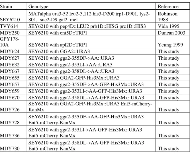
Table 2.1: Yeast stains used in this study ... 34
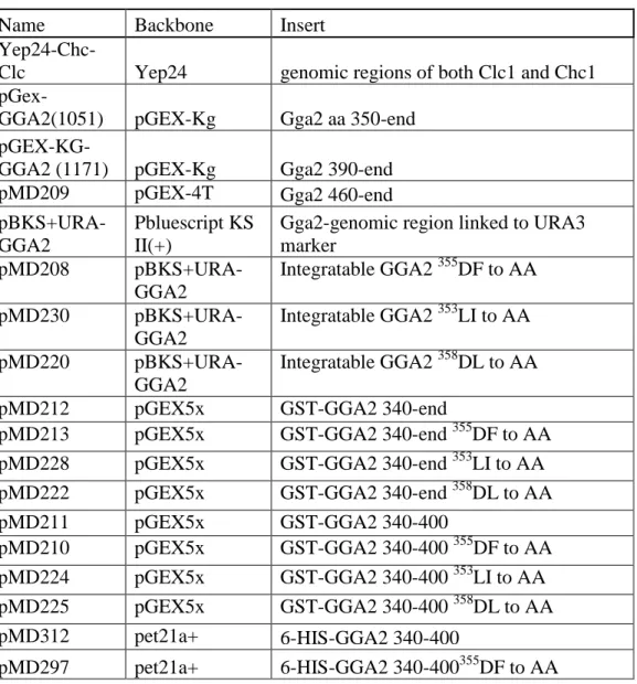
Table 2.2: Plasmids used in this study ... 35
Table 3.1: Stains and plamisds used in this study ... 63
LIST OF FIGURES
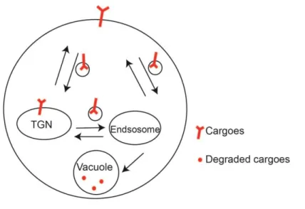
Figure 1.1: The TGN and endosomes are major proteins sorting stations... 10
Figure 1.2: Clathrin adaptors are important for traffic ... 11
Figure 2.1. Gga2 interaction with clathrin is altered in adaptor deletion strains.. ... 36
Figure 2.2. Mutations of the clathrin box region alter interaction of Gga2 with clathrin in vitro. ... 37
Figure 2.3. Mutations of the clathrin box region alter interaction of Gga2 with clathrin in cell lysates... 38
Figure 2.4. The clathrin box region of Gga2 interacts with the Gga2 -ear. ... 39
Figure 2.5. CAB mutations alter co-localization of Ent5 and Gga2.. ... 40
Figure 2.6. Clathrin box region mutations alters the recruitment differential between Gga2 and Ent5 ... 42
Figure 2.7. Schematic of possible adaptor recruitment models. ... 43
Figure 3.1: Loss of Ent5 disrupts traffic at the TGN and/or endosomes.. ... 66
Figure 3.2: Loss of ENT5 prolongs coat life-span.. ... 68
Figure 3.3: Schematic of Ent5 and mutations generated.. ... 69
Figure 3.4: Clathrin is required for maximal interaction of Ent5 with Gga2 and localization of Ent5 to membranes. ... 71
Figure 3.5: Mutation of clathrin-binding or adaptor-binding domains affect the lifespan of Ent5-Gga2 structures and the timing of the recruitment of Ent5.. ... 73
Figure 3.6: Model of coat assembly ... 74
Supplemental Figure 3.1: Mutations in Ent5 do not affect the steady state protein expression levels of Ent5.. ... 75
Figure 4.1. Snf1 is required for localization of the TGN-endosomal clathrin adaptors Gga2 and Ent5 during prolonged glucose starvation.. ... 109
Figure 4.2. Inhibition of cellular energy production induces adaptor redistribution... ... 111
Figure 4.4. Cellular ATP concentration decrease significantly during glucose
starvation and is regulated by glucose repression pathways.. ... 113
Figure 4.5. Cellular ATP concentrations correlate with the association of adaptors to membranes... ... 114
Figure 4.6. ATP is sufficient to recruit adaptors in permeabilized cells... 115
Figure 4.7. ATP and GTP both contribute to adaptor recruitment.. ... 117
Figure 4.8. Glucose starvation alters the localization of Arf1, Pik1 and PI4p... ... 118
Figure 4.9 Arf1, Pik1 and PI4p show differential responses to exogenous nucleotides in permeabilized cells. ... 120
Figure 4.10. Arf1, Pik1 and Sac1 modulate adaptor localization during glucosestarvation... 121
Figure 4.11 Model of energy dependent steps in adaptor recruitment.... ... 122
Supplemental Figure 4.1. Adaptors partially co-localize with Sec7 during prolonged glucose starvation.... ... 123
CHAPTER 1. GENERAL INTRODUCTION Section 1: Adaptor and Membrane Traffic
1.1 Biological Significance of Membrane Traffic
Proteins regulate many essential biological activities. However, they must be delivered to the right place at the right time in order to exercise their activities properly. One major mechanism by which cells regulate the localization and the distribution of proteins is through the process of vesicular membrane traffic. By delivering proteins to different cellular compartments, vesicular membrane traffic plays an important role in cell polarity, proliferation and motility (Reviewed in [1]). Disruption of membrane traffic may contribute to diseases such as heart attack, Alzheimer’s disease, type II diabetes and cancer [2-5].
The trans-Golgi network (TGN) and endosomes represent a major intracellular sorting station that mediates the traffic of plasma membrane proteins (Figure 1.1). For proteins to be sorted to the plasma membrane or targeted for secretion, proteins must first be delivered to TGN. Conversely, proteins or pathogens that are targeted for internalization must first be directed to endosomes. In conclusion, traffic at the TGN and endosomes allows proteins to gain entry into the cells or be targeted to other cellular compartments. Disruption of TGN and endosomal traffic can affect the distribution of proteins distribution and secretion and preventing cell from responding to extracellular signals.
delivery to plasma membranes. Glut4, the main glucose transporter in mammalian cells, is one of many plasma membrane proteins regulated by TGN-endosomal traffic. Under non-stimulating conditions, Glut4 is packaged into Glut4 storage vesicles (GSV) and resides in the TGN and endosomes. Glut4 is only targeted to plasma membranes when cells are stimulated by insulin [6-8]. One of the major mechanisms by which Glut4 is trapped in its intracellular compartments is a futile cycle between GSV and the TGN and/or recycling endosomes. This futile cycle prevents Glut4 translocation to plasma membranes prior insulin stimulation (Reviewed in [8]). Defects in the regulation of Glut4 transporter translocation is linked to the onset of type II insulin resistance diabetes (Reviewed in [8]). Other important plasma membrane proteins, such as Aqp2, a water channel in renal epithelium, and Gap1, a general amino acid permease found in yeast, are also cycled through the TGN-endosomes constantly. Their targeting to the plasma membrane is triggered only when stimuli are present [9, 10]. By regulating the distribution of plasma membrane proteins, the TGN and endosomes play an essential role in cellular response to environmental signals.
TGN-endosomal traffic is regulated by clathrin. The next section will discuss clathrin mediated traffic (CMT) and clathrin adaptors in detail.
1.2 Introduction to Clathrin Adaptors
In Saccharomyces cerevisiae, there are three major types of clathrin adaptors that function at the TGN and endosomes: the heterotetrameric AP-1 complex; the monomeric Golgi localized Gga proteins; and epsin family proteins, which can be divided into ANTH family protein and ENTH family protein.
AP-1:
The AP-1 heterotetrameric complex consists of γ, β1, μ1 and σ1 subunits. The globular core domain includes μ1 and σ1. The N terminal of γ and β1 are also part of the core domain. The appendages of AP-1 include the C terminals of γ, β1 subunits which are consisted of unstructured hinge domains that are followed by small globular domains known as γ and β1 ear. The core domain is important for AP-1 attachment to the membrane of Golgi, while the appendage regions are indispensable for AP-1 to recruit clathrin and other factors that regulate trafficking [14-16].
Gga:
The Gga proteins (Golgi-localized, ear containing, ADP-ribosylation factor binding), consist of a VHS, a GAT, a hinge and an ear domain, which is a homolog to the γ ear found in AP-1. Gga proteins play roles in mannose 6-phosphate trafficking, cargo recognition, ubiquitin binding, and interaction with other clathrin adaptors [16-20]. Humans have three Golgi proteins, while yeasts only have Gga1 and 2; the expression of Gga2 is substantially higher than that of Gga1 [21].
Epsins:
Ent3 and Ent5 contain multiple gamma ear binding motifs [23]. Unlike Ent5, Ent3 does not possess canonical clathrin binding motifs; however, evidence has shown that Ent3 is capable of binding to clathrin directly in vivo, although with less efficiency than Ent5 [25]. Both Ent5 and Ent3 have been shown to interact with cargo [25, 26].
Roles of Adaptors in Traffic
Clathrin adaptors form a complicated interaction network. Many clathrin adaptors share structural similarities and functional redundancies. However, evidences suggest that many adaptors have specific functions or mediate a specific route of traffic. For example, Ent3 plays a role in the Gga2 mediated trafficking pathway, while Ent5 and AP-1 mediate an alternative trafficking pathway [27]. Additionally, adaptors seem to be directional specific: AP-1 can function in traffic from TGN to endosomes and retrieving cargo from endosomes to TGN, while Gga proteins strictly regulate TGN to endosome traffic [12, 27].
In the Chapter 1 and Chapter 2 of this dissertation, I investigated the role of interactions between clathrin, Ent5 and Gga2 on traffic regulation. I discovered a dual function, autoregulatory motif on Gga2 that mediates the interaction between Gga2, Ent5 and clathrin. This novel autoregulation plays an important role in the temporal and spatial regulation of CMT. I also explored the role Ent5 in traffic. We revealed that, unlike many clathrin adaptors, Ent5 is not involved in the initiation or recruitment of clathrin. Rather, it plays an important role during the late stage and promotes the maturation of clathrin traffic structures.
Section 2: Regulation of Membrane Traffic by Energy Metabolism
2.1 Energy Availability and Traffic Regulation during Energy Stress
while their differentiated counterparts prefer energy production by pathways that involve mitochondria [28]. Metabolic remolding not only occurs in response to developmental signals, but is also triggered in response to changes in nutrients. When glucose is present, yeasts, Saccharomyces cerevisiae, produce ATP via glycolysis and undergo exponential growth. Additionally, mitochondrial activity is actively inhibited by cell signaling response to glucose. During glucose starvation, yeast will activate mitochondrial activities and use oxidative phosphorylation to metabolize alternative carbon sources [29].
Glucose is the preferred carbon source for many eukaryotes from yeasts to human. Additionally, glucose is the regulatory molecule to many cellular pathways. For example, translation is inhibited during glucose starvation [30]. In many cell types and organisms, glucose starvation represents a major survival challenge that cells must deal immediately. Knowing the immediate responses and the long term cellular adaptations to glucose starvation can provide insight into understanding mechanisms by which cells survive under stressful conditions.
modifications [47]. Understanding the metabolic regulation on traffic at the TGN and endosomes will improve the yield of pharmaceutical proteins.
Our lab has discovered evidence that suggests that metabolism affects cellular behavior through remodeling traffic. We have demonstrated that traffic remodeling is one of the immediate cellular response to starvation [31]. Upon an acute glucose starvation, many adaptors that function on the TGN and endosomes are mislocalized from the membranes. However, adaptors relocalize to membranes during a prolonged starvation [31]. This observation suggests that membrane traffic is highly sensitive to change of metabolism.
starvation. I then developed a permeabilized cell assay to demonstrate that ATP and GTP work synergistically to recruit clathrin adaptors. This chapter shows that energy and metabolism play an important role in the regulation of membrane traffic. We proposed a multi-steps energy dependent mechanism that regulates traffic in response to the availability of energy.
Our lab demonstrated that clathrin adaptors localization is highly sensitive to cellular ATP levels. However, clathrin adaptors do not have ATP binding domains or ATPase activities. Thus, ATP must control adaptor recruitment by activating other traffic factors. The following section will describe several ATPases that are known to play essential role in traffic
2.2 Roles of ATPases in Traffic
Roles of Flippases in Traffic
One possible ATPase that is important for energy dependent regulation of traffic is flippases. Flippases are essential for catalyzing the trans-bilayer movement of lipid molecules. This movement, also known as “flip-flop”, of lipid molecules between two leaflets maintains the asymmetry of lipid bilayers. The energy independent flip-flop catalyzed by scramblases, which is a flippases that functions in ER, is a very fast process with half times range from seconds to minute [32]. At the plasma membrane and the Golgi, flip-flop is catalyzed by energy dependent flippases, a subfamily of P-type ATPases [33].
Yeast flippases are known to regulate many trafficking pathway that include: endocytosis, TGN to endosomes, and TGN to ER. Yeast have five flippases: DNF1 and DNF2 works in the endocytic pathway, DNF3 and DRS2 works in the TGN trafficking pathway, while NEO1 is
involved in Golgi to ER trafficking [33-35]. The deletion of DRS2 is synthetically lethal with
arf1Δ [36]. This result is significant because Arf1 is a small GTPase that functions at the TGN,
membrane curvature [14, 18, 37]. Additionally, drs2Δ delays the processing of CPY, which is a
yeast protease that has to travel from the TGN to endosomes and then the vacuoles, in order to be
fully processed [35]. Finally, Drs2 has a role in the formation of secretory clathrin coated
vesicles, and the formation of these coated vesicles is dependent on the hydrolysis of ATP by
Drs2 [38]. Thus, flippases are lively involved in the regulation of membrane traffic by energy
dependent mechanisms.
Roles of V-ATPase in Traffic
Another ATPase that is important for the traffic is V-ATPase. The V-ATPase is a major proton pump that is comprised of a peripherally associated V1 domain and a membrane associated V0 domain. Hydrolysis of ATP by V-ATPases enables H+ to be transported to the lumen of endosomes against the gradient. This process induces the luminal acidification, which appears to be important for the trafficking from endosomes to vacuoles. However, it is unclear whether the acidification is required for early to late endosome traffic or late endosome to vacuole trafficking [39-40]. The luminal acidification is required for the recruitment of βCOP,
which is a coat protein that is involved in the formation of vesicles that mediate transport from
early to late endosomes [41]. Finally, the V-ATPase has been shown to interact directly with
ARNO, a GEF for ARF6 [42], which is a small GTPase that mediate endocytosis, further
highlighting its role in trafficking. By using ATP and maintaining the pH gradient across
membranes, V-ATPases likely plays a role in traffic regulation in response to the availability of
energy.
Roles of Phosphatidylinositol 4-Kinase in Traffic
mediate the trafficking is by synthesizing phosphoinositides (PIs) on the surface of membranes. PIs are important membrane phospholipids that are required for organelles functions from yeast to humans. There are seven kinds of PIs that differ by their sites of phosphorylation [43]. Many proteins are targeted to a specific membrane by recognizing the PIs that are present on the membrane. Essentially, PIs define the identity of membranes, allowing the right proteins to be targeted to the right compartment.
FIGURES
REFERENCES
1. Mosesson, Y., G.B. Mills, and Y. Yarden, Derailed endocytosis: an emerging feature of cancer. Nat Rev Cancer, 2008. 8(11): p. 835-50.
2. Shepherd, P.R. and B.B. Kahn, Glucose transporters and insulin action--implications for insulin resistance and diabetes mellitus. N Engl J Med, 1999. 341(4): p. 248-57.
3. Suzuki, T., et al., Trafficking of Alzheimer's disease-related membrane proteins and its participation in disease pathogenesis. J Biochem, 2006. 139(6): p. 949-55.
4. Kimura, T., et al., Involvement of the Ras-Ras-activated Rab5 guanine nucleotide exchange factor RIN2-Rab5 pathway in the hepatocyte growth factor-induced endocytosis of E-cadherin. J Biol Chem, 2006. 281(15): p. 10598-609.
5. Karlsson, H.K., et al., Kinetics of GLUT4 trafficking in rat and human skeletal muscle. Diabetes, 2009. 58(4): p. 847-54.
6. Holman, G.D., L. Lo Leggio, and S.W. Cushman, Insulin-stimulated GLUT4 glucose transporter recycling. A problem in membrane protein subcellular trafficking through multiple pools. J Biol Chem, 1994. 269(26): p. 17516-24.
7. Suzuki, K. and T. Kono, Evidence that insulin causes translocation of glucose transport activity to the plasma membrane from an intracellular storage site. Proc Natl Acad Sci U S A, 1980. 77(5): p. 2542-5.
8. Leto, D. and A.R. Saltiel, Regulation of glucose transport by insulin: traffic control of GLUT4. Nat Rev Mol Cell Biol. 13(6): p. 383-96.
9. Roberg, K.J., N. Rowley, and C.A. Kaiser, Physiological regulation of membrane protein sorting late in the secretory pathway of Saccharomyces cerevisiae. J Cell Biol, 1997. 137(7): p. 1469-82.
10. Gustafson, C.E., et al., Recycling of AQP2 occurs through a temperature- and bafilomycin-sensitive trans-Golgi-associated compartment. Am J Physiol Renal Physiol, 2000. 278(2): p. F317-26.
11. Brodsky, F.M., et al., Biological basket weaving: formation and function of clathrin-coated vesicles. Annu Rev Cell Dev Biol, 2001. 17: p. 517-68.
12. Robinson, M.S., Adaptable adaptors for coated vesicles. Trends Cell Biol, 2004. 14(4): p. 167-74.
13. Boettner, D.R., R.J. Chi, and S.K. Lemmon, Lessons from yeast for clathrin-mediated endocytosis. Nat Cell Biol. 14(1): p. 2-10.
15. Mills, I.G., et al., EpsinR: an AP1/clathrin interacting protein involved in vesicle trafficking. J Cell Biol, 2003. 160(2): p. 213-22.
16. Shiba, T., et al., Structural basis for recognition of acidic-cluster dileucine sequence by GGA1. Nature, 2002. 415(6874): p. 937-41.
17. Zhdankina, O., et al., Yeast GGA proteins interact with GTP-bound Arf and facilitate transport through the Golgi. Yeast, 2001. 18(1): p. 1-18.
18. Scott, P.M., et al., GGA proteins bind ubiquitin to facilitate sorting at the trans-Golgi network. Nat Cell Biol, 2004. 6(3): p. 252-9.
19. Collins, B.M., P.J. Watson, and D.J. Owen, The structure of the GGA1-GAT domain reveals the molecular basis for ARF binding and membrane association of GGAs. Dev Cell, 2003. 4(3): p. 321-32.
20. Lui, W.W., et al., Binding partners for the COOH-terminal appendage domains of the GGAs and gamma-adaptin. Mol Biol Cell, 2003. 14(6): p. 2385-98.
21. Ghaemmaghami, S., et al., Global analysis of protein expression in yeast. Nature, 2003. 425(6959): p. 737-41.
22. Wendland, B., K.E. Steece, and S.D. Emr, Yeast epsins contain an essential N-terminal ENTH domain, bind clathrin and are required for endocytosis. EMBO J, 1999. 18(16): p. 4383-93.
23. Duncan, M.C., G. Costaguta, and G.S. Payne, Yeast epsin-related proteins required for Golgi-endosome traffic define a gamma-adaptin ear-binding motif. Nat Cell Biol, 2003. 5(1): p. 77-81.
24. Duncan, M.C. and G.S. Payne, ENTH/ANTH domains expand to the Golgi. Trends Cell Biol, 2003. 13(5): p. 211-5.
25. Copic, A., T.L. Starr, and R. Schekman, Ent3p and Ent5p exhibit cargo-specific functions in trafficking proteins between the trans-Golgi network and the endosomes in yeast. Mol Biol Cell, 2007. 18(5): p. 1803-15.
26. Chidambaram, S., et al., Specific interaction between SNAREs and epsin N-terminal homology (ENTH) domains of epsin-related proteins in trans-Golgi network to endosome transport. J Biol Chem, 2004. 279(6): p. 4175-9.
27. Costaguta, G., et al., Distinct roles for TGN/endosome epsin-like adaptors Ent3p and Ent5p. Mol Biol Cell, 2006. 17(9): p. 3907-20.
29. Castelli, L.M., et al., Glucose depletion inhibits translation initiation via eIF4A loss and subsequent 48S preinitiation complex accumulation, while the pentose phosphate pathway is coordinately up-regulated. Mol Biol Cell. 22(18): p. 3379-93.
30. Ashe, M.P., S.K. De Long, and A.B. Sachs, Glucose depletion rapidly inhibits translation initiation in yeast. Mol Biol Cell, 2000. 11(3): p. 833-48.
31. Aoh, Q.L., L.M. Graves, and M.C. Duncan, Glucose regulates clathrin adaptors at the trans-Golgi network and endosomes. Mol Biol Cell. 22(19): p. 3671-83.
32. Buton, X., et al., Ultrafast glycerophospholipid-selective transbilayer motion mediated by a protein in the endoplasmic reticulum membrane. J Biol Chem, 1996. 271(12): p. 6651-7.
33. Pomorski, T., et al., Drs2p-related P-type ATPases Dnf1p and Dnf2p are required for phospholipid translocation across the yeast plasma membrane and serve a role in endocytosis. Mol Biol Cell, 2003. 14(3): p. 1240-54.
34. Graham, T.R., Flippases and vesicle-mediated protein transport. Trends Cell Biol, 2004. 14(12): p. 670-7.
35. Hua, Z., P. Fatheddin, and T.R. Graham, An essential subfamily of Drs2p-related P-type ATPases is required for protein trafficking between Golgi complex and endosomal/vacuolar system. Mol Biol Cell, 2002. 13(9): p. 3162-77.
36. Chen, C.Y., et al., Role for Drs2p, a P-type ATPase and potential aminophospholipid translocase, in yeast late Golgi function. J Cell Biol, 1999. 147(6): p. 1223-36.
37. Beck, M.A., Influenza and Obesity: Will Vaccines and Antivirals Protect? J Infect Dis. 38. Gall, W.E., et al., Drs2p-dependent formation of exocytic clathrin-coated vesicles in vivo.
Curr Biol, 2002. 12(18): p. 1623-7.
39. Clague, M.J., et al., Vacuolar ATPase activity is required for endosomal carrier vesicle formation. J Biol Chem, 1994. 269(1): p. 21-4.
40. van Weert, A.W., et al., Transport from late endosomes to lysosomes, but not sorting of integral membrane proteins in endosomes, depends on the vacuolar proton pump. J Cell Biol, 1995. 130(4): p. 821-34.
41. Aniento, F., et al., An endosomal beta COP is involved in the pH-dependent formation of transport vesicles destined for late endosomes. J Cell Biol, 1996. 133(1): p. 29-41.
42. Hurtado-Lorenzo, A., et al., V-ATPase interacts with ARNO and Arf6 in early endosomes and regulates the protein degradative pathway. Nat Cell Biol, 2006. 8(2): p. 124-36.
44. Strahl, T. and J. Thorner, Synthesis and function of membrane phosphoinositides in budding yeast, Saccharomyces cerevisiae. Biochim Biophys Acta, 2007. 1771(3): p. 353-404.
45. Daboussi, L., G. Costaguta, and G.S. Payne, Phosphoinositide-mediated clathrin adaptor progression at the trans-Golgi network. Nat Cell Biol. 14(3): p. 239-48.
46. Walsh, G. and R. Jefferis, Post-translational modifications in the context of therapeutic proteins. Nat Biotechnol, 2006. 24(10): p. 1241-52.
CHAPTER 2. ADAPTOR AUTOREGULATION PROMOTES COORDINATED BINDING WITH CLATHRIN COATS11
Introduction
Clathrin acts in many processes, including endocytosis and transport between the trans-Golgi Network (TGN) and endosomes. (Reviewed in[11]). Clathrin is a multimeric protein complex that forms a polyhedral lattice on the outer surface of some transport vesicles [46]. Formation of the clathrin lattices in vivo is controlled by a class of proteins called adaptors. In both endocytosis and transport between TGN and endosomes, adaptors perform three general functions: membrane association, clathrin binding and assembly, and cargo collection. The adaptors that function in endocytosis and at the TGN are encoded by different genes; however the adaptors play the key role in directing formation of clathrin-coated vesicles in both of the processes.
Clathrin dependent traffic requires seemingly redundant adaptors [25, 26, 47]. At least three different classes of adaptors act at the TGN and endosomes, the heterotetrameric AP-1 complex, the GGA proteins Gga1 and Gga2 in yeast and Gga1, Gga2 and Gga3 in mammals and the epsin-like proteins Ent3 and Ent5 in yeast, and EpsinR in mammals [15, 23, 48-54]. Multiple adaptors can be recruited to the same transport event ([55] and reviewed in [56]). The involvement of multiple adaptors is a hallmark of clathrin dependent traffic, although the functional significance of this complexity remains unclear. Initially it was proposed that adaptor
1
redundancy allows the transport of distinct subsets of cargo. Recent work in endocytosis also suggests that potentially redundant adaptors play different mechanical roles within a single endocytic event [57].
In addition to shared function, different adaptors utilize the same molecular interfaces to interact with clathrin. One common mechanism relies on an interaction with the globular domain at the N-terminus of clathrin known as the terminal domain (reviewed in [58]). Many adaptors contain “clathrin box motifs” which bind to a pocket in the terminal domain of clathrin. Clathrin box sequences are characterized by the consensus L φX φ [D/E](where X is any amino acid and φ is a hydrophobic amino acid). Some adaptors contain an additional motif with the consensus sequence [D/E]LL. This DLL-type motif was first characterized in the endocytic adaptors, where, in multiple copies, it mediates the interaction of adaptors with the clathrin triskelion and clathrin cages [59]. Importantly, DLL-type motifs interact with cages formed of triskelia that lack the terminal domain, suggesting that the DLL-type motifs can bind to a different region of clathrin than the clathrin box[60]. The presence of different adaptors, each capable of binding to clathrin at the same sites, adds to the complexity of clathrin coats.
The physical interactions between TGN/endosome adaptors raises the possibility that adaptors all act coincidently, however genetic results in yeast suggest they function in separate events. In particular, analysis of deletion mutants provides evidence that, although Ent5 cooperates with both AP-1 and Gga proteins in vivo, AP-1 and Gga proteins seem to play roles in distinct transport functions [27].Only in the case of Ent3 has an adaptor-adaptor interaction been shown to influence function. In this case, Ent3 requires Gga proteins for recruitment to clathrin rich structures. Thus, although evolutionary conservation implies that adaptor-adaptor interactions are important in TGN/endosome traffic in yeast, in most cases the functional significance of such interactions has not been addressed.
In the present study, we describe a previously unrecognized auto-regulatory sequence within yeast Gga2 that controls interaction with Ent5. This sequence encodes overlapping motifs that can bind to clathrin or to the Gga2 -ear. We find that in vivo the auto-regulatory sequence modulates interactions with both clathrin and Ent5 and is important for establishing a temporal delay between recruitment of Gga2 and Ent5 to clathrin-rich structures. These findings reveal a highly specialized mechanism that regulates the location/timing of the interaction of Gga2 with Ent5.
Results
Binding of Gga2 to clathrin requires Ent5
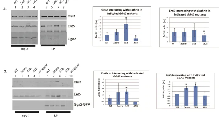
clathrin box motifs in different adaptors. In addition, Gga2 and AP-1 contain multiple DLL-type clathrin interaction surfaces that do not bind to the terminal domain. To determine whether one adaptor influences clathrin binding to other adaptors, we performed clathrin immunoprecipitations from non-denatured lysates of different deletion mutants.
In wild-type cells, both Gga2 and Ent5 co-immunoprecipitated with native clathrin (Figure 2.2.1a). In cells lacking Ent5, substantially less Gga2 immunoprecipitated with clathrin (Figure 2.2.1a & b). In contrast, deletion of the gamma subunit of AP-1 enhanced the amount of Gga2 associated with clathrin. We observed a minor effect of GGA2 deletion on the interaction of Ent5 with clathrin in some immunoprecipitation reactions. However in other reactions, deletion of GGA2 did not produce an effect, suggesting that the effect of Gga2 on Ent5 binding to clathrin is at most minor (Figure 2.2.1a). These results suggest that maximal interaction of Gga2 with clathrin requires Ent5 and also that AP-1 may compete with Gga2 for clathrin. Due to the strong influence of other adaptors on Gga2 binding, we investigated clathrin binding of Gga2 in more detail.
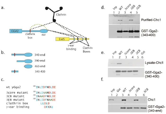
The clathrin box motif in Gga2 is the major contributor to its interaction with clathrin
whereas the ∆EB mutations reduced but did not eliminate clathrin binding (Figure 2.2c). These results confirm previous studies demonstrating that the clathrin box residues are required for direct interactions with clathrin [67]. Furthermore, these results demonstrate residues just after the clathrin box are not required for clathrin binding but that these residues contribute to clathrin affinity in the context of a complex cytosol.
We next investigated the effect of the mutations in the context of a larger Gga2 C-terminal fragment that contains the hinge and ear of Gga2. The Δcore and ΔCB mutations prevented interaction with clathrin while the ΔEB mutation had no effect on clathrin interaction. Thus in cell lysates, only the canonical clathrin box residues are required for clathrin binding (Figure 2.2d). Within the hinge and ear fragment are three DLL-type motifs in addition to the clathrin box motif. The inability of this fragment to bind clathrin when the clathrin box motif is non-functional confirms that stable interaction between Gga2 and clathrin requires the clathrin box.
The clathrin box motif in Gga2 regulates Ent5-clathrin interaction
that sequence elements in the clathrin box region of Gga2 influence clathrin interaction with both Ent5 and Gga2.
We also investigated the effects of the mutations on Gga2 interactions detected by co-immunoprecipitations of GFP-tagged alleles of Gga2. Similar to the results from the clathrin immunoprecipitations, gga2-ΔCB-GFP exhibited a severe reduction in interaction with clathrin, however Ent5 binding to Gga2 was not significantly changed (Figure 2.3b lane 9). Also similar to the clathrin immunoprecipitations, the ΔEB mutation augmented interaction of Gga2p with both clathrin and Ent5 (Figure 2.3b lane 8). gga2-Δcore-GFP displayed a possible increase in clathrin binding and a clear elevation in Ent5 binding. Together our results provide evidence for a network of pairwise interactions between Gga2, Ent5 and clathrin that can influence assembly of the adaptors with clathrin. In particular, the evidence for increased binding of Ent5 and clathrin to ΔEB and Ent5 to Δcore suggest that the clathrin box region is involved in an inhibitory interaction.
An auto-regulatory motif in Gga2 modulates binding to both clathrin and Ent5
In light of these findings, we reviewed residues in and around the clathrin box. The canonical clathrin box motif partially overlaps with a sequence similar to the characterized -ear
binding motif fro Figure 2.2a). The residues targeted
in the ΔCB mutation are specific for the clathrin binding motif, the ΔEB residues are specific for the -ear binding motif, and the ∆core residues are shared by the two motifs. Importantly, because both binding motifs share the central residues, ϒ-ear interaction could prevent binding to clathrin and vice versa.
Gga2 ear fragment and a 6x-histidine fragment of the Gga2 hinge containing the clathrin box motif. Upon crosslinking, a slow migrating species was detected by antibodies against Gga2 (Figure 2.4a lane 8). This species was not observed in reactions containing only the Gga2 ear fragment, only the Gga2 hinge fragment, or in reactions lacking the cross-linker. Furthermore, when reactions were performed with a hinge fragment encoding the Δcore mutation, the level of the specific crosslinked product was substantially reduced compared to wild-type reactions (Figure 2.4a lane 10). These results provide evidence that the clathrin box region of Gga2 also encodes a sequence that can interact with the Gga2 -ear domain.
Next, we tested whether the clathrin box region in Gga2 mediates an intramolecular interaction. Gga2 fragments encompassing the hinge and ear with or without the Δcore mutation were subjected to gel-filtration followed by quasi-elastic and multi-angle-light scattering. Both fragments eluted as a single peak, however the mutant form eluted earlier than the wild-type indicating a slightly larger size (Figure 2.4b). The molecular mass of both the wild-type and mutant fragments were determined to be that of a monomer by light scattering. These results are consistent with a model in which the core residues mediate an intramolecular interaction that compacts the structure of the hinge-ear fragment. Without this intramolecular interaction, the ∆core protein assumes a more extended form.
encompassing both the clathrin box and -ear binding motif as the clathrin and adaptor binding motif (CAB).
Mutation of an auto-regulatory motif in Gga2 alters Ent5 recruitment in vivo
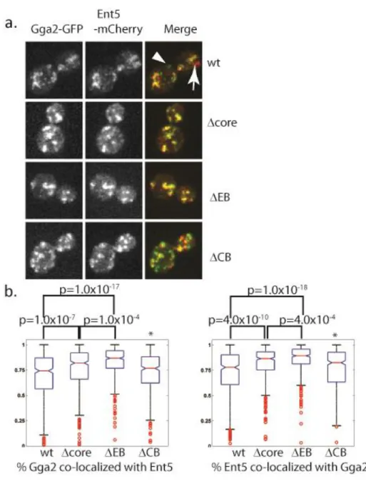
We tagged Gga2 alleles with S65T-GFP and Ent5 with mCherry at the endogenous loci to assess the function of the Gga2 CAB in cells. Ent5 and Gga2 co-localize on a subset of structures in wild-type cells (Figure 2.5a). Because both Ent5 and Gga2 structures are highly motile within the cell, we fixed cells and analyzed the area of co-localization in >100 individual cells for each genotype to obtain a quantitative measure of the effect of mutations on co-localization (Figure 2.5b). In the ΔCB mutant, in which Gga2 shows a reduced interaction with both clathrin and Ent5, co-localization of Ent5 and Gga2 is unaffected. In contrast, in the Δcore mutant, in which the CAB does not bind to the -ear or to clathrin and Ent5 binding is enhanced, there was a statistically significant increase in the co-localization of Ent5 with Gga2. Furthermore, in the ΔEB mutant, in which the CAB retains some ability to bind to clathrin, and interactions with both clathrin and Ent5 are elevated, the extent of colocalization between Ent5 and Gga2 was even greater. Indeed, very few structures could be identified that did not contain both Ent5 and Gga2. Thus when Gga2 has enhanced binding to Ent5, the two proteins almost always localize. These results suggest that the auto-regulatory motif of Gga2 limits co-localization of Ent5 and Gga2 in vivo, consistent with the inhibitory role detected by biochemical assays.
To better understand the effects of Gga2 autoregulation on Ent5 and Gga2 localization in vivo, we monitored adaptor recruitment dynamics in live cells. Similar to previous reports, Gga2
acquired significant Ent5 fluorescence. In contrast, very few events were observed where structures rich in Ent5 but lacking Gga2 then acquired Gga2. In wild-type cells, Ent5 and Gga2 co-localized at some point in approximately 60% of events. Events were observed where Gga2 disappeared without ever recruiting Ent5 and vice versa. To describe the relationship between Ent5 and Gga2 in live cells, we determined the time differential between recruitment of Gga2 and Ent5. In events where Gga2 was recruited first the value is positive, where Gga2 was recruited second the value is negative. Gga2 rich structures that did not recruit Ent5 were omitted from this analysis and made up 23% of all events. In wild-type cells the median recruitment differential is 8 seconds with a broad standard deviation (Figure 2.6 b).
Discussion
Our study reveals a complex interplay of interactions between Ent5, Gga2 and clathrin. In particular, we have identified a novel bifunctional motif in Gga2p, the CAB, which consists of overlapping sequences for clathrin and -ear binding. The evidence supports formation of an intramolecular interaction between CAB and the Gga2 -ear that inhibits binding of Gga2p to clathrin and Ent5. Based on these results we propose that CAB acts as an autoinhibitory element to restrict Gga2 interaction with Ent5 and impose a temporal delay in recruitment of Ent5 relative to Gga2 during formation of clathrin-coated vesicles.
Most, but not all, of the effects of CAB mutations can be explained by the finding that the motif binds to the -ear of Gga2. The two CAB mutations that prevent interaction with the ϒ-ear, ∆EB and ∆core, enhance binding of Gga2 to Ent5. Furthermore, the ΔEB mutation, which leaves the clathrin box intact, also stimulates binding to clathrin. Binding of CAB to the -ear domain would occlude both the CAB clathrin box and the DFxx binding pocket in the -ear. The ΔEB mutation prevents interaction between the CAB and -ear which allows clathrin unrestricted access to the clathrin box motif in CAB and frees the -ear to interact with Ent5, thereby enhancing the interaction of Gga2 with both proteins. In a similar fashion, the ∆core mutation releases the -ear for Ent5 binding but clathrin binding would not be enhanced because of the change in the clathrin box. It is not evident why this mutant retains clathrin binding; perhaps the enhanced binding to Ent5 allows an increase in indirect binding to clathrin that compensates for the loss of direct binding or supplements the weak interactions provided by the DLL-type interaction surfaces on Gga2.
inactivates the clathrin box, accounting for the significant decrease in clathrin binding. Because the ΔCB retains residues required for -ear binding, interaction of the CAB with the -ear occludes interaction with Ent5 thus reducing interaction with Ent5 as well. However, the effects of the ΔCB mutation on the interactions of Ent5 with clathrin and Gga2 are not as easily explained by a simple three protein interaction scheme. This suggests that a more complicated network involving clathrin, Ent5 and Gga2 may contribute to the core interactions defined in our studies.
small structures suggests that Ent5 and Gga2 act together in some instances in vivo. Indeed a functional role of Ent5 in Gga2 mediated traffic was previously indicated by genetic analysis [27]. Based on these findings and the results presented here, the role of CAB-mediated autoregulation is likely to be two-fold: first, to prevent premature interaction between Gga2 and Ent5 in the cytoplasm prior to assembly into clathrin coats; second, to limit levels of Ent5 recruited to Gga2-enriched clathrin coats at the TGN so that most Ent5 is free to assemble independently into later-forming AP-1-containing coats.
In this model, the intramolecular interaction between Gga2-CAB and Gga2 -ear domain limits Gga2 interaction with Ent5 outside of the context of assembling coats. Once Gga2 associates with the membrane to initiate coat formation, clathrin can be recruited through interactions with DLL motifs and limited interaction with the CAB clathrin box. In this nascent coat, higher concentrations of clathrin can compete with the -ear for binding to the CAB. Binding of clathrin to the clathrin box would release the -ear from the CAB, allowing Ent5 to be recruited to Gga2 structures through cooperative binding to the Gga2 -ear and clathrin. Ent5 recruitment would stabilize the coat assembly, both by binding directly to clathrin and by promoting the “open” Gga2 conformation with exposed clathrin box (Figure 2.7b). Supporting this view, deletion of Ent5 reduced co-immuoprecipitation of Gga2 with clathrin. In addition to the interactions described here it is also possible that interactions with other proteins and post-translational modifications contribute to the auto-regulatory process.
of the delay between Gga2 and both AP-1 and Ent5 recruitment resulted in subtle alpha factor maturations defects, providing evidence that the temporal regulation of assembly is important for optimal function of the TGN[68]. We therefore speculate that the function of Gga2 autoregulation of Ent5 recruitment is most important for TGN function in the complex natural environment of yeast growing in the wild.
Materials and Methods
Yeast strains
Yeast strains are listed in Table 2.2.1[69, 70]. Replacement of the genomic alleles used a full gene replacement strategy, in which a full gene deletion was replaced by a DNA fragment excised from a plasmid carrying the desired allele. Clones were then screened by PCR followed by restriction digestion to confirm integration of the desired alleles.
Fluorescent tags were added using a PCR-based strategy with either the pFA6a-S65TGFP-HIS3Mx plasmid or pKS390 (pFA6a-mCherry-KanMx) as described previously [71, 72].
Plasmids
Plasmids used in this study are described in Table 2.2. Point mutations were generated with the Quik-change site directed mutagenesis kit (Stratagene) according to manufacturer’s instructions.
Antibodies
Affigel-10 (Biorad) cross-linked to purified GST to deplete GST-signal. Depleted serum was bound to 2mls of Affigel-10 cross-linked to purified GST-tagged full-length protein and eluted from the matrix with glycine according to manufacturer’s instructions.
Protein purification
Protein expression was induced in BL21-DE2 pLysS (Promega) in mid-log phase at 30°C in LB-Amp (ISC bioexpress) for four hours with 0.1mM IPTG. Cells were pelleted and resuspended in minimal water and frozen. Pellets were quick thawed in 1x PBS with 1:100 protease inhibitor cocktail (Sigma) and sonicated in 0.5 sec pulses for 1 min on ice. Lysates were incubated with 1% Triton-X100 for 30 min at 4°C and pelleted at 12krpm for 20min in a ss34 rotor at 4°C. For GST-tagged proteins, supernatants were incubated with glutathione sepharose (GE Life sciences) for 1 hour at 4°C, washed with PBS and eluted with HSE (100mM Tris pH9, 200mM NaCl, 5mM DTT, 20mM reduced glutathione), or cleaved in place with Factor X (New England Biolabs) in 50mM Tris pH 7.5, 150mM NaCl, 1mM CaCl. Factor X was removed with p-aminobenzamidine agarose (MP Biomedicals). For 6-His tagged proteins, supernatants were incubated with Talon Resin (Clonetech) and eluted with 150mM imidazole in PBS final pH 7.0. For light scattering experiments, cleaved proteins were further purified by passage over tandem Hightrap Q and SP (GE Life-Sciences) 1ml columns. Buffer exchange was performed by several rounds of concentration in Ultracel 10 concentration device (Millipore) at room temperature or several rounds of dialysis with Spectra/Por dialysis tubing MWCO 3500 (Spectrumlabs).
thawed with equal volume to weight in 2x Triskelia buffer (100mM TRis pH 7.5, 100mM NaCl, 2mM EDTA with protease inhibitors). High speed supernatants were generated at 4C in a Ti 70 at 60Kprm for 30 min. 20% weight to volume ammonium sulfate was added and high speed pellets were generated by centrifugation at 60Krpm for 15 min in a Ti70 rotor. Pellets were resuspended in 1X Triskelia buffer (50mM Tris pH 7.5, 50mM NaCl, 1mM EDTA) and dialyzed against two buffer changes of Triskelia buffer. Medium speed supernatants were generated by spinning at 13Krpm for 10 minutes and were fractionated on a Superose 12 column pre-equilibrated with 10mM KPO4 pH 7.5 1mM EDTA. Clathrin fractions were identified by SDS-PAGE analysis followed by coomassie blue staining and found to be free of adaptors (Ent5, Ent3, Gga2, AP-2 and AP-1) by immuno-blotting.
Yeast cell lysates and binding studies
Cell lysates for GST-tagged protein interaction studies were generated by culturing TVY614 in yeast rich media (10g/L yeast extract, 20 g/L peptone, 2 g/L glucose, 20mg/L Uracil, Adenine sulfate and L-tryptophan) followed by liquid nitrogen blending as described for clathrin purification. Powder was thawed with an equal volume to weight in Buffer A (100mM MES pH 6.5, 0.5mM MgCl2, 1mM EGTA). The lysate was clarified by centrifugation at 13 Krpm for 10min and resulting supernanants were used for binding studies. For binding studies, GST-tagged proteins were prebound to glutathione sepharose in 1ml of PBS for 1 hour at room temperature, washed twice with Buffer A and resuspended in lysate. Lysate binding was allowed to proceed for 1 hour at 4°C and beads were washed twice with Buffer A and once with Triskelia buffer. Beads were resuspended in HSE and incubated for 20 minutes at room temp. Supernatants were taken as the bound fraction.
9.4, 2% glucose and 5mM DTT. Cells were then resuspended in digestion buffer (Yeast rich media without additional amino acids or glucose and 0.5% glucose, 10mM Tris, 1.2M Sorbitol and 120 U lyticase) and gently agitated for 30 min at 30°C. Cells were washed in 1.2M sorbitol and lysed with the addition of Buffer A with protease inhibitors followed by glass bead lysis and addition of 1% Triton X-100. Lysates were clarified by centrifugation at 13Krpm for 10 min. Lystates were incubated overnight with 100 uls 20% protein A-agarose slurry and 3 ul of clc1 antibody or 0.5ul of GFP antisera. Lysates were washed three times with Buffer A and bound proteins were eluted in SDS-sample buffer.
For crosslinking studies, reactions were performed at room temperature in Buffer B (100mM HEPES, 50mM NaCl, pH 7.5)[15]. GST-Gga2 460-end was added to 3.78µM, wild-type 6-His hinge fragments to 55.42µM ΔEB fragment to 57.64µM and DTSSP (Thermo Scientific) was added to 1mM. After 30 min, crosslinking was quenched by the addition of 37mM final concentration of Tris pH 7.5. 14% of each reaction was loaded for immuno-blot analysis and for coomassie staining.
Microscopy
For fixed images, cells grown to mid-log phase were fixed by rapid mixing with equal volume of fixative (100mM KPO4 pH 6.5, 2mM MgCl2, 8% formaldehyde) and incubating for 1h in the dark. Cells were washed twice with PBS. Acid-washed 22x22mm coverslips were treated with 5 mg/ml Concanavalin A and allowed to dry. Six microliters of fixed cells were spotted onto a treated coverslip, placed on a clean glass slide and immediately sealed with VALAP (equal parts Vaseline, Lanolin and paraffin).
the GFP and mCherry channels (1200 ms integration per image for each mCherry image and 400 ms integration per EGFP image in that order) were set so that relatively high and similar signal to noise ratio was maintained for both channels.
cells, although analysis of small-budded cells obtained similar results.
For live cell imaging, the fixation and PBS washing steps were omitted. A single central plane was imaged with a 2 second interval. Analysis was performed in ImageJ, first the threshold command was applied to all images uniformly to generate a mask. Structures smaller than 2 squared pixels or that persisted less than five frames were omitted from analysis. Using the threshold masked Gga2 images, non-overlapping Gga2 structures were identified. The first frame in which a structure was visible was considered T=0. Preceding and subsequent frames from the Ent5 masked images were analyzed to identify if and when Ent5 showed above threshold fluorescence within the defined Gga2 mask. The data reported in the figure are from >70 individual events from at least 10 cells.
Gel filtration and light scattering
Purified Gga2 fragments were separated in Buffer A on a WTC-030s5 column (Wyatt) and subjected to multi-angle light scattering and quasi-elastic light scattering on a DAWN EOS light scattering instrument (Wyatt) interfaced to an Akta FPLC (GE life Sciences). Weighted molar mass was calculated with Astra software. Data shown is intensity from detector at position 11.
FIGURES AND TABLES
Table 2.1. Yeast strains used in this study.
Strain Genotype Reference
SEY6210
MATalpha ura3-52 leu2-3,112 his3-D200 trp1-D901, lys2-801, suc2-D9 gal2 mel
Robinson 1988 TVY614 SEY6210 with pep4D::LEU2 prb1D::HISG prc1D::HIS3 Vida 1995
MDY250 SEY6210 with ent5D::TRP1 Duncan 2003
GPY178-10A SEY6210 with apl2D::TRP1 Yeung 1999
MDY624 SEY6210 with GGA2::URA3 This study
MDY627 SEY6210 with gga2-355DF->AA::URA3 This study
MDY632 SEY6210 with gga2-353LI->AA::URA3 This study
MDY667 SEY6210 with gga2-358DL->AA::URA3 This study
MDY655 SEY6210 with GGA2-GFP-His3Mx::URA3 This study
MDY657 SEY6210 with gga2-355DF->AA-GFP-His3Mx::URA3 This study MDY659 SEY6210 with gga2-353LI->AA-GFP-His3Mx::URA3 This study MDY670 SEY6210 with gga2-358DL->AA-GFP-His3Mx::URA3 This study MDY726
SEY6210 with GGA2-GFP-His3Mx::URA3
Ent5-mCherry-KanMx This study
MDY728
SEY6210 with gga2-355DF->AA-GFP-His3Mx::URA3
Ent5-mCherry-KanMx This study
MDY736
SEY6210 with gga2-353LI->AA-GFP-His3Mx::URA3
Ent5-mCherry-KanMx This study
MDY730
SEY6210 with gga2-358DL->AA-GFP-His3Mx::URA3
Ent5-mCherry-KanMx This study
Table 2.2. Plasmids used in this study
Name Backbone Insert
Yep24-Chc-Clc Yep24 genomic regions of both Clc1 and Chc1
pGex-GGA2(1051) pGEX-Kg Gga2 aa 350-end
pGEX-KG-GGA2 (1171) pGEX-Kg Gga2 390-end
pMD209 pGEX-4T Gga2 460-end
pBKS+URA-GGA2
Pbluescript KS II(+)
Gga2-genomic region linked to URA3 marker
pMD208
pBKS+URA-GGA2
Integratable GGA2 355DF to AA
pMD230
pBKS+URA-GGA2
Integratable GGA2 353LI to AA
pMD220
pBKS+URA-GGA2
Integratable GGA2 358DL to AA
pMD212 pGEX5x GST-GGA2 340-end
pMD213 pGEX5x GST-GGA2 340-end 355DF to AA pMD228 pGEX5x GST-GGA2 340-end 353LI to AA pMD222 pGEX5x GST-GGA2 340-end 358DL to AA
pMD211 pGEX5x GST-GGA2 340-400
pMD210 pGEX5x GST-GGA2 340-400 355DF to AA pMD224 pGEX5x GST-GGA2 340-400 353LI to AA pMD225 pGEX5x GST-GGA2 340-400 358DL to AA
pMD312 pet21a+ 6-HIS-GGA2 340-400
Figure 2.2. Mutations of the clathrin box region alter interaction of Gga2 with clathrin in vitro. a. (Top) Schematic of Gga2 and interacting partners. Black dotted lines indicate
Figure 2.4. The clathrin box region of Gga2 interacts with the Gga2 -ear. a. The clathrin
Figure 2.7. Schematic of possible adaptor recruitment models. A. Different types of
REFERENCES
1. Brodsky, F.M., et al., Biological basket weaving: formation and function of clathrin-coated vesicles. Annu Rev Cell Dev Biol, 2001. 17: p. 517-68.
2. Woodward, M.P. and T.F. Roth, Coated vesicles: characterization, selective dissociation, and reassembly. Proc Natl Acad Sci U S A, 1978. 75(9): p. 4394-8.
3. Copic, A., T.L. Starr, and R. Schekman, Ent3p and Ent5p exhibit cargo-specific functions in trafficking proteins between the trans-Golgi network and the endosomes in yeast. Mol Biol Cell, 2007. 18(5): p. 1803-15.
4. Chidambaram, S., et al., Specific interaction between SNAREs and epsin N-terminal homology (ENTH) domains of epsin-related proteins in trans-Golgi network to endosome transport. J Biol Chem, 2004. 279(6): p. 4175-9.
5. Black, M.W. and H.R. Pelham, A selective transport route from Golgi to late endosomes that requires the yeast GGA proteins. J Cell Biol, 2000. 151(3): p. 587-600.
6. Dell'Angelica, E.C., et al., GGAs: a family of ADP ribosylation factor-binding proteins related to adaptors and associated with the Golgi complex. J Cell Biol, 2000. 149(1): p. 81-94.
7. Wasiak, S., et al., Enthoprotin: a novel clathrin-associated protein identified through subcellular proteomics. J Cell Biol, 2002. 158(5): p. 855-62.
8. Kalthoff, C., et al., Clint: a novel clathrin-binding ENTH-domain protein at the Golgi. Mol Biol Cell, 2002. 13(11): p. 4060-73.
9. Bonifacino, J.S., The GGA proteins: adaptors on the move. Nat Rev Mol Cell Biol, 2004. 5(1): p. 23-32.
10. Panek, H.R., et al., Suppressors of YCK-encoded yeast casein kinase 1 deficiency define the four subunits of a novel clathrin AP-like complex. EMBO J, 1997. 16(14): p. 4194-204.
11. Rad, M.R., et al., Saccharomyces cerevisiae Apl2p, a homologue of the mammalian clathrin AP beta subunit, plays a role in clathrin-dependent Golgi functions. J Cell Sci, 1995. 108 ( Pt 4): p. 1605-15.
12. Duncan, M.C., G. Costaguta, and G.S. Payne, Yeast epsin-related proteins required for Golgi-endosome traffic define a gamma-adaptin ear-binding motif. Nat Cell Biol, 2003. 5(1): p. 77-81.
13. Hirst, J., et al., EpsinR: an ENTH domain-containing protein that interacts with AP-1. Mol Biol Cell, 2003. 14(2): p. 625-41.
15. Brady, R.J., Y. Wen, and T.J. O'Halloran, The ENTH and C-terminal domains of Dictyostelium epsin cooperate to regulate the dynamic interaction with clathrin-coated pits. J Cell Sci, 2008. 121(Pt 20): p. 3433-44.
16. Boettner, D.R., R.J. Chi, and S.K. Lemmon, Lessons from yeast for clathrin-mediated endocytosis. Nat Cell Biol, 2011. 14(1): p. 2-10.
17. Schmid, E.M., et al., Role of the AP2 beta-Appendage Hub in Recruiting Partners for Clathrin-Coated Vesicle Assembly. PLoS Biol, 2006. 4(9).
18. Owen, D.J., B.M. Collins, and P.R. Evans, Adaptors for clathrin coats: structure and function. Annu Rev Cell Dev Biol, 2004. 20: p. 153-91.
19. Morgan, J.R., et al., A conserved clathrin assembly motif essential for synaptic vesicle endocytosis. J Neurosci, 2000. 20(23): p. 8667-76.
20. Scheele, U., et al., Molecular and functional characterization of clathrin- and AP-2-binding determinants within a disordered domain of auxilin. J Biol Chem, 2003. 278(28): p. 25357-68.
21. Boman, A.L., et al., A family of ADP-ribosylation factor effectors that can alter membrane transport through the trans-Golgi. Mol Biol Cell, 2000. 11(4): p. 1241-55.
22. Hirst, J., et al., A family of proteins with gamma-adaptin and VHS domains that facilitate trafficking between the trans-Golgi network and the vacuole/lysosome. J Cell Biol, 2000. 149(1): p. 67-80.
23. Costaguta, G., et al., Yeast Gga coat proteins function with clathrin in Golgi to endosome transport. Mol Biol Cell, 2001. 12(6): p. 1885-96.
24. Mattera, R., et al., Definition of the consensus motif recognized by gamma-adaptin ear domains. J Biol Chem, 2004. 279(9): p. 8018-28.
25. Doray, B., et al., Cooperation of GGAs and AP-1 in Packaging MPRs at the Trans-Golgi Network. Science, 2002. 297(5587): p. 1700-1703.
26. Bai, H., B. Doray, and S. Kornfeld, GGA1 Interacts with the Adaptor Protein AP-1 through a WNSF Sequence in Its Hinge Region. J. Biol. Chem., 2004. 279(17): p. 17411-17417.
27. Costaguta, G., et al., Distinct roles for TGN/endosome epsin-like adaptors Ent3p and Ent5p. Mol Biol Cell, 2006. 17(9): p. 3907-20.
28. Vida, T.A. and S.D. Emr, A new vital stain for visualizing vacuolar membrane dynamics and endocytosis in yeast. J Cell Biol, 1995. 128(5): p. 779-92.
30. Snaith, H.A., I. Samejima, and K.E. Sawin, Multistep and multimode cortical anchoring of tea1p at cell tips in fission yeast. EMBO J, 2005. 24(21): p. 3690-9.
31. Longtine, M.S., et al., Additional modules for versatile and economical PCR-based gene deletion and modification in Saccharomyces cerevisiae. Yeast, 1998. 14(10): p. 953-61.
32. Yeung, B.G., H.L. Phan, and G.S. Payne, Adaptor complex-independent clathrin function in yeast. Mol Biol Cell, 1999. 10(11): p. 3643-59.
33. Lemmon, S., V.P. Lemmon, and E.W. Jones, Characterization of yeast clathrin and anticlathrin heavy-chain monoclonal antibodies. J Cell Biochem, 1988. 36(4): p. 329-40.
34. Phan, H.L., et al., The Saccharomyces cerevisiae APS1 gene encodes a homolog of the small subunit of the mammalian clathrin AP-1 complex: evidence for functional interaction with clathrin at the Golgi complex. Embo J, 1994. 13(7): p. 1706-17.
35. Maddox, P.S., et al., Spinning disk confocal microscope system for rapid high-resolution, multimode, fluorescence speckle microscopy and green fluorescent protein imaging in living cells. Methods Enzymol, 2003. 360: p. 597-617.
36. Mullins, C. and J.S. Bonifacino, Structural requirements for function of yeast GGAs in vacuolar protein sorting, alpha-factor maturation, and interactions with clathrin. Mol Cell Biol, 2001. 21(23): p. 7981-94.
CHAPTER 3. CLATHRIN BINDING BY THE ADAPTOR ENT5 PROMOTES LATE STAGES OF CLATHRIN COAT MATURATION2
Introduction
Clathrin dependent traffic is a central facet of all eukaryotic cell biology. It mediates traffic at multiple locations including endocytic traffic that originates at the plasma membrane and endosomal traffic that originates at trans-Golgi Network (TGN) or at the endosomes (reviewed in [11]). It regulates nearly every aspect of cellular behavior through effects on the localization of transmembrane, extracellular and organellar proteins (reviewed in [77]). To perform these many different functions, the clathrin coat must bind to many different protein cargo and package these into transport carriers. This cargo selection is performed by clathrin adaptors.
There are more than a dozen different clathrin adaptors encoded in the genomes of most eukaryotes (reviewed in [58]). Each clathrin adaptor acts as a complex interaction hub. In addition to binding to transmembrane cargo, most bind phospholipids, small GTPases or other membrane associated proteins that confer specificity to adaptor recruitment. Adaptors also directly interact with clathrin, the major structural component of the clathrin coat. This interaction links transmembrane cargo to the forming transport carrier. This function is the minimum definition of a clathrin adaptor. However, many adaptors perform additional
2
mechanistic roles in the coat such as bending membranes or stimulating clathrin polymerization [59, 78-83]. However, it is unclear whether all adaptors perform such central mechanistic roles or whether some act solely as linkers between cargo and clathrin. Determining which adaptors perform mechanistic roles and what those roles are is important to understanding the regulation of clathrin function in vivo.
Elucidating the roles of clathrin adaptors at the TGN, early and late endosome has been particularly challenging due, in part, to difficulties in imaging individual transport events, and because cells can adapt to disruption in clathrin dependent traffic by upregulating other pathways [84]. In the yeast Saccharomyces cerevisiae, five clathrin adaptors are known to function at the TGN and endosomes: the heteromeric AP-1 complex; the Golgi localized gamma-adaptins, which are encoded by the paralogs GGA1 and GGA2; Ent3, a protein belonging to the ENTH-A sub-family of epsins; and Ent5, a protein belonging to the ENTH-D sub-family of epsins [23, 27, 48, 53, 61, 62, 85]. Genetic studies suggest that AP-1 and Gga proteins act in distinct pathways [61]. Ent3 appears to act exclusively with Ggas [27, 86]. In contrast, the role of Ent5 has been unclear. Deletion of Ent5 causes only minor defects in traffic, suggesting its role may be minor or cargo specific [27]. However, it localizes to every Gga2 or AP-1 structure in vivo, and is required for maximal Gga2 interaction with clathrin [86, 87]. These data suggest that it has a more central role.
important for Ent5’s function but not the turn-over of Gga2 containing structures. Together these results suggest that clathrin binding by Ent5 plays a key mechanistic role in the maturation of Gga2 containing transport carriers, whereas adaptor binding by Ent5 does not.
Results
Ent5 provides a central function in endosomal clathrin dependent traffic
Ent5 alters the maximal recruitment of Gga2 or clathrin heavy chain (Chc1). To do this, we monitored the intensity of Clc1-GFP and Gga2-GFP expressed from their endogenous loci in wild type and cells lacking Ent5. Surprisingly, the intensity of Clc1-GFP was unaffected by deletion of ENT5, suggesting that Ent5 is not required for the maximal recruitment of clathrin (Fig. 3.1D). Similarly, Gga2-GFP was recruited to punctate structures in the absence of Ent5. However, the intensity of Gga2 structures was increased by 1.4 fold in cells lacking Ent5. These results suggest that Ent5 is not required for the association of clathrin and Gga2 with membranes.
unlikely to be caused by reduced movement of the structures into or out of the plane of focus because the mean speed of movement of structures determined from mean square displacement measurements were indistinguishable from wild-type (data not shown). These results suggest that loss of Ent5 causes a defect in coat formation after clathrin and Gga2 recruitment but before coat disassembly.
Multiple domains of Ent5 contribute to its function and localization
The extension in lifespan of Gga2 and Chc1 structures in cells lacking Ent5 suggests that Ent5 acts as more than a cargo linker. To better understand the role of Ent5 in ECT, we investigated the importance of different Ent5 domains and motifs in Ent5 function. To do this, we mutated each of the known domains and/or motifs in Ent5 (Fig. 3.3A). ENT5 encodes an N-terminal ANTH domain. This domain is thought to bind cargo and/or lipids. To disrupt the function of the ANTH domain, we mutated several positively charged residues that are predicted
to lie on the surface of the ANTH domain and are similar to residues that interact with lipids in
other ANTH domains to generate a Ent5 ANTH domain charge reversal mutant (Ent5-CR) [80,
90]. Ent5 also binds the γ-ear of clathrin adaptors Gga2 and AP-1. We disrupted this activity by