SURVEY AND SUMMARY
The delivery of therapeutic oligonucleotides
Rudolph L. Juliano
*UNC Eshelman School of Pharmacy and UNC School of Medicine, University of North Carolina, Chapel Hill, NC 27599, USA
Received February 23, 2016; Revised March 25, 2016; Accepted March 28, 2016
ABSTRACT
The oligonucleotide therapeutics field has seen re-markable progress over the last few years with the ap-proval of the first antisense drug and with promising developments in late stage clinical trials using siRNA or splice switching oligonucleotides. However, effec-tive delivery of oligonucleotides to their intracellular sites of action remains a major issue. This review will describe the biological basis of oligonucleotide delivery including the nature of various tissue bar-riers and the mechanisms of cellular uptake and in-tracellular trafficking of oligonucleotides. It will then examine a variety of current approaches for enhanc-ing the delivery of oligonucleotides. This includes molecular scale targeted ligand-oligonucleotide con-jugates, lipid- and polymer-based nanoparticles, an-tibody conjugates and small molecules that improve oligonucleotide delivery. The merits and liabilities of these approaches will be discussed in the context of the underlying basic biology.
AN OVERVIEW OF OLIGONUCLEOTIDE THERAPEU-TICS
The initial advent of antisense and siRNA oligonucleotides sparked high hopes for their eventual use in treatment of disease. However, these early expectations remained largely unfulfilled as first generation oligonucleotides failed to meet therapeutic end points in a number of clinical trials. After a period of disappointment, the field of oligonucleotide ther-apeutics has now been re-invigorated (1). This is due to the convergence of several developments including improved chemistries, better understanding of the basic biology of oligonucleotides, more sophisticated delivery systems and most importantly, increasing success in the clinic. The 2013 approval of the first major antisense drug, KynamroR (2), an inhibitor of apolipoprotein B expression, was accompa-nied by promising clinical trials involving siRNA (3) and
splice switching oligonucleotides (SSOs) (4). More recently, a number of clinical trials utilizing various types of oligonu-cleotides have reported impressive results. Some examples might include a use of a receptor-targeted siRNA conju-gate (5), strong effects on liver diseases using antisense with novel chemical modifications (6,7), anti-cancer effects with a miRNA (8) and treatment of a neurodegenerative disease via intrathecal administration of a SSO (9). More detailed summaries of selected current clinical studies are provided in several recent reviews (10–13).
Despite these advances at the clinical level, effective de-livery of oligonucleotidesin vivoremains a major challenge, especially at extra-hepatic sites (13–15). Various strategies are being pursued including chemical modification of the oligonucleotide itself, use of various lipid or polymeric nanocarriers, linking oligonucleotides to receptor targeting agents such as carbohydrates, peptides or aptamers, and use of small molecules to enhance oligonucleotide effectiveness. The intent of the current article is to provide a broad but an-alytic review of the oligonucleotide delivery area. The em-phasis will be on basic biological aspects rather than recent clinical developments. There are an enormous number of publications in this area, far too many to be cited in their entirety. Thus the focus in this review will be on reports that stand out because of their novelty, or that provide im-portant mechanistic information, or that display significant translational potential. This article will also convey the au-thor’s personal view on the future evolution of the oligonu-cleotide delivery area.
BASIC INFORMATION UNDERLYING
OLIGONU-CLEOTIDE THERAPEUTICS
The scope of the oligonucleotide therapeutics field has ex-panded substantially over the last few years as additional types of nucleic acids are used and as new targets are ad-dressed. One of the most exciting developments is the real-ization that thousands of non-coding RNAs play important roles in cellular function (16) and that these entities can be readily manipulated using oligonucleotides (17). A
contin-*To whom correspondence should be addressed. Tel: +1 919 966 4383; Email: [email protected]
C
The Author 2016. Published by Oxford University Press on behalf of Nucleic Acids Research.
uing thrust in the field is the pursuit of clinical problems that are not easily addressed with small molecule drugs. Thus there has been emphasis on relatively rare disorders for which no current therapy exists. The various therapeu-tic approaches currently under investigation involve several types of nucleic acids with different chemistries and mech-anisms of action; therefore it seems worthwhile to briefly review some basic aspects of oligonucleotide biology and chemistry.
Basic mechanisms of oligonucleotide actions
Classic single stranded antisense oligonucleotides (ASOs) primarily act in the nucleus by selectively cleaving pre-mRNAs having complementary sites via an RNase H de-pendent mechanism (18). Although ASOs can also act by translation arrest, they are currently primarily used as ‘gap-mers’, having a central region that supports RNase H activ-ity flanked by chemically modified ends that increase affin-ity and reduce susceptibilaffin-ity to nucleases (19). SSOs are a form of ASO; however they are fully modified so as to ablate RNase H activity and allow interaction with nuclear pre-mRNA during the splicing process. SSOs can be designed to bind to 5or 3splice junctions or to exonic splicing en-hancer or silencer sites. In doing so they can modify splicing in various ways such as promoting alternative use of exons, exon exclusion or exon inclusion (20). SSOs are very flex-ible tools and are seeing increasing use in therapeutic ap-proaches (21).
RNA interference (RNAi) is a fundamental endogenous mechanism for control of gene expression (22). It can in-volve selective message degradation, translation arrest or modulation of transcription (23). Both endogenous miR-NAs and chemically synthesized externally administered siRNAs utilize Argonaute-containing RISC complexes to regulate gene expression (24,25). With siRNA, selective cleavage of mRNA in the cytosol involves Argonaute 2-containing complexes and requires essentially complete complementarity between the siRNA ‘guide’ strand and the target, usually within the coding region of the message. Be-cause of their selectivity, siRNAs have seen widespread use in the laboratory and there is great interest in their po-tential therapeutic applications (26). With miRNA, partial complementarity, often in 3-untranslated regions, leads to translation arrest followed by message degradation; this in-volves Argonaute proteins and largely takes place in cyto-plasmic P-bodies. As mentioned above, miRNA can also regulate transcription within the nucleus by utilizing other forms of RISC complexes (27). Since miRNA recognition involves only partial complementarity, a single miRNA can influence expression of multiple mRNAs. This lack of selec-tivity can be a problem, but it may also be an advantage in that it can provide coordinate regulation of an entire set of genes. ASOs can act as miRNA antagonists (antagomirs) thus potentially increasing expression of miRNA-regulated genes.
Observations from the ENCODE project indicate that non-coding RNAs (ncRNAs) account for up to 75% of the transcripts from the human genome. There is a bewildering variety of short and long ncRNAs and understanding of their biological functions is still at an early stage (28).
How-ever, in many cases non-coding RNAs are involved in nega-tive regulation of gene expression (17). Thus ASOs comple-mentary to a ncRNA sequence can act as antagonists and promote upregulation of expression of genes regulated by the ncRNA. While attempts to therapeutically exploit ncR-NAs are just beginning, there is a great deal of interest in the potential of this approach (29).
Another emerging thrust involves possible therapeutic use of chemically modified mRNA.In vitrotranscribed mR-NAs incorporating modified bases can effectively express proteinsin vivowhile having reduced effects on the innate immune system (30). In effect this serves as a transient form of gene therapy. This technology may be particularly useful in the context of stem cell therapies (31).
Thus a variety of nucleic acids are now being consid-ered as potential agents for disease therapy. However, there are inevitably problems associated with any therapeutic ap-proach. For antisense and siRNA off-target actions due to partial complementarity remain a concern, although chem-ical modifications can be helpful in this regard. Further, the extent of this type of problem is easily evaluated using contemporary methods for quantitating mRNA expression such as ‘gene chips’ or RNA-Seq. A more complex issue in-volves interaction with the innate immune system (32). Ex-ogenous nucleic acids can trigger inflammatory responses via interactions with pattern recognition receptors includ-ing membrane-bound Toll-like receptors (TLRs) or cytoso-lic RIG-I family receptors (33). While undesired effects on innate immunity are a major problem for use of ASOs and siRNAs in therapeutics, the converse aspect is that oligonu-cleotides can be used to modulate the innate immune system in useful ways by acting as agonists or antagonists of TLRs or RIG-I (34–37). Other problems for oligonucleotides in-clude potentially undesirable interactions with blood com-ponents (38), or with intracellular proteins (39), and rapid clearance via the kidney (40).
Chemical modifications of oligonucleotides
Recent advances in oligonucleotide therapeutics have heav-ily depended on progress in the medicinal chemistry of these molecules. A number of excellent reviews provide a compre-hensive account of oligonucleotide chemistry (41–43); thus this section is simply a brief recapitulation designed to set the stage for discussion of the delivery of the various types of oligonucleotides.
Phosphorothioates. The phosphorothioate (PS) backbone modification has been the keystone for contemporary work on ASOs and SSOs (44). Although it creates a modest re-duction in binding affinity, in compensation it provides two important advantages. First, it improves stability to nucle-ases in the blood and tissues. Second, it promotes protein binding and thus supports interactions with albumin and other blood proteins thereby retarding renal clearance. A disadvantage is that there are significant toxicities associ-ated with the protein binding capabilities of PS oligonu-cleotides (44). The PS modification is fully consistent with RNase H activity.
modifi-cations provide neutral backbones and high resistance to nucleases; however, they do not support RNase H activity. Thus PMOs, and to a lesser degree PNAs, have primarily seen use as SSOs (45).
2modifications. The most widely used alterations at the 2 sugar position are the 2-O-Me and 2-O-(2-methoxyethyl) (MOE) modifications. Both promote an A-form or RNA-like conformation and considerably increase binding affin-ity to RNA, as well as providing enhanced nuclease re-sistance. Oligonucleotides fully modified at the 2position do not support RNase H activity and thus can be used as SSOs. However, RNase H dependent antisense effects can be achieved by use of ‘gapmers’ that contain a central un-modified section of about seven residues flanked by 2 mod-ified regions. KynamroR, the first FDA approved ASO, is a MOE gapmer with a PS backbone. Modification of the 2 position is also widely used in siRNA with 2-O-Me and 2– F being the most common. An important aspect for siRNA is that 2modifications can reduce both immunostimulatory effects (33) and off target effects (26).
Bridged rings. The locked nucleic acid (LNA) (46) chem-istry as well as constrained ethyl (cEt) and tricyclo-DNA (tc-DNA) modifications involve bridging of the sugar ring. They each promote an RNA-like structure, display nuclease resistance and most importantly, provide dramatic increases in binding affinity. They do not support RNase H activity, but can be used effectively in antisense gapmers or as SSOs.
Novel approaches. Recently several highly novel ap-proaches to oligonucleotide chemistry have been developed. A strategy pursued by Dowdyet al. entailed a complete re-design of the synthesis of siRNA so as to reversibly mask the negative charges of the phosphate backbone thus creat-ing neutral siRNAs (47). Although this did not in itself al-low increased delivery to cells, it enhanced binding to serum protein thus reducing renal clearance. Further, the neutral siRNAs could be effectively delivered to tissues by conjuga-tion with a targeting ligand. A development of far reaching significance is the advent of XNAs, polymers formed from building blocks not found in nature that mimic many of the properties of RNA and DNA (48). Although this technol-ogy has yet to find therapeutic application, it clearly opens up many exciting possibilities.
CHALLENGES FOR NUCLEIC ACID DELIVERY
The key problem for oligonucleotide-based therapeutics is to deliver the active oligonucleotide to its site of action in the cytosol or nucleus of cells within tissues. There are really two parts to this problem. The first is to convey the oligonu-cleotide to the tissue of therapeutic interest while minimiz-ing exposure of other tissues. The second is to convey the oligonucleotide to the right intracellular compartment. The delivery problem can be usefully considered in terms of bar-riers to movement of oligonucleotides within the body. The relative importance of the various barriers will depend on the chemical and physical properties of the oligonucleotide therapeutic being employed. For example, the biodistribu-tion of antisense or siRNA oligonucleotides when used as
individual molecules will obviously be quite different from that attained when some type of nanoparticle carrier is used.
Tissue barriers to delivery
The first challenge concerns getting the oligonucleotide to the tissue of therapeutic interest. In this section we will con-sider several barriers that influence oligonucleotide access to tissue sites, as schematically illustrated in Figure1.
The vascular endothelial barrier. In most tissues the cap-illary lumen is surrounded by a layer of endothelial cells that tightly abut upon each other and are joined together by VE-cadherin containing adherence junctions and by oc-cludin and claudin-containg tight junctions, thus forming a barrier between blood and the parenchymal space (49,50). Molecules in the blood can be transported across the en-dothelial barrier by two routes. The first is paracellular transport that occurs through the junctions between cells and is limited to molecules of ∼6 nm diameter or less. The second is caveolar-mediated transcytosis that carries albumin and other large proteins across the endothelium within vesicles of about 70 nm. Both forms of transport are tightly regulated by various signaling systems. In most tis-sues neither transport system is capable of efficiently con-veying typical ∼100 nm nanoparticles. However, in some tissues, such as liver and spleen, there are gaps or fenes-trations between the endothelial cells, thus allowing egress of larger macromolecules and particles. Endothelial per-meability is also increased in sites of inflammation and in some tumors. This last is a basis of the ‘Enhanced Perme-ation Retention’ (EPR) effect that has evoked much inter-est among proponents of nanoparticle-based drug delivery for cancer therapy. The concept is that the increased leak-iness of tumor vasculature will allow nanoparticles to se-lectively accumulate at these sites (51). While this is clearly true for a number of rapidly growing xenograft tumors, not all xenografts display a strong EPR effect, and the extent of the effect in human tumors is rather unclear (52,53). Thus there are concerns regarding reliance on the EPR effect as a delivery strategy. In summary, the vascular endothelium allows ready passage of molecules the size of individual oligonucleotides into many tissues, but limits the passage of nanoparticles, except in certain sites such as the liver.
Endothelial Cell
Kupffer Cell
Astrocyte foot
Pericyte
A
B
C
Figure 1. Tissue barriers to oligonucleotide delivery. Barriers for blood to parenchyma transfer are depicted. The star-shaped forms represent ‘free’ oligonu-cleotides or molecular scale oligonucleotide conjugates. The blue circles represent oligonuoligonu-cleotides incorporated in nanoparticles. Tissue parenchyma is represented as pink or tan (brain) coloration. (A)Blood brain barrier. The tightly apposed endothelial cells as well as pericytes and astrocyte processes present an essentially impenetrable barrier for both free oligonucleotides and oligonucleotides in nanoparticles. (B)Blood tissue barrier. In many tissues oligonucleotides can readily cross the endothelium by diffusion through paracellular routes. Permeation of nanoparticles is much more limited and may take place via transcytosis. (C)Blood liver barrier. The fenestrated endothelium in liver and spleen is easily permeated by both free oligonucleotides and nanoparticles. However, the liver kupffer cells avidly take up nanoparticles.
a large fraction of the material being taken up by the cells of the RES, particularly the kupffer cells (57). Additionally, the RES also plays an important role in uptake and clear-ance of individual ‘free’ oligonucleotides. Thus mononu-clear phagocytes express a number of cell surface receptors, including integrins and scavenger receptors, which can po-tentially be involved in uptake (19,58,59). The role of scav-enger receptors in uptake of free PS oligonucleotidesin vivo
is somewhat controversial (60). Nonetheless, such receptors have clearly been implicated in the uptake of morpholino oligonucleotides conjugated with cell penetrating peptides (CPPs) (61). In summary, the phagocytic cells of the RES are important modulators of the biodistribution of both ‘free’ and nanoparticulate oligonucleotides (62).
Renal excretion and effects on pharmacokinetics and biodis-tribution. The kidney plays an important role in the phar-macokinetics and biodistribution of oligonucleotides. Typ-ically molecules with sizes of 3–6 nm or less can be ultra-filtered by the kidney (63); many types of oligonucleotide fall in this range and thus can be rapidly excreted by the re-nal route. PS ASOs bind to plasma proteins thus slowing their renal clearance and permitting broad distribution to tissues, with accumulation to the highest levels in liver and kidney (64). However, the kidney is also the primary route of excretion of phosphorothioates although this is a rela-tively slow process and mainly involves nuclease degrada-tion products. In contrast, siRNA and uncharged oligonu-cleotides do not bind extensively to plasma proteins (40,65). Thus they are cleared by the kidney much more readily than
phosphorothioates and tend to accumulate at lower levels in tissues. For siRNAs, the liver and kidney are the major sites of accumulation (66). Uncharged morpholino oligonu-cleotides are rapidly cleared by the kidney largely as intact molecules and display lower levels of tissue accumulation than phosphorothioates (65); the kidney and the liver are the primary tissues of distribution for these molecules. Thus renal clearance plays an important role in the pharmacoki-netics and biodistribution of essentially all types of ‘free’ oligonucleotides.
ASOs have been used to treat models of Huntington’s dis-ease (69) as well as familial forms of Alzheimers and ALS (70). Correction of the defect in spinal muscular atrophy in-volves the use of SSOs (71) and has progressed from animal experiments to phase II clinical trials (72). A particularly impressive recent study in a mouse model of Angelman syn-drome used ASOs to reduce levels of a lncRNA resulting in ‘un-silencing’ of the key gene in this disease (73).
Obviously systemic administration is preferable to di-rect CNS administration and thus there have been many attempts to convey oligonucleotides across the BBB. Per-haps the most promising involve conjugates of PMOs with CPPs (74). There have been several reports of CPP-PMO conjugates reaching the brain (67,75). However, there re-main concerns about the possible systemic and CNS toxici-ties of the polycationic CPPs. Another interesting develop-ment is a recent report that systemically administered tri-cyclic SSOs had an effect in the brain (76). Although the tricyclic modification provides increased affinity it is un-clear why these molecules should cross the BBB while other oligonucleotides with similar backbones do not. It has been suggested that the tricyclos aggregate to form nanopar-ticles, however there are also problems with nanoparticle delivery to the brain. Thus there have been many studies using various nanoparticles seeking to deliver drugs, pep-tides or oligonucleopep-tides across the BBB (77), but usually with limited success. One potentially interesting approach is to link nanoparticles to transferrin receptor ligands or to anti-receptor antibodies, thus making use of a transfer-rin receptor-mediated transcytotic route across the vascu-lar endothelium (78–80). However, as yet there are no re-ports of functionalin vivo delivery of oligonucleotides by this approach. Another interesting strategy involves use of a rabies virus peptide to target siRNA nanoparticles to neurons (81,82). However, since the receptors for this pep-tide are on neurons rather than brain capillary endothe-lium it is unclear how this would help to traverse the BBB. A further consideration is that even if nanoparticles cross the brain endothelium their relatively large size will re-strict their diffusion through the extracellular matrix of brain parenchyma (83) whereas ‘free’ oligonucleotides read-ily spread throughout the brain. Thus while there are a num-ber of reports in the literature purporting to achieve deliv-ery across the BBB with nanoparticles, it is important to ask whether the BBB was intact in these studies or was it com-prised by infection, cancer, inflammation or the toxic prop-erties of the delivery vehicle itself. In summary, systemic de-livery of oligonucleotides to the CNS remains a challenge that is largely unresolved.
Receptors and cell-selective targeting
There is increasing interest in ‘targeting’ oligonucleotides to specific cell types within the body. Perhaps the best way to do this is to conjugate the oligonucleotide (or its nanocar-rier) to a ligand that interacts selectively with a cell sur-face receptor. Ideally, one would like to utilize a recep-tor that is expressed only in a single tissue, that is abun-dant, that rapidly and extensively internalizes, and for which high affinity ligands are readily available. Obviously, no re-ceptor fully meets this ideal. However, there are many
in-stances where receptor mediated targeting can greatly assist oligonucleotide delivery. Experience with targeting of anti-bodies and nanoparticles suggests that the key beneficial ef-fect of targeting relates primarily to increased uptake at the cellular level rather than to overall changes in biodistribu-tion (84,85). The paragraphs below briefly describe some of the basic characteristics of several important receptor fam-ilies, emphasizing the aspects that are relevant to targeted delivery. A later section of the review will discuss studies that use these receptors to target oligonucleotides to specific cells.
Integrins. The integrins comprise a family of het-erodimeric cell surface receptors that are differentially expressed on a variety of cell types. The 18␣and 8chains give rise to 24 distinct integrins in mammals. Integrins serve both as structural proteins and as components of the signal transduction machinery (86,87). Thus integrins link the cytoskeleton to large extracellular matrix proteins such as fibronectin and laminin. They also directly generate intracellular signals themselves, primarily through focal adhesion kinase (88). As well they can modulate other sig-naling processes, including the MAP Kinase pathway (89). Integrins are expressed at relatively high levels, typically in the range of hundreds of thousand of copies per cell (90). Integrins are actively internalized by clathrin-dependent and independent endocytotic mechanisms and usually recycle to the cell surface via Rab4- or Rab-11 medi-ated trafficking pathways (91). While many integrins are rather ubiquitously expressed, there are several examples of tissue or disease state selective expression including alphaIIbbeta3 on platelets, beta2 integrins in leukocytes and alphavbeta3 expression in angiogenic endothelia and in certain tumors (92,93). This last has engendered a great deal of interest in using alphavbeta3 selective ligands for tumor targeting (94). In summary, integrins offer many potential advantages for targeting including relatively high expression levels, rapid recycling and the availability of well-defined peptide and small molecule ligands.
cases, efficient internalization takes place only when the re-ceptor is presented with an agonist ligand but not an antag-onist.
RTKs. The human receptor tyrosine kinase (RTK) fam-ily is comprised of 58 members grouped into multiple sub-families. The basic mechanism of activation of RTKs in-volves binding of the specific growth factor causing recep-tor oligomerization thus activating the tyrosine kinase and triggering cell signaling (100). The intracellular trafficking of RTKs is a key aspect of their function (101); the EGFR is well studied in this regard and can serve as an example for other receptors. After ligand binding, the EGFR enters early endosomes and is subsequently trafficked to late endo-somes (LEs) and then to lysoendo-somes, where both ligand and receptor are degraded thus terminating signaling (100). Dif-ferential expression of certain RTKs is observed in various tissues or disease states including over-expression of HER2 in some forms of breast cancer (102), overexpression of Trk family members in neuronal tissues (103), and enhanced expression of VEGFR2 in vascular endothelial cells (104). The expression of RTKs can vary widely, ranging from 103 to 106copies per cell (105,106). The endogenous ligands for RTKs are all relatively large polypeptides and thus are not ideally suited for delivery approaches. However, there are many high affinity monoclonal antibodies for RTK external domains (107) that can be used as targeting reagents, con-verted to Fab fragments, or reconfigured as scFv reagents (108). In summary, the RTKs offer a mixed picture for tar-geting purposes. An advantage is that they are often highly expressed. However, this is offset by the fact that, upon in-ternalization, the receptor is largely degraded rather than recycling to the cell surface. Another disadvantage is the lack of relatively small ligands that can be readily coupled to oligonucleotides or nanocarriers.
TLRs. The 10 members of the TLR family in humans pri-marily respond to ligands that contain pathogen associated molecular patterns derived from bacterial cell walls or mem-branes and from bacterial or viral nucleic acids (109,110). The TLRs are comprised of an external ligand binding domain, a single transmembrane segment and a cytosolic TIR (Toll/IL-1R) domain. There are two groups of TLRs; members of the first group reside at the plasma membrane as monomers and respond to lipid and protein ligands by dimerization to initiate signaling. Members of the second group are found as dimers within the endoplasmic retic-ulum and endosomes and are activated by exogenous nu-cleic acids in a process that involves conformational alter-ation of the receptor. Thus TLR 9 is activated by DNA with unmethylated CpGs, TLR 7, 8 are activated by sin-gle stranded RNAs, while TLR3 is activated by ds-RNA. The issue of how TLRs discriminate exogenous and endoge-nous RNA is an area of active investigation. Signal trans-duction by TLRs involves interaction with cytosolic pro-teins such as Myd88 and TRIF that also contain TIR do-mains. Downstream responses include induction of genes for inflammatory cytokines as well induction of interferons. TLRs are most highly expressed in macrophages and den-dritic cells but many cell types express at least one member of this receptor family (111). However, there seems to be
lit-tle quantitative information in the literature regarding TLR expression at the protein level. The plasma membrane TLRs are internalized by clathrin mediated endocytosis; this both downregulates receptor availability and is important to as-pects of the signaling process (112). There is little informa-tion on the trafficking of the endosomal TLRs subsequent to the ligand binding event. In terms of targeted delivery of oligonucleotides, TLRs offer one important advantage, their ability to bind nucleic acids. Thus the investigator can synthesize ‘chimeric’ oligonucleotides that contain an active segment such as an siRNA and a delivery segment such as a CpG motif.
Scavenger receptors. This cohort of transmembrane re-ceptors constitutes a functional family rather than one de-marcated by common sequence or structure (59,113). Their role is to remove modified or damaged endogenous molecules or cells and to clear the body of foreign macro-molecules or particles. The various scavenger receptors tend to bind a wide variety of ligands with extensive overlap of binding between different receptors. However, a com-monality is the tendency to bind polyanions, probably via patches of cationic residues on the receptor external do-main. The scavenger receptors have been grouped into eight sub-families (A-I) of which the class A (SCARA) subfam-ily is most widely studied. Despite having very short cy-toplasmic domains several scavenger receptors are known to participate in signal transduction processes, probably via formation of complexes with other cell surface recep-tors. Scavenger receptors efficiently internalize via endocy-tosis or phagocyendocy-tosis thus conveying their ligands into en-domembrane compartments. It is difficult to use these re-ceptors for targeting purposes because of their widespread expression and diverse and overlapping ligand binding abili-ties. However, whether intended or not, it seems increasingly likely that scavenger receptors play a substantial role in the cellular uptake of both ‘free’ oligonucleotides and nanocar-riers (62).
po-tential role of the ASGR in signaling. However, it has now become clear that the ASGR is a signaling receptor and in-deed is crucially involved in platelet homeostasis through a JAK-STAT signaling pathway that regulates thrombopoi-etin production (115).
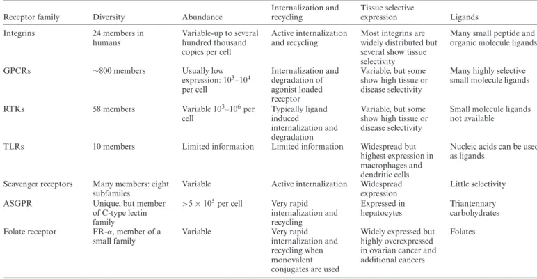
In addition to the receptors mentioned above there are obviously many other receptor families that might be used for oligonucleotide targeting. Interleukin and interferon re-ceptors, Wnt-family rere-ceptors, Transforming growth factor (TGF)-/activin receptors, the immunoglobulin family re-ceptors found in lymphoid cells, and the numerous recep-tors involved in neuronal cell recognition all come to mind as possibilities. As well, the folate/folate receptor system has been widely used to target ovarian cancer cells (116). Each of these receptors must be considered in terms of tis-sue selectivity, expression levels, rate of internalization and recycling, and availability of ligands. The properties of sev-eral receptor families relevant to oligonucleotide delivery are summarized in Table1.
Unintended consequences of targeting. While targeted de-livery of oligonucleotides, drugs and imaging agents has been the focus of thousands of publications over the last couple of decades, there has been a surprising lack of em-phasis on one of the basic consequences of the targeting process. A ligand that provides effective delivery by virtue of its high affinity binding to a specific receptor will also serve as an agonist or antagonist of that receptor. In doing so it will strongly affect the downstream signal transduction cascade. Thus the net effect will combine both the sequence specific actions of the oligonucleotide and the signaling ef-fects of the targeting agent. It is possible that this may be of modest importance in short term laboratory experiments, but if ligand-conjugated oligonucleotides (or nanoparticles) are to be used for therapy of human disease, the conse-quences of chronic modulation of key signaling processes must be considered. While the need to be concerned about signaling when dealing with GPCRs or RTKs is rather ob-vious, recent studies reveal that receptors not usually as-sociated with signaling, including SCARAs and ASGR, can nonetheless participate in important signal transduc-tion cascades.
Cellular uptake, intracellular trafficking and endosomal bar-riers
Upon reaching the cell surface, ‘free’ oligonucleotides, oligonucleotide conjugates or nanocarriers bearing oligonucleotides all share essentially the same fate; they are internalized by endocytosis and then traffic through mul-tiple membrane-bound intracellular compartments. Thus most of the oligonucleotide accumulated by cells remains separated from the cytosol and nucleus by membrane barriers. The concept of an endosome escape barrier has become prominent in the literature over the last few years and is now generally regarded as perhaps the most im-portant impediment to effective use of oligonucleotides in therapeutics. Several recent reviews have dealt in detail the mechanisms of endocytosis and trafficking and how these impact oligonucleotide pharmacology (117–119). Thus here we will briefly outline some of the basic aspects most
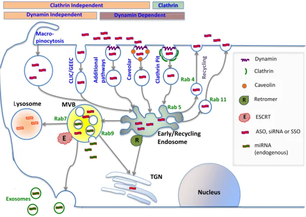
relevant to oligonucleotide delivery. Figure2schematically depicts some of the processes described below.
Basic aspects of endocytosis. The simple term endocyto-sis encompasses a variety of complex events whereby cells take up materials from their surroundings (120). The best-known internalization mechanism is the coated pit path-way that utilizes adaptor proteins, a clathrin network, and the GTPase dynamin to concentrate ligand-bound recep-tors at the cell surface and then convey them into cells. Several important physiological processes utilize clathrin-mediated endocytosis including uptake of transferrin and low-density lipoproteins, as well as internalization of ago-nist activated GPCRs (121). Caveolae originate from mem-brane structures enriched in cholesterol, sphingolipids and the transmembrane protein caveolin (122). While the role of caveolae in endocytosis has been questioned at times, it now seems clear that these compact structures (100 nm) do play a role in the internalization of certain receptors and their ligands. For example, some members of the integrin family as well as certain sodium channels are internalized via the caveolar pathway (123,124). Multiple additional endocy-totic pathways occur in cells including ones that do not rely on clathrin, caveolin or dynamin (125). An important exam-ple is the CLIC/GEEC pathway that results in the forma-tion of tubular endosomes that make a large contribuforma-tion to fluid phase endocytosis. Another high volume pathway is macropinocytosis whereby the cells use an actinomyosin-driven process to pinch off and engulf large amounts of ex-tracellular fluid. All of these processes have been implicated to varying degrees in the initial uptake of oligonucleotides. Increasing evidence indicates that the initial route of endo-cytosis can be an important determinant of oligonucleotide pharmacology and that there are both productive and non-productive paths of cellular uptake (126,127).
Basic aspects of intracellular trafficking. After initial in-ternalization by endocytosis, oligonucleotides, like all inter-nalized materials, must traffic through a complex network of endomembrane compartments each with distinct charac-teristics and functions. Major membrane-bound compart-ments include early and recycling endosomes, LEs/ multi-vesicular bodies (LEs/MVBs), lysosomes, the Golgi appa-ratus and the endoplasmic reticulum. Irrespective of the original pathway of endocytosis, most substances entering the cell are initially delivered to early endosomes. Subse-quently there are two basic fates for materials that reach the early endosome; they can be shunted to lysosomes for degradation, or they can be recycled to the plasma mem-brane and cell exterior (128).
Table 1. Receptor properties relevant to oligonucleotide targeting
Receptor family Diversity Abundance
Internalization and recycling
Tissue selective
expression Ligands
Integrins 24 members in humans
Variable-up to several hundred thousand copies per cell
Active internalization and recycling
Most integrins are widely distributed but several show tissue selectivity
Many small peptide and organic molecule ligands
GPCRs ∼800 members Usually low
expression: 103–104 per cell
Internalization and degradation of agonist loaded receptor
Variable, but some show high tissue or disease selectivity
Many highly selective small molecule ligands
RTKs 58 members Variable 103–106per
cell
Typically ligand induced
internalization and degradation
Variable, but some show high tissue or disease selectivity
Small molecule ligands not available
TLRs 10 members Limited information Limited information Widespread but highest expression in macrophages and dendritic cells
Nucleic acids can be used as ligands
Scavenger receptors Many members: eight subfamiles
Variable Active internalization Widespread expression
Little selectivity
ASGPR Unique, but member
of C-type lectin family
>5×10⁵per cell Very rapid internalization and recycling
Expressed in hepatocytes
Triantennary carbohydrates Folate receptor FR-␣, member of a
small family
Variable Very rapid
internalization and recycling when monovalent conjugates are used
Widely expressed but highly overexpressed in ovarian cancer and additional cancers
Folates
Some receptor characteristics important for oligonucleotide delivery are listed in the table.
The next stage of the trafficking process involves LEs/MVBs that are the primary stepping-stone on the road to lysosomes (130). These structures are morphologically and biochemically distinct from early endosomes, having a non-tubulated appearance, a perinuclear location and a lu-men filed with small intraluminal vesicles (ILVs) that have pinched off from the boundary membrane of this organelle. Although the major task for LEs is to convey internalized material to lysosomes, an interesting detour involves the ex-ternalization of the ILVs as exosomes that can convey ma-terials to other cells (131).
Ultimately most material internalized by endocytosis is delivered to lysosomes. These dense organelles are rich in hydrolases that function in the low pH environment (pH 4.5–5.5) that is maintained by an active V-ATPase proton pump, thus allowing the hydrolases to degrade proteins, lipids, carbohydrates and nucleic acids to their constituent building blocks (132). Besides degrading internalized ma-terials, lysosomes are also a key part of the machinery for autophagy whereby cells degrade damaged proteins and or-ganelles and recycle the constituents (133).
It is important to note that the early endosome to lyso-some pathway is not linear and that there are several branches and loops. An important one is the retrograde trafficking pathway that links endosomes to the trans-Golgi. A classic example of retrograde transfer is the recap-ture of mannose-6 phosphate receptors from endosomes to the Golgi, while their hydrolase ligands are delivered to lyso-somes (134). Interestingly, several pathogens have ‘hijacked’ this pathway; for example, certain bacterial toxins reach the cytosol by following the retrograde pathway to the trans-Golgi and thence to the endoplasmic reticulum (135).
In summary, the pathways of endocytosis and intracel-lular trafficking are complex and dynamic. Contemporary imaging technologies (136) are starting to provide detailed insights into these pathways that will be very helpful in un-derstanding the fundamental basis of oligonucleotide phar-macology. However, it remains a challenging problem to link microscopic observations on the intracellular traffick-ing of a fluorescent oligonucleotide to the pharmacological effects of that molecule. Visualization of the bulk distribu-tion may not reveal minor compartments that are key to biological activity.
The machinery of intracellular trafficking. There are two basic ways by which materials can move through the intra-cellular trafficking network (128,136,137). One involves rel-atively small shuttle vesicles that convey both luminal and membrane material between larger endomembrane com-partments. The other is endosome maturation whereby one compartment gradually assumes the characteristics of a sec-ond compartment.
com-ASO, siRNA or SSO
Dynamin Independent Dynamin Dependent
Clathrin Independent
ndent Clathrin
Macro- pinocytosis
CLIC/GEE
C
Ca
v
eolar
Clathrin
Pit
Lysosome MVB
Early/Recycling Endosome
Rab9 Rab7
Rab 5 Rab 4
Rab 11
R
E
Ly
Additional path
w
a
y
s
R
ecy
cling
TGN
E
Dynamin
Clathrin
Caveolin
E
R Retromer
ESCRT
miRNA (endogenous)
Exosomes
ASO, siRNA or SSO Dynamin
Clathrin
Caveolin
E
R Retromer
ESCRT
miRNA (endogenous)
Nucleus
Figure 2. Cellular uptake and intracellular trafficking of oligonucleotides. Oligonucleotides enter cells via several endocytotic pathways that vary in terms of their dependence on clathrin, caveolin or dynamin. These pathways all initially lead to the early/re-cycling endosome compartment; nonethe-less molecules entering via different pathways can traffic to different downstream destinations. Most internalized oligonucleotide accumulates in late endosomes/multivesicular bodies (MVBs) and in lysosomes; however, some trafficking to other membrane bound compartments does occur. Oligonu-cleotides within endomembrane compartments are pharmacologically inert, but a very small portion of internalized oligonucleotide can spontaneously escape to the cytosol and nucleus. Intracellular trafficking is highly regulated by a large number of proteins and protein complexes. Thus the Rab family of GTPases regulates many aspects of trafficking and individual members serve as markers for distinct endomembrane compartments. The formation of MVBs is regulated by the multi-protein ESCRT complex that has recently been demonstrated to play a key role in the effectiveness of oligonucleotides. This complex also plays a role in exosome formation. The retromer complex may deliver oligonucleotides to the trans-Golgi instead of to lysosomes.
plexes, tethering complexes, the ESCRT complex and the Retromer complex. These various proteins/protein com-plexes play important functional roles, but they also serve as easily recognizable markers for specific endomembrane compartments.
Members of the numerous (>60) Rab GTPase protein family serve as molecular switches that regulate many as-pects of trafficking including vesicle uncoating, movement along cytoskeletal tracks and the ultimate membrane fusion events involving tethers and SNARES (137,141). Rab pro-teins also serve as excellent markers of individual membrane compartments and trafficking pathways. For example, Rab5 is a marker for early endosomes while Rab7 identifies LEs.
The shuttle vesicle trafficking process involves coat pro-teins that assist in the initial formation of the vesicle (140,142). The generation of clathrin-coated vesicles at the plasma membrane is a good example, but other types of coats exist including the COPI proteins involved in Golgi to ER transport and the COPII proteins involved in the reverse process. Disjunction of the clathrin-coated vesicle from the donor membrane is accomplished by the dynamin GTPase, but other pinching off mechanisms exist for other types of vesicles.
Tethering proteins impart selectivity to trafficking by pro-moting preferential interactions between the vesicle and the recipient compartment. The coiled-coil tethers and the multi-subunit tethers comprise the two broad classes of tethering proteins (143,144). By associating with both Rab proteins and SNARES, tethers are thought to physically link the two membranes destined for fusion. However, some tethers clearly have multiple functions including possibly ‘proof-reading’ SNARE complexes to assure fusion of the correct partners.
An important detour on the pathway between endosomes and lysosomes is retrograde trafficking between early en-dosomes and the trans-Golgi (134,135,147). This process is driven by the retromer that includes a trimeric complex (Vps26–Vps35–Vps29) that binds to the cytoplasmic tails of potential cargo proteins. It also includes SNX proteins that have PX domains that recognize membrane phospho-inositides and BAR domains that can affect membrane cur-vature. This results in the tubulation of the EE membrane and eventual formation of shuttle vesicles that traffic to the trans-Golgi.
In addition to the shuttle vesicle mechanism, intracellu-lar trafficking also utilizes processes involving maturation of one major endomembrane compartment into another (128,148). Perhaps the best example is the conversion of EEs to LEs. The Rab5 GTPase plays a key role in the iden-tity and function of early endosomes (137,149). The ac-tivating proteins Rabex-5 and Rabaptin-5 stimulate Rab5 on the cytosolic surface of the EE resulting in the recruit-ment of Rab5 effectors including the tethering factor EEA1 and the PI 3-OH kinase Vps34. Initially this process is self-sustaining allowing the EE to interact with other EEs and to recycle receptors and other membrane constituents to the cell surface. Eventually other proteins are recruited to the EE that drive displacement of Rab5 and association with Rab7. Two sets of effectors, the SAND-1/Mon complex and the HOPS complex, seem to work in tandem in the EE to LE maturation (128). In parallel to the Rab5 to Rab7 conver-sion, the endosome loses ability to interact with EE partners and instead acquires the ability to associate with LE part-ners. One of the key aspects of the EE to LE maturation is the formation of ILVs (150). This process helps to concen-trate selected proteins and lipids in the LE lumen directing them to lysosomal degradation. The five multi-protein com-plexes of the ESCRT (endosomal sorting complex required for transport) machinery, ESCRTs 0–III and Vps4–Vta1, recognize ubiquitinated membrane proteins and drive them into invaginations that ultimately form ILVs within the lu-men of the LE/MVB (151).
How is all this trafficking complexity linked to oligonu-cleotide delivery? As an example, in an extremely impor-tant recent investigation of basic aspects of oligonucleotide trafficking, Wagenaaret al. (152) used shRNA libraries to identify TSG101, a component of the ESCRT machinery, as having a key role in the uptake and intracellular traf-ficking of oligonucleotides. Silencing of this gene led to a dramatic increase in the effectiveness of an antisense antag-onist of miR-21. This publication establishes two critically important points. First, that the endomembrane trafficking machinery plays a key role in the pharmacology of oligonu-cleotides, and second that the machinery can be manipu-lated so as to improve oligonucleotide delivery and actions. Thus, the complex and dynamic pathways of intracellular trafficking are regulated by an equally complex set of pro-teins whose interactions vary in time and space throughout the process. While this makes the investigation of subcel-lular trafficking rather complicated it also opens up many opportunities to manipulate the machinery of trafficking using molecular and chemical probes. As shown by Wage-naaret al., this can have important implications for oligonu-cleotide pharmacology. Thus increasing understanding of
the mechanistic basis of oligonucleotide trafficking will no doubt provide important new avenues to manipulate oligonucleotide delivery.
Breaching the endosomal barrier. While the trafficking machinery is usually quite efficient in moving internal-ized material to the appropriate intracellular destination, nonetheless opportunities for molecules to escape from en-domembrane compartments to the cytosol do exist. Traf-ficking involves a plethora of membrane fusion and fission events. These events create localized membrane stress that can result in the formation of non-bilayer lipid domains (153). Typically non-bilayer regions can be much more per-meable to solutes than bilayer regions (154,155). Thus there is an inherent relationship between the fusion/fission events essential to intracellular trafficking and the potential for leakage of vesicle contents. There are several loci in the intracellular trafficking network that may be particularly susceptible to increases in permeability that would allow release of oligonucleotides to the cytosol. The first is in early/sorting endosomes where there is extensive tubula-tion and formatubula-tion of vesicles for return of receptors to the plasma membrane. A second locus is in LE/MVBs where the ESCRT complex distorts the endosome membrane to form ILVs. Third, retrograde traffic from early or LEs to trans-Golgi offers another possibility for membrane insta-bility. Finally, SNARE driven membrane fusions at multi-ple sites afford opportunities for partial leakage of vesic-ular contents (156). The role of specific trafficking events in oligonucleotide delivery can be explored using molecu-lar techniques such as siRNA or vectors expressing domi-nant negative proteins to perturb these events. Another in-teresting approach is to use cell lines with defects in traf-ficking processes. There are a growing number of exam-ples of both approaches in the oligonucleotide literature (127,152,157,158). In addition to endogenous escape of oligonucleotides from endosomes, a substantial portion of the recent literature on oligonucleotide delivery is focused on approaches to disrupt or alter the endosomal barrier. Thus cationic lipids and polymers have been used to desta-bilize the endosome membrane. Titratable peptides or poly-mers have been used to alter intra-endosomal pH thus af-fecting endosome stability and trafficking. Recently a vari-ety of small molecules have emerged that seem to assist in endocytosis of oligonucleotides or that selectively perme-abilize endosomal compartments leading to oligonucleotide release to the cytosol. It is important to note that once an-ionic single stranded oligonucleotides reach the cytosol they readily enter the nucleus (159,160).
APPROACHES TO DELIVERY
There are two broad strategies for oligonucleotide deliv-ery. One is to incorporate the oligonucleotide into some form of nanocarrier that then determines the tissue dis-tribution and cellular interactions of the oligonucleotide. The other is to chemically modify the oligonucleotide it-self, most commonly with a targeting ligand, while preserv-ing the molecular nature of the conjugate. A fundamental difference between the two approaches lies in the size of the delivery moiety, nanoscale versus molecular scale; this has profound effects on the biodistribution and biological actions of the oligonucleotide. Several of the delivery ap-proaches that will be discussed in detail below are depicted in Figure3.
Delivery at the nanoscale
Lipid nanoparticles. The delivery approach that is both most widely used and most clinically advanced is to com-plex anionic oligonucleotides with cationic lipids thus form-ing lipid nanoparticles (LNPs)(161–163). This approach has been especially important for therapeutic use of siRNA (164) and good accounts of current clinical trials using siRNA LNPs are found in recent reviews (10,165). In ad-dition to the cationic lipid, LNPs typically include a neu-tral lipid such as cholesterol, but a variety of compositions are possible. While simple LNPs are effective as cell cul-ture transfection agents, they cannot be used in vivo be-cause extensive interactions with opsonic proteins in blood lead to rapid clearance by RES phagocytes. LNPs for in vivouse are usually 100–200 nm in size and include a sur-face coating of a neutral polymer such as PEG to mini-mize protein binding and uptake by RES cells. This allows greater persistence in the circulation and the opportunity to interact with other cell types. However, because of their size, LNPs can only exit the circulation at sites where the endothelial barrier is fenestrated, particularly liver, spleen and certain tumors having a high EPR effect. Because of this, much of the work with siRNA-LNPs has focused on liver diseases including transthyrethrin-mediated amyloido-sis, clotting disorders, liver cancer and disorders of lipid metabolism (11,166).
The action of LNPs involves initial uptake by endocy-tosis. In some cases this is mediated by the binding of apolipoprotein E and interaction with the LDL receptor (167). Once in endosomes, the cationic lipids of the LNP in-teract with anionic membrane lipids to disrupt membrane structure through the formation of a non-bilayer lipid phase termed inverted hexagonal (HII). This leads both to in-creased membrane permeability and to dissolution of the LNP and is the basis for conveying the oligonucleotide to the cytosol (168). However, the interaction of cationic lipids with cellular membranes is also the basis for possible toxic-ities of LNPs (169,170).
A great deal of effort has been expended on optimizing the delivery properties of LNPs. One problem concerns the PEG coating (which is usually attached to the LNP by a lipid anchor). A dense coat is beneficial for increasing cir-culation time, but it also reduces uptake by cells such as the hepatocytes that are the intended destination of the oligonucleotide. Several approaches have been tried to at-tain dynamic control of PEG levels, including use of
cleav-able linkers or short lipid anchors, with the intent of main-taining a dense coating of PEG in the circulation while al-lowing release in the cellular environment (161). Studies of how PEG density and characteristics affect the pharma-cokinetics, biodistribution and function of LNPs remains an active area of investigation (171).
There has also been progress in optimizing the delivery characteristics of the cationic lipids themselves. A widely used type of nanocarrier for siRNA delivery is the SNALP (stable-nucleic-acid lipid particle), a PEG stabilized LNP. Two important steps for optimizing the cationic lipids were (i) altering the pKa so that the lipids were almost uncharged in the circulation but became charged in the low pH endo-some and (ii) using linkages that were readily biodegrad-able (172,173). This resulted in dramatic improvement in effectiveness, allowing siRNA doses as low as 0.005 mg/kg to achieve significant silencing of hepatic targets in ani-mal models, accompanied by low toxicity (174). Additional work on the chemistry of lipids continues. One powerful ap-proach is the testing of chemical libraries of ‘lipoids’ (lipid like molecules) for their delivery capabilities (175). Another interesting strategy is the design of multifunctional lipids that include a pH responsive head group and well as SH moieties. The multifunctional LNPs provide increased sta-bility in the blood but then promote endosome escape in the low pH and reducing environment of the cell interior (176). Another approach that may avoid some of the toxi-cities of cationic LNPs involves using siRNA entrapped in neutral liposomes (165,177). There has also been manipula-tion of the physical structure of lipid delivery systems. Thus ‘cuboplexes’ are novel lipid structures designed to promote interaction with endosome membranes and thus allow es-cape of siRNA to the cytosol (178). Another interesting ap-proach uses liposomes termed ‘SmarticlesR’ made from dialkyl cationic amino acids; these undergo a pH sensitive conversion to a HIIphase in endosomes allowing oligonu-cleotide escape (179).
The precise mechanisms of uptake, intracellular traffick-ing and ultimate delivery of oligonucleotides by LNPs has been a matter of some debate (157,180). However, two arti-cles appearing simultaneously in 2013 used advanced imag-ing techniques to provide unprecedented insights into the intracellular fates of siRNA LNPs. Thus Gilleron et al. (181) found that LNPs were initially taken up by clathrin-mediated endocytosis but further accumulation involved macropinocytosis. The LNPs accumulated in an EE-LE hybrid compartment; however, only 1–2% of the siRNA reached the cytosol. Sahayet al. (158) also demonstrated a role for macropinocytosis. However, they found that much of the siRNA was re-exported from LEs/lysosomes using a process involving the NPC1 lipid transporter protein. A more recent study using highly sensitive fluorescence mi-croscopy techniques documented release of siRNA from individual endosomes (182). Release took place primarily at the EE/LE conversion step rather than from lysosomes. These studies provide important insights into the mecha-nistic basis of oligonucleotide delivery via LNPs and attest to the overall inefficiency and transience of oligonucleotide delivery even when using effective nanocarrier systems.
Oligonucleotide
Ligand
PEG
Cellular Barriers
A
B
C
D
E
F
Figure 3. Oligonucleotide delivery strategies. Several approaches to oligonucleotide delivery are depicted. (A) Antibody-oligonucleotide conjugate. (B) Polymer-oligonucleotide conjugate with PEGylation and targeting ligand. (C) Molecular scale ligand-oligonucleotide conjugate with triantennary carbohy-drate ligand. (D) Lipid nanoparticle with PEGylation. (E) Gold nanoparticle with dense oligonucleotide coat. (F) DNA nanostructure with oligonucleotide and targeting ligand incorporated. Images are not to scale.
is to decorate the LNP surface with antibodies. However, long experience with targeting of drug-loaded liposomes suggests that it is quite difficult to find the right balance be-tween PEG shielding and availability of the antibody (183). Recently however, there have been some interesting reports using antibody-targeted siRNA LNPs. Thus a scFv was used to target LNPs to dendritic cellsin vivo. Good ‘knock down’ of several co-stimulatory surface antigens and inhi-bition of mixed lymphocyte reactions were observed (184). In another study LNPs decorated with antibody to CD20 delivered Bcl-2 ASO to B-cell tumorsin vivowith good an-titumor effect (185). Additionally an anti CD4 monoclonal was used to target siRNA LNPs to CD4 positive T-cells re-sulting in knock-down of the intended mRNAs both in cell culture andin vivo(186).
Progress has also been made using small molecule ligands to target LNPs. For example, in a series of publications, Huanget al. have used anisamide as a ligand to assist in the delivery of siRNA LNPs to tumors (187,188). Finally aptamers are also being explored as a targeted delivery ap-proach for LNPs (189). Some of the above mentioned stud-ies on LNP targeting are also of interest because they in-volve delivery of oligonucleotides to non-hepatic sites.
Finally, there have been some impressive recent pre-clinical studies using LNPs. Thus LNPs prepared using a novel lipid from a chemical library selectively delivered oligonucleotides to the lung. When used with si-KRAS and
miR-34a, significant inhibition of lung cancer was attained in a genetically engineered mouse tumor model (190). In an-other study, siRNA LNPs showed therapeutic efficacy ver-sus Marburg virus (an Ebola relative) in non-human pri-mates (191). These studies are both good examples of non-hepatic delivery using LNPs.
In summary, over the last few years there have been im-pressive advances in the LNP delivery technology. Current formulations allow highly effective delivery of siRNA to hepatocytes using doses of oligonucleotide and of lipid car-rier that display only minimal toxicity. This has allowed sev-eral liver-based diseases to be addressed. The greater chal-lenge lies with non-hepatic delivery. Although there have been some interesting reports using LNPs to deliver siRNA to other tissues and to tumors, there remain questions re-garding efficacy and toxicity particularly in the context of long term use in human therapy.
There are several types of polymeric nanocarriers. Early studies in this area primarily used well-known, biomedically compatible polymers such as poly (lactic-co-glycolic acid) (PLGA) to form solid nanoparticles through various oil-in-water emulsion techniques. However, since PLGA is an-ionic, a common approach was to incorporate positive side chains in the polymer or to complex the anionic oligonu-cleotide with a positively charged moiety such as polyethy-lene imine (PEI) (195,196). Polymeric micelles, sometimes called ‘core-shell’ nanoparticles, have also been widely used for oligonucleotide delivery. These are formed by self-assembly of amphiphilic polymers in a water environment. Typically a tri-block polymer might be used, including a hydrophobic portion to drive self-assembly, a cationic por-tion to bind the oligonucleotide and PEG or other neutral polymer to provide a protective coating (197,198). A recent report described a novel self-assembled hybrid nanocarrier comprised of a PLGA core and a lipid-PEG shell. This sys-tem provided impressive results in terms of a long circula-tion lifetime and funccircula-tionally effective delivery of siRNA to tumor xenografts in mice (199). A third type of polymeric nanocarrier is the nanohydrogel. These nanoparticles have an open, water-filled polymer lattice that can easily incorpo-rate bio-macromolecules such as polypeptides and oligonu-cleotides, whose release kinetics can be controlled by the de-gree of cross linking of the lattice. A particularly interest-ing form is the PRINT nanohydrogel whose size and shape can be precisely controlled by a nano-molding technique (200,201). A virtue of many polymeric nanocarriers is their ready ability to convey both an oligonucleotide and a small molecule drug; this is especially interesting in the context of cancer chemotherapy. Thus there are several promising re-ports ofin vivoco-delivery of siRNA and anticancer drugs (194,197,198).
Another common approach involves the direct complex-ation of anionic oligonucleotides with ccomplex-ationic molecules that have some degree of endosome escape capability in-herent in their chemistry. This would include formation of nanoscale polyplexes involving CPPs (202,203), cationic dendrimers such as PAMAMs (194,204) or linear or cross-linked PEI (205).
CPPs have been extensively studied for oligonucleotide delivery (74,206). Starting with the original Transactivator of Transcription (TAT) and penetratin structures, a wide variety of short polycationic CPPs have been synthesized and used as delivery agents for drugs, peptides, proteins and nucleic acids (203,207). In many cases short amphipathic sequences are also included with the intent of promoting endosome escape. While some work has been done with chemical conjugation of CPPs to anionic oligonucleotides, in most cases delivery is via formation of nanocomplexes. These are usually in the 100–200 nm range with a surface charge that depends on the chemistry of the CPP and the ratio of cationic peptide to oligonucleotide. Perhaps the most advanced CPP for oligonucleotide delivery remains PepFect6 that has been used to deliver siRNA in an ani-mal model resulting in ‘knock down’ of target gene expres-sion in several tissues (208). Additional work on this type of CPP has included the incorporation of endosome dis-rupting moieties such as quinoline derivatives (209). An-other promising study used a cationic peptide derived from
bee melittin to form nanocomplexes with siRNA directed against NF-kB; these were used to treat a mouse model of rheumatoid arthritis (210). In addition to their direct use as polyplexes, CPPs have been used to augment the properties of other delivery systems such as LNPs (211). An interesting variant of the CPP approach entailed making a chimera of an RNA binding protein and a CPP. This entity could bind and deliver siRNA in cell culture and in an animal model (212).
Dendrimers are branched polymers with well-defined ar-chitectures. By controlling the degree of branching, differ-ent ‘generations’ of dendrimer can be made thus varying size and the number of potentially reactive surface moi-eties. PAMAMs (213), the dendrimer type most commonly used for oligonucleotide delivery, shares with PEI the abil-ity to exert a strong ‘proton sponge’ effect. Thus the titrat-able amines on the PAMAM bind protons in endosomes and lysosomes leading to increased pumping of protons by the membrane V-ATPase; this is accompanied by Cl− transport, water accumulation and swelling and rupture of the organelle. This makes PAMAM dendrimers very effec-tive agents for nucleic acid delivery (204). Unfortunately the strength of the protein sponge effect, along with lack of biodegradability and multiple non-specific interactions with blood and tissue proteins, all contribute to the substan-tial toxicities observed within vivouse of PAMAMs (214). By virtue of their multiple surface amino groups PAMAMs are easily conjugated to a variety of ligands. For example, PEG has been used to reduce toxicity and improvein vivo
biodistribution properties (215). It is also possible to simul-taneously conjugate both targeting ligands and therapeuti-cally active small molecules to PAMAM dendrimers (216). Thus there is a great deal of flexibility inherent in this deliv-ery system but concerns remain about toxicity.
PEIs are linear or branched polymers that have multi-ple titratable amino groups. Thus they can readily form nanocomplexes with oligonucleotides (217). Like PAMAM dendrimers, PEI exerts a strong ‘proton sponge’ effect mak-ing it quite effective for nucleic acid delivery. It is also read-ily modified by conjugation with a variety of ligands. For example, a branched PEI modified with PEG and Arg-Gly-Asp (RGD) ligands was used to deliver siRNA to tumors in mice (218). In addition to its direct use as an oligonu-cleotide carrier, as noted above, PEI is often incorporated into other types of nanoparticles to provide binding for oligonucleotides and as an endosome escape agent. Unfor-tunately, PEIs are poorly biodegradable.
There has been a great deal of interest in methods for targeting nanoparticles with a plethora of research publi-cations and multiple reviews on this broad topic (219–221). Recent reviews have focused particularly on siRNA or ASO delivery using targeted polymeric nanocarriers (222,223). A widely used approach has been to couple folate to the nanocarrier surface to promote interaction with cancer cells that overexpress the folate receptor (224,225). However, al-though the first demonstrated siRNA effects in man in-volved a targeted polymeric nanocarrier (192), there has been little further clinical progress with this approach.
cel-lular level. A recent study showed that the celcel-lular uptake of CPP/oligonucleotide nanocomplexes involves scavenger re-ceptors (226). Another interesting report demonstrated that the NPC1 lipid transport system, previously shown to be in-volved in LNP processing (158), is also inin-volved in the pro-cessing of certain types of polymeric nanoparticles (227). However, a reduction in NPC1 levels had the opposite effect on delivery of siRNA using polymers as in the case of de-livery using lipids. There have been only a few comparisons of uptake mechanisms for LNPs and polyplexes (157,228); both of the cited reports suggest that the mechanisms of up-take are different and one report indicated that polyplexes are released from endosomes via a rapid bursting process. A recent study sought to incorporate endosome-disruptive he-lical peptides into siRNA nanoparticles and demonstrated release into the cytosol; this system was used to knock down TNF-␣in an animal model of inflammation (229).
In contrast to the lack of information at the cellular level, there is substantial data on the pharmacokinetics and biodistribution of polymeric nanoparticles (193,230). In mice, as well as in patients, polymeric nanoparticles tend to be cleared quickly (231) with accumulation in the liver predominating (232,233). However, as mentioned above, a recent study with a novel polymeric nanoparticle formula-tion demonstrated an extended lifetime in circulaformula-tion and impressively high levels of tumor uptake as compared to liver uptake (199). Thus it may be possible to substan-tially improve the biodistribution characteristics of poly-meric nanoparticles. Nonetheless, polypoly-meric nanoparticles have not yet advanced as far as LNPs in terms of pre-clinical and clinical development.
An interesting offshoot for polymeric nanoparticles in-volves siRNA delivery via the gastrointestinal tract. Thus siRNA nanoparticles have been used orally to block inflam-matory effects mediated by TNF-␣(234), or have been ad-ministered into the colon to knock down apolipoprotein B mRNA (235), or via the rectum to suppress inflammatory cytokines (236).
Advantages and liabilities of nanoscale delivery. There is an interesting divergence in the oligonucleotide therapeu-tics field in terms of approaches to delivery of single strand molecules and of siRNA or miRNA. For the most part de-livery of ASOs and SSOs, at least at the clinical level, has relied on ‘free’ oligonucleotides. By contrast, with one ma-jor exception, siRNA delivery has relied on nanocarriers, particularly SNALPS. The reasons for this divergence are easy to understand. ASOs and SSOs can readily be chemi-cally modified to resist nuclease degradation and, in the case of phosphorothioates, they have a long circulation lifetime and are readily taken up by cells. By contrast, early ver-sions of siRNA had none of these advantages; they were very unstable, rapidly cleared by the kidney, and poorly ac-cumulated by cells. Based on these problems, a huge ef-fort has gone into the development of siRNA nanocarri-ers. Although a bewildering variety of lipids and polymers have been tried, they almost all share the weakness of being positively charged and therefor highly interactive with an-ionic biological macromolecules and cells. This inevitably leads to toxicity especially upon chronic use. As discussed above, extensive work on the lipid components of SNALPs
has both increased their efficacy for siRNA delivery and reduced their toxicity. Thus SNALP siRNA formulations seem a sound approach for siRNA delivery to the liver. It is not clear that the same can be said about the various types of polymeric nanoparticles. Although attempts have been made to reduce the toxicity of dendrimers, PEI or cationic peptides it is difficult to avoid this entirely because of their inherent chemical nature.
A potential advantage of nanocarrier systems is that a large bolus of oligonucleotide can be delivered during one cellular uptake event. However, the converse is that entry of a nanoparticle into the cells entails the delivery of a large mass of carrier material. Even with the best nanocarriers only a few percent of the total mass is siRNA with the rest being carrier (172). Thus nanoparticle delivery is inevitably associated with the accumulation of substantial amounts of potentially toxic material; this is especially problematic for lipids or polymers that are not readily biodegradable. It seems unlikely that such materials will ever be clinically useful.
A final concern regarding nanocarriers involves their lim-ited biodistribution. As mentioned early in this review, typ-ical nanoparticles with diameters of∼100 nm are excluded from most tissues by the barrier presented by the capillary endothelium. Accumulation occurs primarily in liver and spleen where the endothelium is fenestrated, and to a lesser degree in certain rapidly growing tumors that have a strong EPR effect. The parenchyma of most other normal tissues are virtually inaccessible, thus limiting the range of thera-peutic applications for nanocarriers.
Delivery at the molecular scale: ligand-oligonucleotide conju-gates
Partly because of the many concerns about using nanopar-ticle delivery systems, there has recently been a surge of in-terest in molecular scale ligand-oligonucleotide conjugates. These offer the possibility of selective delivery to specific cells or tissues via receptor mediated mechanisms coupled with an avoidance of the toxicities often associated with nanocarriers. Another advantage of conjugates is that they are well-defined molecular entities as opposed to the het-erogeneity characteristic of nanoparticles. Thus while the conjugate approach is still in its infancy, it seems to offer a promising path forward for oligonucleotide therapeutics.