Vol 1, No 1 (2013) ISSN 2167-8677 (online) DOI 10.5195/d3000.2013.10
http://dentistry3000.pitt.edu
This work is licensed under a Creative Commons Attribution 3.0 United States License.
This site is published by the University Library System, University of Pittsburgh as part of its D-Scribe Digital Publishing Program and is cosponsored by the University of Pittsburgh Press.
Sexual dimorphism involved in the mesiodistal and buccolingual
di-mensions of permanent teeth
Ticiana Medeiros Sabóia1, Patricia Nivoloni Tannure2, Ronir Raggio Luiz5, Marcelo de Castro Costa1, Jose Mauro Granjeiro3, Erika Calvano Küch-ler3, Leonardo Santos Antunes4
1
Department of Pediatric Dentistry and Orthodontics, School of Dentistry, Federal University of Rio de Janeiro, RJ, Brazil
2Discipline of Pediatric Dentistry, School of Dentistry, Veiga de Almeida University, RJ, Brazil 3
Unit of Clinical Research, Fluminense Federal University, Niterói, RJ, Brazil
4
Department of Specific Formation, School of Dentistry, Fluminense Federal University, Nova Friburgo, RJ, Brazil
5 Institute of Public Health Studies, Federal University of Rio de Janeiro, RJ, Brazil
Abstract
Studies indicate that tooth crown diameters are clinical markers for sex differentiation. Therefore, the aim of this study was to assess the degree of sexual dimorphism in different teeth. Maximum mesiodistal (MD) and buccolingual (BL) dimensions of 2400 permanent teeth from 100 pretreatment orthodontic dental study casts and clinical records (50 males and 50 females) from the Department of Pediatric Dentistry and Orthodontics, Federal Uni-versity of Rio de Janeiro, Brazil, were examined. Comparison of the MD and BL dimensions between males and females was performed using the Student’s t test with alpha 0.05, ef-fect size, and discriminant function analysis. Comparisons in MD and BL widths between sexes demonstrated that the combined mean in the female group presented reduction when compared with the male group, except for the BL dimension of tooth 26. In regard to the MD dimensions, statistically significant differences were observed in various dental groups. The greatest sexual dimorphism was observed in the left mandibular canine (p<0.001) with effect size over 0.8 (0.94), which characterizes large effect. In BL dimension, numerous teeth demonstrated statistical differences between the sexes. Our findings rein-forced the magnitude of sexual dimorphism in tooth size, and, in addition, highlighted the differences in specific dental groups.
Citation: Sabóia TM, Tannure PN, Luiz RR, Costa MC, Granjeiro JM, Küchler EC, Antunes LS. (2013) Sexual dimorphism involved on the mesiodistal and buccolin-gual dimensions of permanent teeth. Dentistry 3000 1:a003 doi: 10.5195/d3000.2013.10
Received: May 06, 2013 Accepted: November 01, 2013 Published: November 19, 2013
Copyright: ©2013 Sabóia et al. This is an open-access article licensed under a Creative Commons Attribution 3.0 United States license.
License.
Email: leonardoantunes@id.uff.br
Introduction
The sexual dimorphism is explained by dif-ferent effects of the human X and Y chromo-some genes on various somatic features, such as the frequency of some dental anom-alies and the tooth crown size [1-3]. Various explanations for tooth-size dimor-phism between males and females have been proposed: the differences in hormonal balance [4], the effect of the Y chromosome in increasing mitotic activity within the developing dental lamina, and the fact that the chromosome X is known to be involved in the enamel formation [2]. Some authors suggested that this difference is due to the amount of enamel [5], while others found significant differences in the amounts of dentine [6].
The tooth crown size is a valuable tool and provides significant information on human evolution [7] and biological alterations [8], a in forensic evaluation [9,10] and clinical odontology [11]. Tooth crown diameters are reasonably accurate predictors of sex and are good adjuncts for sex differentiation [12]. Although the degree of dimorphism varies within different populations, general-ly, males have larger tooth crowns than females [13-16].
Therefore, the present study was conducted to assess the degree of sexual dimorphism in permanent teeth of patients recruited at the Federal University of Rio de Janeiro, Brazil.
Materials and Methods
A total of 2400 permanent teeth from 100 pretreatment orthodontic dental study casts
and clinical records from the Department of Pediatric Dentistry and Orthodontics, Fed-eral University of Rio de Janeiro, Brazil, were examined. Patients with underlying syndromes and oral clefts were not included in this study. Patients who had their pre-treatment orthodontic records performed between 2000 to 2010 were available for this study. Fifty males and 50 females were selected at random. Ethical approval was obtained from the Human Ethics Committee of the Health Department of the city of Rio de Janeiro, Rio de Janeiro, Brazil (113/09). Written informed consent was obtained from all participating individuals or par-ents/legal guardians.
ndants (53.6%) and African descendants (33.6% obviously mixed Portuguese, 12.3% not obviously mixed Africans). The remain-ing 0.5% of the population is Amerindian or Asian descendants. The ethnicity was based on the self-reported description.
Assessment of tooth dimensions
Dental casts were used to obtain data re-garding tooth dimensions. A dental diagram with the tooth nomenclature used in this work is presented in Figure 1. Dental casts were excluded from assessment if they fell into one of the following criteria: teeth with restorations extending both for the mesi-odistal and for the buccolingual surfaces, teeth displaced or crowded, and teeth not fully erupted. Second and third molars were excluded due to the young age of many of the subjects. Maximum mesiodistal (MD) and buccolingual (BL) dimensions of fully erupted permanent teeth were measured to the nearest 0.1 millimeters using the digital Mitutoyo caliper. MD is defined as the max-imum distance between the most mesial and the most distal point of the crown, whereas BL is defined as the maximum dis-tance between the most lingual/palatal and the most buccal/labial point of the crown. The dimensions were recorded for each tooth using the method proposed by Moor-rees and Reed [17].
All measurements were undertaken by one operator (T.M.S.), with a strict criterion in order to reduce variation. Five models were analyzed per day during two months to prevent depletion of the operator. Each tooth was measured three times and, if the
measurements differed by more than 0.2 millimeters, was measured three times again.
Statistical Analysis
An intra-class correlation coefficient was calculated to assess random error of intra-observer variability. The dental casts of ten subjects were selected and all tooth meas-urements were assessed on two occasions at least two weeks apart. The level of agreement was equal to 0.99, indicating an excellent level of reproducibility of the tooth dimension measurements. The de-scriptive values of MD and BL dimensions
(means and standard deviations) were rec-orded. Comparison between MD and BL dimensions of the male and female perma-nent teeth was performed using the Stu-dent’s t test with an alpha of 0.05. Effect sizes were calculated as the difference be-tween the means (measurements for each tooth in relation to sex) divided by standard deviation of either group. The widely used benchmarks, suggested by Cohen [18], indi-cated the magnitude of the change ob-served. That is, effect sizes of 0.2 were taken to be small, 0.5 to be moderate, and 0.8 or above to be large. Comparison between MD
Table1.Characteristics of the individuals studied.
Male Female p-value
Mean age (Standard Deviation) 17.2(±4.6) 19.8(±6.3) 0.02*
Ethnicity (%)
Caucasian 37 (74%) 30 (60%)
0.1**
African descendant 13 (26%) 20 (40%)
Note: * T-test; ** Chi-square test; bold emphasis indicates statistical significance (p≤0.05).
Figure 1. Dental diagram. This diagram repre-sents the permanent dentition. Second molars are presented in gray color since they were not evaluated in this work. Third molars are not presented in this diagram. This diagram is la-beled according to the Federation Dentaire In-ternationale Numbering System.
Figure 2. Box plot showing quartiles and mean of the mesiodistal dimensions of upper and lower
http://dentistry3000.pitt.edu
and BL dimensions of the Caucasian and African descendants’ permanent teeth was also performed using the Student’s t test with an alpha of 0.05. For the dimensions that were different between ethnicities, logistic regression analyses were performed including ethnicity as covariates. Results were reported according to the STROBE guidelines for cohort.
Results
The univariate analysis showed that there are no significant differences in ethnicity between male and female (p=0.1). The de-mographic characteristics of the studied individuals are summarized in Table 1.
In both comparisons, MD and BL widths between sex demonstrated that the com-bined mean of each tooth in the female group presented reduction when compared with the male group. Out of the 48 variables measured, male teeth exceeded female teeth significantly in 26 variables (p<0.05). Of
these, 13 belonged to the maxillary and 13 were BL dimensions.
In regard to the MD dimensions, statistically significant differences were observed in various teeth, the greatest of which were observed in the mandibular left canine (33) (p<0.001), with effect size over 0.8 (0.94), and in the mandibular right canine (43) (p<0.001), with effect size over 0.8 (0.88). In BL dimension, many teeth demonstrated statistical differences between the sexes. These results were observed in Table 2. A graphical representation of MD and BL di-mensions are also provided in Figure 2 and Figure 3.
In regard to the differences between the ethnicities, Caucasians presented statistical-ly significant reduced dimensions in specif-ics cases. In males, differences were ob-served in the MD dimension of upper right second premolar (15) and in the BL dimen-sion of upper right (12) and left (22) lateral incisor. In females, differences were
ob-served in the MD dimension of the first mo-lars (16, 26, 36 and 46) and the lower left lateral incisor (32). Differences were also observed in the BL dimensions of the lower second right premolar (45). The results of the logistic regression analyses for these dimensions, using the ethnicity as covariant, are presented in Table 3.
DISCUSSION
The general structure and morphology of the teeth are similar in both men and wom-en, however, there are subtle differences, such as variation in dental size, that can give a clue about differences between the sexes. Following this pattern, teeth can be consid-ered an important step for sex determina-tion as they are resistant to postmortem destruction and fragmentation. The accessi-bility for measuring the dimensions of the teeth using morphometric devices would be a reliable method for solving medicolegal investigations and to identify victims of crime, natural disasters, and severe acci-dents.
In this study, we analyzed the degree of the sexual dimorphism in different teeth by measuring the maximum diameters, mesi-odistal and buccolingual, of fully erupted permanent teeth from study casts. Our re-sults raise an interesting possibility that could be used to clarify this difference. Measurement of the MD width of the man-dibular and maxillary canines provides good evidence of sex identification due to dimorphism, since this measurement showed higher effect sizes than other teeth. These results are consistent with previous studies that found the canine to be one of the most dimorphic teeth [19-21].
It was previously proposed that the magni-tude of the difference of canine size is not isolated and that there is a “field” of sexual dimorphism that includes the teeth adjacent to the canines (incisors and premolars) [22, 23]. Interestingly, this study provided evi-dence of strong difference between the first premolars (BL dimensions of 14, 24, 34, 44) and lateral incisors (MD dimension of 12 and BL dimension of 22) of males and fe-males.
Regarding the difference in tooth size be-tween men and women, MD and BL dimen-sions were smaller in females than males in almost all teeth, corroborating with previ-ous studies [20, 24].
Several studies have investigated the possi-ble reasons for the morphological and de-velopmental difference in teeth between men and women. Animal model studies suggested that specific genetic factors might be involved with specific types of tooth de-velopment [25]. According Schwartz and
Figure 3. Box plot showing quartiles and mean of the buccolingual dimensions of upper and lower
Dean [6], sex hormone concentrations dur-ing development could relate to dental tis sue proportions in teeth forming at different moments. Smith et al. [26] obtained histo-logical sections from molars and observed
that males showed significantly greater dentine area, enamel—dentine junction length, and bi-cervical diameters in certain tooth types, while women presented significantly thicker average enamel. These
results are consistent with the study of Saunders et al. [23], who noted that male canines and premolars have significantly more dentine than their female counter-parts, as well as relatively more dentine with respect to overall crown size. The fe-male canines and premolars have signifi-cantly more enamel relative to overall crown area than those of the males. Follow-ing this pattern, we presented some evi-dence that in humans, different dental groups might respond differently to the influence of sex chromosomes/hormones on crown development.
Another important aspect that should be taken into considerations is the ethnicity differences. Caucasian subjects presented some reduced dimensions when compared with African descendants. This should be taken into consideration in mixed popula-tions, like Brazilians.
In summary, our findings reinforced the magnitude of sexual dimorphism in tooth size, and, in addition, highlighted the differ-ences in specific dental groups. The ap-proach taken in this manuscript (oversim-plified with only tooth size variant ana-lyzed) is a limitation to properly assess the explanations for tooth-size dimorphism between males and females.
Thus, further investigations, based on ge-netic, evolutionary, and metabol-ic/hormonal reasons for sexual dimor-phism, will hopefully further clarify the etiology of sexual dimorphism in tissue proportions and dental development. In conclusion, our results established the de-gree of sexual dimorphism in permanent teeth of Brazilian individuals.
Conflict of interest: There are no conflicts of interest to report.
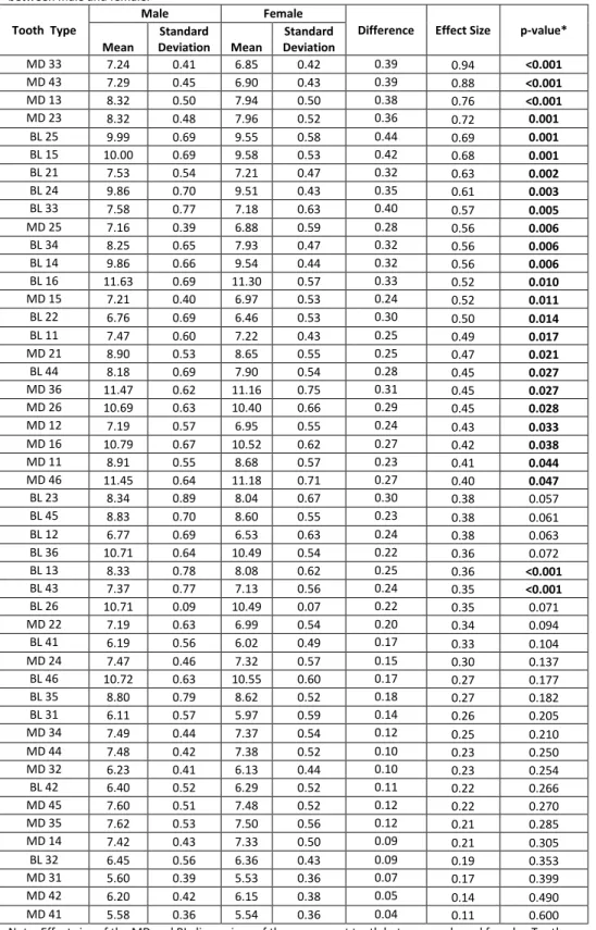
Table2. Effect size of the mesiodistal (MD) and buccolingual (BL) dimensions of the permanent teeth
between male and female.
Tooth Type
Male Female
Difference Effect Size p-value*
Mean
Standard
Deviation Mean
Standard Deviation
MD 33 7.24 0.41 6.85 0.42 0.39 0.94 <0.001
MD 43 7.29 0.45 6.90 0.43 0.39 0.88 <0.001
MD 13 8.32 0.50 7.94 0.50 0.38 0.76 <0.001
MD 23 8.32 0.48 7.96 0.52 0.36 0.72 0.001
BL 25 9.99 0.69 9.55 0.58 0.44 0.69 0.001
BL 15 10.00 0.69 9.58 0.53 0.42 0.68 0.001
BL 21 7.53 0.54 7.21 0.47 0.32 0.63 0.002
BL 24 9.86 0.70 9.51 0.43 0.35 0.61 0.003
BL 33 7.58 0.77 7.18 0.63 0.40 0.57 0.005
MD 25 7.16 0.39 6.88 0.59 0.28 0.56 0.006
BL 34 8.25 0.65 7.93 0.47 0.32 0.56 0.006
BL 14 9.86 0.66 9.54 0.44 0.32 0.56 0.006
BL 16 11.63 0.69 11.30 0.57 0.33 0.52 0.010
MD 15 7.21 0.40 6.97 0.53 0.24 0.52 0.011
BL 22 6.76 0.69 6.46 0.53 0.30 0.50 0.014
BL 11 7.47 0.60 7.22 0.43 0.25 0.49 0.017
MD 21 8.90 0.53 8.65 0.55 0.25 0.47 0.021
BL 44 8.18 0.69 7.90 0.54 0.28 0.45 0.027
MD 36 11.47 0.62 11.16 0.75 0.31 0.45 0.027
MD 26 10.69 0.63 10.40 0.66 0.29 0.45 0.028
MD 12 7.19 0.57 6.95 0.55 0.24 0.43 0.033
MD 16 10.79 0.67 10.52 0.62 0.27 0.42 0.038
MD 11 8.91 0.55 8.68 0.57 0.23 0.41 0.044
MD 46 11.45 0.64 11.18 0.71 0.27 0.40 0.047
BL 23 8.34 0.89 8.04 0.67 0.30 0.38 0.057
BL 45 8.83 0.70 8.60 0.55 0.23 0.38 0.061
BL 12 6.77 0.69 6.53 0.63 0.24 0.38 0.063
BL 36 10.71 0.64 10.49 0.54 0.22 0.36 0.072
BL 13 8.33 0.78 8.08 0.62 0.25 0.36 <0.001
BL 43 7.37 0.77 7.13 0.56 0.24 0.35 <0.001
BL 26 10.71 0.09 10.49 0.07 0.22 0.35 0.071
MD 22 7.19 0.63 6.99 0.54 0.20 0.34 0.094
BL 41 6.19 0.56 6.02 0.49 0.17 0.33 0.104
MD 24 7.47 0.46 7.32 0.57 0.15 0.30 0.137
BL 46 10.72 0.63 10.55 0.60 0.17 0.27 0.177
BL 35 8.80 0.79 8.62 0.52 0.18 0.27 0.182
BL 31 6.11 0.57 5.97 0.59 0.14 0.26 0.205
MD 34 7.49 0.44 7.37 0.54 0.12 0.25 0.210
MD 44 7.48 0.42 7.38 0.52 0.10 0.23 0.250
MD 32 6.23 0.41 6.13 0.44 0.10 0.23 0.254
BL 42 6.40 0.52 6.29 0.52 0.11 0.22 0.266
MD 45 7.60 0.51 7.48 0.52 0.12 0.22 0.270
MD 35 7.62 0.53 7.50 0.56 0.12 0.21 0.285
MD 14 7.42 0.43 7.33 0.50 0.09 0.21 0.305
BL 32 6.45 0.56 6.36 0.43 0.09 0.19 0.353
MD 31 5.60 0.39 5.53 0.36 0.07 0.17 0.399
MD 42 6.20 0.42 6.15 0.38 0.05 0.14 0.490
MD 41 5.58 0.36 5.54 0.36 0.04 0.11 0.600
Note: Effect size of the MD and BL dimensions of the permanent teeth between male and female. Tooth numbers coorelate to the numbers in Figure 1.
*Student’s t test was used to compare the means between males and females.
Table 3. Logistic regression analysis using ethnic-ity as covariates.
Tooth Type p-value
BL 12 0.029
MD 15 0.005
BL 22 0.010
MD 16 0.021
MD 26 0.017
MD 32 0.150
MD 36 0.012
BL 45 0.029
MD 46 0.024
http://dentistry3000.pitt.edu
References
1. Studies of dental anomalies in a large group of school children. Küchler EC, Risso PA, Costa Mde C, Modesto A, Vieira AR. Arch Oral Biol.
2008 Oct;53(10):941-6. doi:
10.1016/j.archoralbio.2008.04.003. PMID: 18490001
2. Human sex chromosomes in oral and craniofa-cial growth. Alvesalo L. Arch Oral Biol. 2009 Dec;54 Suppl 1:S18-24. doi: 10.1016/j.archoralbio.2008.06.004. PMID: 18657798
3. Side of dental anomalies and taurodontism as potential clinical markers for cleft subpheno-types. Küchler EC, da Motta LG, Vieira AR, Granjeiro JM. Cleft Palate Craniofac J. 2011 Jan;48(1):103-8. doi: 10.1597/09-159. PMID: 20507240
4. Sex chromosomes and human growth. A dental approach. Alvesalo L. Hum Genet. 1997 Nov;101(1):1-5. PMID: 9385359
5. 47,XXX females, sex chromosomes, and tooth crown structure. Alvesalo L, Tammisalo E, Therman E. Hum Genet. 1987 Dec;77(4):345-8. PMID: 3692479
6. Sexual dimorphism in modern human perma-nent teeth. Schwartz GT, Dean MC. Am J Phys Anthropol. 2005 Oct;128(2):312-7. PMID: 15861426
7. Sexual dimorphism in primate evolution. Plavcan JM. Am J Phys Anthropol. 2001;Suppl 33:25-53. PMID: 11786990
8. The effect of prenatal factors on crown dimen-sions. Garn SM, Osborne RH, McCabe KD. Am J Phys Anthropol. 1979 Nov;51(4):665-78. PMID: 574721
9. Use of diagonal teeth measurements in predict-ing gender in a Turkish population. Karaman F. J Forensic Sci. 2006 May;51(3):630-5. PMID: 16696712
10. Gender determination by odontometrics in a Swedish population. Lund H, Mörnstad H. J Fo-rensic Odontostomatol. 1999 Dec;17(2):30-4. PMID: 10709560
11. Mesiodistal crown diameters of permanent teeth in Jordanians. Hattab FN, al-Khateeb S, Sultan I. Arch Oral Biol. 1996 Jul;41(7):641-5. PMID: 9015564
12. Sex discrimination potential of buccolingual and mesiodistal tooth dimensions. Acharya AB, Mainali S. J Forensic Sci. 2008 Jul;53(4):790-2. doi: 10.1111/j.1556-4029.2008.00778.x. PMID: 18557797
13. Standards for mesiodistal and buccolingual crown size and height of primary molars in a sample of Spanish children. Barbería E, Suárez MC, Villalón G, Maroto M, García-Godoy F. Eur J Paediatr Dent. 2009 Dec;10(4):169-75. PMID: 20073541
14. The correlation of sexual dimorphism in tooth size and arch form. Haralabakis NB, Sifakakis I, Papagrigorakis M, Papadakis G. World J Orthod. 2006 Fall;7(3):254-60. PMID: 17009475 15. Comparison of tooth size and dental arch
widths in contemporary Japanese and American
preschool children. Yonezu T, Warren JJ, Bishara SE, Steinbock KL. World Journal of Orthodontics 2:356-360, 2001.
16. Mesiodistal crown diameters of the primary and permanent teeth in southern Chinese--a longitudinal study. Yuen KK, So LL, Tang EL. Eur J Orthod. 1997 Dec;19(6):721-31. PMID: 9458605 17. Correlations among crown diameters of human teeth. Moorrees CF, Reed RB. Arch Oral Biol. 1964 Nov-Dec;9:685-97. PMID: 14219519 18. Cohen J (1988) Statistical power analysis for the
behavioral sciences (2nd ed.). Hillsdale, NJ: Lawrence Earlbaum Associates.
19. Determining dental sex dimorphism in South Indians using discriminant function analysis. Anuthama K, Shankar S, Ilayaraja V, Kumar GS, Rajmohan M, Vignesh. Forensic Sci Int. 2011
Oct 10;212(1-3):86-9. doi:
10.1016/j.forsciint.2011.05.018. PMID: 21664775
20. Odontometric sex assessment in Indians. Prab-hu S, Acharya AB. Forensic Sci Int. 2009 Nov
20;192(1-3):129.e1-5. doi:
10.1016/j.forsciint.2009.08.008. PMID: 19744808
21. Sexual dimorphism in permanent teeth of mod-ern Greeks. Zorba E, Moraitis K, Manolis SK. Fo-rensic Sci Int. 2011 Jul 15;210(1-3):74-81. doi: 10.1016/j.forsciint.2011.02.001. PMID: 21371836
22. Genetic control of sexual dimorphism in tooth size. Garn SM, Lewis AB, Swindler DR, Kerewsky RS. J Dent Res. 1967 Sep-Oct;46(5):963-72. PMID: 5234039
23. Sexual dimorphism of the dental tissues in human permanent mandibular canines and third premolars. Saunders SR, Chan AH, Kahlon B, Kluge HF, FitzGerald CM. Am J Phys Anthro-pol. 2007 May;133(1):735-40. PMID: 17295299 24. Sexual dimorphism in the buccolingual tooth
diameter. Garn SM, Lewis AB, Kerewsky RS. J Dent Res. 1966 Nov-Dec;45(6):1819. PMID: 5226551
25. Oral clefts and syndromic forms of tooth agene-sis as models for genetics of isolated tooth agenesis. Vieira AR. J Dent Res. 2003 Mar;82(3):162-5. PMID: 12598542