Please cite this article as:V. Hajihashemi, K. Borna,An Adaptive Hierarchical Method Based on Wavelet and Adaptive Filtering for MRI Denoising, International Journal of Engineering (IJE), TRANSACTIONS A: Basics..Vol. 29, No. 1, (January 2016) 31-39
International Journal of Engineering
J o u r n a l H o m e p a g e : w w w . i j e . i rAn Adaptive Hierarchical Method Based on Wavelet and Adaptive Filtering for
Magnetic Resonance Imaging
Denoization
V. Hajihashemia, K. Bornab*
a Faculty of Engineering, Kharazmi University, Tehran, Iran
b Faculty of Mathematics and Computer Science, Kharazmi University, Tehran, Iran
P A P E R I N F O
Paper history:
Received 08 December 2015
Accepted in revised form 07 January 2016
Keywords: Adaptive Filtering Denoising Gaussian pdf
Magnetic Resonance Imaging Rician pdf
Structural Similarity Index (SSIM)
A B S T R A C T
Magnetic resonance imaging (MRI) is one of the most powerful techniques to study the internal structure of the body. MRI image quality is affected by various noises. Noises in MRI are usually thermal and mainly due to the motion of charged particles in the coil. Noise in MRI images also cause a limitation in the study of visual images as well as computer analysis of the images. In this paper, first, it is proved that probability density function (PDF) of MRI images is rician because of the process of image capturing and MRI hardware. Based on the review of later works in this area, it is determined that rician denoising in wavelet domain is better. Then, it is concluded that the remaining noise in the final output of the conventional methods in wavelet domain, is Gaussian and can be greatly reduced with a Gaussian adaptive filter. Based on this estimation, a Gaussian filter designed and the output image was filtered again. The results showed that the final image quality will improve considerably. As a conclusion, in similar situations, our proposed algorithm is always better than the others.
doi: 10.5829/idosi.ije.2016.29.01a.05
1. INTRODUCTION1
MRI is a method which is developed about 50 years ago. But within this period, many improvements and numerous Nobel prizes have been awarded for the achievements. On 2003, the Nobel prize in physiology and medicine was awarded to Paul C. Lauterbur and Sir Peter Mansfield for their interesting work on MRI improvements. In recent years, however, signal to noise ratio (SNR) and MRI images has increased but also images are polluted with noise.
Noise reduction can be divided into two parts: noise reduction while imaging methods and after imaging methods. Ways which eliminate noise while imaging increase time (for example, averaging for measuring), or reducing the spatial resolution (such as larger voxel sizes). Imaging time period due to restrictions such as patient comfort, system power and physical limitations in dynamic applications such as cardiac imaging and
1*Corresponding Author’s Email: [email protected] (K. Borna)
functional magnetic resonance imaging (FMRI) is limited. So in fact, there is a limit to SNR of MRI data and after imaging denoising methods were replaced.
Perona and Malik [4] proposed a multi-scale plan for edge detection and image smoothing which is called diffusion filter. This method can improve image quality with preserving edges and able to remove noise in homogeneous regions and edges. Because of the problem in partial differential heat equation in an anisotropic space this filter should be convolved in the direction of vertical gradient.
This technique with an improvement for denoising 2D and 3D was used by Gerig et al. [5] but sharpening the edges cause a region with a constant gray slope. In this article, image noise is regarded Gaussian with zero mean. Sharpening the edges in diffusion filter is not considered [6]. This filter is corrected, and does its work with combining rician filter and estimating filter parameters by Aja-Fernandez et al. [7].
Tang [8] proposed an improvement of diffusion filter completed Gerig’s work for reducing rician noise in MRI Images. Krissian and Aja-Fernandez [9] proposed noise driven anisotropic filter for diffusion MRI images. This filter relies on accurate prediction of the noise standard deviation. Noise prediction filter parameters are selected adaptively. Senra Filho et al. [10] is one of the last implementation of this technique that used an adaptive diffusion filter technique in MRI images. Partial differential equations filter will be used to obtain new matrix. Zhang and Ma [11] proposed anisotropic coupled relations to eliminate noise in MRI images. You and Kaveh [12] proposed a technique using 4th order partial differential equations to avoid the effects of various components in images processed with anisotropic diffusion filter. This method uses energy minimization performance of Laplacian image. Lysaker [13] used his technique for removing MRI images noise in space and time. You [12] expanded Lysaker view for denoising. This method can process signals with linear changes in intensity and protect edges of the image. Each technique above talked about, is based on the principle that noise intensity is relatively fixed and a 3D uniform distribution can be used for modeling it. Due to recent developments in parallel imaging (PI) in MRI images, the noise cannot be spatially uniform. Samsonov and Johnson [14] proposed a non-linear adaptive noise filtering for three-dimensional MRI images with different levels of noise. Given the sensitivity of the coil, basic information with regard to noise levels of the three-dimensional distribution is obtained and the non-isotropic diffusion filters to prepare the local settings are used. Tomasi et al. [15] have used a bidirectional filter based on wavelet and laplacian operator and tried to reduce SSIM and MSE between main and processed image. In another research [16], it is proved that Ricianpdf of noise in the MRI image will become a Gaussian distribution if the SNR is high and will become a probability distribution function Riley If SNR is low. With the distribution model in both
SNR has noise. In addition, according to the distribution for the noise level in two states has gain an adaptive threshold. To remove noise due to two-noise density functions assumption, the noise of background image is used [15].
Gudbjartsson and Patz [16] investigation was similar to Tomasi et al. [15] work in different aspects; based on background noise estimation, the locally an adaptive noise were accurately estimated.
In addition, the Bayesian criterion used for modeling and decision-making which rises decision accuracy and minimizes errors. Magnetic domain thermal noise in coils was modeled [15]. This noise obeys Chi-square probability density function. In especial state n=1 this pdf lead to rician function.
Awate et al, [17] have provided a non-parametric method which is based on non-parametric features of the neighborhood, and model the statistical query properties and tries to remove the noise. Other investigator [18] has worked on statistical properties of Markov and Bayesian statistical function.
Lopez-Rubio et al. [19] have used three-dimensional regression to estimate the actual amount of pixels and remove excess noise in the pixels. A good three-dimensional image has many pixel data parameters. Therefore, if we can use locally linear regression, we can guess the noise to an acceptable accuracy and remove it from the image. Zhu et al. [20] developed a method which is more focused on application of linear regression to remove noise. If the filters used in wavelet domain can help us as well. The drawback of means filter is edge destruction. It means that when using it, the image becomes smoother and so the edges fade. In literature [21], images are three-dimensional which non-local means is used and blocking is done. Blocking three-dimensional images analysis the original image and discrete wavelet is used too. In literature [22] worked based on discrete wavelet and assumed a multi-resolution state in the image. This state breaks the images to multidimensional images, images with fewer dimensions and images with more dimensions. Each one shows a series of features and we finally can eliminate noise with combination of them. Works on wavelet domain and remove noise with a bi-directional filtering [23]. In another research [24] used principle component analysis (PCA) and tried to map features to vertical space and gain good results comparing to wavelet. Haar transform is one of the wavelet branches. You have [12] used Haar wavelet and did analysis and comparing with DCT transforms and their filters.
stage of noise removing is done based on wavelet coefficients. The coefficients correlations have a key role in the threshold detection and removing the noise.
One of the weaknesses of wavelet transform is poor performance in edge detection which is very important in MRI images. One of the proposed methods to cover this disadvantage is curvelet transform. In the denoising MRI image algorithms, curvelet coefficients correct with the aim of improving the edges and so improving contrast and image quality. In other words, the presence of a function which changes properly curvelet coefficients is essential. Modified adaptive function parameters [27], is based on some statistical characteristics of curvelet in input images which cause denoising and are effectively applied on curvelet coefficients. Curvelet transform for denoising has used [22]. A five-stage algorithm with a strict threshold level proposed to correct curvelet levels. Anand et al. [23] have used previous [22] idea for MRI images denoising and Zaroubi et al. [24] combines curvelet transform with non-linear diffusion algorithm.
Pižurica et al. [26] helped Placidi et al. [25] and combined the method with anisotropic diffusion method to remove Gibbs jump caused by the lack of continuous changes and occured in discontinuity points.
Do et al. [28] tried to denoise directly and use special features. Direct methods use pixel value features and try to image denoising by estimate the actual image structure using neighbor pixels and predict pixel values. In other research [29] use minimizing least square error (MMSE) method to estimate rician noise model parameters in MRI images with mean zero and variance σ2 . This method is linear. Given the importance of the neighborhood, the neighborhood window size is very important. If the image assume three-dimensional, the neighborhood could also be time or frame. Bamberger et al. [30] has provided a method based on differential equations and solve the noise problem and try to denoising. The drawback of this method is that if the partial derived of differential equations parameters have miscalculated, the performance of the system minimize. In low image quality, Monir et al. [31] has provided an adaptive diffusion method which directly use rician model. An adaptive nonlinear diffusion method [32] is provided which is better and more effective of the linear method [33]. All of these methods directly attacked to image noise by Rician noise assuming, and tried to remove unwanted parts without removal of important image details. In some research [34], Monte Carlo estimator designed based on statistical rician process. Wong et al. [33] Used maximum likelihood method for noise estimation and denoising according to rician noise probability model. This is one of good methods for estimating parameters and pattern recognition. Sijbers et al. [35] used this criteria and background histogram with Riley probability density function to estimate noise
variance. In another work [36], a method based on linear least mean square error is used to estimate the rician noise model. In this two references, rician noise model parameters approximated locally and using a number of neighborhoods. Local parameters including mean and variance indicated a neighborhood of pixels. He et al. [36] used total image estimation to calculate noise parameters. Coupe et al. [32] used the combination of local methods and total image so that results have improved. Monir et al. [31] is assumed that three-dimensional images have enough sample size to estimate noise function parameters.
Methods based on signal amplitude, similar to two recent methods has the problem which may destroy signal amplitude and eliminate important edge data. In methods based on phase, amplitude data did not destroyed, so there is no problem in this regard. Tisdall et al. [37] used this method to keep data and noise parameters approximation simultaneously. Awate et al. [17] used a non-parametric method to estimate noise level. In the proposed method [19], higher-order statistical parameters 1, 2 (parameters other than mean and variance) of noisy pixels estimated from neighborhood noisy pixels. Finally, the higher torque is used for denoising and actual pixels estimation. [38] Use the methods for correcting image damaged using intact parts features in addition to higher-order statistic estimation and Bayesian criterion and properly do denoising. Luo et al. [38] used analysis of singularities in the frequency spectrum of the image to remove noise. Due to noise structure, the frequency spectrum is distributed uniformly in entire frequency band, while this relationship is not established in the original image. Analysis of singularities helps from spectrum difference and tries to remove the noise.
Hu et al. [39] and Coupe et al. [40] have used DCT transform for denoising which is very practical in image processing. They [39] worked by an optimization algorithm and had good results by going backward in denoising.
noise from original image. This method analyzes each component separately; therefore, it has good flexibility and customizability on image processing. Ketabchi et al. [44], have analyzed the image by moving least square method to reduce noise and zooming some areas of image. In fact, in this algorithm the structure of noise is supposed gaussian probability density function which in MRI images is not applicable. Hassanpour and Ghadi [45] showed various types of impairments; which had different effects on the illumination and reflectance of image components. They showed impairment effect on an image depending on the type of the impairment on one component is more to another component. Finally, unlike conventional methods [45] reduce the impairment effects from image components. Results showed that image enhancement had better results comparing to conventional methods. Ehsaeyan et al. [46] are used contourlet transform based on its benefit to capture the oriented geometrical structures of images. By incorporating Stein’s Unbiased Risk Estimator (SURE) approach in Nonsubsampled Contourlet Transform (NSCT) domain, they [46] proposed a new method and utilize the characteristics of NSCT coefficients in high and low subbands. They applied SURE shrinkage, bilateral filter and SURE-LET strategy to minimize the estimation of the Mean Square Error between the clean image and the denoised one in the NSCT domain. All the above explanation can be attributed to the algorithms which are features based and do not attack directly to tissue image.
The paper is organized as follows. In part two, two proposed algorithms is described. In part three, database, implementation details and results is explained, and the last part of the paper is summary and conclusion.
2. OUR PROPOSED ALGORITHM
Since the output sub-bands of wavelet includes noise and edge features, obviously proper analysis can be very effective. According to denoising methods in MRI images using wavelet transform, in proposed method, a proper threshold level is very important. Higher coefficient of threshold level will protect important signal data and lower coefficient of threshold will correct noising probability.
This method known as neighshrink threshold is widely used [23]. For the first time, Zhou [47] used neighshrink for denoising. Zhou proposed method [47]using optimal selected parameters and obtained much better results than that of other previous methods in MRI denoising.
2. 1. Soft Threshold Level Wavelet coefficients
Wij with greater amplitude than a determined threshold
λ, shoud be maintained to reconstruct of signal. Noise free coefficients ˆijestimated using soft threshold level as follows [23]:
0 ˆ
ij ij
ij ij
ij ij
W W
W
W W
(1)
λ considered as a general threshold level and is calculated by the following equation:
2log
n n
(2)
where, n is the number of samples and σnis the estimated noise variance. In our proposed method, appropriate noise estimation are taken from MRI image background according to literature [23].
2. 2. Neighshrink Threshold Assume that the size of the neighborhood window is L × L. For
2 2
, ij
ij k I B ki
S
W
Neighshrink formula [43] ispresented as follows:
ij ij i
2 2
j, max(1 ,0)
w B ij
ij B
S
(3)
In the above equation: ˆij is uncertain signal coefficient
estimation and λ is threshold level. Optimal value of λ and L for each high frequency subbands is estimated by using Stein's unbiased risk estimate (SURE) algorithm [43]. Optimal threshold λs with window size Ls minimize SURE (Ws, λ, L) value as follow:
,
,
, ,
s s
L s
L arg min SURE w L
(4)Before calculating optimal neighshrink parameters, estimated coefficients are normalized in nonsingular variance data σn.
2. 3. Summary of WD-BF Algorithm (Wavelet Domain Bilateral Filtering) Anand et al. [23] calculate noisy MRI image square to detrmine square magnitude of Isq.
Separate N level for Isq using UDWT for getting approximation and coefficients details.
1- Bias in coefficients approximate is obtained at level j by subtracting from σn
2
2j+1 [23]
2- These unbiased coefficients will be passed from bilateral filter.
3- Denoising detail coefficients using neighshrink technique.
4- UDWT filter and its detail coefficients calculation to get an estimate for Isq.
2. 4. Applying Adaptive Gaussian Filter Since the output image is filtered against rician noise in this stage and correction is done on it, it should assumed that retained noise due to filtering error and wavelet coefficients correction, probably is a Gaussian noise. It should be explained that according to central limit theorem in statistic, combination of all natural processes which excess certain limit and alos have uncertain distribution create Gaussian process.
Variance and means of this Gaussian noise is uncertain and therefore it should be help by an optimal adaptive method to approximate it. Such test parameters estimation from a Gaussian distribution is named as parametric test. In the first stage, chi-square test was used for prove Gaussian distribution of remainder noise. Chi-square test or Goodness of fit is used to compare observed distribution and theoretical distribution. Structural equation modeling where the researcher has outlined a theoretical model based on variables relationships can be used. A significance level of 0.05 (confidence level 95%) showed on remaining test images which had Gaussian distribution in all noise level percentages.
In next stage, the following methods in background of image were used to estimate mean and variance according to normal distribution of noise.
Single-sample t test: this method used for hypothesis test around expected value on average noise. Based on the Likert scale, we can estimate mean of each window from background in a significant level using this method. Paired t-test: different image points were used to compare two backgrounds means difference. For example, mean difference between right and left corner of the image.
Two independent samples t-test: was used to compare mean noise in bilateral image. The noise variance in two independent samples t-test is assumed equal bilaterally.
Welch’s t-test: This test like two independent samples t-test used for comparison of bilateral image mean. Welch's t-test assumes that variance is not equal. According to results of four above tests, it is obvious that noise distribution in total image parts has a significance level 0.01 and noise variance is equal in all parts with significance level 0.05. In this way, only discussed, correct estimation of mean and variance of output image background noise of previous method that it will be easy. Finally, after estimation, it is enough that the image is filtered again with Gaussian filter using these parameters.
3. IMPLEMENTATION AND RESULTS
Implementation is exactly according to part 2 of the article. The database used in implementation was
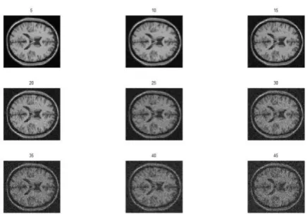
database [48]. First, some sample MRI images from several brain layers of this database show for this purpose which reader know the form of database images. Figure 1 shows several MRI images. Number above each images represents number of each layer which is MRI images. These images taken directly from database and are not noisy.
After showing various layers of noise-free images to determine the effect of noise in output image, 90th image (approximately in the middle of a group of MRI images) are combined with rician noise in levels 5 to 45% with step 5, which is shown in Figure 2. As Figure 2 depicts, the image has no difference with the base image at noise levels 5%. Nevertheless, with increasing the percentage of noise, the image quality will be reduced. After this, Figure 3 shows denoising images by using [48] according to different percentage of noise level. Although naked eye cannot detect upgrading, but tables and SSIM criteria show that proposed method [48] could reduce nonlinear noise of images to an acceptable level and we have quality increasing even comparing with new and strict SSIM. Figure 4 shows the output images based on our proposed method.
Figure 1. Image samples of several brain layers.
Figure 3. Result of Denoising in Figure 2 by method [48]
Figure 4. Denoising using our proposed method
Comparing Figures 4 and 3 in literature [48] without applying our proposed method shows significance improvement with further processing stage.
To confirm visual improvement by standard criteria, Table 1 shows SSIM which is one of standard image quality evaluation parameters for output images [51, 53] and our proposed method. Shyam Anand [51] study was one of the fundamental researches in the MRI denoising based on wavelet method. Manjon et al. [53] is one of the most recent investigations on denoising image field.
It can be easily seen from our proposed method with SSIM criteria, in all cases, had significant improvements rather than other works [23, 49]. Estimated mean and variance of retained Gaussian noise of output images presented in separate columns for the evaluation of our proposed method validity. While rician noise increases, mean and variance of Gaussian noise of output images [23, 49] increase too. This matter fully justifies use of our proposed method and shows that the proposed idea is correct. It should be noted that reference [49] is one of the latest work on this field which is done in 2015 using principal components
analysis. Since noises in images are naturally Rician, our comparison has just been done for this noise.
TABLE 1. Comparison of output images in proposed method and [23, 49] with SSIM criteria
Noise level%
SSIM
Noise mean
Noise variance
reference
53 reference 51
Proposed method
5 0.989 0.9 0.989 0.06 5.289 10 0.964 0.621 0.969 -1.45 10.403 15 0.934 0.453 0.952 -3.32 15.581 20 0.897 0.325 0.935 -5.59 20.455 25 0.861 0.321 0.917 -8.25 25.331 30 0.820 0.270 0.9 -11.01 29.922 35 0.783 0.233 0.883 -14.15 34.326 40 0.710 0.199 0.870 -17.27 38.843 45 0.680 0.169 0.852 -20.74 43.242
Figure 5. Our proposed method and [23, 49] methods in SSIM (bar chart)
Figure 6. Comparison of our proposed method and [50] in SSIM.
For more accurate and easier comparison of proposed method and methods in literatures [23, 49], SSIM parameters variations of three methods presented
0 0.2 0.4 0.6 0.8 1 1.2
1 2 3 4 5 6 7 8 9
method [53] method [51] proposed method
0 0.2 0.4 0.6 0.8 1 1.2
1 2 3 4 5 6 7 8 9 10 11 12
in Figure 5. It is completely obvious that our proposed method and methods in research [49] are almost similar around origin with lower noise level but with higher noise level, proposed method is completely more efficient.
In Figure 6, the difference between proposed method and method of literature [49] presented as a line chart to show a significant difference between two methods in low SNR. Vertical axis shows SSIM and horizontal axis shows percentage of noise divided by five.
4. CONCLUSION
This paper proposed a method to improve quality of medical MRI images. At First, we proved that according to process occurring in image capture hardware MRI, noise follows rician probability density function. Based on the review of works in this area, it is determined that denoising according to rician probability density function in wavelet space is done much better. After that the denoising principles in wavelet space completely is explained and a new high quality work based on wavelet is implemented. The quality reduction analysis in final output image results showed that output retained noise has Gaussian PDF in these methods and we can reduce it with an adaptive Gaussian filter. According to histograms in output images and using an adaptive estimation of Gaussian noise in it, a Gaussian filter designed and output image filtered again using this filter. The results showed that, final image have better quality than prior outputs. In addition, SSIM used to prove the validity of our proposed method. The final image compared with original noise free image and the best output of prior methods showed that, the output image of our proposed method is better than prior results.
6. REFERENCES
1. McVeigh, E., Henkelman, R. and Bronskill, M., "Noise and filtration in magnetic resonance imaging", Medical physics, Vol. 12, No. 5, (1985), 586-591.
2. Bird, R.E., Wallace, T.W. and Yankaskas, B.C., "Analysis of cancers missed at screening mammography", Radiology, Vol. 184, No. 3, (1992), 613-617.
3. Arodź, T., Kurdziel, M., Popiela, T.J., Sevre, E.O. and Yuen, D.A., "Detection of clustered microcalcifications in small field digital mammography", computer methods and programs in biomedicine, Vol. 81, No. 1, (2006), 56-65.
4. Perona, P. and Malik, J., "Scale-space and edge detection using anisotropic diffusion", Pattern Analysis and Machine Intelligence, IEEE Transactions on, Vol. 12, No. 7, (1990), 629-639.
5. Gerig, G., Kübler, O., Kikinis, R. and Jolesz, F., "Nonlinear anisotropic filtering of mri data", Medical Imaging, IEEE Transactions on, Vol. 11, No. 2, (1992), 221-232.
6. Coakley, K., Quintarelli, F., Van Doorn, T. and Hirst, C., "Classification of equivocal mammograms through digital analysis", The Breast, Vol. 3, No. 4, (1994), 222-226. 7. Aja-Fernández, S., Alberola-López, C. and Westin, C.-F., "Noise
and signal estimation in magnitude mri and rician distributed images: A lmmse approach", Image Processing, IEEE Transactions on, Vol. 17, No. 8, (2008), 1383-1398.
8. Tang, J., Sun, Q., Liu, J. and Cao, Y., "An adaptive anisotropic diffusion filter for noise reduction in mr images", in Mechatronics and Automation. ICMA. International Conference on, IEEE. (2007), 1299-1304.
9. Krissian, K. and Aja-Fernández, S., "Noise-driven anisotropic diffusion filtering of mri", Image Processing, IEEE Transactions on, Vol. 18, No. 10, (2009), 2265-2274. 10. Senra Filho, D.S., Carlos, A., Jinzenji Duque, J., Junior, M. and
Luiz, O., "Isotropic anomalous filtering in diffusion-weighted magnetic resonance imaging", in Engineering in Medicine and Biology Society (EMBC), 35th Annual International Conference of the IEEE, (2013), 4022-4025.
11. Zhang, F. and Ma, L., "Mri denoising using the anisotropic coupled diffusion equations", in Biomedical Engineering and Informatics (BMEI), 3rd International Conference on, IEEE. Vol. 1, (2010), 397-401.
12. You, Y.-L. and Kaveh, M., "Fourth-order partial differential equations for noise removal", Image Processing, IEEE Transactions on, Vol. 9, No. 10, (2000), 1723-1730.
13. Lysaker, M., Lundervold, A. and Tai, X.-C., "Noise removal using fourth-order partial differential equation with applications to medical magnetic resonance images in space and time",
Image Processing, IEEE Transactions on, Vol. 12, No. 12, (2003), 1579-1590.
14. Samsonov, A.A. and Johnson, C.R., "Noise‐adaptive nonlinear diffusion filtering of mr images with spatially varying noise levels", Magnetic Resonance in Medicine, Vol. 52, No. 4, (2004), 798-806.
15. Tomasi, C. and Manduchi, R., "Bilateral filtering for gray and color images", in Computer Vision,. Sixth International Conference on, IEEE. (1998), 839-846.
16. Gudbjartsson, H. and Patz, S., "The rician distribution of noisy mri data", Magnetic Resonance in Medicine, Vol. 34, No. 6, (1995), 910-914.
17. Awate, S.P. and Whitaker, R.T., "Nonparametric neighborhood statistics for mri denoising", in Information Processing in Medical Imaging, Springer. (2005), 677-688.
18. Awate, S.P. and Whitaker, R.T., "Feature-preserving mri denoising: A nonparametric empirical bayes approach", Medical Imaging, IEEE Transactions on, Vol. 26, No. 9, (2007), 1242-1255.
19. López-Rubio, E. and Florentín-Núñez, M.N., "Kernel regression based feature extraction for 3d mr image denoising", Medical image analysis, Vol. 15, No. 4, (2011), 498-513.
20. Zhu, H., Li, Y., Ibrahim, J.G., Shi, X., An, H., Chen, Y., Gao, W., Lin, W., Rowe, D.B. and Peterson, B.S., "Regression models for identifying noise sources in magnetic resonance images", Journal of the American Statistical Association, Vol. 104, No. 486, (2009), 623-637.
21. Coupé, P., Yger, P., Prima, S., Hellier, P., Kervrann, C. and Barillot, C., "An optimized blockwise nonlocal means denoising filter for 3-d magnetic resonance images", Medical Imaging, IEEE Transactions on, Vol. 27, No. 4, (2008), 425-441. 22. Anand, C.S. and Sahambi, J., "Mri denoising using bilateral
23. Anand, C.S. and Sahambi, J.S., "Wavelet domain non-linear filtering for mri denoising", Magnetic Resonance Imaging, Vol. 28, No. 6, (2010), 842-861.
24. Zaroubi, S. and Goelman, G., "Complex denoising of mr data via wavelet analysis: Application for functional mri", Magnetic Resonance Imaging, Vol. 18, No. 1, (2000), 59-68.
25. Placidi, G., Alecci, M. and Sotgiu, A., "Post-processing noise removal algorithm for magnetic resonance imaging based on edge detection and wavelet analysis", Physics in Medicine and Biology, Vol. 48, No. 13, (2003), 1987.
26. Pižurica, A., Philips, W., Lemahieu, I. and Acheroy, M., "A versatile wavelet domain noise filtration technique for medical imaging", Medical Imaging, IEEE Transactions on, Vol. 22, No. 3, (2003), 323-331.
27. Ashamol, V., Sreelekha, G. and Sathidevi, P., "Diffusion-based image denoising combining curvelet and wavelet", in Systems, Signals and Image Processing,. IWSSIP. 15th International Conference on, IEEE. Vol., No. Issue, (2008), 169-172. 28. Do, M.N. and Vetterli, M., "The contourlet transform: An
efficient directional multiresolution image representation",
Image Processing, IEEE Transactions on, Vol. 14, No. 12, (2005), 2091-2106.
29. Parthiban, L. and Subramanian, R., "Medical image denoising using x-lets", in India Conference, Annual IEEE, (2006), 1-6. 30. Bamberger, R.H. and Smith, M.J., "A filter bank for the
directional decomposition of images: Theory and design",
Signal Processing, IEEE Transactions on, Vol. 40, No. 4, (1992), 882-893.
31. Monir, S.M.G. and Siyal, M.Y., "Denoising functional magnetic resonance imaging time-series using anisotropic spatial averaging", Biomedical Signal Processing and Control, Vol. 4, No. 1, (2009), 16-25.
32. Coupé, P., Manjón, J.V., Gedamu, E., Arnold, D., Robles, M. and Collins, D.L., An object-based method for rician noise estimation in mr images, in Medical image computing and computer-assisted intervention–miccai. Springer (2009), 601-608.
33. A wong, a k mishra, quasi-monte carlo “estimation approach for denoising mri data based on regional statistics”, IEEE trans.
Biomedical Engineering Vol. 58, (2011), 1076–1083
34. Sijbers, J. and Den Dekker, A., "Maximum likelihood estimation of signal amplitude and noise variance from mr data", Magnetic Resonance in Medicine, Vol. 51, No. 3, (2004), 586-594. 35. Sijbers, J., Poot, D., den Dekker, A.J. and Pintjens, W.,
"Automatic estimation of the noise variance from the histogram of a magnetic resonance image", Physics in Medicine and Biology, Vol. 52, No. 5, (2007), 1335.
36. He, L. and Greenshields, I.R., "A nonlocal maximum likelihood estimation method for rician noise reduction in mr images",
Medical Imaging, IEEE Transactions on, Vol. 28, No. 2, (2009), 165-172.
37. Tisdall, D. and Atkins, M.S., "Mri denoising via phase error estimation", in Medical imaging, International Society for Optics and Photonics. (2005), 646-654.
38. Luo, J., Zhu, Y. and Hiba, B., "Medical image denoising using one-dimensional singularity function model", Computerized Medical Imaging and Graphics, Vol. 34, No. 2, (2010), 167-176.
39. Hu, J., Pu, Y., Wu, X., Zhang, Y. and Zhou, J., "Improved dct-based nonlocal means filter for mr images denoising",
Computational and Mathematical Methods in Medicine, Vol. 2012, No., (2012), 232-685
40. Coupé, P., Yger, P. and Barillot, C., Fast non local means denoising for 3d mr images, in Medical image computing and computer-assisted intervention–miccai , Springer (2006). 33-40. 41. Rajan, J., Poot, D., Juntu, J. and Sijbers, J., "Noise measurement from magnitude mri using local estimates of variance and skewness", Physics in Medicine and Biology, Vol. 55, No. 16, (2010), 441-449.
42. Nadernejad, E., Hassanpour, H. and Miar, H., "Image restoration using a pde-based approach", International Journal of Engineering Transactions B: Applications, Vol. 20, No. 3, (2007),225-236.
43. Khosravi, M. and Hassanpour, H., "Image denoising using anisotropic diffusion equations on reflection and illumination components of image", International Journal of Engineering-Transactions C: Aspects, Vol. 27, No. 9, (2014), 1339-1348. 44. S. Ketabchi, M. Kianpour, r. Valizadeh, m.J. Mahmoodabadi,
a new technique for image zooming based on the moving least squares, International Journal of EngineeringTransactions C: Aspects, Vol. 25, No. 2, (2012), 105–109.
45. Hassanpour, H. and Ghadi, A.R., "Image enhancement via reducing impairment effects on image components",
International Journal of Engineering-Transactions B: Applications, Vol. 26, No. 11, (2013), 1267-1274.
46. E. Ehsaeyan, A robust image denoising technique in the contourlet transform domain, International Journal Of Engineering Transactions B: Applications, Vol. 28, No. 11 (2015), 1589-1596.
47. Yu, H. and Zhao, L., "An efficient denoising procedure for magnetic resonance imaging", in Bioinformatics and Biomedical Engineering. ICBBE. The 2nd International Conference on, IEEE. (2008), 2628-2630.
48. Kwan, R.K., Evans, A.C. and Pike, G.B., "Mri simulation-based evaluation of image-processing and classification methods",
Medical Imaging, IEEE Transactions on, Vol. 18, No. 11, (1999), 1085-1097.
49. Manjón, J.V., Coupé, P. and Buades, A., "Mri noise estimation and denoising using non-local pca", Medical Image Analysis, Vol. 22, No. 1, (2015), 35-47.
50. H Yu, L Zhao, “An efficient denoising procedure for magnetic resonance imaging”, in: Proceedings of IEEE 2nd International Conference on Bioinformatics and Biomedical Engineering, (2008) .
51. C Shyam Anand, S Jyotinder Sahambi, “Wavelet domain non-linear filtering for MRI denoising”, Magnetic Resonance Imaging, Vol. 28, )2010(, 842-86.
52. RK Kwan, AC Evans, GB Pike, “MRI simulation-based evaluation of image-processing and classification methods”, IEEE Trans. Med Image, Vol. 18, (1999), 1085-1097.
An Adaptive Hierarchical Method Based on Wavelet and Adaptive Filtering for
Magnetic Resonance Imaging
Denoization
V. Hajihashemia, K. Bornab
aFaculty of Engineering, Kharazmi University, Tehran, Iran
bFaculty of Mathematics and Computer Science, Kharazmi University, Tehran, Iran
P A P E R I N F O
Paper history:
Received 08 December 2015
Accepted in revised form 07 January 2016
Keywords: Adaptive Filtering Denoising Gaussian pdf
Magnetic Resonance Imaging Rician pdf
Structural Similarity Index (SSIM)
ديكچ ه
MRI
یاه سکع تیفیک .دشاب یم ندب یلخاد یاه تمسق یراتخاس هعلاطم یاهکینکت نیرتیوق زا یکی
MRI
ریثات تحت
رد زیون .دنتسه یفلتخم یاهزیون
MRI
سناکرف رد رادراب تارذ تکرح رطاخ هب هک دشاب یم یترارح زیون عون زا اتدمع
ب چیپ میس ییویدار ریواصت رد زیون .دیآ یم دوجو ه
MRI
نینچمه و ریواصت یرهاظ یسررب رد تیدودحم داجیا ثعاب
رازفا تخس رد هداد خر دنیآرف ساسا رب دوش یم تابثا ادتبا هلاقم نیا رد .دوش یم رتویپماک طسوت ریواصت نیا زیلانآ ریوصت یرادربسکع
MRI
متحا یلاگچ عبات زا ریواصت نیا رد دوجوم زیون ، اب و ساسا نیا رب .دنک یم یوریپ نیسیار لا
یاضف رد نیسیار لامتحا یلاگچ عبات هب هجوت اب زیون فذح دش صخشم هنیمز نیا رد هدش ماجنا یاهراک هعومجم رورم ییاهن یجورخ رد هدنام یقاب زیون هک دش یریگ هجیتن روطنیا هتفرگ تروص یاه لیلحت اب .دریگ یم تروص رتهب کجوم رم یاهشور ات یسوگ یقیبطت رتلیف کی اب ار نآ ناوت یم هک دراد یسوگ تیهام یرتلیف یاطخ لیلد هب ،کجوم هزوح موس ریوصت رد دوجوم یسوگ زیون یقیبطت دروآرب و لضافت ریواصت رد دوجوم یاه مارگوتسیه ساسا رب .داد شهاک یدایز دح
ریوصت نآ زا هدافتسا اب و یحارط یسوگ رتلیف کی ییاهن یجورخ ناشن جیاتن .دیدرگ رتلیف هرابود هدش زیون فذح یجورخ
هولاع شور ییآراک و تحص تابثا یارب نآ رب هولاع .دنک یم ادیپ یا هظحلام لباق دوبهب تیفیک رظن زا ییاهن ریوصت داد رایعم زا هدافتسا اب یمشچ رایعم رب
SSIM
هشور یجورخ نیرتهب و زیون نودب هیلوا ریوصت اب ییاهن ریوصت ، دروم لبق یا
دهاوخ رتهب یلبق جیاتن زا هراومه یداهنشیپ شور یجورخ تیفیک ناسکی تلاح رد دش هداد ناشن و تفرگ رارق هسیاقم .دوب
![Table 1 shows SSIM which is one of standard image To confirm visual improvement by standard criteria, quality evaluation parameters for output images [51, 53] and our proposed method](https://thumb-us.123doks.com/thumbv2/123dok_us/220974.2016612/6.595.309.535.154.497/standard-confirm-improvement-standard-criteria-evaluation-parameters-proposed.webp)