*Corresponding author: Raghuveer H.P ISSN: 0976-3031
Review Article
BASAL CELL CARCINOMA-A CASE REPORT AND REVIEW
Raghuveer H.P*
1., Sudeeptha Raghuveer
2., Shobha E.S
3., Prashanth N.T
4.,
Vinod Rangan
5and Nikhila G
61, 3,4,5,6
Department of Oral and Maxillofacial Surgery, Dayananda Sagar
College of Dental Sciences, Bangalore
2
MBBS, Junior Resident
DOI: http://dx.doi.org/10.24327/ijrsr.2017.0811.1069
ARTICLE INFO ABSTRACT
Although Basal cell carcinoma is only locally aggressive, if not completely removed, recurrence may occur and be troublesome, especially in the head and neck. This review aims to present with a brief and inclusive overview of basal cell carcinoma in terms of etiology, clinical features, diagnosis and classification, and treatment modalities.
INTRODUCTION
Basal cell carcinoma (BCC) is locally malignant tumour of the basal layer of the epidermis occurring probably due to the prolonged exposure to sun.1 This tumour generally involve persons of fair complexion, making dark coloured individuals somewhat immune to this disease. The incidence rates of this tumour in Indian population are very less. Treatment of this tumour is usually done by wide local excision. Recurrence rates, even after complete cure, are high, so the patient has to remain in long term follow-up
Case Report
A 64 years old female patient presented to our department, with an ulcerated exophytic growth on the lateral aspect of the ala of the nose on the left side of the patient’s face, measuring approximately 1x1cm in size. On examination the mass was firm in consistency, non-tender, fixed to the skin and with irregular margins Patient was advised biopsy for the lesion. An incisional biopsy specimen was taken from the margin of the ulcerative growth and its adjoining area. The report gave the diagnosis as Basal cell carcinoma.
Patient was advised excision of the lesion with reconstruction under general anaesthesia. After obtaining informed consent from the patient, pre-anaesthetic workup was done. After induction of general anaesthesia, the lesion was excised with an elliptical incision with healthy margins in all dimensions. The excised tissue was sent for histopathological examination. A triangular advancement flap was used to close the defect.
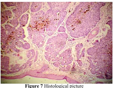
The histopathological report of the excised specimen confirmed the diagnosis of basal cell carcinoma. The final result at three weeks was satisfactory. There was minimal deformity of the nose with no morbidity to the patient. Patient was followed up for recurrence for the next six months. There was no evidence of recurrence in the follow up period
DISCUSSION
Nonmelanoma skin cancers (NMSCs) mainly comprise of squamous cell carcinoma (SCC) and basal cell carcinoma (BCC). These neoplasms are common in white-skinned individuals but are rare in dark skin phenotypes The NMSCs compriseof 1%-2% of cutaneous neoplasms in Indians, in contrast to one-third in whites2,4.
International Journal of
Recent Scientific
Research
International Journal of Recent Scientific Research
Vol. 8, Issue, 11, pp. 21412-21416, November, 2017
Copyright © Raghuveer H.P et al, 2017, this is an open-access article distributed under the terms of the Creative Commons Attribution License, which permits unrestricted use, distribution and reproduction in any medium, provided the original work is properly cited.
DOI: 10.24327/IJRSR CODEN: IJRSFP (USA)
Article History:
Received 06th August, 2017 Received in revised form 14th September, 2017
Accepted 23rd October, 2017
Published online 28th November, 2017
Key Words:
Basal cell carcinoma, also known as “rodent ulcer” was termed first by Jacob and Dublin in 1827. It is considered to be the most common skin malignancy in adults of fair-complexion. It is a locally invasive, slow growing malignancy that arises from the basal layer of the epidermis and its appendages. Studies have shown that 75 - 85% of the tumours occur in head and neck region and remaining occurs on the trunk and lower limbs, particularly in women.
Tumours can occur on nose (32.3%), orbital (19.1%) and cheek (18.1%) regions as these areas are comparatively more exposed to sunlight1,5,6,7. Lesions of frontotemporal, scalp and auricular region are more common in men, which can be due to short hair or androgenic alopecia, perioral lesions especially more on the upper lip are found to be less in men1,4. BCC is more prevalent in 6th and 7th decade with the mean age of occurrence being 62 years1. In the last decade incidence of BCC has substantially increased and also shows geographical variations. The size of the lesion may vary from 0.2 mm to 9 cm with a mean size of 2 cm. Clinical subtypes seen are nodular, superficial, fibroepithelial and morpheaform1,2,3. The most frequently occurring subtype is the nodular variety6.
Aetiology
Aetiology of BCC may be attributed to depletion of ozone layer due to environmental pollution and increased exposure to UV light, which damage the DNA resulting in development of skin cancer12. The type of UV exposure (intermittent vs. chronic), the point in time (childhood vs. adulthood), the source (natural vs. artificial), and the quantity (number of severe sunburns) appear to vary the risk. Patients with immunosuppression are more prone to the disease. A study shows that the incidence in transplant recipients was 10 times higher than in the generalpopulation. BCC is also associated with various syndromes like Nevoid Basal Cell Carcinoma Syndrome (Gorlin's syndrome), Darier’s disease, albinism, xeroderma pigmentosa, Bazex's syndrome
Clinical Features
Early BCC appear as small, translucent or pearly, with raised areas in which dilated vessels can be seen (telangiectasia). The classical appearance of rodent ulcer is in which edges are indurated and centre is ulcerated. Typical BCC are indolent with slow progression, but if neglected can become locally destructive and result in fatal outcomes due to locally destructive growth into vital structures. But however they have a very limited potential for metastasis. The tumours that metastasize maybe those which were neglected lesions and also recurred lesions despite repeated treatment. The metastatic rate
Illustrations
Figure 1 Pre- op
Figure 2 Excision of the lesion
Figure 3 Elliptical defect post excision
Figure 4 Triangular advancement flap
Figure 5 Triangular advancement flap
Figure 6 Immediate Post- op
ranges from 0.0028% to 0.55%. An increased risk of developing further BCC is in patients. Risk of developing SCC is increased after BCC with a 6% risk
Diagnosis and Classification
WHO, classified basal cell carcinoma into the following subtypes:
Superficial basal cell carcinoma,
Nodular (solid) basal cell carcinoma,
Micronodular basal cell carcinoma,
Infiltrative basal cell carcinoma,
Fibroepithelial basal cell carcinoma (Pinkus tumor),
Basal cell carcinoma with adnexal differentiation,
Basosquamous carcinoma,
Keratotic basal cell carcinoma.
Superficial basal cell carcinoma is characterized by apparently separate and usually round buds of tumor cells pushing into the papillary dermis. There is a characteristic retraction artifact between the tumor cells and the surrounding connective tissue. In case of longstanding tumors, the tumor buds can extend into deeper layers of the dermis and exhibit secondary nodular growth.
Nodular BCC consists of large nodules of basaloid cells with peripheral palisading, which push into the reticular dermis or deeper. Mucinous cystic spaces may be present inside the tumor cell aggregates because of degenerative changes. Peritumoral retraction artifacts and fibromucinous stroma can also be found. Micronodular BCC shows smaller tumor cell nodules compared with nodular BCC. The typical peritumoral retraction artifacts are missing.
Infiltrative BCC consists of small strands of basaloid tumor cells, which may be only one to two cell layers thick. Peritumoral retraction and peripheral palisading of tumor cells are absent. The sclerodermiform (morphea-like) variant is characterized by marked stromal fibrosis or sclerosis. Fibroepithelial BCC, also known as Pinkus tumor, is marked by narrow reticulate strands of neoplastic cells arising from the epidermis, which show hair germ-like structures and are surrounded by a fibrovascular stroma.
Basal cell carcinoma with adnexal differentiation is characterized by tumor components that show differentiation towards adnexal structures. Here, apocrine, eccrine, sebaceous, and follicular differentiation may occur. Basosquamous carcinoma is a rare basal cell carcinoma subtype with squamous differentiation. Histologically, the tumor cells are pleomorphic and show a broader cytoplasm and more open chromatin. BCC-typical palisading as well as immunohistochemical BerEP4 expression is not consistently present. The prognosis of basosquamous carcinoma is poorer, and corresponds more to that of squamous cell carcinoma than BCC.
Keratotic basal cell carcinoma has the architecture of nodular BCC and is characterized by prominent keratinization and formation of horn cysts within the tumor cell clusters. Dystrophic calcification is also a frequent finding.
Differential Diagnoses
Includes malignant melanoma, squamous cell carcinoma, melanocytic naevi (pigmented), Bowen's disease (especially superficial basal cell carcinoma), psoriasis (superficial), sebaceous hyperplasia, molluscum contagiosum, and eczema (superficial)
Treatment
The goal of treating BCC is to eradicate the tumour with the safest and most effective method available and to provide an aesthetically and functionally pleasant outcome. Treatment modalities include curettage and cautery, cryosurgery, and surgical excision. Curettage, cautery and cryosurgery leaves behind wounds which may take longer time to heal than those wounds of surgical excision. However adequate tissue samples to examine the margins for clearance is not available in these techniques. In case of management of recurrent, large tumours at high risk sites, curettage and cautery is not recommended13.
The most common preferred treatment of BCC is surgical excision1,2. The surgical excision margin depends on the clinical features, size and location of the lesion. The main advantage of this treatment modality is that the excision margins can be histologically examined to check for clearance. A surgical margin of 3 mm is enough for clearance of 85% of small, well defined BCCs. Currently surgical excision margin to 4 - 5mm is recommended as its provides a complete excision rate of approximately 95%1, 6.
Moh’s micrographic surgery is a specialised technique. It provides high cure rates at high risk sites (central face), morphoiec tumours, and recurrent tumours, with maximum preservation of normal tissues. In this technique, adequate marginal clearance can be achieved as serial sections can be taken and histologically examined till margins are clear6,23. Thus, subclinical tumor extensions can be topographically mapped and excised until the outer margins of the excised specimen are tumor free. This procedure can also be beneficial for small and unproblematic tumors, as healthy skin may be spared and the excision can be limited to tissues with histologically proven tumor infiltration. Overall 5 year cure rate with Moh’s micrographic surgical technique is approximately 99% for primary tumours and upto 95% for recurrent lesions18,19.
Radiation therapy is a therapeutic option when surgery is not feasible or reasonable because of tumor location or extent or due to comorbidities12. Radiotherapy is generally used in elderly patients, but it is not recommended in younger patients as its results are inferior to those obtained by surgery. 5 year cure rate obtained by this treatment modality is approximately 90%20.
thickness which affects the uptake of photosensitiser and penetration of light source.
Another treatment option available is topical application of fluorouracil 5% cream. It is used in management of multiple superficial basal cell carcinoma lesions and is applied twice daily over a period of 3–6 weeks21. Treatment should be continued until an erosion occurs; Its use may be extended if necessary. Erythema, pruritus, pain, erosions, and ulcerations are certain side effects seen. A newer topical immunomodulatory treatment is imiquimod 5% cream.It is used in the treatment of small superficial and multiple basal cell carcinomas in immunocompetent adults22. A maximum tumor size of 2 cm outside the “risk locations” (nose, ears, eyelids, anogenital region, hands and feet) can be treated by this method. It is used 5 times a week over a period of 6 weeks. Therapeutic success should be checked 12 weeks after discontinuation of treatment.It has shown a clearance of 70 – 100%, when used for superficial lesions15.Local reactions with erythema, pruritus, burning, erosions, ulcerations, and scabs may be the side effects seen.
When compared to the lesions on the trunk and extremities, lesions of head and neck region are at more risk for recurrence 6,9,10,11
. Studies have shown that most of the recurrences were in the median parts of the head and neck region, this may be due to high recurrence rates in embryonic fusion planes. Mostly micronodular and sclerotic histopathological subtype is seen to recur and is more difficult to eradicate1. These lesions have lower cure rates with most treatment modalities compared to primary tumours. Cryosurgery or curettage and cautery is not recommended for recurrenttumours13. The overall five year cure rate for recurrent basal cell carcinoma treated by curettage and cautery is estimated at 60%. In general, recurrent tumours, especially morphoeic tumours or recurrences at high risk sites, are best treated by Mohs' micrographic surgery.
Follow UP and Prevention
The development of new basal cell carcinomas maybe prevented or delayed by oral retinoid treatment. It is mainly used in patients with Gorlin’s syndrome, renal transplant patients who are at high risk, actinically damaged patients.
Given that patients are at risk of developing further basal cell carcinoma it may be wise to follow up high risk patients with multiple or truncal lesions. Educating the patients about sun avoidance and tumour detection may help to prevent further malignancies and facilitate diagnosis of smaller basal cell carcinomas, which, in general, are easier to treat and have less morbidity.
CONCLUSION
It is important to define the surgical margins to achieve clearance and thereby reduce recurrence rate. Educating and motivating patients for follow up visits, helps to evaluate outcomes and diagnose recurrences.
References
1. Duriye DD, Candemir C, Berrak A, Mustafa ED, Ahmet M. Basal Cell Carcinoma of the Head and Neck Region: A Retrospective Analysis of Completely Excised 331 Cases. J Skin Cancer. 2014;
2. Soyer HP, Rigel DS, Wurm EMT. Actinic keratosis, basal cell carcinoma and squamous cell carcinoma. In: Bolognia JL, Jorizzo JL, Schaffer JV, editors. Dermatology. Beijing, China: Elsevier Saunders; 2012. pp. 1773–1793.
3. Rippey JJ. Why classify basal cell carcinomas? Histopathology. 1998;32(5):393-398.
4. Bastiaens MT, Hoefnagel JJ, Bruijn JA, Westendorp RGJ, Vermeer BJ, Bavinck JNB. Differences in age, site distribution, and sex between nodular and superficial basal cell carcinomas indicate different types of tumors. Journal of Investigative Dermatology. 1998;110(6):880-884.
5. Tiftikcioğlu YO, Karaaslan O, Aksoy HM, Aksoy B, Koçer U. Basal cell carcinoma in Turkey. The Journal of Dermatology. 2006;33(2):91-95.
6. Janjua OS, Qureshi SM. Basal cell carcinoma of the head and neck region: an analysis of 171 cases. Journal of Skin Cancer. 2012;2012:4 pages.943472
7. Seretis K, Thomaidis V, Karpouzis A, Tamiolakis D, Tsamis I. Epidemiology of surgical treatment of nonmelanoma skin cancer of the head and neck in Greece. Dermatologic Surgery. 2010; 36(1):15-22. 8. Cho S, Kim MH, Whang KK, Hahm JH. Clinical and
histopathological characteristics of basal cell carcinoma in Korean patients. Journal of Dermatology. 1999;26(8):494-501.
9. Sartore L, Lancerotto L, Salmaso M, et al. Facial basal cell carcinoma: analysis of recurrence and followup strategies. Oncology Reports. 2011;26(6):1423-1429. 10. Smith V, Walton S. Treatment of facial basal cell
carcinoma: a review. Journal of Skin Cancer. 2011;2011:7 pages.380371
11. Sussman LAE, Liggins DF. Incompletely excised basal cell carcinoma: a management dilemma? Australian and New Zealand Journal of Surgery. 1996;66(5):276-278. 12. Venura S, Vishal, John TL. Focus on Basal Cell
Carcinoma. J Skin Cancer. 2011; 2011: 328615.
13. Silverman MK, Kopf AW, Grin CM, Bart RS, Levenstein MJ. Recurrence rates of treated basal cell carcinomas: part 2: curettage- electrodesiccation. Journal of Dermatologic Surgery and Oncology. 1991;17(9):720-726.
14. Morton CA, McKenna KE, Rhodes LE. Guidelines for topical photodynamic therapy: update. British Journal of Dermatology. 2008; 159(6):1245-1266.
15. Gollnick H, Barona CG, Frank RGJ, et al. Recurrence rate of superficial basal cell carcinoma following treatment with imiquimod 5% cream: conclusion of a 5year longterm followup study in Europe. European Journal of Dermatology. 2008;18(6):677-682.
16. Peng Q, Warloe T, Berg K, Moan J, Kongshaug M, Giercksky KE, et al. 5Aminolevulinic acidbased photodynamic therapy. Cancer 1997;79: 2282308. 17. Svanberg K, Andersson T, Killander D, Wang I,
Stenram U, Andersson-Engels S, et al. Photodynamic therapy of non-melanoma malignant tumours of the skin using topical delta-amino levulinic acid sensitization and laser irradiation. Br J Dermatol 1994;130: 74351. 18. Rowe DE, Carroll RJ, Day CL Jr. Longterm recurrence
carcinoma: implications for patient followup. J Dermatol Surg Oncol 1989;15: 31528.
19. Rowe DE, Carroll RJ, Day CL Jr. Mohs surgery is the treatment of choice for recurrent (previously treated) basal cell carcinoma. J Dermatol Surg Oncol 1989;15: 42431
20. Silverman MK, Kopf AW, Gladstein AH, Bart RS, Grin CM, Levenstein MJ. Recurrence rates of treated basal cell carcinomas. Part 4: xray therapy. J Dermatol Surg Oncol 1992;18: 54954.
21. Goette DK. Topical chemotherapy with 5fluorouracil. J Am Acad Dermatol 1981;6: 63349.
22. Marks R, Gebauer K, Shumack S, Amies M, Bryden J, Fox TL, et al. Imiquimod 5% cream in the treatment of superficial basal cell carcinoma: results of a multicentre 6week doseresponse trial. J Am Acad Dermatol 2001;44: 80713.
23. C S M Wong, R C Strange, J T Lear. Basal cell carcinoma. BMJ. 2003 Oct 4; 327(7418): 794–798. 24. Khullar G, Saikia UN, De D, Radotra BD.
Nonmelanoma skin cancers: An Indian perspective. Indian J Dermatopathol Diagn Dermatol 2014;1:55-62
*******
How to cite this article: