Heat-Induced Synthesis of
δ
32in Flexibacter chinensis:
The Role and Function in Cell Division
J. Raheb,
1,*S. Naghdi,
1and K.P. Flint
21
National Institute for Genetic Engineering and Biotechnology (NIGEB), Tehran, Islamic Republic of Iran
2
Department of Biological Sciences, University of Warwick, Coventry CV4 7AL, UK
Abstract
A Sigma factor gene which is important in the global regulation of heat shock
responses in prokaryotes,
rpoH,
was isolated from
Flexibacter chinesis
by PCR,
sequenced and compared to the
rpoH
genes of a variety of other organisms. The
rpoH
gene was 98% similar to other previously characterized genes. A mutant of
rpoH
was produced (JR102) which had a reduced growth rate at both low and
high temperature. JR102 also produced filaments when grown at temperatures
greater than the organism’s optimum growth temperature. During the stationary
phase when the wild-type cell size was reducing, the cell size of JR102 did not
change, suggesting that cells division was inhibited in the mutant. The results
presented in this paper suggest that
F. chinensis
subjected to starvation and other
stresses reduces its cell size by miniaturization or cell division to conserve energy.
Anything which prevents the formation of these miniaturized cells reduces the
chances of the bacterium surviving under stress conditions.
Keywords: Flexibacter chinensis, δ32, Heat shock protein, RpoH, SOS response
*
E-mail: jam@nrcgeb.ac.ir
Introduction
The heat shock response in Escherichia coli depends primarily on the increased synthesis and stabilization of otherwise scarce and unstable sigma32 (rpoH gene product), which is required for the transcription of heat shock genes [1]. RpoH (δ32) was originally identified in Escherichia coli as a sigma factor that responds to heat shock. In response to a sudden increase in temperature or other stresses, the levels of RpoH rise transiently, inducing transcription of a subset of genes encoding heat shock proteins (HSPs). HSP includes DnaK, DnaJ and GrpE, and GroEL and GroES, and proteases.
Although RpoH and HSPs were identified as part of the heat shock response, these proteins are present at low temperature and play important roles in cellular processes under non stress conditions [2].
They constitute the two major chaperone systems of E. coli. They are important for cell survival, since they play a role in preventing aggregation and refolding proteins.
of the heat shock response occurs as a consequence of declining sigma 32 levels and inhibition of sigma 32 activities [3].
In this study, the role of the δ32 dependent heat shock regulon on cell size of F. chinensis was investigated. For this purpose, mutant strains of F. chinensis was generated which were deficient in the expression of the first alternative sigma factor as a regulator of heat shock chaperons and protease enzymes.
Materials and Methods
Bacterial Strains and Plasmids
The bacterial strains used were, the Flexibacter chinensis strain and the Escherichia coli strains including ML30, TG1, S17-1 and the plasmids used, were listed in Table 1.
Bacterial Growth Media and Conditions
All bacterial strains were routinely grown in Luria Broth (10 g/l Bacto tryptone, 5 g/l yeast extract, 5 g/l NaCl, pH 7.2) or on Luria Agar (10 g/l bacto tryptone, 5 g/l yeast extract, 5 g/l NaCl, 15 g/l Agar).
Plasmid Mobilization
The biparental method has been described by Wolk et al. [4] but was used with slight modifications. For the mobilization of recombinant plasmid pLRPOH32, E. coli S17-1 was used as the donor for the transfer of the recombinant plasmid, pLRPOH32 into F. chinensis. The plasmid RP4 is an incP type plasmid which is integrated in the chromosome of E. coli S17-1 and the plasmid, pLYLO3, contains an oriT (transfer origin) from PK2, an incP1 plasmid, which is recognized by IncPα plasmids, such as RP4, but not by Incpβ plasmids. The recombinant plasmid, pLRPOH32, was transferred into the E. coli S17-1 by transformation and ampicillin resistant cells were isolated on LB plates containing 100 µg/ml ampicillin. The donor strain, E. coli S17-1, was grown to mid exponential phase in LB containing ampicillin as a selective agent for construct (Erythromycin resistance is not expressed in E. coli strains) at 37°C and F. chinensis as the recipient in LB containing kanamycin as a selective agent at 30°C. Both donor and recipient strains were harvested by centrifugation, mixed together (1:1 ratio), and approximately 109 cells were spotted onto LB agar plates without antibiotics. After incubation for 18 hr at 30°C, the cells were scraped off the plates, diluted in LB and plated onto LB agar containing 200 µg
Erythromycin to select for transconjugants. The plates were incubated for 2 to 3 days at 30°C. Fifteen erythromycin-resistant colonies were single recom-binants with disruption of the target gene at one site which also resistant to ampicillin. A single recombinant clone carrying pLRPOH38 in the chromosome was named F. chinensis JR 102.
Sequencing
The dideoxy chain termination DNA sequencing method was carried out. Samples were prepared and sent to MWG-Biotech Co. (Germany).
Southern blot protocol
The DNA was digested and separated on a 0.7% agarose gel, then incubated in 0.25 M HCl at 20°C for 20 min, in denaturation solution (50 ml 10 M NaOH and 87.66 g/l NaCl in 1 liter distilled water) for 45 min and in neutralization solution (121.14 g/l Tris and 87.66 g/l NaCl in 1 liter distilled water) for 30 min. The transfer buffer (20 x SSC) contained 175.3 g/l NaCl and 88.2 g/l Tri-sodium citrate in 1 liter distilled water.
The southern hybridization method was carried out using the 32P-labeled probe (Amersham,UK) and immobilized DNA on the membrane was using the method of Sambrook et al. [11].
Preparation of Random-Labeled DNA Probes
A commercially available random- primed DNA labelling kit (Boehringer Mannheim) was used to label the DNA. The DNA was purified by phenol: chloroform extraction, precipitated and resuspended in TE buffer (50 mM Tris pH 8.0, 5 mM EDTA, 50 mM NaCl). The method was carried out per the manufacturer’s instructions.
Polymerase Chain Reaction Technique (PCR)
The PCR technique was used to amplify the rpoH gene, using two primers, 5′-GGGGAATTCTG / TGTTA / CAACCAGC / GCCAGA / GCGCGCTG / TC-3′ and 5′-GGGGGATCCCGA / CTGTTGTCGG / CCTGAC / AGAGGAGC-3′. A total of 35 cycles consisted of 94°C for 45 sec, 55°C for 1 min and 72°C for 45 sec was repeated.
Viable Count
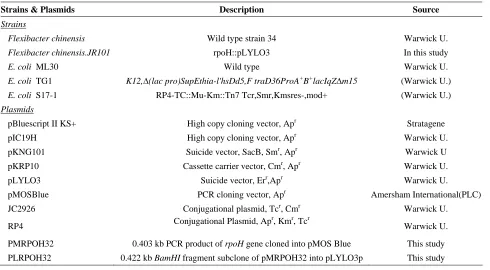
Table 1. Strains, plasmids used in this study
Strains & Plasmids Description Source
Strains
Flexibacter chinensis Wild type strain 34 Warwick U.
Flexibacter chinensis.JR101 rpoH::pLYLO3 In this study
E. coli ML30 Wild type Warwick U.
E. coli TG1 K12,∆(lac pro)SupEthia-l'hsDd5,F traD36ProA+B+lacIqZ∆m15 (Warwick U.)
E. coli S17-1 RP4-TC::Mu-Km::Tn7 Tcr,Smr,Kmsres-,mod+ (Warwick U.)
Plasmids
pBluescript II KS+ High copy cloning vector, Apr Stratagene
pIC19H High copy cloning vector, Apr Warwick U.
pKNG101 Suicide vector, SacB, Smr, Apr Warwick U
pKRP10 Cassette carrier vector, Cmr, Apr Warwick U.
pLYLO3 Suicide vector, Err,Apr Warwick U.
pMOSBlue PCR cloning vector, Apr Amersham International(PLC)
JC2926 Conjugational plasmid, Tcr, Cmr Warwick U.
RP4 Conjugational Plasmid, Ap
r
, Kmr, Tcr
Warwick U.
PMRPOH32 0.403 kb PCR product of rpoH gene cloned into pMOS Blue This study
PLRPOH32 0.422 kb BamHI fragment subclone of pMRPOH32 into pLYLO3p This study
flasks and serial dilutions prepared as a 10-fold serial dilution in Quarter-strength Ringers solution (2.25 g/l NaCl, 0.12 g/l CaCl2, 0.05 g/l NaHPO4 and 0.105 g/l KCl in 1 liter Distilled water). 100 µl of the diluted samples were spread on duplicate plates. The plates were incubated at 30°C for at least 48 hr. Plates were counted manually by using an illuminated colony counter. The results were expressed as cfu/ml.
Total Counts
The total count was determined using a Coulter counter ZM (Coulter Euro Diagnostics GMBH). The data were analyzed using Coulter channelyzer software to estimate the size distribution. The samples were diluted in an isotonic buffer containing 0.4% (v/v) glutaraldehyde to fix the cells. Total count is expressed as total particles/ml. The software also determines mean cell size and volume.
Transmission Electron Microscopy
Transmission electron microscopy was used after samples were negatively stained with phosphotungstic acid. Samples were placed onto a Formvar-coated copper grid (100 segment mesh; Agar scientific) for 30 sec. After drying, the grid was negatively stained with
one drop of 1% (w/v) phosphotungstic acid. The samples were examined using the Jeol JEM-100S transmission electron microscope with an 80 kV accelerating voltage. Photographs were taken using Kodak Panasonic film, which was developed in Kodak D-19 developer at 20°C for 3 min and fixed in Kodak fixer. Final pictures were printed on Kodak Veribrom paper.
Results
Construction of a Vector, Carrying rpoH Gene
Figure 1. Restriction digestion of pMRPOH32. Lane 1; λ ladder of 1 kb DNA as a molecular size marker. Lane 2 to 7; pMRPOH32 digested with EcoRI and BamHI.
Figure 2. Schematic illustration of integration of pLROPH32 in the F. chinensis wild type chromosome.
Figure 3. Southern blot analysis. Chromosomal DNA was digested with EcoRV. Lane 1; wild-type F. chinensis, Lane 2; the single recombinant strain F. chinensis JR102.
Analysis of rpoH Mutant Strain
Southern blot analysis of the F. chinensis strain JR102 was carried out to confirm the interruption of the rpoH gene in the chromosome by a single crossover event between disrupted internal parts of the rpoH gene cloned in pLYLO3 and the intact rpoH gene in the chromosome of F. chinensis.(Fig. 2) Chromosomal DNA was purified from F. chinensis wild type and JR102 and the subclone of pLRPOH32 was used as a probe. Chromosomal DNA from both the wild type and JR102 were digested with EcoRV. Southern blot analysis showed that the probe hybridized with a band in strain JR102 approximately 6 kb larger than the wild-type (Fig. 3). The probe hybridized with the wild-wild-type chromosomal DNA digested with EcoRV with a band slightly less than 1.636 kb, while there was no hybridization at this site with strain JR102. The probe hybridized with mutant chromosomal DNA digested with EcoRV slightly larger than 7.126 kb. The result also proved that there was no wild-type copy of rpoH gene in strain JR102 due to the result of single crossover recombination.
Growth Characteristics of Mutant Strain
The growth rate of wild type and strain JR102 were investigated in Luria broth at 20°C, 30°C and 37°C using changes in optical density as measure of growth. Strain JR102 grew more slowly and had a longer lag phase than the wild type at both 20°C and 30°C. At 30°C, JR102 did not show a stable stationary phase and the optical density began to decline immediately, as a result of cell lysis or of cell miniaturization. The photomicrographs of both the wild type and strain JR102 was carried out after 24 hr incubation at the different temperatures. Samples were directly applied to slides, fixed; Gram stained and observed using a Zeiss microscope at 600 times magnification. At 20°C, there was little difference between the cell size of the wild-type and strain JR102. There was little visible evidence of a change in cell size over a 48 hr incubation period at 20°C for strain JR102. At 30°C (Fig. 4) the cell size was different in the mutant and wild type organisms after 24 hr incubation with the cells of strain JR102 being longer than the wild-type cells. The change in cell size with increasing temperature shows that the longer size must be a direct result of the temperature affecting the mutant.
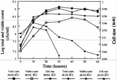
morphology occurred in strain JR102 lacking the δ32, when grown at higher temperatures. Both strains were grown at different temperatures (20°C and 30°C) and the total count, viable count and cell size were measured for and incubation period of up to 60 hr. The results are shown in Figure 5 for the wild-type, and show that although the viable count at 30°C declined slightly faster than at 20°C, there was no significant difference in the total cell counts at either temperature. At 30°C the cell size was slightly greater than that at 20°C the reduction in cell size between 20 and 60 hr of incubation was slightly faster at 30°C than at 20°C.
Strain JR102 showed considerable differences in total count, viable count and cell size compared to the data obtained for the wild type strain. Figure 6 shows that cell viability declined more quickly in the wild type so that after 40 hr incubation the viable count had declined below the limits of detection at 30°C, although for the wild-type viable counts were still high even after 60 hr incubation. At 20°C the viable count for strain JR102 did not show a similar decline however it did not reach the same maximum size as for the wild type strain. The total cell count at 30°C and 20°C did not show similar decline so the decrease in cell viable counts was not due to cell lysis. The total count at 20°C was slightly higher than at 30°C unlike with the wild type strain where the counts were very similar. At both temperatures the total counts were lower for the wild type at the same temperature. The cell size declined rapidly at 20°C after about 10 hr of incubation and failed to reach the same maximum size as the wild type. At 30°C the cell size did not decrease and reached a maximum size almost identical to that reached by the wild type. Again this demonstrates a close link between cell size reduction and maintenance of cell viability under starvation conditions. In this case the cells are entering the stationary phase of growth. These results also clearly demonstrate that a mutation in the rpoH gene of F. chinensis results in inhibition of cell division.
Discussion
The rpoH gene has been mapped immediately downstream of a cell division operon in E. coli and the genes which map in this region are the cell division genes ftsy, ftsE, ftsX and ftsS, the heat shock regulatory gene rpoH, the lipoprotein biogenesis gene fam-715, and another essential gene, dnaM. The rpoH gene lies immediately downstream of the last gene ftsX of a cell division operon and is transcribed in the same direction [6]. It is possible that in E. coli the mutation in the rpoH gene affects cell division because of the overlapping of rpoH and ftsX gene but this is considered unlikely.
However as we do not know the location of the rpoH gene in F. chinensis, it seems likely that rpoH mutants reported here could be located immediately after the promoters of a cell division operon, and could therefore have affected cell division not directly through the involvement of rpoH.
One possible suggestion why the mutant of rpoH gene results in filament formation is the role of the Lon gene. The Lon protease was the first ATP-dependent protease to be identified and characterized in detail. It has the ability to hydrolyze casein and a variety of other protein substrates [7]. It is known that one class of UV-sensitive mutants which form long non-separated filaments and die after treatment, have Lon mutations. Suppressors of the UV-sensitivity of Lon mutations can be isolated at two sites in E. coli, sulA and sulB (sfiA and sfiB) [8].
It has been reported that sulB is an allele of ftsZ. A sulB mutation leads to an altered ftsZ gene product which is slightly temperature sensitive. This altered ftsz gene product is resistant to the SulA inhibitor which allows cells division after induction of the SOS response [9].
Lon protease is produced at a detectable level in normally growing cells. Its synthesis is increased transiently when cells are subjected to a heat shock stress. A consensus sequence for this is found in the promoter region of the Lon gene [8]. It is known that the Lon protease is the part of the δ32 regulon which plays an important role in protein degradation and cells lacking the protease show a 50% decrease in ATP dependent degradation of abnormal proteins [10]. In the absence of the Lon protease two proteins which are substrates for Lon are synthesized at a high level. In E. coli, mutation in the Lon gene produces two detectable phenotypes [11]. One of these has a mucoid appearance due to the over-production of the RcsA protein. This is a natural substrate for the Lon protease and is a positive regulator of capsular polysaccharide synthesis.
Figure 5. The total counts, viable counts and cell size of wild-type F. chinensis. F. chinensis wild-type cells were grown with shaking at 20°C and 30°C in Luria broth from an initial viable count of 107 cfu/ml. Viable counts were determined on nutrient agar plates after incubation at 30°C for 48 hr. The total counts and cell size were determined using a CellFacts analyzer after dilution of he samples in the appropriate electrolyte solution to which 0.4% (v/v) glutaraldehyde had been added to fix the cells.
Figure 6. The total counts, viable counts and cell size of F. chinensis strain JR102. F. chinensis strain JR102 cells were grown with shaking at 20°C and 30°C in Luria broth from and initial viable count of 107 cfu/ml. Viable counts were determined on nutrient agar plates after incubation at 30°C for 48 hr. The total counts and cell size were determined using a CellFacts analyser after dilution of the samples in the appropriate electrolyte solution to which 0.4% (v/v) glutaraldehyde had been added to fix the cells.
The other phenotype is sensitive to DNA damaging agents. DNA damage induces the synthesis of a set of genes which is regulated by the LexA repressor following induction of the SOS response. One of the
induced genes, sulA, is a reversible inhibitor of cell division. Over-production of SulA leads to elongation of cells due to the inhibition of cell septation through the interaction with the cell division protein FtsZ [10]. This protein accumulates in annular rings at the points of septation for correct septum formation. SulA interacts with FtsZ and forms a SulA-FtsZ complex which prevents the proper formation of the septum. This inhibition is reversible as degradation of either free SulA or the SulA-FtsZ complex leads to the formation of a new septum [11].
Lon mutants which are exposed to stress and have the SOS response induced can repair their DNA normally. However, they are unable to recover from transient filamentation and instead they form long non-viable filaments. It has been suggested that following DNA damage, the RecA protein (which belongs to the SOS regulon) is activated. The RecA protein in turn inactivates the LexA protein which is a repressor of SulA synthesis in normally growing cells. After the inactivation of this repressor, the SulA protein accumulates in the absence of the LexA protein. In wild-type cells the Lon protease degrades SulA and cell division will resume. However in the mutants, SulA will react with FtsZ and will prevent septum formation. These results occur in the transient inhibition of cell division and consequently lethal filamentation of the cells [11].
All things considered, although the rpoH mutants produced here had phenotypes similar to other mutants the exact mechanism of the formation of these filaments remains to be elucidated. However, the purpose of producing these mutants was to investigate the effects of such mutations on bacterial survival. The inability to miniaturize leads to loss of survivability once the cell enters the stationary or starvation state. Again the molecular mechanisms behind this enhanced death rate remain to be investigated.
References
1. Nonaka G., Blankschien M., Herman C., Gross C.A., and Rhodius V.A. Regulon and promoter analysis of the E. coli heat shock factor, δ32, reveals a multifaceted cellular response to heat stress. Genes and Development, 20: 1776-1789 (2006).
2. Bittner A.N. and Oke V. Multiple groESL operons are not key targets of RpoH1 and RpoH2 in Sinorhizobium meliloti, J. of Bacteriology, 188: 3507-3515 (2006). 3. Aresene F, Tomoyasu T., Bukau B. The heat shock
response of Escherichia coli. Int. J. Food Microbiol., (2000).
filamentous cyanobacteria. Proceeding of the National Academy of the Sciences USA, 81: 1561-1565 (1984). 5. Sambrook J., Fritsch E.J., and Maniatis T. Molecular
Cloning; A Laboratory Manual. 2nd Ed., Cold Spring Harbor Laboratory Press, New York (1989).
6. Crickmore N. and Salmond G.P.C. The Escherichia coli
heat shock regulatory gene is immediately downstream of the cell division operon: The fam mutation is allelic with
rpoH. Molecular and General Generics, 205: 535-569 (1986).
7. Miller C.G. Protein degradation and proteolyic modification. In: Cutris R., Ingraham J.L., Lin E.C.C. Low K.B., Magasanik B., Rezniikoff W.E., Riley M., Schaechter M., and Umbarger H.E., Fredrick C., and Neidhardt F.C. (Eds.), Escherichia coli and Salmonella.
1, 1382-1399. Washington DC, USA (1996).
8. Gottesman S. Genetic of proteolysis in Escherichia coli.
Annual Review of Genetics, 23: 163-198 (1989).
9. Lutkenhaus J.F. Coupling of DNA replication and cell division: sulB is an allele of ftsZ. Journal of Bacteriology, 154: 1339-1346 (1983).
10. Gross C.A. Function and regulation of the heat shock proteins. In: Neidhardt F.C., Curtis R., Ingraham J.L., Lin E.C.C., Low K.B., Magasanik B., Reznikoff W.E., Riley M., Schaechter M., and Umbarger H.E. (Eds.),
Escherichia coli and Salmoneila. 1, 1382-1399. Washington DC, USA (1996).
11. Gottesman S. and Maurizi M.R. Regulation by proteolysis: Energy-dependent proteases and their targets.