RESEARCH NOTE
A MATHEMATICAL MODEL FOR BLOOD FLOW THROUGH
NARROW VESSELS WITH MILD-STENOSIS
M. Jain, G.C. Sharma* and S. Kumar Sharma Department of Mathematics, Institute of Basic Science
Khandari, Agra-282002, India
[email protected] - [email protected] - [email protected]
*Corresponding Author
(Received: January 17, 2008 – Accepted in Revised Form: September 25, 2008)
Abstract In this paper we examine the effect of mild stenosis on blood flow, in an irregular axisymmetric artery with oscillating pressure gradient. The Herschel-Bulkley fluid model has been utilized for this study. The combined influence of an asymmetric shape and surface irregularities of constriction has been explored in this computational study. An extensive quantitative analysis has been performed for narrowing of vessels through numerical computations on the flow velocity, plug flow rate and the apparent fluidity. The graphical representations have been made to validate the analytical findings with a view of its applicability to stenotic diseases. Velocity profiles, plug flow rate, and apparent fluidity along the radius of the obstructed tube are determined to give the flow characteristics, for diagnostic point of view. The effects of viscosity on the flow field are examined numerically and are shown graphically.
Keywords Mathematical Model, Mild-Stenosis, Micropolar Fluid, Shearing Stress, Apparent Fluidity
ﻩﺪﻴﮑﭼ
ﺮـﺑ ﻲﻧﺎـﺳﻮﻧ ﺭﺎﺸـﻓ ﺮـﻴﻴﻐﺗ ﺎـﺑ ﻱﺩﺎﻋﺮﻴﻏ ﻥﺭﺎﻘﺘﻣ ﮒﺮﺧﺮﺳ ﻚﻳ ﺭﺩ ﻲﻳﺰﺟ ﻥﺪﺷ ﮓﻨﺗ ﺮﻴﺛﺎﺗ ﻪﻟﺎﻘﻣ ﻦﻳﺍ ﺭﺩ
ﻲﻣ ﻲﺳﺭﺮﺑ ﻥﻮﺧ ﺵﺩﺮﮔ
ﺩﻮﺷ . ﻪﺑ ﻞﺷﺮﻫ ﻝﺎﻴﺳ ﻝﺪﻣ ﺯﺍ ﺭﻮﻈﻨﻣ ﻦﻴﻤﻫ
ﻲـﻣ ﻩﺩﺎﻔﺘـﺳﺍ ﻲﻠﻜﻟﺎﺑ ﺩﻮـﺷ
. ﻲـﺒﻴﻛﺮﺗ ﺮﻴﺛﺎـﺗ
ﻥﺭﺎﻘﺘﻣﺎﻧ ﻞﻜﺷ
ﻲﻤﻈﻧ ﻲﺑ ﻭ
ﻲﻣ ﺭﺍﺮﻗ ﻲﺳﺭﺮﺑ ﺩﺭﻮﻣ ﻲﮕﻨﺗ ﻲﺤﻄﺳ ﻱﺎﻫ
ﺩﺮﻴﮔ .
ﻩﺩﺮﺘﺴﮔ ﻱﺩﺪﻋ ﺰﻴﻟﺎﻧﺁ ﻲﺳﺭﺮﺑ ﻱﺍﺮﺑ ﻱﺍ
ﮒﺭ ﻥﺪﺷ ﮓﻨﺗ ﮒﻼﭘ ﻥﺎﻳﺮﺟ ﺥﺮﻧ ،ﻥﺎﻳﺮﺟ ﺖﻋﺮﺳ ﻱﻭﺭ ﻱﺩﺪﻋ ﺕﺎﺒﺳﺎﺤﻣ ﻡﺎﺠﻧﺍ ﻖﻳﺮﻃ ﺯﺍ ﺎﻫ
ﻱﺮﻫﺎﻇ ﺖﻴﻟﺎﻴﺳ ﻭ ﺎﻫ
ﻲﻣ ﻡﺎﺠﻧﺍ ﺩﺮﻴﮔ .
ﻞﻜﺷ ﻭ ﺎﻫﺭﺍﺩﻮﻤﻧ
ﻲﻜﻴﻓﺍﺮﮔ ﻱﺎﻫ ﯽﺸﺨﺑ ﺭﺎﺒﺘﻋﺍ ﯼﺍﺮﺑ
ﻪﺘﻓﺎﻳ ﺎﻫ
ﺮﺑﺭﺎـﻛ ﻩﺎﮔﺪـﻳﺩ ﺯﺍ
ﻱﺭﺎـﻤﻴﺑ ﺭﺩ ﺩ ﻱﺎـﻫ
ﻕﻭﺮﻋ ﻲﮕﻨﺗ
ﯽﻣ ﻪﺘﻓﺮﮔﺭﺎﮐ ﻪﺑ ﻮﺷ
ﺪﻧ .
ﮒﻼﭘ ﻥﺎﻳﺮﺟ ﺥﺮﻧ ،ﻥﺎﻳﺮﺟ ﺖﻋﺮﺳ ﮒﺭ ﻉﺎﻌﺷ ﻦﻴﻨﭽﻤﻫ ﻭ ﻱﺮﻫﺎﻇ ﺖﻴﻟﺎﻴﺳ ،ﺎﻫ
ﻲﻣ ﻦﻴﻴﻌﺗ ﻩﺪﺷ ﺩﻭﺪﺴﻣ ﻲﮔﮋﻳﻭ ﻥﺍﻮﺘﺑ ﺎﺗ ﺩﺩﺮﮔ
ﺩﻮﻤﻧ ﻪﺋﺍﺭﺍ ﻱﺭﺎﻤﻴﺑ ﺺﻴﺨﺸﺗ ﺮﻈﻧ ﻪﻄﻘﻧ ﺯﺍ ﺍﺭ ﻥﺎﻳﺮﺟ ﻱﺎﻫ .
،ﻪﻤﺗﺎﺧ ﺭﺩ
ﻪﺑ ﻥﺎﻳﺮﺟ ﻥﺍﺪﻴﻣ ﺮﺑ ﻪﺘﻳﺯﻮﻜﺴﻳﻭ ﺮﻴﺛﺎﺗ
ﺩﺭﻮﻣ ﻱﺩﺪﻋ ﺕﺭﻮﺻ
ﻪﺑ ﻭ ﻪﺘﻓﺮﮔ ﺭﺍﺮﻗ ﻲﺳﺭﺮﺑ
ﻩﺩﺍﺩ ﻥﺎﺸـﻧ ﻲـﻜﻴﻓﺍﺮﮔ ﺕﺭﻮﺻ
ﻲﻣ ﺩﻮﺷ .
1. INTRODUCTION
It is known that a severe constriction of a coronary artery significantly alters the mean resting coronary flow. Cardiac ischemia is caused due to the constriction, which is responsible for insufficient flow of blood through the coronary arteries into the heart. This insufficiency is usually caused by atherosclerotic plaque, which builds up in the coronary arteries, gradually diminishing the flow of blood through the said arteries. Such occlusion of the arteries increases the risk of heart attack.
dependent on its minimum cross-sectional area rather than its length (Chakravarty, et al [3]). These types of studies have been confounded to a description of the overall behavior of blood flow in the presence of a stenosis through experimental investigation.
Analytical models also have been developed in an effort to predict the pressure drop, caused by a given stenotic area. The minimum lumen areas created in stenosed tube were about 65 % and 90 %, including a model without stenosis, respectively (Zohdi, et al [4]). Ischemic heart disease, which results from high grade stenosis, is the single most common cause of death all over the world. Approximately 35 percent of all deaths are resulted by this cause. High grade stenosis increases flow resistance in arteries, which forces the body to raise the blood pressure in order to maintain the necessary blood supply. Both high pressure and narrowing vessels cause high flow velocity, high shear stress and low or even negative pressure, at the throat of the stenosis (Wille, et al [5]). These may be related to thrombus formation, atherosclerosis growth and plaque cap rupture, leads directly to stroke and heart attack. The exact mechanism of this complicated process is still not well understood. A more comprehensive study in this physiological process is of great importance for diagnosis, prevention and treatment of stenosis related diseases. A considerable number of experimental and numerical researches have been conducted to study the flow dynamics and stresses in collapsible elastic tube (Tang, et al [6]).
There are a number of studies, which suggest the existence of link between arteriosclerosis and micro polar fluid flow. In some studies, Young, et al [7,8] found that resistance is greater for asymmetric than for axisymmetric stenosis. They also performed some experiments on some unsteady flow with similar results. Wille, et al [9] investigated pressure and flow in arterial stenosis. Wille, et al [10] extended the problem of pulsatile pressure and flow in an arterial stenosis in their mathematical model. Krishan, et al [11] developed a mathematical model for unsteady flow of a micropolar fluid, through a constricted channel by using perturbation method for solution of slope parameters. Siouffi, et al [12] obtained the effect of unsteadiness of the flow through stenosis and bifurcations. Kapoor, et al [13] illustrated some
mathematical models in medical sciences. Misra, et al [2] examined the problem of the blood flow in arteries in presence of stenosis. Numerical study on the flow of a non-Newtonian fluid through an axisymmetric stenosis was made by Nakamura, et al [14]. However Chakravarty, et al [3] examined the effects of stenosis on arterial rheology through a mathematical model. Johnston, et al [15] investigated a mathematical model of blood flow through an irregular arterial stenosis. Finite element simulation of pulsatile flow through arterial stenosis can be found in the work of Tu, et al [16]. A model for blood flow through a stenotic tube has been developed by Tandon, et al [17]. Chakravarty, et al [18] did the mathematical modeling of blood flow through an overlapping arterial stenosis. In recent past, Sharma, et al [19] gave the finite element technique for two dimensional arterial flows in the presence of a transverse magnetic field. Cavalcanti, et al [1] did numerical simulation to examine the hemodynamics in a mild stenosis with consideration of pulsatile wall motion. Tang, et al [6] used axisymmetric models to investigate steady/unsteady viscous flow in elastic stenotic tubes with various stenosis stiffness and pressure conditions. Tu, et al [20] studied pulsatile flow of non-Newtonian fluids through arterial stenoses. Ang, et al [21] made the mathematical modeling of three dimensional flows through an asymmetric stenosis. Bathe, et al [22] suggested a fluid-structure interaction by using finite element analysis of pulsatile blood flow through a compliant stenotic artery. Dash, et al [23] analyzed flow in a catheterized curved artery with stenosis.
Figure 1. Schematic diagram of a mild-stenotic tube equation of continuity.
for the behavior of the flow and the wall in a mildly stenosed tube. Tang, et al [29] showed the effect of stenosis asymmetry on blood flow and artery compression. In their study they considered a three-dimensional fluid-structure interaction model. A simple model for shear stress mediated lumen reduction in blood vessels was given by Zohdi, et al [4]. Johnston, et al [30] described the non-Newtonian blood flow in human right coronary arteries, which showed the transient simulations. Lorenzini, et al [31] calculated blood velocity field with numerical assessment using a GPL code in case of intravascular doppler catheter effects. This study was a comparative analysis of different rheological models.
Recently, Christofidis, et al [32] have shown the influence of a convergent nozzle on the flow field of a mild stenosis located in a T-junction. Cuniberti, et al [33] gave the development of mild aortic valve stenosis in a rabbit model with hypertension. Jung, et al [34] suggested a hemodynamic computation using multiphase flow dynamics in a right coronary artery. Banks, et al [35] described the turbulence modeling in three-dimensional stenosed arterial bifurcations. Liu, et al [36] has examined the effect of the Reynolds number on the flow pattern in a stenotic right coronary artery. Matar, et al [37] studied the dynamics and stability of flow down a flexible incline.
While much work has been reported, the mathematical models for flow in stenotic collapsible tubes were primarily limited. But, most researches were focused on elastic tubes, in which stress, produces its characteristic strain instantaneously, and strain vanishes immediately upon the removal of the stress. In fact for realistic modeling channels have been considered porous as in human physiological tissues in the arteries suck the nutrients flowing within the blood. All the above studies are devoted in the wake of the new models for blood flow over the stenosis. Modeling of blood flow over the mild stenosis with medium degree of constriction through Herschel-Bulkley fluid model for blood flow with oscillating pressure gradient is considered in the present study. Further more we consider blood as non-Newtonian fluid. The rest of the paper is organized in various sections as follows. In Section 2, we describe the basic model with assumed notations,
which are used for mathematical formulation purpose. In Section 3, we explore the design parameters for numerical illustration. The variation of viscosity, shearing stress and velocity over the stenosis are also explained, descriptively as well as graphically. Finally, the conclusions are drawn in the Section 4.
2. MODEL DESCRIPTION
We consider axisymmetric steady flow in a mild stenotic tube. The flow is assumed to be laminar, non-Newtonian, viscous and incompressible. The shape of the tube is under zero pressure and the tube wall is assumed to have no axial motion, that is, no slipping takes place between the fluid and the wall. The pressure gradient is oscillatory in nature, which is compatible with a pumping heart motion. The complex nature of blood with various parameters is approximated here. The blood is in a uniform circular tube with an axisymmetric mild stenosis takes place whose boundary is specified by Krogh model, (Kapoor, et al [13]);
d 0 L z d , )} 20 L d (z
0 L
2π
cos {1 0 2R
δ
1 0 R
R
+ ≤ ≤ −
−
+ −
=
= 1, otherwise. (1)
Where R0 is the radius of unobstructed tube and R is the radius of obstructed tube. L0 is the length of the stenosis and d is the location of the stenosis. The maximum height of stenotic growth is taken as
δ. The schematic diagram is shown in Figure 1. 0
V
. =
∇ (2)
τ
. p Dt Dv
ρ =−∇ +∇ (3)
Where ρ is the density, p is the pressure, and τ is the shearing stress tensor.
Herschel-Bulkley law to model the fluid behavior of blood flow, taking into account two characteristic features, which has emerged from the experimental data namely:
• The presence of a yield stress,
• The dependence of the viscosity with respect to the shear rate. (see. ref. Kapoor, et al [13]) Let τ0 be the yield stress, the coefficient of viscosity is μ and γ′ be the strain rate.
Then constitutive equation in one dimensional form for Herschel-Bulkley pulsatile fluid with the shearing stress τ, is given by
⎪⎭ ⎪ ⎬ ⎫ < = ≥ + = [31]) al et Tu, ref. (see. 0 τ τ , 0 γ' 0 τ τ , 0 τ n )
μ(γ'
τ
(4) The governing equation of motion for steady
incompressible blood flow with pressure gradient through the mild stenosis in an artery reduces to the following form:
r ) rτ ( r 1 P ∂ ∂ =
− (5)
Where,
P z p = − ∂ ∂
, (6)
P being a constant. Integrating Equation 5 with respect to r which is the radial co-ordinate, we have
2 r P
τ = − (7)
From Equations 4 and 7, we have
n ) γ' ( ) μ 0 τ 2 r μ P ( + =
− (8)
For the Herschel-Bulkley fluid in circular tube, we have γ′ = 0 when τ≤τ0 and there is a core region which flows as a plug.
Let the radius of this plug region be rp. At the surface of this plug, the stress is τ0, so that considering the force on the plug, we get
p r 2π 0 τ 2 p r π
P× = ×
or
0 2τ
p r
P× = (9)
Then Equation 8 becomes as
) μ p r P 2 1 μ r P 2 1 ( n ) '
(γ =− + (10)
We know that
γ' dr dv =
(11)
Then Equation 10 to
n 1 ) p r r ( n 1 ) μ P 2 1 ( dr
dv = − − − (12)
The relevant conditions are
v = 0, at r = R and R0 (13)
Integrating (10) and using conditions (13), we get
} n n 1 β) R r ( n n 1 β) {(1 n n 1 R) ( n 1 ) μ P 2 1 ( 1 n n v + + − + + + − + = (14) Where . β R p r
= (15)
Plug flow exists whenever the shear stress does not exceed yield stress. The velocity of the plug flow can be obtained by putting
r = βR (16)
Then we get
⎪ ⎭ ⎪ ⎬ ⎫ ⎪ ⎩ ⎪ ⎨ ⎧ + − + + + − + = n n 1 β) (2 n n 1 β) (1 n n 1 R) ( n 1 ) μ P 2 1 ( 1 n n p v (17) Flow rate Q is obtained as follows:
β) 1) (2n n 1 3n (1 3 c n 1 ) μ P 2 1 ( 1 3n nπ Q + + − +
= (18)
Where n n 1 R) ( c + −
= (19)
Apparent fluidity φα at maximum height of stenosis
i.e. at Z = L0/2 is obtained as follows:
β) 1 2n 1 3n (1 ) c δ 3n (1 2 ) μ 1 ( α φ + + − −
= (20)
From Equation 16, we get the shear stress τω as
follows: ) β 1 2n 1 3n 1 ( ) c δ 3n 1 ( n ) 3 c π n 1/n μ Q 1) (3n ( ω τ + + + + + = (21)
3. NUMERICAL ILLUSTRATION
In this section, we present the numerical results for velocity profiles, volume flow rate, apparent fluidity and walls' shear stress. All these profiles provide detailed description of flow field. In the presence of mild-stenosis the flow exhibits a resistance and increases the shear stress. These are the quantities of physiological relevance. The
computation was programmed by MATLAB 6.5 software and run on P-IV for default parameter values β = 1.29; r0 = 0.01; τω = 0.02; n = 2; r =
0.15; rp = 0.003; P = 0.5; and δ = 0.2. These values have been chosen in consultation with medical practitioner having long clinical experience.
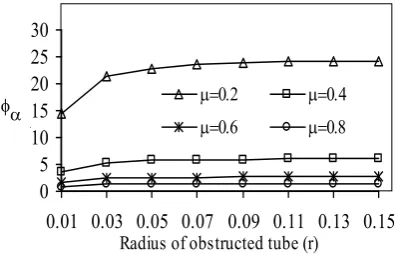
Figure 2 depicts the velocity profiles of fluid flow with respect to the radius of the obstructed tube for different value of μ. It is observed that the velocity of fluid decreases with increasing r in the presence of mild-stenosis. Also as we increase the values of μ, the velocity decreases. In Figure 3, we see the trend of flow rate in the plug region for different values of μ. It is observed that the velocity in plug region increases gradually at first and then it becomes rapid with the increase in r; by increasing the values of μ the flow rate decreases. For different values of μ, the pattern of the apparent fluidity in the direction of radius is shown in Figure 4. It is seen that the apparent fluidity slightly increases first with r and then attains almost constant value. The findings are quite close to the experimental results (Cuniberti, et al [33]) done on rabbit.
4. CONCLUSION
0 0.001 0.002 0.003 0.004
0.01 0.03 0.05 0.07 0.09 0.11 0.13 0.15 Radius of obstructed tube (r)
ve
lo
ci
ty
(v
)
μ=0.2 μ=0.4 μ=0.6 μ=0.8
Figure 2. Profile of velocity vs. radius of obstructed tube (r) for different values of μ.
0 0.002 0.004 0.006 0.008 0.01
0.01 0.03 0.05 0.07 0.09 0.11 0.13 0.15 Radius of obstructed tube (r)
ve
lo
ci
ty
(v
p)
μ=0.2 μ=0.4 μ=0.6 μ=0.8
Figure 3. Velocity profile of plug region vs. radius of obstructed tube (r) for different values of μ.
0 5 10 15 20 25 30
0.01 0.03 0.05 0.07 0.09 0.11 0.13 0.15
Radius of obstructed tube (r)
φ
μ=0.2 μ=0.4 μ=0.6 μ=0.8
Figure 4. Variation of apparent fluidity φα vs. radius of obstructed tube (r) for different values of μ.
vessels. The long-term application of our mathematical model is to provide the quantitative tool for gaining insights into the pathology of arterial diseases.
6. REFERENCES
1. Cavalcanti, S., “Hemodynamics of an Artery with Mild Stenosis”, J. Biomech., Vol. 28, No. 4, (1995), 387-399.
2. Misra, J.C. and Chakravarty, S., “Flow in Arteries in the Presence of Stenosis”, J. Biomech., Vol. 19, No. 11, (1986), 907-918.
3. Chakravarty, S. and Datta, A., “Effects of Stenosis on Arterial Rheology through a Mathematical Model”, Math. Comput. Model, Vol. 12, No. 12, (1989), 1601-1612.
4. Zohdi, T.I., “A Simple Model for Shear Stress Mediated Lumen Reduction in Blood Vessels”, Biomech. Model Mechanobio., Vol. 4, No. 1, (2005), 57-61.
5. Yao, H., Ang, K.C., Yeo, J.H. and Sim, E. K.W., “Computational Modeling of Blood flow through Curved Stenosed Arteries”, J. Med. Engg. Tech., Vol. 24, No. 4, (2000), 163-168.
6. Tang, D., “An Axisymmetric Model for Viscous flow in Tapered Collapsible Stenotic Elastic Tubes”, Adv. Bioengi., Vol. 33, (1996), 87-88.
7. Young, D.F. and Tsai, F.Y., “Flow Characteristics in Models of Arterial Stenoses-I: Steady Flow”, J. Biomech., Vol. 6, (1973), 395-410.
8. Young, D.F. and Tsai, F. Y., “Flow Characteristics in Models of Arterial Stenoses-II. Unsteady Flow”, J. Biomech., Vol. 6, (1973), 547-559.
9. Wille, S.O., “Pressure and Flow in Arterial Stenosis Simulated in Mathematical Models”, Appl. Math. Model, Vol. 4, (1980), 483-488.
10. Wille, S.O. and Walloe, L., “Pulsatile Pressure and Flow in an Arterial Stenosis Simulated in a Mathematical Model”, J. Biomed. Eng., Vol. 3, (1981), 17-24.
11. Krishan, B., Gupta, G.D. and Sharma, G.C., “Unsteady Flow of Micropolar Fluid through a Constricted Channel”, Jour. M. A. C. T., Vol. 16, (1983), 29-35. 12. Siouffi, M., Pelissier, R., Farahifar, D. and Rieu, R.,
“The Effect of Unsteadiness on the Flow through Stenoses and Bifurcations”, J. Biomech., Vol. 17, No. 5, (1984), 299-315.
13. Kapoor, J.N., “Some Mathematical Models in Medical Sciences”, Internat. J. Math. Edn. Sc. Tech., Vol 15, (1984), 587-600.
14. Nakamura, M. and Sawada, T. “Numerical Study on the Flow of a Non-Newtonian Fluid through an Axisymmetric Stenosis”, J. Biomech. Engg., Vol. 110, (1988), 137-143.
15. Johnston, P.R. and Kilpatrick, D., “Mathematical Modelling of Flow through an Irregular Arterial Stenosis”, J. Biomech., Vol. 24, No. 11, (1991), 1069-1077. 16. Tu, C., Deville, M., Dheur, L. and Vanderschuren, L.,
“Finite Element Simulation of Pulsatile Flow through Arterial Stenosis”, J. Biomech., Vol. 25, No. 10, (1992), 1141-1152.
17. Tandon, P.N., Kawahara, M., Rana, U.S. and Katiyar, V.K., “A Model for Blood Flow through a Stenotic Tube”, Int. J. Biomed. Comput., Vol. 32, (1993), 61-78. 18. Chakravarty, S. and Mandal, P.K., “Mathematical
Modelling of Blood Flow through an Overlapping Arterial Stenosis”, Math. Comput. Model, Vol. 19, No. 1, (1994), 59-70.
19. Sharma, G.C. and Kapoor, J., “The Finite Element Computations of Two Dimensional Arterial Flows in the Presence of a Transverse Magnetic Field”, Int. J. Nume. Method Fluid Dyna., Vol. 20, (1995), 1153-1161.
20. Tu, C. and Deville, M., “Pulsatile Flow of Non-Newtonian Fluids through Arterial Stenoses”, J. Biomech., Vol. 29, No. 7, (1996), 899-908.
21. Ang, K.C. and Mazumdar, J.N., “Mathematical Modeling of Three Dimensional Flow through an Asymmetric Stenosis”, Math. Comput. Model, Vol. 25, No. 1, (1997), 19-29.
22. Bathe, M. and Kamm, R.D., “A Fluid-Structure Interaction Finite Element Analysis of Pulsatile Blood Flow through a Compliant Stenotic Artery”, J. Biomech. Engg., Vol. 121, No. 4, (1999), 361-369. 23. Dash, R.K., Jayaraman, G. and Mehta, K.N., “Flow in a
Catheterized Curved Artery with Stenosis”, J. Biomech., Vol. 32, No. 1, (1999), 49-61.
24. Liu, B. and Tang, D., “A Numerical Simulation of Viscous Flows in Collapsible Tubes with Stenoses”, Appl. Num. Math., Vol. 32, No. 1, (2000), 87-101. 25. Steinman, D.A., Poepping, T.L., Tambasco, M., Rankin,
R. N. and Holdsworth, D. W., “Flow Patterns at the Stenosed Carotid Bifurcation: Effect of Concentric Versus Eccentric Stenosis”, Ann. Biomed. Engg., Vol. 28, No. 4, (2000), 415-423.
26. Banks, H.T., Barnes, J.H., Eberhardt, A., Tran, H. and Wynne, S., “Modeling and Computation of Propagating Waves from Coronary Stenosis”, Comput. Appl. Math., Vol. 21, (2002), 767-788.
27. Stroud, J.S., Berger, S.A. and Saloner, D., “Numerical Analysis of Flow through a Severely Stenotic Carotid Artery Bifurcation”, J. Biomech. Engg., Vol. 124, No. 1, (2002), 9-20.
28. Lee, K.W. and Xu, X.Y., “Modeling of Flow and Wall Behaviour in a Mildly Stenosed Tube”, Med. Eng. Phys., Vol. 24, No. 9, (2002), 575-586.
29. Tang, D., Yang, C., Kobayashi, S., Zheng, J. and Vito, R.P., “Effect of Stenosis Asymmetry on Blood Flow and Artery Compression: a Three-Dimensional Fluid-Structure Interaction Model”, Ann. Biomed. Engg., Vol. 31, No. 10, (2003), 1182-1193.
30. Johnston, B.M., Johnston, P.R., Corney, S. and Kilpatrick, D., “Non-Newtonian Blood Flow in Human Right Coronary Arteries: Transient Simulations”, J. Biomech., Vol. 39, (2005), 1116-1128.
31. Lorenzini, G., “Blood Velocity Field Numerical Assessment Using a GPL Code in Case of Intravascular Doppler Ctheter Affections: Comparative Analysis of Different Rheological Models”, J. Biomech., Vol. 38, (2005), 2058-2069.
32. Christofidis, C.C., Papaioannou, T.G. and Mathioulakis, D. S., “The Influence of a Convergent Nozzle on the Flow Field of a Mild Stenosis Located in a T-Junction”, J. Biomech., Vol. 39, No. 1, (2006), S310.
33. Cuniberti, L.A., Stutzbach, P.G., Guevara, E., Yannarelli, G.G., Laguens, R.P. and Favaloro, R. R., “Development of Mild Aortic Valve Stenosis in a Rabbit Model of Hypertension”, J. Amerc. College Cardio, Vol. 47, No. 11, (2006), 2303-2309.
34. Jung, J., Hassanein, A. and Lyczkowski, R.W., “Hemodynamic Computation using Multiphase Flow Dynamics in a Right Coronary Artery”, Ann. Biomed. Engg., Vol. 34, No. 3, (2006), 393-397.
Three-Dimensional Stenosed Arterial Bifurcations”, J. Biomech. Engg., Vol. 129, No. 1, (2007), 40-42. 36. Liu, B., “Effect of the Reynolds Number on the Flow
Pattern in a Stenotic Right Coronary Artery”, ICCES,
Vol. 1, No. 1, (2007), 35-40.