Activity of peroxisomal enzymes, and levels of polyamines in
LPA-transgenic mice on two different diets
Knut A Eliassen*
1, Bjørn P Brodal
2, Aud Svindland
3, Harald Osmundsen
2,
Helle Rønning
1, Srdjan Djurovic
4,5and Kåre Berg
4,5Address: 1Department Basic Sciences and Aquatic Medicine, Norwegian School of Veterinary Science, P.O. Box 8146 Dep. No-0033 Oslo, Norway, 2Department of Oral Biology, University of Oslo, P.O. Box 1052 Blindern, No-0316 Oslo, Norway, 3Department of Pathology, Aker University
Hospital, No-0514 Oslo, Norway, 4Institute of Medical Genetics, University of Oslo, P.O. Box 1036 Blindern, Oslo, Norway and 5Department of
Medical Genetics, Ullevål University Hospital, No-0407 Oslo, Norway
Email: Knut A Eliassen* - knut.eliassen@veths.no; Bjørn P Brodal - bbrodal@odont.uio.no; Aud Svindland - a.d.svindland@ioks.uio.no; Harald Osmundsen - haraldo@odont.uio.no; Helle Rønning - wkhr@weifa.no; Srdjan Djurovic - serdjan.djurovic@ioks.no;
Kåre Berg - k.berg@genova.no * Corresponding author
Abstract
Background: In man, elevated levels of plasma plipoprotein (a)(Lp(a)) is a cardiovascular risk factor, and oxidized phospholipids are believed to play a role as modulators of inflammatory processes such as atherosclerosis. Polyamines are potent antioxidants and anti-inflammatory agents. It was therefore of interest to examine polyamines and their metabolism in LPA transgenic mice.
Concentration of the polyamines putrescine, spermidine and spermine as well as the activity of peroxisomal polyamine oxidase and two other peroxisomal enzymes, acyl-CoA oxidase and catalase were measured. The mice were fed either a standard diet or a diet high in fat and cholesterol (HFHC). Some of the mice in each feeding group were in addition given aminoguanidine (AG), a specific inhibitor of diamine oxidase, which catalyses degradation of putrescine, and also inhibits non-enzymatic glycosylation of protein which is implicated in the aetiology of atherosclerosis in diabetic patients. Non-transgenic mice were used as controls.
Results: Intestinal peroxisomal polyamine oxidase activity was significantly higher in LPA transgenic mice than in the non-transgenic mice, while intestinal peroxisomal catalase activity was significantly lower. Hepatic β-oxidation increased in Lp(a) transgenic mice fed the HFHC diet, but not in those on standard diet.
Hepatic spermidine concentration was increased in all mice fed the HFHC diet compared to those fed a standard diet, while spermine concentration was decreased. With exception of the group fed only standard diet, transgenic mice showed a lower degree of hepatic steatosis than non-transgenic mice. AG had no significant effect on hepatic steatosis.
Conclusion: The present results indicate a connection between peroxisomal enzyme activity and the presence of the human LPA gene in the murine genome. The effect may be a result of changes in oxidative processes in lipid metabolism rather than resulting from a direct effect of the LPA construct on the peroximal gene expression.
Published: 04 October 2005
Lipids in Health and Disease 2005, 4:23 doi:10.1186/1476-511X-4-23
Received: 10 August 2005 Accepted: 04 October 2005
This article is available from: http://www.lipidworld.com/content/4/1/23 © 2005 Eliassen et al; licensee BioMed Central Ltd.
Background
Elevated levels of plasma lipoprotein (a) Lp(a) are a sig-nificant cardiovascular risk factor in man [1]. We have ear-lier reported development of arteriosclerosis in aorta of mice transgenic for cDNA representing the human gene for Lp(a), hLPA, on a standard diet [2,3], while the non-transgenic mice only sporadically developed arteriosclero-sis. In the LPA cDNA-transgenic animals, apolipopro-tein(a), (apo(a)), occurs free in plasma, and we found a significant correlation between the plasma apo(a) con-centration and the size of aortic lesions.
The present investigation, based on tissue samples from the animals in the above-mentioned study, was under-taken to uncover if polyamines could influence on the atherosclerotic development. There were no leftovers of blood and the aortic wall for polyamine measurements. The liver and kidney were therefore chosen.
The rationale for examining the polyamines, spermidine and spermine, is that their positive charges strongly inter-act with phospholipids and inhibit lipid peroxidation [4], and to a certain extent protect liposome from oxidation [5]. In addition, spermine exhibits an anti-inflammatory effect [5] and exerts an antagonistic action on platelet aggregation [6]. Since cell proliferation in the vascular walls is important in the development of atherosclerotic lesions, knowledge of levels and metabolism of polyamines are per se of interest because of their well-known importance for cell growth and differentiation. In addition it was of interest to measure the activity of the peroxisomal polyamine oxidase, an enzyme important in converting polyamines [7]. Examination of the activity of peroxisomal enzymes was of interest also because the per-oxisomal proliferators activated receptors (PPARs) are known to be involved in the development of arterioscle-rosis (for review, see [8]. We have earlier shown that feed-ing rats a diet enriched in polyamines resulted in a decrease in hepatic polyamine oxidase and catalase activ-ity, which could be restored by simultaneously supple-menting the diet with clofibrate, a peroxisomal proliferator and a hypolipidemic drug [9].
The fact that the hypolipidemic drug, clofibrate, changed the activity of polyamine oxidase indicates that the fat content of tissues may influence polyamine metabolism. It was therefore of special interest to examine if fat loading had any effect on liver polyamine content, and on polyamine oxidase activity.
In the present study we found that polyamine oxidase activity was higher in transgenic mice than in non-trans-genic animals. In order to examine whether this reflects a general increase in peroxisomal enzymes, we measured the activity of two other peroxisomal enzymes, catalase
and β-oxidase. Catalase decomposes H2O2, a product of oxidase activity, and thereby protects the cell against the toxic effect of H2O2, while peroxisomal β-oxidation plays the important physiological role of oxidation very long fatty acids and the side chain of cholesterol.
Some groups of mice were treated with AG because AG inhibits the formation of non-enzymatic glycosylation of proteins [10], which are implicated in the aetiology of dia-betic complications, including arteriosclerosis [10,11], and because AG is a well-known specific inhibitor of diamine oxidase, which catalyses degradation of putrescine, the precursor of the polyamines; spermidine and spermine.
We here report that the introduction of the human LPA gene into FVB mouse resulted in no significant changes in the polyamine concentration in liver and kidneys, but it changed the activity of three peroxisomale enzymes namely: intestinal polyaminoxidase and catalase and hepatic acyl-CoA oxidase (responsible for β-oxidation). There seemed to be less hepatic steatosis in the transgenic mice compared to the control FVB mice.
In mice fed a HFHC diet compared to those fed a standard low fat diet, the hepatic spermidine and spermine concen-trations were different, resulting in a lower ratio between spermidine and spermine concentrations in the HFHC fed mice.
Results
The results did not suggest a sex difference with respect to any of the measured parameters.
Body weight
Body-weight decreased by 2–5% during the experimental period. Animals treated with aminoguanidine showed the highest weight-loss. The reduction in the body weight may be related to the relatively high age of the mice. Mice on the high fat diet, showed an intermittent increase in body weight of 2–9%, with a peak about 3 weeks after start. However, mean body weight at the end of the treat-ment period, was not significantly different from those of the groups fed a standard diet (data not shown).
Polyamine oxidase activity
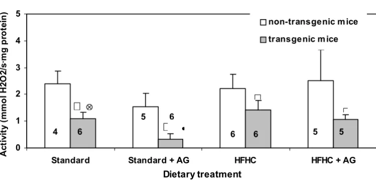
Catalase activity
Figure 2 shows the catalase activity measured in homoge-nates from the small intestine. In all treatment groups the catalase activity in the transgenic animals was significant lower than in the non-transgenic mice. In transgenic mice, given aminoguanidine in the drinking water, the catalase activity was significantly higher in those on the HFHC diet than in those on the standard diet (Fig. 2). AG treatment of transgenic mice on the standard diet reduced the intes-tinal catalase activity significantly.
There were only minor and insignificant differences in hepatic catalase activity between transgenic and non-transgenic mice and between the two diets. The activity
was roughly the same as in the small intestine of non-transgenic mice.
AG in the drinking water to mice on the HFHC diet resulted in an increased hepatic catalase activity from about 1.5 to 3.0 µmol H2O2/s·mg protein.
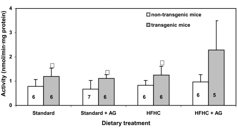
Peroxisomal β-oxidation (hepatic acyl-CoA oxidase activity)
Peroxisomal β-oxidation in livers of transgenic mice fed the HFHC diet and aminoguanidine in the drinking water was significantly (p < 0.05) higher than in the non-trans-genic mice on the same treatment (Fig. 3). In correspond-ing experiment with mice on the same diet, but without
Intestinal polyamine oxidase activity in LPA transgenic and non-transgenic mice
Figure 1
Intestinal polyamine oxidase activity in LPA transgenic and non-transgenic mice. The mice were fed two different diets, one high in fat and cholesterol (HFHC diet), the other a standard diet low in fat. Some of the animals from each group (see Table 1) were treated with 0.5% AG (aminiguanidine) in the drinking water. After 100 days of treatment the activity of the enzyme was assayed. The number of mice in each group is given with numbers within each column. Experimental details are given in Mate-rials and Methods. The histograms represent means with SD indicated. Population means that were significantly different from the non-transgenic mice are denoted with asterisk, * (p < 0.05).
Intestinal polyamine oxidase activity
0 1 2 3 4
Standard Standard + AG HFHC HFHC + AG
Dietary treatment
Ac
ti
vi
ty (nm
o
l/
m
in·
m
g
pr
o
tei
n
)
non-transgenic mice
transgenic mice
6 6 7 6 6 6 6 5
aminoguanidine (Fig. 3), the difference observed did not reach statistical significance.
Intestinal diamine oxidase activity
The diamine oxidase activity in the proximal part of the small intestine was powerfully inhibited in animals given AG, irrespective of their genetic status. The mean ± SD val-ues of diamine oxidase activity expressed as amount putrescine turnover pr. min and g tissue was 220 ± 160 and 17 200 ± 6200 pmol for the aminoguanidine treated and the non-treated animals, respectively. This is in agree-ment with the established inhibitory effect of aminogua-nidine on diamine oxidase activity [12].
Tissue polyamines
The hepatic concentrations of putrescine wearied around the level of detection of the assay. Evaluation of these data could therefore not be carried out.
With exception of mice on standard diet and aminoguani-dine treatment, transgenic and non-transgenic mice exhibited no significant differences in hepatic spermidine and spermine concentrations (Figs. 4 and 5)
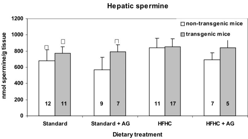
The hepatic concentration of spermidine in mice fed the atherogenic diet, HFHC; was significantly (p < 0.05) lower than in mice given standard diet (Fig. 4). On the other hand, a slight, but significant increase (p < 0.05) in sper-mine concentration was observed in mice fed the HFHC diet without AG in the drinking water, compared to those fed the standard diet (Fig. 5). The spermidine/spermine ratio was 0.76 for mice on the HFHC diet, and 1.32 for those on standard diet.
In kidney there were no differences between transgenic and non-transgenic mice. Thus for the non-transgenic mice the putrescine, spermidine and spermine concentra-tions were (73 ± 36) nmol/g, (363 ± 83) nmol/g and (606 ± 101) nmol/g, respectively, and the corresponding values for the transgenic animals were 66 ± 22, 398 ± 170 and 642 ± 154 nmol/g. Furthermore, there was no difference between mice on the two diets, or between AG treated and untreated mice.
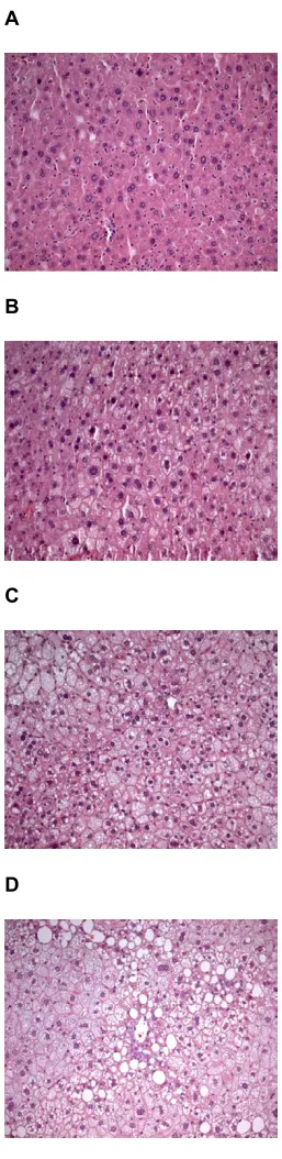
Hepatic steatosis
In the fatty infiltrations, there was no sign of inflamma-tory cells. Hepatic steatosis was graded from 1 to 3.
Intestinal catalase activity in LPA transgenic and non-transgenic mice
Figure 2
Intestinal catalase activity in LPA transgenic and non-transgenic mice. The histograms represent means with SD indicated. Pop-ulation means that were significantly different from the non-transgenic mice (p < 0.05) are denoted with *. PopPop-ulation means denoted with • indicate difference from the corresponding mice fed the HFHC diet and 丢 indicate significant differences from the corresponding aminoguanidine (AG) treated mice. Experimental details are given in the legend to Fig. 1 and in Materials and Methods section.
Intestinal catalase activity
0 1 2 3 4 5
Standard Standard + AG HFHC HFHC + AG
Dietary treatment
A
c
ti
v
it
y
(
m
m
o
l H
2
O
2
/s
·m
g pr
ot
e
in) non-transgenic m ice
transgenic m ice
4 6 6 6 5 5
5 6
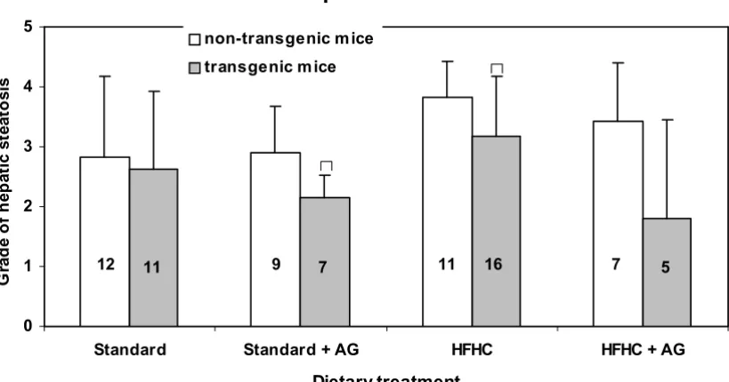
Typical sections of liver with steatosis of grade 1, 2 and 3, together with sections of a normal liver are shown in Fig. 6. Steatosis occurred in the liver of all, but four mice. The grade of steatosis was lower in the transgenic mice than in non-transgenic mice, but the difference was statistical sig-nificant (p < 0.05) only for those on a standard diet with AG in the drinking water and for those fed only a high fat diet (Fig. 7).
Discussion
Enzyme activitiesIntroduction of cDNA representing the LPA gene together with the transferrin promotor, into the mouse genome resulted in changes in activity of the peroxisomale enzymes polyamine oxidase and catalase in the small intestine, but not in the liver (Figs. 2, 3, 4). This is in con-trast to the fact that the LPA gene is expressed in liver, but not in the intestines [13], indicating that the effect on the intestinal enzymes must be secondary. Since the genes coding for polyamine oxidase (Locus ID no Mm 212503) and for acyl-CoA oxidase (Locus ID no. Mm 11430) are located on different mouse chromosomes, 7 and 11,
respectively, it is unlikely that the observed changes in these enzyme activities were caused by the introduction of the LPA gene with a transferrin promotor, in or near the regulator region of the gene for these enzymes.
The increased intestinal polyamine oxidase activity in LPA transgenic mice compared to non-transgenic mice (Fig. 1), indicates an altered peroxisomal polyamine metabolism. When the activity of the H2O2 producing polyamine oxi-dase enzyme is increased in the transgenic mice, an increase in the catalase activity should be anticipated. However, the opposite was the case.(Fig. 2) Increased polyamine oxidase activity can therefore not be explained by a general increase in activity of peroxisomal enzymes. The increase in H2O2 production from enhanced polyamine oxidase activity is probably marginal, com-pared with H2O2 production from other sources. The observed changes in hepatic peroxisomal β-oxidation (Fig. 3) cannot explain the decrease in the catalase activity because peroxisomal β-oxidation did not change in the control mice and even increased in the LPA-transgenic mice on HFHC diet. The increased peroxisomal β
-oxida-Hepatic peroxisomal β-oxidation activity in LPA transgenic mice and non-transgenic mice
Figure 3
Hepatic peroxisomal β-oxidation activity in LPA transgenic mice and non-transgenic mice. Experimental details are given in the legend to Fig. 1 and in Materials and Methods section. The histograms represent means with SD indicated. Population means that were significantly different (p < 0.05) from non-transgenic mice are denoted with *. Population means denoted with ♦ indicate significant differentce from the corresponding mice fed a HFHC diet.
Hepatic peroxisomal ß-oxidation
0.0 0.5 1.0 1.5 2.0 2.5
Sta
A
c
tivit
y (nmol/mi
n
·mg prot
ein)
ndard Standard + AG HFHC HFHC + AG
Dietary treatment
non-transgenic mice
transgenic mice
6 6 7 5 6 6 6 5
¡
tion in LPA transgenic mice on the HFHC diet is in agree-ment with reports showing that diets high in fat, particular those containing hydrogenated fat, cause increased of per-oxisomal β-oxidation [14,15]. In non-transgenic mice the same diet caused no significant increase of peroxisomal β -oxidation. It follows that mice, transgenic with respect to LPA may be more prone than non-transgenic mice to increase of peroxisomal β-oxidation probably because of the low fat content (Fig. 2).
The observed increase in polyamine oxidase and β -oxi-dase activity together with a decrease in catalase activity indicate that also other oxidative processes are changed as a result of the introduction of the LPA gene. This seems interesting in the view of the possible role of oxidized phospholipids as modulators of inflammatory processes. Thus, modified phospholipids accumulate at sites of inflammation such as atherosclerotic lesions [16].
Fur-thermore, oxidized phospholipids are shown to induce expression of atherosclerosis-related genes [17]. Addi-tional evidence has been provided by the use of murine natural monoclonal IgM antibody, EO6, which binds to oxidation-specific epitopes on oxidized low-density lipo-protein. A high correlation between plasma Lp(a) and EO6 was then found [18].
The conclusion so far must be that changes in the three peroxisomal enzyme activities in LPA-transgenic mice is more likely to be caused by altered lipid metabolism in these animals, rather than by a direct effect of the LPA con-struction on the expression of the peroxisomal genes.
Polyamines
In the kidneys, neither the LPA gene nor the HFHC diet and AG treatment seemed to influence the polyamine lev-els significantly (data not shown).
Hepatic spermidine concentration in LPA transgenic and non-transgenic mice
Figure 4
Hepatic spermidine concentration in LPA transgenic and non-transgenic mice. The columns represent means with SD indi-cated. Population mean that is significantly different from that of non-transgenic mice is denoted with asterisk, *. ∆ Indicate sig-nificant difference from the corresponding groups fed a standard diet. Experimental details are given in the legend to Fig 1 and in the Materials and Methods section.
Hepatic spermidine
0 200 400 600 800 1000 1200
Standard Standard + AG HFHC HFHC + AG
Dietary treatment
n
m
ol s
p
e
rm
idi
ne
/g
t
is
s
ue
non-transgenic m ice
transgenic m ice
1 11 9 7 11 17 7 5
The hepatic spermidine and spermine concentration were almost unaffected by aminoguanidine treatment (Fig. 4 and 5). The same was true for the kidneys. Although the polyamine concentration in the standard diet was much higher than in the HFHC diet, the HFHC diet resulted in higher hepatic spermidine concentration. It seems not likely that differences in the intestinal micro flora caused the observed differences in hepatic polyamine content between the two feeding groups.
Clofibrate treatment, that decreases plasma lipid levels has earlier been found to increase the hepatic content of spermidine and reduce that of spermine [9], which is con-trary to the effect of the HFHC diet.
The HFHC fed group had higher levels of hepatic fat, as judged by the amount of fat filled vacuoles (steatosis) than those on the standard diet. However, we found no significant correlation between hepatic steatosis and hepatic levels of spermidine or spermine. In spite of that,
we suggest that hepatic lipid level may be of importance for the hepatic polyamine level. The fact that spermine binds to negatively charged lipids, e.g. phospholipids [19] may in part explain the increased spermine concentration in liver.
Spermidine in plasma can bind to HDL [20], which is known to inhibit the formation of oxidized low-density lipoprotein (LDL) [21]. Polyamines, especially spermine, are also potent antioxidants and anti-inflammatory agents [5]. The polyamine concentrations have further been reported to inhibit platelet aggregation in hypercholester-olemic rabbits [6]. Studies of the role of polyamines in atherogenesis/thrombogenesis are therefore called for.
The spermidine/spermine ratio is known to be high in fast growing tissue. Thus, in rat liver at birth the ratio is 4.5, whereas this ratio at 9 months is 0.8 [22]. One can only speculate if the cell turnover rate for liver cells in mice fed the HFHC diet, that have a hepatic spermidine/spermine
Hepatic spermine concentration in LPA transgenic and non-transgenic mice
Figure 5
Hepatic spermine concentration in LPA transgenic and non-transgenic mice. The columns represent means with SD indicated. Population mean that is significantly different (p < 0.05) from that of the non-transgenic mice is denoted with asterisk, *. ∆ Indi-cate significant difference (p < 0.05) from the corresponding HFHC fed groups. Experimental details are given in the legend to Fig. 1 and in the Materials and Methods section.
Hepatic spe rmine
0 200 400 600 800 1000 1200
Standard Standard + AG HFHC HFHC + AG
Dietary treatment
n
m
ol
s
p
e
rm
ine
/g t
is
s
u
e
non-transgenic m ice
transgenic m ice
12 11 9 7 11 17 7 5
ratio on 0.76, is reduced compared to that of mice on a standard diet where the corresponding ratio was 1.32.
Liver steatosis
For two of the feeding groups there was significantly lower degree of hepatic steatosis in the transgenic mice than in the non-transgenic animals (Fig 7) suggesting differences in lipid metabolism. We have earlier reported a signifi-cantly higher rate of aortic lesions in LPA transgenic mice than in non-transgenic animals and a positive correlation between apo(a) level and size of aortic lesions [2,3]. This, together with the fact that inflammatory cells only could be seen in the aortic fat infiltrations, not in that of the liver, indicate that there are different mechanisms involved in the deposition of lipids in aortic lesions and in liver cells.
The AG treated mice on the HFHC diet had less steatosis than the untreated animals but the differences were far from significant. This indicates that glycosylation is not participating in the formation of hepatic steatosis in mice.
Conclusion
An increase in polyamine oxidase activity in hLPA trans-genic mice, and an increase in β-oxidation in transgenic mice fed a HFHC diet, together with a decrease in catalase activity may indicate that also other oxidative processes are changed as a result of the introduction of the LPA gene. Such changes would be of great interest because oxi-dation products are known to be important in the devel-opment of arteriosclerosis.
Changes in the polyamine pattern upon increased fat intake are also noteworthy. However, in this study we have not been able to show any connection between polyamines/polyamine-metabolism and the arterioscle-rotic lesion seen in the same mice [2,3]. Further studies are warranted.
Materials and methods
ReagentsHorseradish peroxidase (EC 1. 11. 1. 7) (Type VI. RZ between 250 and 330 U/mg), N1-acetyl-spermine, 4-ami-noantipyrine, palmitoyl-CoA, NAD+, FAD, HEPES, hexan-ediamine, putrescine 2HCl, spermidine 3HCl, spermine 4HCl, and mannitol were purchased from Sigma Chemi-cal Co. (St. Louis, MO., USA). Perhydrol (H2O2) was obtained from E. Merck (Darmstadt, Germany). All other reagents were of analytical grade. [1,4-14C]-Putrescine dihydrochloride (spes.act. 2.11 GBq/mmol) was obtained from Amersham Pharmacia Biotech, Ltd. (Rainham, Essex, UK).
Scoring of hepatic steatosis
Figure 6
Scoring of hepatic steatosis. A) Normal liver with no lipid deposits, grade 0, B) Liver tissue with steatosis grade 1 (slight steatosis), C) grade 2, that between slight and serve stearo-sis, D) Liver tissue with steatosis grade 3 (serve steatisis), almost all cells are affected and many with large fat vacuoles. Magnification 1000x.
A
B
C
Animals and feeding
This stock of mice have also been used in a parallel inves-tigation of effects of the LPA gene on the development of aortic lesions [2,3]. Forty-one transgenic mice with cDNA representing the human LPA gene linked to the mouse transferrin promotor were studied. The mice were of a hybrid genetic background of strain C57BL/6 crossed with strain SJL [2,3]. The original transgenic breeder mice were a generous gift from Dr. Richard M. Lawn, Falk Cardiovas-cular Research Center, Stanford University, CA., USA. Twenty-nine non-transgenic littermates from breeding between transgenic and non-transgenic mice were used as controls. DNA analyses of white blood cells were per-formed to ascertain the LPA transgenic status of the mice. The mice were of both sexes and between 49 and 67 weeks of age when the study started (see Table 1).
Half of the animals were fed a standard mouse diet (RMI (E) SQS, Special Diets Services, Witham, Essex, England) containing 2.6% fat (crude oil), where saturated fat accounted for 20%. The other half of the animals, were fed an atherogenic semi-purified diet high in fat and
cho-lesterol (HFHC diet), containing 1.25% chocho-lesterol, 18.4% regular butter and 0.5% sodium cholate (ICN Pharmaceuticals, Inc. 1731 Asse-Relegem, Belgium). Sev-eral of the animals on each diet were given 0.1% (w/v) aminoguanidine in the drinking water (Table 1). All the treatments lasted for 100 days.
The amounts of polyamines in the standard diet were: 110 nmol/g putrescine, 240 nmol/g spermidine and 45 nmol/ g spermine. The corresponding figures for putrescine and spermidine in the HFHC diet were 1.0 and 3.5 nmol/g, respectively. Spermine could not be detected.
Females and males were housed separately, with 1–5 mice per cage. They had a 12 hrs. light/dark cycle, a constant temperature of 21°C and a relative humidity of 65%. Sen-tinel mice were used to run a FELASA-style health-moni-toring scheme. The mice were clinically healthy throughout the experimental period. The mice were raised in The Laboratory Animal Unit at Norwegian School of Veterinary Science (accredited by the Association for Assessment and Accreditation of Laboratory Animal Care
Hepatic steatosis (grade 0 to 3) in LPA transgenic and non-transgenic mice
Figure 7
Hepatic steatosis (grade 0 to 3) in LPA transgenic and non-transgenic mice. The columns represent means with SD indicated. Population means that are significantly (p < 0.05) different from the non-transgenic mice are denoted with asterisks, *. Experi-mental details are given in the legend to Fig. 1 and in the Materials and Methods section.
Hepatic steatosis
0 1 2 3 4 5
Standard Standard + AG HFHC HFHC + AG
Dietary treatment
G
rad
e o
f
h
ep
at
ic st
e
at
o
sis
non-transgenic m ice
transgenic m ice
12 11 9 7 11 16 7 5
and Use International, Brussels, Belgium), and they were kept according to the regulations of the Norwegian Gene Technology Act of 1994. The study was approved by the Norwegian Animal Research Authority.
Blood sampling and preparation of tissue samples for enzyme measurements
The mice were anaesthetized by a subcutaneous injection of 0.05 ml/10 g body-weight of a 1:1 mixture of Fenatyl/ Midazolum (Hypnorm "Janssen" diluted 10x with water and Dormicum "Roche" 5 mg/ml) prior to blood sampling, which was done by an incision in vena saphena. The mice were afterwards killed by neck dislocation, and tissues for analysis were removed as quickly as possible. After removal, the liver was weighed, and transferred into ice-cold mannitol-medium (300 mmol/L mannitol, 25 mmol/L HEPES, 1 mmol/L EGTA, pH 7.2). A 10% (w/v) homogenate was prepared by 2 strokes in a Potter-Elve-hjem homogenizer equipped with a Teflon® piston. The homogenate was centrifuged for 1 min at 1075 × gav. The resulting supernatants were transferred into several small vials, and stored at -20°C until analyzed.
A 20 cm long proximal segment of the small intestine, starting about 0.5 cm from the pyloric sphincter, was pre-pared. It was cut open longitudinally, rinsed with ice-cold 0.9% (w/v) NaCl, blotted against filter paper, and homog-enized for 15 s in ice-cold mannitol medium with an Ultra-Turrax® at 25,000 rpm. The resulting homogenate was subsequently centrifuged at 1075 × gav for 15 min and the supernatant was stored at -20°C until it was analyzed. Prior to assays, one part of the homogenate was mixed with one part of ice-cold mannitol medium, and then Tri-ton X-100 was added to a resulting concentration of 0.05% (v/v).
Enzyme assays
Catalase (EC 1.11.1.6) activity was assayed by monitoring the decomposition of H2O2 at 240 nm and 25°C essen-tially as described by Bergmeyer et al [23]. Catalase is labile in dilute solution. Therefore, homogenates were
diluted 10 times with 20 mmol/L potassium phosphate buffer, pH 7.4 immediately prior to analysis.
Peroxisomal β-oxidation was assayed as palmitoyl-CoA-dependent NAD+-reduction, according to Hovik and Osmundsen [24]. Polyamine oxidase (polyamine: oxygen oxidoreductase, EC 1.5.3.3) was assayed spectrophoto-metrically as described by Hayashi et al. [25]. Diamine oxidase, EC 1.4.3.6 was determined radiometrically, as described by Okuyama and Kobayashi [26].
Catalase activity, peroxisomal β-oxidation and polyamine oxidase activity were measured in 6 randomly selected mice from each of 7 groups of animals, whereas all 5 mice in the transgenic group on HFHC diet and AG were exam-ined. As seen in Fig. 3 the numbers of mice in some other groups were also less than 6, due to accidental loss of some samples.
Polyamine assays
Tissue specimens were weighed and thereafter homoge-nized in 4 volumes of 5% trichloroacetic acid. Hexane diamine was added as an internal standard. The homoge-nates were kept on ice for 1 hr, followed by centrifugation at 2°C for 10 min at 5000 gav. The supernatant was stored at -20°C until analyzed. The polyamines were dansylated [27,28] and separated by HPLC [29] on a Radial PAK-A column (Waters, Milford, Ma., USA).
Assay of proteins
Protein assay were carried out using the biuret method [30] with Boehringer Precimat (Boehringer Mannheim, Mannheim, Germany) as protein standard.
Liver steatosis
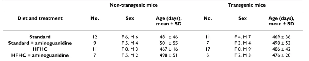
Liver tissue for morphometric studies were removed and immediately placed in a buffered formaldehyde solution. The examination was made routinely on sections of liver, in paraffin blocks, stained with Oil red O and counter-stained with hematoxylin. Hepatic steatosis was identified by light microscopy as empty vacuoles in the cytoplasm of Table 1: Diet, treatments, age and sex distribution of LPA transgenic and non-transgenic mice
Non-transgenic mice Transgenic mice
Diet and treatment No. Sex Age (days), mean ± SD
No. Sex Age (days), mean ± SD
Standard 12 F 6, M 6 481 ± 46 11 F 4, M 7 469 ± 36 Standard + aminoguanidine 9 F 5, M 4 501 ± 55 7 F 3, M 4 498 ± 53 HFHC 11 F 8, M 3 467 ± 16 17 F 8, M 9 486 ± 42 HFHC + aminoguanidine 7 F 5, M 2 498 ± 51 5 F 2, M 3 476 ± 20
with Oil Red O. The same pathologist examined the sec-tions blindly, and all measurements were performed four times. The evaluation of visible fat in the liver cells was assessed using a semi quantitative scale from 0 to 3. Zero indicates that no vacuolated liver cells could be detected. In the slightest degree of steatosis (grade 1) tiny vacuoles are found in small parts of the lobule, whereas in the severe case (grade 3) almost every liver cell is affected and most cells have large vacuoles. "Moderate" (grade 2) denotes fatty changes between that of slight and severe steatosis (Fig. 7).
Statistical analysis
To calculate the significance of differences between non-transgenic and non-transgenic mice on the same treatment we used an unpaired, two tailed t-test, employing the Graph-Pad Prism v.2.0 program (GraphGraph-Pad software Inc., San Diego, USA).
The values given in the text and legends to Figures are means, with SD as indicated.
List of abbreviations
AGE, advanced glycosoationtion end products; AG, aminoguanidine; HFHC, diet high in fat and cholesterol; LPA, gene encoding apo(a).
Authors' contributions
SD was responsible for the testing of transgenicity of ani-mals used in this study. BPB and HO were responsible for measurements of β-oxidation, polyamine oxidase and cat-alase activity. AS did the pathological examinations. HR measured the polyamines. KB and KAE conceived the study and performed its design and coordination. KAE drafted the manuscript. All authors read and approved the final version of the manuscript.
Acknowledgements
We gratefully thank Dr. Richard M. Lawn for the generous gift of LPA trans-genic mice. The skillful technical assistance of Patrica A. Engen, Bente B. Gehrken, Tove Norén, Inger E. Nossen and Toril Woldene is highly appreciated.
References
1. Djurovic S, Berg K: Epidemiology of Lp(a) lipoprotein: its role in atherosclerotic/thrombotic disease. Clinical Genetics 1997,
52:281-292.
2. Svindland A, Berg K, Eliassen K, Lawn RM, Djurovic S, Alestrom P, Noren T, Smith A: Histopathology of arterial lesions in LPA transgenic mice on cholesterol-enriched chow. Atherosclerosis 2000, 153:349-354.
3. Berg K, Svindland A, Smith AJ, Lawn RM, Djurovic S, Alestrom A, Alestrom P, Eliassen K: Spontaneous atherosclerosis in the proximal aorta of LPA transgenic mice on a normal diet.
Atherosclerosis 2002, 163:99-104.
4. Tadolini B, Cabrini L, Landi L, Varani E, Pasquali P: Polyamine bind-ing to phospholipid-vesicles and inhibition of
lipid-peroxida-5. Lovaas E, Carlin G: Spermine – an antioxidant and anti inflam-matory agent. Free Radical Biology and Medicine 1991, 11:455-461. 6. de la Pena HC, Sosa-Melgarejo JA, Ramos RR, Mendez JD: Inhibition of platelet aggregation by putrescine, spermidine, and sper-mine in hypercholesterolemic rabbits. Archives of Medical Research 2000, 31:546-550.
7. Seiler N, Bolkenius FN, Rennert OM: Interconversion, catabo-lism and elimination of the polyamines. Medical Biology 1981,
59:334-346.
8. Duval C, Chinetti G, Trottein F, Fruchart JC, Staels B: The role of PPARs in atherosclerosis. Trends in Molecular Medicine 2002,
8:422-430.
9. Brodal BP, Eliassen KA, Ronning H, Osmundsen H: Effects of die-tary polyamines and clofibrate on metabolism of polyamines in the rat. Journal of Nutritional Biochemistry 1999, 10:700-708. 10. Brownlee M, Vlassara H, Kooney A, Ulrich P, Cerami A:
Aminogua-nidine prevents diabetes-induced arterial-wall protein cross-linking. Science 1986, 232:1629-1632.
11. Li YM, Steffes M, Donnelly T, Liu C, Fuh H, Basgen J, Bucala R, Vlassara H: Prevention of cardiovascular and renal pathology of aging by the advanced glycation inhibitor aminoguanidine. Proceed-ings of the National Academy of Sciences of the United States of America 1996, 93:3902-3907.
12. Schayer RW, Kennedy J, Smiley RL: Studies on histamine-metab-olizing enzymes in intact animals II. Journal of Biological Chemistry 1953, 205:739-748.
13. Teivainen PA, Eliassen KA, Rubin EM, Djurovic S, Berg K: Human apoB contributes to increased serum total apo(a) levels in LPA transgenic mice. Lipids in Health and Disease 2004, 3:8. 14. Neat CE, Thomassen MS, Osmundsen H: Effects of high-fat diets
on hepatic fatty acid oxidation in the rat. Isolation of rat liver peroxisomes by vertical-rotor centrifugation by using a self-generated, iso-osmotic, percoll gradient. Biochemical Journal 1981, 196:149-159.
15. Thomassen MS, Christiansen EN, Norum KR: Characterization of the stimulatory effect of high-fat diets on peroxisomal beta-oxidation in rat liver. Biochemical Journal 1982, 206:195-202. 16. Furnkranz A, Leitinger N: Regulation of inflammatory responses
by oxidized phospholipids, structure-function relationships.
Current Pharmaceutical Design 2004, 10:915-921.
17. Furnkranz A, Schober A, Bochkov VN, Bashtrykov P, Kronke G, Kadl A, Binder BR, Weber C, Leitinger N: Oxidized Phospholipids Trigger Atherogenic Inflammation in Murine Arteries. Arte-rioscler Thromb Vasc Biol 2005, 25:633-638.
18. Bergmark C: Oxidized Phospholipids and Lp(a) Lipoprotein, a role in Innate Immunity? "Genomics and Genetic Applications in Medicine" Symposium organized by S.K Thoresens foundation and The Norwegian Academy of Sciences, Oslo, May 19 – May 21, 2005 :44-46. Abstract
19. Tadolini B, Cabrini I, Varani E, Sechi AM: Spermine binding and aggregation of vesicles of different phospholipid composition. Biogenic Amines 1985, 3:87-96.
20. CatrosQuemener V, Leray G, Moulinoux JP, Havouis R, deCertaines JD, Chapman J: Tumour growth modifies intravascular polyamine transport by plasma lipoproteins in the mouse.
Biochimica et Biophysica Acta-Lipids and Lipid Metabolism 1997,
1346:30-37.
21. Navab M, Hama SY, Cooke CJ, Anantharamaiah GM, Chaddha M, Jin L, Subbanagounder G, Faull KF, Reddy ST, Miller NE, Fogelman AM:
Normal high density lipoprotein inhibits three steps in the formation of mildly oxidized low density lipoprotein: step 1.
Journal of Lipid Research 2000, 41:1481-1494.
22. Russell DH: Polyamines as biochemical markers of normal and malignant growth. Progress in cancer Research and Therapy 1973, 9:1-13.
23. Bergmeyer HU, Grassl M, Walter HE: Catalase. Verlag Chemie, Weinhein, Germany 1983, II:165-167.
24. Hovik R, Osmundsen H: Peroxisomal beta-oxidation of long-chain fatty acids possessing different extents of unsaturation.
Biochemical Journal 1987, 247:531-535.
Publish with BioMed Central and every scientist can read your work free of charge
"BioMed Central will be the most significant development for disseminating the results of biomedical researc h in our lifetime."
Sir Paul Nurse, Cancer Research UK
Your research papers will be:
available free of charge to the entire biomedical community
peer reviewed and published immediately upon acceptance
cited in PubMed and archived on PubMed Central
yours — you keep the copyright
Submit your manuscript here:
http://www.biomedcentral.com/info/publishing_adv.asp
BioMedcentral
26. Okuyama T, Kobayashi Y: Determination of diamine oxidase activity by liquid scintillation counting. Archives of Biochemistry and Biophysics 1961, 95:242-250.
27. Smith TA, Best GR: Polyamines in barley seedlings. Phytochemis-try 1977, 16:841-843.
28. Seiler N, Wiechmann M: Microdetermination of spermine and spermidine as 1-dimethylaminonaphthalene-5-sulfonic acid derivatives. Hoppe Seylers Z Physiol Chem 1967, 348:285-1290. 29. Seiler N, Knodgen B, Eisenbeiss F: Determination of di- and
polyamines dimethylaminonaphthalene-1-sulfonyl derivatives. Journal of Chromatography 1978, 145:29-39.