Winter & Spring 2016, Volume 13, Number 1
Role of E-Learning in Teaching Anatomical Sciences
Amin Zand1, Hojjat Allah Abbaszadeh2, Mohammad Amin Abdolahifar1, Abbas Ali Aghaee1, Abdollah Amini1, Reza Mastery Farahni1*
1. Department of Biology and Anatomical Sciences, Faculty of Medicine, Shahid Beheshti University of Medical Sciences, Tehran, Iran.
2. Hearing Disorders Research Center & Department of Biology and Anatomical Sciences, School of Medicine, Shahid Beheshti University of Medical Sciences, Tehran, Iran.
* Corresponding Author: Reza Mastery Farahani, PhD
Address: Department of Biology and Anatomical Sciences, Faculty of Medicine, Shahid Beheshti University of Medical Sciences, Tehran, Iran.
Tel: +98 (21) 23872555 Fax: +98 (21) 22439976
E-mail: [email protected]
A B S T R A C T
Article info:Received: 11 Feb. 2015 Accepted: 26 Oct. 2015 Available Online: 01 Jan 2016
Key Words:
Electronic learning, Teaching, Anatomical Sciences, Anatomy, Histology
Introduction: Medical undergraduates usually understand and memorize anatomical course
material with difficulty. Also, the current text books and atlases of anatomy and histology do not fulfill all the learning needs of the undergraduates. Therefore, it is necessary to consider the role
of internet websites and computer programs (i.e. the role of electronic learning) in teaching. The
present research aimed to introduce the new teaching facilities in the field of anatomical sciences to improve learning among medical undergraduates and ameliorate the present teaching deficiencies.
The present research also aimed at facilitating the continual training of graduates and lecturers in the anatomical sciences.
Methods: In this research, content analysis was conducted on 9 internet websites and 4 projects and computer programs related to anatomical sciences on the basis of those introduced by the lecturers of the anatomical sciences department of medicine faculty of Shahid Beheshti University of Medical Sciences. This review was conducted on the basis of teaching processes and technologies needed in electronic learning environment and also the necessary teaching facilities in learning of the anatomical sciences.
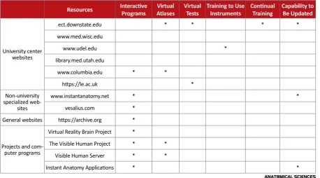
Results: Having interactive programs, atlases and virtual tests, the capability of updating material and continual education are some of the properties of electronic learning environments in the teaching of anatomical sciences. According to this research, 100% of the investigated projects and the computer programs and 44% of the websites, had interactive programs. Furthermore, 50% of the projects and computer programs and 22% of websites had virtual updated atlases, and 22% of websites had virtual tests.
Conclusion: By using the facilities in reliable educational sites of anatomical sciences and also
the interactive learning computer programs, difficulties in learning and understanding anatomical sciences syllabus can be reduced and the level of knowledge in the field raised so that the way can be paved for improving clinical skills in the areas of diagnosis and treatment. Besides, the use of these technologies is effective in updating the knowledge of graduates and lecturers in the field of
anatomical sciences.
Citation: Zand A, Abbaszadeh HA, Abdolahifar MA, Aghaee AA, Amini A, Mastery Farahni R. Role of e-learning in teaching anatomical sciences. Anatomical Sciences. 2016; 13(1):55-60.
ly on online learning. Facilitation of learn-ing occurs in the e-learnlearn-ing environment via the use of diverse technologies like in -teractive applications (including multime-dia applications such as movies, photos, audio materials, simulated environments, and applications installed on cell phones or tablets) and via links with other learners (e.g. e-mail, video-conferencing, and chat) which pro-vide suitable environments for group discussion [1].
Courses in basic anatomy for medical undergraduates have always been hard to understand and memorize. The commonly available resources namely anatomical and histological textbooks and atlases do not meet all their needs regards accessing all anatomical structures and related topics in physiology and other basic or clinical sciences, as well as having access to images, interactive samples, and the latest updates. Bearing in mind ad-vances in technology and the availability of new teach-ing technologies in the virtual space, explorteach-ing the role of electronic learning, including websites and computer programs for resolving these deficits is recommended.
2. Materials and Methods
One of the objects of the current research was to examine the role of e-learning in presenting the processes and new training facilities in the field of anatomical sciences (anat -omy and histology), to enhance undergraduate learning in basic medicine, to alleviate the deficits, and to fill in the gaps in the teaching of the anatomical sciences. Furthermore, this study by introducing these training prospects sets the scene for continual training of graduates and lecturers in this field.
In this review, 9 websites and 4 projects and computer programs related to the field of anatomical sciences were investigated according to proposals made by the lectur-ers of anatomy at the Anatomy Group of Shahid Beheshti University. This review was based upon the educational processes and technologies required in the e-learning en-vironments and also the necessary training facilities in learning the anatomical sciences. The studied websites consisted 6 cases belonged to university centers, 2 cases to non-university specialized training websites, and 1 website contained sorted information on various scientific fields.
Websites belonging to university centers
• ect.downstate.edu (SUNY Downstate Medical Cen-ter, Office of Educational Computing & Technology),
• www.udel.edu (University of Delaware, Department of Biological Sciences),
• library.med.utah.edu (University of Utah, Health sci-ences library),
• www.columbia.edu (Columbia University, College of Physicians & Surgeons, Department of Anatomy and Cell Biology),
• https://le.ac.uk (University of Leicester).
• Specialized non-university training websites:
• www.instantanatomy.net (website for anatomical education with numerous applications for training in anatomy) and,
• vesalius.com (the Internet resource for surgical edu-cation).
• A database website in various fields of science:
• https://archive.org (A non-profit library of millions of free books, movies, software, music, and more).
The examined programs and computer projects were as follows
• Virtual Reality Brain Project (a project consisting of images of various cross-sections of the human brain with the capability of interactive use),
• The Visible Human Project (a project consisting of real images of extremely thin slices of the human body),
• Visible Human Server (A collection of 3D images based on data from The Visible Human Project) and,
• Instant Anatomy Applications (A collection of appli-cations that can be installed on cell phones and tablets that contain flash cards for learning anatomy which allow the user to review important anatomical points).
3. Results
Winter & Spring 2016, Volume 13, Number 1
comprehending these structures, their interconnections, and spatial structures. In such cases, using various cross-sectional images of the organ at various positions helps towards a better comprehension of its structure. A case in point is brain structure. This facility is presented in the relevant computer programs.
Accordingly, in a program entitled “Virtual Reality Brain Project”, which uses the computer technology of “Quick Time”, the facility has been provided for observing the various cross-sections of the human brain interactively allowing change of the size of these cross-sections, their rotation, and selection of a specific region for observing its structure [2]. Regarding some anatomical areas of the body, textbooks and atlases are unable to display them in a way that can be understood. In these cases, using com-puter programs and websites that display the relevant spaces from different directions in 3D are helpful. Some examples of these displays are the pterygopalatine fossa
[3] and the peritoneal space [4].
Computer simulation of a number of complex anatomi -cal structures can be helpful in the comprehending such organs. In such simulations, the user can rotate and ob-serve the relevant organ or take away and remove some parts so as to observe the underlying structures and sim-plify the organ in order to gain a better comprehension. For instance, under the Visible Human Server using the images obtained from The Visible Human Project and 3D technology; an interactive computer program is created
and a collection of images and 3D videos of the various body organs together with supplementary information is incorporated [5]. Obviously, such capabilities do not exist in anatomy textbooks or atlases. In addition, these interac -tive programs give one the opportunity to interac-tively in-spect organs despite not having physical access (through a corpse or a model) to them. Also, such programs indi-rectly reduce the costs of training centers for the provision and retaining of various models or parts of the body.
Images or concrete descriptions and likening the ana -tomical structures lead to their better comprehension. For instance, in one of the websites affiliated to a uni -versity center, by using metaphorical descriptions and images, the anatomical structure of the larynx has been likened to a collection of pipes and elastic bands [6].
In such cases, the utilization of images of real corpses can assist learning process. Likewise, in cases such as scarcity of an organ or lack of necessary resources, the option of dissection of some organs such as the brain does not exist. In this situation, using real images of brain cross-sections in such internet atlases can assist comprehension.
A project entitled The Visible Human Project was under-taken in the year 2000 in the US with the goal of achiev -ing a better comprehension of human anatomy. In this project, after fixation, the corpse of a man and a woman were axially cut in thin slices. Then, images of very high resolution were prepared out of them. Finally, a website Table 1. Training processes and the technologies employed used in the studied e-learning resources.
Resources Interactive Programs AtlasesVirtual Virtual Tests Training to Use Instruments Continual Training Capability to Be Updated
University center websites
ect.downstate.edu * * * *
www.med.wisc.edu
www.udel.edu *
library.med.utah.edu
www.columbia.edu * *
https://le.ac.uk *
Non-university specialized
web-sites
www.instantanatomy.net * *
vesalius.com *
General websites https://archive.org *
Projects and com-puter programs
Virtual Reality Brain Project *
The Visible Human Project * *
Visible Human Server * *
The use of real specimens not only assists comprehend-ing and longer lastcomprehend-ing memory but also assists the basic medical undergraduates in gaining higher diagnostic skills on entering clinical courses at hospital departments. In this regard, they would relate the basic scientific topics with the clinical topics and realize that acquiring the basic sci-entific knowledge is essential for working in the clinical sphere. A few websites affiliated to university centers in -troduce deceased clinical cases together with an account of the disease and patient history, examinations performed, and para-clinical measures (tests and imaging) performed. Then, they discuss the dissection and autopsy results and their anatomic pathologic findings after death and explain and interpret these according to the person's primary dis-ease [8]. The elucidation of anatomic pathologic structures, especially assists the basic medical science undergraduates to better comprehend the clinical topics. Furthermore, such cases should be incorporated alongside training on the nor-mal human anatomy. A few internet resources train these topics by using images and in addition describe surgery techniques for restoring such structures. For instance, pre-sentation of images of inguinal hernia and its variations, its anatomic connections and surgery techniques leads to a better comprehension of such a disease [4].
Because of the high price, volume, and weight of some books and atlases; their inaccessibility; and their shortage in displaying all cross-sections, virtual atlases in computer programs be employed. They can be downloaded from a number of websites. Lecturers and undergraduates are al -ways searching for suitable training resources correspond-ing to their needs and requirements and are sometimes forced to spend much time in libraries or non-specialized search websites for finding these references. However, fa -miliarity with specialized websites in any scientific field that introduces the resources for that course frees one from spending time and money. Also, as regards the anatomical sciences, in a number of specialized websites affiliated to the universities, the references for these sciences are cited and some of them allow users to download freely online
[9]. Access to old and historical references concerning anatomy and histology is sometimes necessary for con-ducting research. Such facilities have been provided in the virtual space, which are certainly less demanding of time and money compared to the usual practice of going to li-braries and searching printed references.
Other ways of updating the sciences include becoming acquainted with distinguished persons in one’s special-ized field and having the opportunity to discuss and ex
-announce creditable conferences and their agendas. Con-sidering the daily advances in various sciences, includ-ing anatomy and histology, updated accessibility to news about these sciences through books and atlases, which are usually published and edited once every few years is not feasible and the use of reliable internet references in this field is mandatory. In a number of these resources, advanc -es in th-ese scienc-es become momentarily available [10].
Continual training, which is in fact necessary for grad-uates in any field requires a organized and well-defined scheme so that by upgrading and updating the knowledge of graduates and lecturers in each field the graduates under training benefit indirectly. Anatomical sciences are no exception to this rule and suitable pro -grams of graduate education are present on a number of websites affiliated to creditable universities and by refer -ring to them the necessary training can be received [11]. Such continual training programs have advantages over the traditional ones in terms of time and money saving on attending lectures, conferences, or workshops; as well as having updates of references and well-organized and purposeful training programs.
In recent years, in some medical schools, including some in Iran, the training of some topics of the basic science courses like anatomy and histology has become system-based (according to various systems of the body such as the nervous system, the digestive system, and other ones) and integrated (i.e. integration of various topics such as anatomy, histology, embryology, physiol-ogy and other topics in the frame of each organ of the body). In this respect, suitable textbooks have not been provided for the undergraduates. This can lead to disori-entation and impairment of this novel training process, whilst there are references in the websites of creditable universities which offer this training system [11].
Winter & Spring 2016, Volume 13, Number 1
For training medical undergraduates, dissection halls are always in need of provision of corpses. Considering the repeated use of these corpses, which have limited and restricted durability, it is indispensable for these centers to be provided with new corpses. Nowadays, in a num-ber of websites affiliated to the university centers, there is a scheme entitled Body Donation Program in which persons above 18 years are requested to sign a form announcing their consent for donating their body after death. They can participate in this way in the training of the forthcoming physicians [13].
Finally, the training processes and the technologies employed in each of these e-learning resources are intro-duced in the following table.
4. Discussion
Devising interactive programs, atlases, and virtual tests, as well as the ability for updating the material and continual training are some of the features of e-learning environments for the training of anatomical sciences. In the present review, 100% of the projects and computer programs (4 cases out of 4) and 44% of websites (4 cases out of 9) under review had interactive programs. Also, 50% of the projects and computer programs (2 cases out of 4) and 22% of websites (2 cases out of 9) had virtual updated atlases and 22% of websites (2 cases out of 9) had virtual tests. Therefore, their utilization in the train-ing of anatomical sciences contributes towards counter-acting the gaps in traditional educational resources (the customary textbooks and atlases), and also towards re -ducing learning difficulties among undergraduates in the basic medical sciences regarding anatomical sciences.
Besides, their clinical skills in the fields of diagnosis and treatment are facilitated by advancing their knowl -edge in these fields. In addition, using these technologies for the continual training and updating the knowledge of graduates and lecturers in the fields of anatomical sci -ences is effective and economic. In this regard, more re-search is essential in the area of novel technologies like e-learning to advance training in disciplines of anatomi-cal sciences and subsequent results could be employed in constructing training processes founded on technology.
Acknowledgements
The current research hasn't received any financial support.
Conflict of Interest
The authors of this study declared no conflict of interests.
References
[1] Moore JL, Dickson-Deane C, Galyen K. E-learning, online learning and distance learning environments: Are they the same? Internet and Higher Education, 2011; 14(2):129-35.
[2] Frick GS, Conyers K, Laurance J, Brett G. Virtual reality brain project [Internet]. Brooklyn, N.Y.: SUNY Downstate Medical Center; 2008. Available from: http://ect.down-state.edu/courseware/vr_brain
[3] Clinical Anatomy Course New Media Resources [Internet]. New York, N.Y.: Columbia University. Available from: https://www1.columbia.edu/sec/itc/hs/medical/anato-my_resources/anatomy/main.html
[4] The internet resource for surgical education Vesalius [In-ternet]. 2015 [Cited 2016 Oct. 19]. Available from: http:// vesalius.com
[5] Hersch R. Visible human server: Ecole Polytechnique Fédé-rale de Lausanne [Internet]. 2000 [Cited 2000 Oct. 1].
Avail-able from: http://visiblehuman.epfl.ch/index.php
[6] Instant anatomy app: Instant Anatomy [Internet]. 2015 [2015 Oct. 8]. Available from: http://www.instantanatomy.net
[7] Ackerman M. Visible Human Project U.S: National Library of Medicine [Internet]. 2000 [Cited 2015 Oct. 8]. Available from: https://www.nlm.nih.gov/research/visible/vis-ible_human.html
[8] Mackay RV, Mark A, Taylor T. The Virtual Autopsy [Inter-net]. Available from: http://www.le.ac.uk/pa/teach/va/ titlpag1.html
[9] Kubie OC. Downloadable Atlases SUNY Downstate
Medi-cal Center, Office of Educational Computing & Technol -ogy, Courseware, Neuroscience, NeuroQuiz Software [In-ternet]. 2014 [Cited 2014 Feb. 20]. Available from: http://
ect.downstate.edu/courseware/neuroscience/neuro-quiz/index.html
[10] Najman R. News releases. SUNY Downstate Medical
Center, Office of Institutional Advancement, News Re
-leases; 2015 [Cited 2015 Oct. 26]. Available from: http:// ect.downstate.edu
[11] Krabbenhoft K. MD Program Curriculum: Integrated Med-ical Anatomy [Internet]. 2015 [Cited 2015 Mar. 12]. Avail-able from: http://www.med.wisc.edu/education/md/ curriculum/year-1/integrated-medical-anatomy/427
[12] Ketcham B. Virtual microscope solves teaching challenge [Internet]. Newark: University of Delaware; 2006.
Avail-able from: http://www.udel.edu/present/profiles/ket
-cham/index.html