Please cite this article as: S. Momtahen, M. Taajobian, and A. Jahanian, Drug Discovery Acceleration Using Digital Microfluidic Biochip Architecture and CAD Flow, International Journal of Engineering (IJE), IJE TRANSACTIONS B: Applications Vol. 32, No. 8, (August 2019) 1169-1176
International Journal of Engineering
J o u r n a l H o m e p a g e : w w w . i j e . i rDrug Discovery Acceleration Using Digital Microfluidic Biochip Architecture and
Computer-aided-design Flow
S. Momtahena, M. Taajobianb, A. Jahanian*c
a Department of Electrical and Computer Engineering, Ryerson University, Toronto, Canada b Department of Computer Engineering Mahdishahr Branch, Azad University, Tehran, Iran c Department of Computer Science and Engineering, Shahid Beheshti University, Tehran, Iran
P A P E R I N F O
Paper history:
Received 25 October 2018
Received in revised form 25 May 2019 Accepted 05 July 2019
Keywords:
Digital Microfluidic Biochip
Lab-on-chip Drug Discovery Machine Learning
A B S T R A C T
A Digital Microfluidic Biochip (DMFB) offers a promising platform for medical diagnostics, DNA sequencing, Polymerase Chain Reaction (PCR), and drug discovery and development. Conventional Drug discovery procedures require timely and costly manned experiments with a high degree of human errors with no guarantee of success. On the other hand, DMFB can be a great solution for miniaturization, integration, automation, and cost reduction of drug discovery. DMFB can improve the parallelism of drug discovery procedures; since most procedures in drug discovery are concurrent and parallel, DMB can reduce the execution time of these bioassays. Therefore, there is a critical need to develop DMFBs to speed up the drug discovery applications and improve cost and error of these reactions. In this paper, a new architecture is used for drug discovery applications. The architecture is evaluated and compared with FPPC architecture. The experimental results prove that the new architecture is faster and cheaper than FPPC; it reduces all the important parameters such as total execution time, number of controlling pins, CAD algorithm execution time, and the area usage and its costs. There is an urgent need for collaboration between experts of drug discovery, microfluidic platform architecture and also machine learning to design a data-driven microfluidic architecture which improves the CAD algorithms by learning from prior knowledge.
doi: 10.5829/ije.2019.32.08b.13
1. INTRODUCTION1
Drug discovery and development are the processes of discovering candidate medications and bringing new drugs. Nowadays, drug discovery and development applications, such as target selection, lead identification, preclinical tests, clinical trials, chemical synthesis, formulations studies, and product management [1], are very important in medicine and pharmacology. However, current drug discovery procedures are time- consuming which are prone to errors. Digital Microfluidic Biochips (DMFBs) are electronic platforms for automation, parallelization, and cost-reduction of the biochemical reactions in point of care (POC) [2], DNA analysis, Polymerase Chain Reaction (PCR), and drug discovery and development, and other
*Corresponding Author Email: [email protected] (A. Jahanian)
biological and chemical procedures. DMFBs integrate fluid-handling operations such as sample preparation, mixing, separation, and detections. Assay protocols run on a digital microfluidic by controlling discrete droplets with volumes in the ranges of nano- to pico-liter using a two-dimensional array of electrodes [3].
DMFBs take advantages of computer-aided-design (CAD) tools for automation of the procedures. Moreover, Digital microfluidic biochips miniaturize the devices and also bioassays; thus DMFBs decrease the consumption of samples, reduce the likelihood of errors, and increase experimental throughput [4].
be accomplished by a sequence of droplet-mix-split steps on a digital microfluidic biochip [5]. Besides, in dilution of a sample, a special case of sample preparation can be performed on DMFB by mixing two different types of fluids with the desired concentration factor (CF) [5].
Moreover, protein determination techniques have widely used in drug discovery applications [6]; but for many of such procedures, the assays are typically performed manually, so the processes are slow, inefficient, expensive, and error-prone. Moreover, studies have focused on the application of digital microfluidic biochips on protein concentration assays. Some researchers have also demonstrated the feasibility of performing a protein concentration assay on a digital microfluidic biochip [7]. A DMFB for the Bradford protein assay using Field-Programmable Pin-Constrained (FPPC) is proposed [7]. The basic fluidic operations in protein concentrator assays, such as transportation, mixing and splitting of droplets can be performed by FPPC. However, no DMFB has been proposed for performing BCA protein assay that is typically preferred over other protein concentration assays. Customized DMFBs are not developed for performing common drug-based assays such as the assays for the synthesis of aspirin and acetaminophen.
Furthermore, some researchers have used microfluidics for enzymatic synthesis, amplification, and application of DNA, such as analyzing DNA assay and enzymatic assay for analysis of new compounds [8]. DMFB technique is also applied in proteomics. As shown in [9], a digital microfluidic platform for the detection of protein biomarkers, which can quantify protein abundance and activity, is proposed. For this purpose, interleukin-6 abundance was quantified from concentrations as low as 50 pM and Abelson tyrosine kinase activity was determined in samples containing 100 pM of kinas [9]. Other studies have shown the use of microfluidic technology for preparing partial samples, performing a portion of the assay for lysosomal storage diseases, and analyzing dried blood spot [10]. A microfluidic chip for the detection of rubella infection by using the magnetic beads is developed [11]. Besides, a DMF platform for multiple detections is introduced by Samiei [8].
In addition, analysis of single cells is very important in studying drug development and many researchers have used microfluidics for studying single-cell analysis. Some microfluidic chips have been reported to capture single cells and to determine the impedance of the cells or circulating tumor cells. Furthermore, a microfluidic chip to trap single cells and to measure the impedance on cell membranes is demonstrated [12]. Recently, as represented in literature [13], a microfluidic technique capable of probing single cells is introduced. An assay of the deformability of native
populations of leukocytes and malignant cells in pleural effusions was enabled on the biochip.
Lab-on-Chip technology as a novel technique offers a wide range of benefits for drug discovery applications. DMFB is capable to improve the controllability in drug discovery; it can process large droplets of biological and chemical solutions. For instance, DMFB is used in drug design by applying the concentration gradient of a drug. For instance, according to literature [5], in bacterial susceptibility tests, samples are needed in multiple concentration factors. It is important to measure the minimum inhibitory concentration (MIC). MIC is the minimum amount of an antibiotic that inhibits the visible growth of isolated bacteria. The drug with the highest dilution is proposed to have satisfied MIC. Therefore, for these tests, drugs with different dilutions are required. In addition, preparation of a given target CF on digital microfluidic biochip needs a sequence of mix-split steps [5]. A new approach for multi- target CFs is proposed [5].
Furthermore, digital microfluidic has the capability of integration with conventional setups. For instance, a droplet-on-demand (DOD) platform was used to produce micro-droplets for high-throughput drug screening. An automated DOD is proposed by Dressler et al. [14] and enzyme kinetics and inhibition were investigated. In addition, using microfluidics, the partitioning of drug molecules in different oil media was investigated. Also, a droplet-based microfluidic platform was used to screen a drug library of more than 700 inhibitors for inhibition of different target enzymes for different diseases [14].
Although microfluidic platforms are valuable techniques, traditional DMFB architectures have drawbacks; especially the current architectures have to be customized for drug discovery acceleration. This work uses and evaluates a new digital microfluidic platform, Programmable Bio-Cell Matrix (PBCM-), for accelerating the execution time of bioassays. Using Can flow algorithms, PBCM performs basic bio-operations in parallel [15]. PBCM is compared by FPPC by our proposed case studies, such as assays of aspirin, acetaminophen, and the BCA protein assay.
The rest of the paper is organized as follows: Section 2 is a review on digital microfluidic biochips. In section 3 related prior art on the biochip architectures are briefly summarized. The performance procedure of case studies is presented in section 4. In Section 5, evaluation and experimental results are discussed. Finally, in section 6 the paper is the conclusion.
2. DIGITAL MICROFLUIIDIC BIOCHIP (DMFB)
electrowetting-on-dielectric (EWOD) phenomenon [16]. As Figure 1 shows a basic cell of a DMFB cnsists of two electrode layers coated with a hydrophobic layer between which droplets are placed [17].
The hydrophobic dielectric insulator is added to the two parallel plates to decrease the wettability of the surface and to increase the capacitance between the droplet and the control electrode [18]. The top plate in the DMFB is coated with a continuous ground electrode. The bottom electrode layer contains an array of individually controllable electrodes, which is patterned to implement reservoirs or channels. These electrodes are derived by a sequence of voltage patterns (actuation sequences) and are used to handle operations, such as transport, mix, split/merge, store, detect, and dilution [17]. Some droplet operations performed on digital microfluidic biochip is shown in Figure 2.
By applying a voltage to an electrode adjacent to the droplet, the droplet is moved and the electrode under the droplet is deactivated [18]. The results of the synthesis are programmed into a microcontroller that controls the voltages of electrodes in the array. By varying the patterns of control voltage activation, basic assay operations such as dispensing, transport, mixing, merging, heating, incubation, and splitting can be performed on-chip in a programmable fashion. For example, a mixing operation can be performed by moving two droplets to the same cell and then turning them about a pivot [19].
As shown in Figure 3, a DMFB needs additional components, which together form a cyber physical digital microfluidic biochip. These components include: a computer for generating and sending actuation sequences to DMFB; sensors for gathering information; actuators add extra processing capabilities like heating [20].
Figure 1. Schematic view of a DMFB: (a) side view (b) top
view: a ring-based DMFB with 2D array and on-chip resources [17]
Figure 2. Basic set of droplet operations performed on a
DMFB: (a) Transport (b) Split/Merge (c) Mixing (d) Storage and Detection [2]
Figure 3. Schematic of a typical cyber physical DMFB system
[20]
The digital microfluidic biochip offers software-based control of multifunctional biochips and uses CAD tools for the automated synthesis and optimization of biochips from bioassay protocols. The platform offers the flexibility of dynamic reconfigurability since the basic operations can be performed anywhere on the array. Other operations such as dispense and output are performed by I/O reservoirs on the chip's perimeter; detectors are placed in a special location of the biochip. As shown in Figure 1, Optical detectors like LEDs can be integrated in digital microfluidics to monitor colorimetric bioassays [21].
DMFBs can be used to perform an assortment of bioassays. Automated design flow is used to compile bioassays on-chip into a software program to activate the electrodes [22]. As shown in Figure 2, three steps must be done before performing an assay on-chip. These steps are scheduling, placing, and routing, which constitute the synthesis flow of any typical DMFB. DMFB Synthesis activate the proper sequence of the electrode for performing operations at any times during assay execution.
Figure 4. Synthesis of DMFB [12]
specifications of the DMFB including dimensions and locations of inputs and output reservoirs.
In the first stage (e.g. scheduling), scheduler ensures the constraint between operations are met and assigns each operation to a start and end times [24]. At the placement step, the location of each operation on the array is determined. Operations are placed to modules (specific set of electrodes) at the specified time steps. The routing step is after placement of modules in which the movement of droplets, between modules, from input to modules, and from modules to output reservoirs are computed [25].
DMFBs enable real-time decision making for sample processing but are not as effective for interfacing to the external world. Thus, for solving this drawback, hybrid microfluidic systems are proposed which can suggest a synthesis method that controls experiments in a dual-domain microfluidic setting [26]. Figure 5 shows an efficient on-chip implementation of a single cell analysis protocol on a hybrid platform [26].
3. DMFB ARCHITECTURES
The Programmable Bio-Cell Matrix (PCB) is a two-dimensional planer array of controllable electrodes in which each electrode is controlled via an external pin. PCB is complex and expensive due to the number of control pins and expensive multi-layer PCBs [27]. FPPC offers a general-purpose architecture that decreases the
Figure 5. The hybrid platform for single-cell analysis [26]
control pins and PCB layers and so reduces the corresponding costs. In FPPC, there is a routing column between modules, the routing paths are vertical and horizontal, operating modules are fixed, and time scheduling is performed at the design time but the placement is determined at the usage time [28].
FPPC is an inexpensive architecture but decreases flexibility on chip because the operations cannot be performed in parallel. PBCM is a general-purpose architecture that maintains flexibility and efficiency for large assays with massive parallel operations. Furthermore, PBCM can be reconfigured for any bioassays without the need to fabricate a new chip. PBCM constitutes an array of Configurable Bio-Cells (CBCs) which contains the necessary modules for carrying out the operations, and the routing paths between CBCs are controlled by some electrodes. Figure 6 is shown a PBCM with 9 cells whose CBCs are connected by using the routing paths. PBCM can perform 9 parallel mixings, splitting, and storage operations [16].
4. DRUG DISCOVERY ACCELERATION ON DMFB
As said before, drug discovery requires long-time experiments of the drug. These experiments are very time-consuming with great human and material costs. Moreover, manned operation of these experiments faces with considerable error rate. In this paper, the impact of using the DMFB to mitigate the problems of drug discovery is evaluated. We selected BCA protein assay and the assays for synthesis of aspirin and acetaminophen as our case studies.
4. 1. BCA Protein Concentration Assay The
transported to some electrodes and are measured using detectors [29]. Finally, a standard curve is produced based on the protein standard concentration, plotting the protein concentration versus the absorbance. Figure 7 shows flow graph of the BCA protein assay [30].
In Figure 7, the colors of light blue, green, orange, blue, and grey are being used to represent dispense, output, mixer, split, and store for all mix and split operations, respectively.
4. 2. Synthesis of Aspirin and Acetaminophen
For aspirin preparation, salicylic acid (C7H6O3) and acetic anhydride (C4H6O3) are dispensed and mixed.
Then, the result is mixed with a droplet of concentrated sulfuric acid (H2SO4). The resulted droplet is mixed with a droplet of water on the biochip.
Figure 6. The PBCM architecture: (a) Schematic view of a
PBCM with 9 CBCs, (b) Internal of CBC structure [15]
Figure 7. Flow graph of the BCA protein assay [30]
Furthermore, acetaminophen is prepared by mixing a droplet of water with an acid anhydride and then mixing the result with an amine (p-aminophenol) on the bio-chip. Figures 8 and 9 demonstrate the assay flow graph of aspirin and acetaminophen, respectively. The time and the color of operations are similar to Figure 7 [30].
5. EXPERIMENTAL RESULTS
In this work, we prove that PBCM improves our three drug-related study cases. Before reporting the simulation results, we propose some assumptions. For evaluating the efficiency of FPPC and PBCM, both architectures are simulated on a 1.60GHz Intel Core i5-5250U, 4GB of RAM, within UCR SSS framework [31], which is an open-source digital microfluidic biochip synthesis framework for implementing the DMFB’s CAD algorithms. Also, for evaluation of the architectures, the results are reported in terms of assay experimental time, characteristics of the architectures, and CAD execution time. We implemented the three study cases in both PBCM and architectures and the results are compared as it is discussed in the next section.
Figure 8. Flow graph of the assay of aspirin [30]
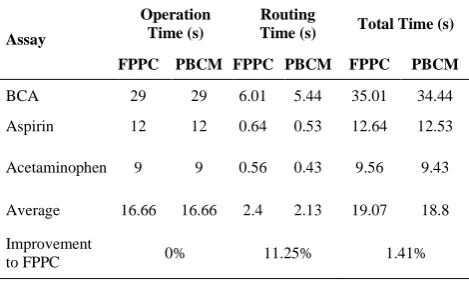
5. 1. Total Experimental Time Table 1 compares the BCA assay experimental time of PBCM with FPPC. In Table 1, column Operation Time shows the maximum time which is required for performing the operations; column #Routing Time demonstrates the cycles used to route a droplet among the modules; column Total Time represents the total execution time of assay.
TABLE 1. Comparison of PBCM with FPPC architectures in
terms of experimental time
Assay
Operation Time (s)
Routing
Time (s) Total Time (s)
FPPC PBCM FPPC PBCM FPPC PBCM
BCA 29 29 6.01 5.44 35.01 34.44
Aspirin 12 12 0.64 0.53 12.64 12.53
Acetaminophen 9 9 0.56 0.43 9.56 9.43
Average 16.66 16.66 2.4 2.13 19.07 18.8
Improvement
to FPPC 0% 11.25% 1.41%
TABLE 2. Comparison of the characteristics of PBCM and
FPPC architectures
Assay
Array
Dimension #Electrodes #Pins
FPPC PBCM FPPC PBCM FPPC PBCM
BCA 12*31 10*37 220 233 59 54
Aspirin 12*9 10*9 69 63 23 22
Acetaminophen 12*9 10*9 69 63 23 22
Average 119.33 119.66 35 32.66
Improvement
to FPPC -0.27% 6.68%
TABLE 3. Experimental results in terms of CAD execution
time
Assay
Scheduling Time (ms)
Placement Time (ms)
Routing Time (ms)
FPPC PBCM FPPC PBCM FPPC PBCM
BCA 9 8 0 0 51 24
Aspirin 1 0 0 0 10 2
Acetaminophen 1 0 0 0 7 1
Average 3.66 2.66 0 0 22.66 9
Improvement
to FPPC 27.32% 0% 60.28%
In PBCM, the total time is improved by 1.41% since the operations are accomplished in parallel. Total experimental time proves which biochip would be capable of performing faster. Thus, our architecture is faster.
5. 2. Characteristic of the Architectures Table 2
shows a comparison of the characteristics of both architectures and has 4 columns: Assay, Array Dimension, #Electrodes, and #Pins. Column Array Dimensions represents rows and columns of chip and columns #Electrodes and #Pins show the number of electrodes and pins on the microfluidic biochip. The results in Table 2 prove that with PBCM, the number of electrodes is increased by 0.27% while the number of controlling pins is improved by 6.68%. Also, in PBCM the area is improved.
5. 3. CAD Algorithm Execution Time Table 3
compares the CAD algorithm execution time of both designs. Columns Scheduling Time, Placement Time, and Routing Time demonstrate the total time that required for completing scheduling, placement, and routing in UCR SSS. Table 3 proves that the scheduling time and routing time in PBCM-DMFB synthesis flow are both improved by 27.32% and 60.28% respectively.
6. CONCLUSION
In this work, a new DMFB architecture for drug discovery and development is introduced and evaluated by several case studies; our simulation proved that this new design can speed up the process and reduce the costs. This work prove that digital microfluidic platform has the potential to improve drug discovery applications greatly. But, there is an urgent need for collaboration between experts of the drug discovery, microfluidic platform architecture, and also machine learning to design a data-driven microfluidic architecture which by learning from prior knowledge, improves the CAD algorithms. In the future, drug discovery process would be revolutionized by combining microfluidic for acquiring data with machine learning for analyzing data.
7. REFERENCES
1. Kang, L., Chung, B.G., Langer, R. and Khademhosseini, A., “Microfluidics for drug discovery and development: from target selection to product lifecycle management”, Drug Discovery
Today, Vol. 13, No. 1-2, (2008), 1-13.
2. Chakraborty, S. and Chakraborty, S., “Module-less Synthesis on Cyberphysical Digital Microfluidic Biochip Ensuring Error Detection and Routing Performance Optimization”, arXiv
3. Fair, R. B., “Digital microfluidics: is a true lab-on-a-chip possible?” Microfluidics and Nanofluidics, Vol. 3, (2007), 245-281.
4. Xu, T. and Chakrabarty, K., “Broadcast electrode-addressing for pin-constrained multifunctional digital microfluidic biochips,” Proc. IEEE/ACM Design Automation Conference, (2008), 173– 178.
5. Sudip, P., Bhattacharjee, S., Nandy, S. C., Chakrabarty, K., and Bhattacharya B. B., "Optimization of Multi-Target Sample Preparation On-Demand with Digital Microfluidic Biochips."
IEEE Transactions on Computer-Aided Design of Integrated
Circuits and Systems, Vol. 38, No. 2, (2018), 253-266.
6. Noble, J. E., Knight, A. E., Reason, A. J., Matola, A. D., and Bailey, M. J. A., “A comparison of protein quantitation assays for biopharmaceutical applications,”Molecular Biotechnology, Vol. 37, No. 2, (2007), 99-111.
7. Grissom, D., Design of Topologies for Interpreting Assays on Digital Microfluidic Biochips (Doctoral dissertation, UC Riverside), (2014).
8. Samiei, E., “Development of advanced operators for enhanced on-chip biosensing in digital microfluidic platforms,” Ph.D. dissertation, Univ. of British Columbia, BC, Canada, (2016). 9. A. Mok, J., Mindrinos, M. N., Davis, R. W. and Javanmard, M.,
"Digital microfluidic assay for protein detection." Proceedings of the National Academy of Sciences, Vol. 111, No. 6 (2014), 2110-2115.
10. Jebrail, M.J., Yang, H., Mudrik, J.M., Lafreniere, N.M., McRoberts, C., Al-Dirbashi, O.Y., Fisher, L., Chakraborty, P. and Wheeler, A.R., “A digital microfluidic method for dried blood spot analysis”,Lab on a Chip, Vol. 11, No. 19, (2011), 3218-3224.
11. Ng, A.H., Lee, M., Choi, K., Fischer, A.T., Robinson, J.M. and Wheeler, A.R., “Digital microfluidic platform for the detection of rubella infection and immunity: a proof of concept”,Clinical
Chemistry, Vol. 61, No.2, (2015), 420-429.
12. He, J.L., Chen, A.T., Lee, J.H. and Fan, S.K., “Digital microfluidics for manipulation and analysis of a single cell,”
International Journal of Molecular Sciences, Vol. 16, No. 9,
(2015), 22319-22332.
13. Gossett, D.R., Henry, T.K., Lee, S.A., Ying, Y., Lindgren, A.G., Yang, O.O., Rao, J., Clark, A.T. and Di Carlo, D., “Hydrodynamic stretching of single cells for large population mechanical phenotyping”, Proc.National Academy of Sciences
Vol.109, No. 20, (2012), 7630-7635.
14. Dressler, O.J., Maceiczyk, R.M., Chang, S.I. and DeMello, A.J., “Droplet-based microfluidics: enabling impact on drug discovery,” Journal of Biomol Screen. Vol. 19, No. 4, (2014), 483-496.
15. Taajobian, M. and Jahanian, A., "Improved experimental time of ultra-large bioassays using a parallelized microfluidic biochip architecture/scheduling."IET Nanobiotechnology, Vol. 12, No. 4 (2018): 484-490.
16. Pollack, M.G., Fair, R.B. and Shenderov, A.D., “Electrowetting-based actuation of liquid droplets for microfluidic applications”,
Applied Physics Letters, Vol. 77, No. 11, (2000), 1725-1726.
17. Keszocze, O., Ibrahim, M., Wille, R., Chakrabarty, K. and Drechsler, R., “Exact synthesis of biomolecular protocols for multiple sample pathways on digital microfluidic biochips”, In
2018 31st International Conference on VLSI Design and 2018 17th International Conference on Embedded Systems (VLSID) (2018), 121-126. IEEE.
18. Srinivasan, V., Pamula, V.K. and Fair, R.B., “An integrated digital microfluidic lab-on-a-chip for clinical diagnostics on human physiological fluids”, Lab on a Chip, Vol. 4, No. 4, (2004), 310-315.
19. Paik, P., Pamula, V.K. and Fair, R.B., “Rapid droplet mixers for digital microfluidic systems”, Lab on a Chip, Vo. 3, No. 4, (2003), 253-259.
20. Tang, J., Ibrahim, M., Chakrabarty, K. and Karri, R., “Secure randomized checkpointing for digital microfluidic biochips”,
IEEE Transactions on Computer-Aided Design of Integrated
Circuits and Systems, Vol. 37, No. 6, (2017), 1119-1132.
21. Srinivasan, V., Pamula, V.K. and Fair, R.B., “An integrated digital microfluidic lab-on-a-chip for clinical diagnostics on human physiological fluids”, Lab on a Chip, Vol. 4, No. 4, (2004), 310-315.
22. Grissom, D. and Brisk, P., “Path scheduling on digital microfluidic biochips”, In DAC Design Automation Conference 2012, (2012), 26-35. IEEE.
23. Su, F. and Chakrabarty, K., “Benchmarks for digital microfluidic biochip design and synthesis”, Duke University Department ECE, (2006).
24. Su, F. and Chakrabarty, K., “High-level synthesis of digital microfluidic biochips,” ACM Journal on Emerging
Technologies in Computing Systems, Vol. 3, No. 4, (2008),
1-32.
25. Ho, T.Y., Chakrabarty, K. and Pop, P., “Digital microfluidic biochips: recent research and emerging challenges”, In Proceedings of the seventh IEEE/ACM/IFIP international conference on Hardware/software codesign and system synthesis (2011), 335-344. ACM.
26. Ibrahim, M., Chakrabarty, K. and Schlichtmann, U., “Synthesis of a cyberphysical hybrid microfluidic platform for single-cell analysis”, IEEE Transactions on Computer-Aided Design of
Integrated Circuits and Systems, (2018).
27. Grissom, D., Curtis, C., Windh, S., Phung, C., Kumar, N., Zimmerman, Z., Kenneth, O.N., McDaniel, J., Liao, N. and Brisk, P., “An open-source compiler and PCB synthesis tool for digital microfluidic biochips”, Integration, the VLSI journal, Vol. 51, (2015), 169-193.
28. Grissom, D., Brisk, P., “A field-programmable pin-constrained digital microfluidic biochip”, ACM/IEEE Design Automation Conf., Aust in, TX, USA, (2013), 1–9.
29. Momtahen, S., Taajobian, M., and Jahanian, A., "A Customized Digital Microfluidic Biochip Architecture/CAD flow for Drug Discovery Applications." IEEE Nanotechnology Magazine, 2019, DOI: 10.1109/MNANO.2019.2927773
30. Momtahen, S., Taajobian, M., and Jahanian, A., “Drug discovery evolution using the customized digital microfluidic biochips”, Electrical Engineering (ICEE), Iranian Conf. on, (2018), 1415-1420. IEEE.
Drug Discovery Acceleration Using Digital Microfluidic Biochip Architecture and
Computer-aided-design Flow
S. Momtahena, M. Taajobianb, A. Jahanianc
a Department of Electrical and Computer Engineering, Ryerson University, Toronto, Canada b Department of Computer Engineering Mahdishahr Branch, Azad University, Tehran, Iran c Department of Computer Science and Engineering, Shahid Beheshti University, Tehran, Iran
P A P E R I N F O
Paper history:
Received 25 October 2018
Received in revised form 25 May 2019 Accepted 05 July 2019
Keywords:
Digital Microfluidic Biochip
Lab-on-chip Drug Discovery Machine Learning
هدیکچ
و یرامعم هئارا اب وراد فشک لحارم رد یشهج دنور
ت یشرافس یحارط هشار
یاه یتسیز لاتیجید لایسزیر هشارت
یاه
لایسزیر یتسیز هک تسا عمتجم یاهرادم تعنص یاهدربراک زا لاتیجید
لرتنک و یزاسراکدوخ یارب یدنمتردق رازبا هب
شیامزآ تاقیقحت و اه ،ییوراد
یکشزپ هدش لیدبت یهاگشیامزآ و دنا
؛ هشارت نیا تیلباق اه و هروظنم دنچ تایلمع ماجنا
زاوم دنراد ار ی ؛ ِنامز و اطخ شهاک بجوم تایلمع
یم و دنوش همانرب لباق ددجم هدافتسا و یزیر دنتسه
. شیامزآ یاه
تیدودحم ،ینونک ییوراد یدایز یاه
نامز هلمج زا یناسنا تلاخد زا یشان یاطخ ،شیامزآ ماجنا یارب ینلاوط
و هنیزه
.دنراد ییایمیش داوم ندوب بایمک و نارگ لیلد هب لااب هاگتسد رد
یم لوط لاس هدزناپ ات هد طسوتم ،لومعم یاه ات دشک
رازاب دراو دیدج یوراد نویلیم کی نیب زا هک تسا یطیارش رد نیا .تسا رلاد درایلیم کی دودح رد طسوتم هنیزه و دوش
یم رازاب دراو وراد ددع کی اهنت ،ییایمیش بیکرت کچوک هکیلاحرد .دوش
هشارت یزاس سزیر یتسیز یاه
ی فرصم ثعاب لا
هنومن زا یرایسب رب هجیتن رد و هدش رتمک یژرنا فرصم ،رتمک هنیزه ،رتمک یاه یدودحم
ت یاه یلعف هبلغ یم دنک نیاربانب .
هشارت تخاس و یحارط هنیزه شهاک ،شیامزآ ماجنا تعرس شیازفا بجوم هک لاتیجید لایسزیر یاه
هاگتسد تخاس یاه
-هنیزه شهاک و شیامزآ یاه اوم یاه
یم هدافتسا دروم یفرصم د ،هلاقم نیا رد .تسا رادروخرب یرایسب تیمها زا ،دوش
یشرافس ییوراد یاهدربراک یارب لاتیجید لایسزیر یتسیز هشارت همانرب یقطنم هیارآ یرامعم( دیدج یرامعم و هدش یزاس
-هشارت نیا یارب نآ یحارط دنور هارمه هب )ریذپ شیامزآ دنچ همادا رد .تسا هدش یسررب اه
دیدج یرامعم یور رب ییوراد
هیبش جیاتن .دوش یبایزرا دیدج یرامعم درکلمع و ییاراک ات دندش ارجا انبم یرامعم نینچمه و یم ناشن یزاس
هک دهد
شیامزآ یارجا نامز و تسا رتنازرا و رتعیرس )انبم یرامعم هب تبسن( دیدج یرامعم ،اه
نامز ا ماجن تابساحم یبایریسم و(
نامز رازبا )یدنب یاهدورتکلا هیارآ تحاسم و هشارت
لایسزیر یتسیز یاه یم شهاک ار لاتیجید
.دبای
![Figure 2. Basic set of droplet operations performed on a DMFB: (a) Transport (b) Split/Merge (c) Mixing (d) Storage and Detection [2]](https://thumb-us.123doks.com/thumbv2/123dok_us/17809.2001821/3.595.62.276.542.690/figure-droplet-operations-performed-transport-mixing-storage-detection.webp)
![Figure 5. The hybrid platform for single-cell analysis [26]](https://thumb-us.123doks.com/thumbv2/123dok_us/17809.2001821/4.595.69.272.599.733/figure-the-hybrid-platform-for-single-cell-analysis.webp)
![Figure 8. Flow graph of the assay of aspirin [30]](https://thumb-us.123doks.com/thumbv2/123dok_us/17809.2001821/5.595.60.293.483.729/figure-flow-graph-assay-aspirin.webp)