Anti-mullerian hormone and antral follicle count in polycystic ovary
syndrome and non-polycystic ovary syndrome women
Received: 20/10/2016 Accepted: 30/3/2017
Abstract
* Maternity Teaching Hospital, Erbil, Iraq.
** Department of Obstetrics and Gynecology, College of Medicine, Hawler Medical University, Erbil, Iraq. *** Department of Community Medicine, College of Medicine, Hawler Medical University, Erbil, Iraq.
Introduction
Polycystic ovary syndrome (PCOS) is the most common endocrine disorder in women. It affects 5–7% of women in their
reproductive ages.1 PCOS is the most
common form of anovulatory infertility. Anovulation in PCOS is due to arrested growth of antral follicles. Women with PCOS often seek care for menstrual
disturbances, clinical manifestations of
hyperandrogenism and infertility.2 The
diagnosis of PCOS based on
the Rotterdam criteria includes
oligomenorrhea /anovulation (O), clinical or biochemical hyperandrogenism (H), and
the presence of polycystic ovaries morphology (P) on ultrasound. According
to these criteria PCOS is diagnosed if at
Background and objective: Although the ultimate pathogenesis of polycystic ovary syndrome remains obscure, the distinctive feature is the failure of follicular maturation resulting in an ovulation and accumulation of preantral and small antral follicles which contribute significantly to the production of the anti-mullerian hormone. This study aimed to compare anti-mullerian hormone concentration and antral follicle count in polycystic ovary syndrome and non-polycystic ovary syndrome women regarding clinical, hormonal and ultrasound parameter in both groups.
Methods: A cross-sectional study with comparison group study was conducted in the fertility and gynecology outpatient clinic in the Maternity Teaching Hospital, Erbil, Kurdistan
region, Iraq from April 1st, 2015, to December 31st, 2015. The study involved a total of 100
infertile women aged 18 - 39 years; 50 polycystic ovary syndrome women based on the Rotterdam criteria and 50 infertile non-polycystic ovary syndrome selected as a comparison group. Anti-mullerian hormone and antral follicle count in both groups were
compared.
Results: A strong, inverse and significant correlation was found between anti-mullerian hormone and age in each of the two study groups. A weak correlation was detected between anti-mullerian hormone with body mass index, luteinizing hormone, follicular
stimulating hormone, and total testosterone, in each of the two study groups. A significant inverse correlation was detected between anti-mullerian hormone and luteinizing hormone/follicular stimulating hormone ratio in the non-polycystic
ovary syndrome group (P <0.001). There was a statistically strong, significant and positive
correlation between anti-mullerian hormone and antral follicle count in each of the study groups.
Conclusion: Anti-mullerian hormone and antral follicle count are higher in polycystic ovary syndrome group than in non-polycystic ovary syndrome group. Elevated levels of the anti-mullerian hormone were associated and related to increased number of follicles in women with polycystic ovary syndrome.
Keywords: Antimullerian hormone; Antral follicular count; Polycystic ovarian syndrome.
least two of the criteria were present.3 Transvaginal ultrasound examination showed that polycystic ovaries are larger and contain more antral follicles and a biopsy study indicated a much-increased density of follicles at primary stages in polycystic ovaries when compared with normal ovaries, suggesting that PCOS women may actually have larger ovarian
reserve at birth than non-PCOS.4 Many
studies have shown a positive correlation
between AFC and serum androgen
levels.5 Anti-Mullerian hormone (AMH) is
considered a useful marker of ovarian
reserves.6 It was proposed that serum AMH
may be the marker of PCOS. AMH was shown to be two to three folds higher in
PCOS than non-PCOS.7 This study aimed
to compare AMH level and AFC in two groups of women with polycystic ovary syndrome and non-polycystic ovary syndrome group regarding clinical, hormonal and ultrasonography parameters.
Methods
>0.481 ng/ml) and ultrasonography morphology of the polycystic ovaries (12
or more follicles in each ovary measuring 2 –9 mm in diameter, and/or increased
ovarian volume >10 ml3.3 The control group
involved 50 infertile women aged 18–39 year who attended the hospital during the period of the study for investigation of infertility and who agreed to participate in the study. The women in the comparison group having no PCOS criteria (Non-PCOS group) had regular menstrual cycles (21-35
days), no evidence of hirsutism and no polycystic ovary morphology on ultrasonography, having male factor infertility, tubal factor, and unexplained infertility. Exclusion criteria for PCOS and non-PCOS groups included women with premature ovarian failure, women on
regular medications for ≥3 months prior to
the study such as oral contraceptives, glucocorticoids, ovulation induction agents, estrogenic or anti-androgenic medication which could alter clinical presentation or hormonal profile and women with other endocrinological abnormalities such as thyroid dysfunction, hyperprolactinemia, Cushing’s syndrome and late-onset adrenal hyperplasia or androgen-producing tumor. Data regarding history and examination were collected in a specially designed questionnaire. Women were asked about their age, menstrual cycle regularity (regular, oligomenorrhea and amenorrhea), age at marriage, duration
and type of infertility (primary or secondary). Women were examined for the
presence of acne, alopecia, and hirsutism. Alopecia was described as male pattern balding, and it is the partial or complete loss of hair on the scalp. Hirsutism was defined as the amount of excess terminal hair growth assessed by using modified Ferriman- Gallwey method looking for terminal hair over nine body areas (upper lip, chin, chest, upper and lower abdomen, thighs, upper and lower back and upper arm). Hair growth was rated from 0 (no growth of terminal hair) to 4 (Extensive hair
growth) in each of the nine locations; This cross-sectional study (with a
comparison group) included a total of 100 infertile women aged 18 - 39 years, visiting the fertility clinic and gynecology outpatient clinic in the Maternity Teaching Hospital,
Erbil city, Iraq from April 1st, 2015, to
December 31st, 2015. The study protocol
was approved by the Scientific Council of Obstetrics and Gynecology, Iraqi Board for Medical Specializations. Informed consent
was obtained from all women who participated in the study. The PCOS group
included 50 women of reproductive age group diagnosed to have PCOS. The diagnosis of PCOS in the participant was based on the Rotterdam-PCOS criteria. According to these criteria, PCOS was diagnosed if at least two of the following criteria were present; oligomenorrhoea/ anovulation (defined as delayed menses >35 days or <8 spontaneous hemorrhagic episodes/year), clinical hyperandrogenism (hirsutism using modified Ferriman–
Gallwey score of ≥8) or biochemical
a score of 8 or higher was regarded as
androgen excess.9 Weight and height were
measured using a clinical balance scale. Height (cm) was measured using a vertical scale with a rigid adjustable arm piece with the women standing erect and without shoes. The body mass index (BMI) was defined according to WHO criteria by dividing the weight in kilograms by height in meters square, BMI was classified into
Underweight <18.5 kg/m2, normal weight
18.5-24.9 kg/m2 , overweight 25-29.9 kg/
m2 and obese as ≥30 kg/m2.10 Waist-to-
Hip Ratio (WHR) was calculated after
measuring the waist circumference at the top of the hip bones, while hip circumference was taken at the level of the greater trochanter. A WHR <0.85 was
regarded normal, while a WHR ≥ 0.85 was
regarded as abnormal. 11 A transvaginal
ultrasound examination was performed using a 6.5 MHz frequency vaginal transducer, probe destination E8CS/E8C, USA. The ultrasound measurement was done by a specialist ultrasonographer at the early follicular phase for ovarian morphology, as well as the number of small follicles in each ovary. Polycystic ovaries (PCO) were diagnosed in the case of an increased follicular count (>12 follicles, 2 - 9 mm in one or both ovaries) and/or an
increased ovarian volume (>10 ml3) for at
least one ovary. Women were asked to provide blood samples on day 2 or 3 of the menstrual cycle in the control group and after spontaneous bleeding in the PCOS group or randomly if they were in a state of amenorrhea. Serum luteinizing hormone (LH), Follicular stimulating hormone (FSH) and total testosterone were measured with electrochemiluminescence immunoassays machine using the E170 kit (Elecys 2010, cobase 601, Modular Analytics E170Roche Diagnostic, Germany). The normal range of FSH in the follicular phase was 3.5-12.5 mIU/mL. The normal range of LH in the follicular phase was 2.4-12.6 mIU/mL. The normal range of total testosterone for women is 0.084-0.481 (ng/mL).
The conversion factor was ng/
mL×3.47=nmol/L). A total testosterone level >0.481 ng/mL was regarded as biochemical Hyperandrogenemia. The anti
-mullerian hormone was measured using an ultrasensitive enzyme-linked immunosorbent assay ELISA (AMH Gen II ELISA, Beckman Coulter, Inc250 S. Kraemer Blvd, Brea, CA 92821 U.S.A.). The unit of measurement used for AMH was ng/mL (1 ng/mL = 7.14 pmol/l). Serum hormonal levels of FSH, LH, total testosterone, AMH were performed at the laboratory of the Maternity Teaching
Hospital.Data analysis was performed by
using the statistical package for the social sciences (version 19). Categorical data were described as count and percentage while numerical data were described as means ± SD. Student’s t-test was used to compare the means of two groups. A Chi-square test of association was used to compare between proportions. When the expected count of more than 20% of the cells of the table was less than 5, the
Fisher's exact test was used. A P value
of ≤0.05 was considered statistically
significant.
Results
The mean age (+ SD) of the sample was 27.61 + 5.23 years, ranging from 19 to 38 years. The mean age of the non-PCOS group was 27.92 + 5.5 years, and that of the PCOS group was 27.3 + 4.98
(P = 0.556). No significant differences
were detected between the two study
groups regarding residency (P = 0.683),
occupation (P = 0.424), age at marriage
(P = 0.799), duration of infertility
(P = 0.317), and type of infertility
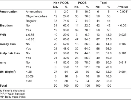
(P = 0.839) (Table 1). Table 2 shows that
the menstruation was regular in 74% of the non-PCOS group compared with 14%
in the PCOS group (P <0.001). The
majority (78%) of the PCOS group had hirsutism compared with 38% in the
non-PCOS group (P <0.001). Most (94%)
association was detected between PCOS
with greasy skin (P = 0.107), scalp hair loss
(P = 0.161), acne (P = 0.617), and BMI
categories (P = 0.904).
Table 1: Distribution of samples by demographic variables, duration, and type of infertility.
Non-PCOS PCOS Total
No. % No. % No. % P value
Residency
Outside Erbil 19 38.0 21 42.0 40 40 0.683
Inside Erbil 31 62.0 29 58.0 60 60
Occupation
Housewife 43 86.0 40 80.0 83 83 0.424
Employed 7 14.0 10 20.0 17 17
Age at marriage
≥ 30 years 10 20.0 9 18.0 19 19 0.799
< 30 years 40 80.0 41 82.0 81 81
Duration of infertility ≥ 2 years 38 76.0 42 84.0 80 80 0.317
< 2 years 12 14.0 8 16.0 20 20
Type of infertility Secondary 21 42.0 20 40.0 41 41.0 0.839
Primary 29 58.0 30 60.0 59 59.0
Total 50 100 50 100 100 100
Table 2: Association between study groups and clinical features.
Non-PCOS PCOS Total
No. % No. % No. % P value
Menstruation Amenorrhea 1 2.0 5 10.0 6 6 < 0.001*
Oligomenorrhea 12 24.0 38 76.0 50 50
Regular 37 74.0 7 14.0 44 44
Hirsutism No 31 62.0 11 22.0 42 42 < 0.001
Yes 19 38.0 39 78.0 58 58
WHR ≤ 0.85 10 20.0 3 6.0 13 13.0 0.037
> 0.85 40 80.0 47 94.0 87 87.0
Greasy skin No 26 52.0 18 36.0 44 44.0 0.107
Yes 24 48.0 32 64.0 56 56.0
Scalp hair loss No 29 58.0 22 44.0 51 51.0 0.161
Yes 21 42.0 28 56.0 49 49.0
Acne No 41 82.0 39 78.0 80 80.0 0.617
Yes 9 18.0 11 22.0 20 20.0
BMI (Kg/m2) < 25 27 54 25 50 52 52.0 0.904
25-29 8 16 8 16 16 16.0
≥30 15 30 17 34 32 32.0
Total 50 100 50 100 100 100
The mean FSH in the non-PCOS group was significantly higher than the mean
of the PCOS group (P <0.001). All the
other means mentioned in Table 3 were significantly higher in the PCOS group compared with the non-PCOS group
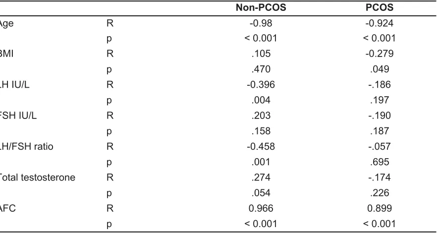
(P <0.001). Table 4 shows strong, inverse,
significant correlation between AMH and
age in each of the two study groups. A weak correlation was detected between
AMH with BMI, LH, FSH, and total testosterone, in each of the two study
groups. A significant inverse correlation was detected between AMH and LH/FSH ratio in the non-PCOS group.
Table 3: Hormonal and ultrasonographic findings of study groups expressed as mean ± SD.
Non-PCOS PCOS
Mean SD Mean SD P value
FSH IU/L 5.74 1.04 4.73 .78 < 0.001
LH IU/L 4.39 1.53 8.02 1.49 < 0.001
LH/FSH ratio .80 .41 1.71 .30 < 0.001
Total testosterone (ng/ml) 49.28 9.82 63.66 11.76 < 0.001
AMH ng/L 4.16 .85 9.22 1.21 < 0.001
AFC 19.78 2.39 32.60 4.42 < 0.001
FSH: Follicular stimulating hormone, LH: Luteinizing hormone, AMH: Antimullerian hormone, AFC: Antral follicular count
Table 4: Correlation between AMH and clinical, hormonal and ultrasonographic parameters in the PCOS and control groups.
Non-PCOS PCOS
Age R -0.98 -0.924
p < 0.001 < 0.001
BMI R .105 -0.279
p .470 .049
LH IU/L R -0.396 -.186
p .004 .197
FSH IU/L R .203 -.190
p .158 .187
LH/FSH ratio R -0.458 -.057
p .001 .695
Total testosterone R .274 -.174
p .054 .226
AFC R 0.966 0.899
p < 0.001 < 0.001
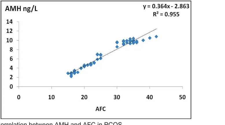
Table 4 shows a strong, significant, positive correlation between AMH and AFC in each of the study groups. Figure 1 presents this correlation in the PCOS group.
abdominal obesity.14 Although there are no
systematic studies to detect the exact prevalence of obesity in women with PCOS. There is evidence showing that
normal weight PCOS patients have
increased intra-abdominal fat.15 There was
no significant difference in body mass index categories between PCOS and non-PCOS in the current study. This finding may reflect the lifestyle of the female population in our region. Azziz et al. found that around 75% - 85% of women
with PCOS had menstrual dysfunction,16
this is in consistent with our study that reported a high frequency of oligomenorrhea (76%) among the PCOS
group. Hirsutism was found in 78% of the study sample in women with PCOS. The prevalence and degree of hirsutism depend on the ethnicity of the patients. Hirsutism is less prevalent in women with PCOS of East Asian region or Pacific
Islanders17 but is more prevalent in women
of Indian origin.18 In this study, no
significant difference was found regarding acne between PCOS and comparison group. Acne was more prevalent among women with PCOS than women without
PCOS. Azziz et al. reported acne incidence
of 12% - 14% among PCOS women.16
The findings of this study regarding serum LH and LH/FSH ratio were consistent with
Figure 1: Correlation between AMH and AFC in PCOS.
Discussion
An overall significant variation in hormonal levels and ultrasound findings between PCOS group and the non-PCOS group has been detected in this study. AMH concentrations could be used as a marker in ovarian pathophysiology, like PCOS. Most women with PCOS present with polycystic ovaries, in which the number of pre-antral and small antral follicles is
increased. This study also gives information on clinical, biochemical differences in both PCOS and those without PCOS. In the current study, there was a non-significant difference in mean age of both groups, a finding which is similar to that reported by Hollinrake et al. where a total of 103 women with PCOS and 103 control women were enrolled in
their study.12 The current study reported
significant higher waist circumference WC and WHR in women with PCOS as compared to control women which is
similar to studies done abroad.13,14 Most
a study conducted by Dewailly et al., who showed that serum LH and LH/FSH ratios were higher in women with PCOS than
controls.19 In a study done by Sung et al.,
showed that women with PCOS exhibited a significantly higher total testosterone
level, than women without PCOS,20 which
is similar to the current study. The present study clearly shows that AMH levels are increased in PCOS patients, and there is a highly significant difference with non-PCOS women which is consistent with the
findings of Woo et al.21 Laven et al. have
demonstrated that serum AMH levels were significantly increased in PCOS than ovulatory women, this was consistent with AFC during ultrasonography examination which is similar to the result of the current
study. 22 Serum AMH levels were positively
correlated with PCOS clinical features like androgen level, ovarian volume, and cycle durations. Therefore, it was proposed that AMH may be a marker of ovarian dysfunction in these women with PCOS as represented by elevated testosterone or LH levels and ovarian volumes by ultrasound
examination.23 Fanchin et al. demonstrated
that AMH and AFC were positively correlated in a study on infertile women
and found that they were superior to other
markers like inhibin B, FSH or estradiol 24
Other studies showed that AMH was 2- 4
folds increase as well as AFC in PCOS.22
Many studies have shown that AFC has
increased diagnostic threshold for PCOS.25
In the current study, the mean AFC in PCOS was 32.60±4.42 versus 19.78±2.39 in the comparison group. Dewaily et al. concluded from their study a higher threshold of up to 19 follicles have showed increased sensitivity and specificity of 81% and 92%, respectively, for PCOS
diagnosis26 but this depends on the quality
of the ultrasound rather than medical aspects. Consistent with this is a recent study by Lujan et al. who suggested
increased threshold to 29.27 The results of
the present study have shown a significant strong positive correlation between AMH and number of follicles <10 mm in the
whole group of patients which is in line
with the findings of other studies. 7,26 Our
findings regarding LH and LH/FSH are comparable with the results of previous
studies.22,28 However, Pigny et al. found no
relationship between AMH and LH and LH/
FSH in PCOS and controls.7 The results of
the present study revealed a significant correlation between AMH and age, Nardo LG, et al. indicated that AMH is generally
decreased with chronological age. In the present study, significant positive correlations were found between AMH and
serum testosterone in PCOS group.29
These findings are in accordance with the
results of the previous studies7,22,28 and
add to the existing evidence for small ovarian follicles in the production of both AMH and androgens.
The means ofserum AMH and AFC in
PCOs group were higher than the means in the comparison group. Elevated levels of AMH were associated and related to the increased number of follicles in women with PCOS.
Conclusion
Competing interests
The authors declare that they have no competing interests.
References
1. Azziz R, Woods KS, Reyna R, Key TJ, Knochenhauer ES, Yildiz BO. The prevalence
and features of the polycystic ovary syndrome in an unselected population. J Clin Endocrinol Metab 2004; 89:2745–9.
2. Farquhar C. Introduction and history of polycystic ovary syndrome. In Kovacs G, Norman R, editors. Polycystic ovary syndrome. 2nd ed. Cambridge, UK: Cambridge University Press; 2007. P. 4–24.
3. Zawadzki JK, Dunaif A. Diagnostic criteria for polycystic ovary syndrome: towards a rational approach. In: Dunaif A, Givens JR, Haseltine FP, Merriam GR, editors. Polycystic Ovary Syndrome. Boston: Blackwell Scientific; 1992. P. 377–84.
4. Escobar-Morreale HF, San Millan JL. Abdominal adiposity and the polycystic ovary
5. Dewailly D, Pigny P, Soudan B, Catteau-Jonard S, Decanter C, Poncelet E, et al. Reconciling the definitions of polycystic ovary syndrome: The ovarian follicle number and serum anti-mullerian hormone concentrations aggregate with the markers of hyperandrogenism. J Clin Endocrinol Metabol 2010; 95(9):4399–405.
6. Kwee J, Schats R, McDonnell J, Themmen A, De Jong F, Lambalk C. Evaluation of anti-Mullerian hormone as a test for the prediction of ovarian reserve. Fertil Steril 2008; 90:737–43. 7. Pigny P, Merlen E, Robert Y, Cortet-Rudelli C,
Decanter C, Jonard S, et al. Elevated serum level of anti-mullerian hormone in patients with polycystic ovary syndrome: relationship to the ovarian follicle excess and to the follicular arrest. J Clin Endocrinol Metab 2003; 88:5957–62. 8. Rotterdam ESHRE/ASRM Sponsored PCOS
Consensus Workshop Group Revised 2003 consensus on diagnostic criteria and long-term health risks related to polycystic ovary syndrome. Fertil Steril 2004; 81:19–25.
9. Hatch R, Rosenfield RL, Kim MH, Tredway D. Hirsutis implications, etiology, and management. Am J Obstet Gynecol 1981; 140:815–30.
10. WHO Expert Consultation. Appropriate body-mass index for Asian populations and its
implications for policy and intervention strategies. Lancet 2004; 363(9403):157–63.
11. Balen AH, Dresner M, Scott EM, Drife JO. Should obese women with polycystic ovary syndrome receive treatment for infertility? Br Med J 2006; 332:434–5.
12. Hollinrake E, Abreu A, Maifeld M, Van Voorhis B, Dokras A. Increased risk of depressive disorders in women with polycystic ovary syndrome. Infertil Steril 2007; 87:1369–76.
13. Ehrmann DA, Lilijenquest DR, Kasza K, Azziz R, Ghazzi MN. Prevalence &predictors of metabolic syndrome in women with polycystic ovary syndrom . J Clin Endocrinol Metab 2006; 91(1):48
–53.
14. Yildiz BO, Knochenhauer ES, Azziz R. Impact of obesity on the risk for PCOS. J Clin Endocrinol Metab 2008; 93:162–8.
15. Yildirim B, Sabir N, Kaleli B. Relation of intra-abdominal fat distribution to metabolic disorders in non-obese patients with polycystic ovary syndrome. Fertil Steril 2003; 79(6):1358– 64.
16. Azziz R, Marin C, Hog L, Badamgarav E, Song P. Health care-related economic burden of the polycystic ovary syndrome during the reproductive life span. J Clin Endocrinol Metabol 2005; 90(8):4650–8.
17. Williamson K, Gunn AJ, Johnson N, Milsom SR. The impact of ethnicity on the presentation of polycystic ovarian syndrome. Aust N Z J Obstet Gynaecol 2001; 41: 202–6
18. Wijeyaratne CN, Balen AH, Barth JH, Belchetz PE Clinical manifestations and insulin resistance
(IR) in polycystic ovary syndrome (PCOS) among South Asians and Caucasians: Is there a difference? Clin Endocrinol 2002; 57:343–50.
19. Dewailly D, Catteau-Jonard S, Reyss AC, Leroy M, Pigny P. Oligoanovulation with polycystic ovaries but not overt hyperandrogenism. J Clin Endocrinol Metabol 2006; 91:3922–92.
20. Sung YA, Oh JY, Lee H, Chung H. Hyperandrogenemia is implicated in both the
metabolic and reproductive morbidities of PCOS. Fertil Steril 2014; 101:840–5.
21.Woo HY, Kim KH, Rhee EJ, Park H, Lee MK. Differences of the association of AMH with clinical and biochemical characteristics between women with and without PCOS. Endocr J 2012; 59:781–90.
22. Laven JS, Mulders AG, Visser JA, Themmen AP, De Jong FH, Fauser BC. Anti-Mullerian hormone serum concentrations in normoovulatory and anovulatory women of reproductive age. J Clin Endocrinol Metab 2004; 89:318–23.
23. Lin Y-H, Chiu W-C, Wu C-H, Tzeng C-R, Hsu C-S, Hsu M-I. Antimüllerian hormone and polycystic ovary syndrome. Fertil Steril 2011; 230:235–96.
24. Fanchin R, Maria SL, Righini C, Guobourdenche J, Frydman R, Taeib JM. Serum AMH is more strongly related to ovarian follicular status than serum inhibin B, estradiol, FSH and LH on day 3. Hum Reprod 2003; 323:327.
25. Peltonen T, Morin-Papunen L, Koivunen R, Perheentupa A, Ruokonen A, Tapanainen JS. Serum anti-Mullerian hormone levels remain high until late reproductive age and decrease during metformin therapy in women with polycystic ovary syndrome. Hum Reprod 2005; 20(7):1820– 6.
26. Dewailly D, Gronier H, Poncelet E, Robin G, Leroy M, Pigny P, et al. Diagnosis of polycystic ovary syndrome( (PCOS): revisiting the threshold values of follicle count on ultrasound and of the serum AMH level for the definition of polycystic ovaries. Hum Reprod 2011; 26:3123–9.
27. Lujan ME, Jarrett BY, Brooks ED, Reines JK, Peppin AK, Muhn N, et al. Updated ultrasound criteria for polycystic ovary syndrome: reliable thresholds for elevated follicle population and ovarian volume. Hum Reprod 2013; 28(5):1361– 8.
28. Eldar-Geva T, Margalioth EJ, Gal M, Ben-Chetrit A, Algur N, Zylber-Haran E, et al. Serum anti-Mullerian hormone levels during controlled ovarian hyperstimulation in women with polycys-tic ovaries with and without hyperandrogenism. Hum Reprod 2005; 20:1814–9.