Experience gained from using three extra oral approaches to the
neck of the mandibular condyle: A comparative study
Received: 4/2/2013 Accepted: 12/5/2013
Reiadh K. Al-Kamali *
Background and objective: Surgical treatment of condylar diseases involves some
prob-lems concerning the choice of the surgical approach to the condylar neck that provides adequate exposure of the area with the least trauma to the facial nerve and parotid tissue. In this paper, personal experience with the surgical treatment of some mandibular condylar neck problems by the preauricular, submandibular and the retromandibular- transmasse-teric approaches is reported.
Methods: Over the last 5 years, 52 condylar neck surgeries were carried out on 41
pa-tients to treat 18 cases of condylar neck fractures, 19 cases of plate and bone graft fixation after resection of mandibular tumours and 4 cases of chronic pain and dysfunction of the TMJ. During follow-up, functions of the mandible and facial nerve branches were monitored as does the presence or absence of sialocele or parotid fistula. The appearance of the scar post- surgically and the quality of the access achieved by each type of the approaches to the condylar neck were also appraised.
Results: The incidence of apparent postoperative scar appeared most after the
preauricu-lar approach to the condypreauricu-lar neck and least after the submandibupreauricu-lar approach. Difficulties in management of condylar neck fractures are found more with the preauricular and least with the retromandibular-transmasseteric approaches. Weaknesses in the branches of the facial nerve are noticed in 50% of the preauricular approaches, 6.89% of the retromandibu-lar-transmasseteric approaches, and 47.36% of the submandibular approaches. In all of the patients, this problem lasted for 3-6 weeks to resolve spontaneously thereafter. All the patients in this work suffered limitation of jaw opening in the early postsurgical period. This problem was a transient one and due to pain and muscle spasm. However, persistent limi-tation of jaw opening is reported in 13.79% of the retromandibular-transmasseteric ap-proaches and 47.36% of the submandibular apap-proaches to the mandibular condyle.
Conclusion: Experience has shown that the retromandibular-transmasseteric approach to
the condylar neck allows for good anatomical repositioning of the fractured condyle and direct access for precise positioning and fixation of the plate or bone graft to achieve satis-factory mandibular function with the least chance of trauma to the facial nerve and parotid tissues.
Keywords: Condylar neck surgery, preauricular, submandibular and
retromandibular-transmasseteric approches.
Abstract
*Department of Oral and Maxillofacial Surgery, College of Dentistry, Hawler Medical University, Erbil, Iraq.
Introduction
There are 4 main extra oral approaches for exposure of mandibular condyle. These include the preauricular, submandibular, preauricular-transparotid and retroman-dibular-transmasseteric approuches. They all expose one or the other of the facial nerve branches to the risk of damage and
extension, this approach allows for limited access to the condylar neck and risk the temporal branch of the facial nerve to dam-age2. The submandibular approach to the
condylar neck risks the marginal mandibu-lar branch of the facial nerve to trauma and does not allow for comfortable exposure of the condylar neck. This often makes ade-quate reduction of the fracture line and the application of fixation screws quite difficult. Furthermore, when this approach is used, fixation of the screws often has to be done through transbuccal route or by the use of an endoscope3. The preauricular-transparotid approach to the condylar neck is usually carried out through the facelift or retromandibular incision. In either case, the skin flap is elevated above the parotid fas-cia which is then cut by knife to expose the parotid tissue. The dissection is then taken blindly through the parotid tissue in a direc-tion parallel to the anticipated branches of the facial nerve to reach the masseteric muscle which is split along its fibers to ex-pose the condylar neck. Although this ap-proach allows for direct access to the condylar neck, but it carries the risk of in-jury to the facial nerve and the parotid gland4. In the retromandibular
-transmasseteric approach, the condylar neck is approached through an incision in the retromandibular area. The skin flap is taken forward to expose the anteroinferior edge of the parotid gland. An incision is then made parallel to this edge to expose the masseter muscle which is split along its fibers to expose the underlying bony ra-mus. The split through the muscle may be extended superiorly as needed to expose the condylar neck5. The aim of this work is
to analyze the experience gained from ap-proaching the condylar neck through three different surgical approaches.
Fourty one adult patients underwent 52 surgeries on the condylar neck in the pe-riod from July 2007 until October 2011. Of these patients, 18 were with fractures of the condyle, 19 were with tumours
involving the mandible, , and 4 were with intractable pain and dysfunction of the tem-poromandibular joint. Those patients with neurological deficit affecting the facial nerve were excluded from this work. Can-cer patients with their submandibular lymph node involved by the tumour were also excluded from this study. The criteria for selecting surgical treatment for the frac-ture condylar neck include more than 5mm shortening of the ramus and/or more than 30 degree angulation at the fracture site6. Presurgical radiological imaging of the mandibular ramus and condyle included the OPT, PA of the mandible (or reverse Towne’s view), and CT scan of the mandi-ble. In the fracture cases, the OPT is used to calculate the ramal height and condylar angulations in the sagittal plane, whereas the reveres Town’s view is used to calcu-late the condylar angulations in the coronal plane. Three types of surgical approaches have been used in this work; they are the retromandibular-transmasseteric, subman-dibular and preauricular.approaches. In
this work the retromandibular
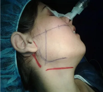
-transmasseteric approach has been modi-fied to reduce the chances of damage to the parotid gland and the facial nerve. This approach is started by surface land mark-ing of the anterior border of the parotid gland as shown in, Figure 1.
Figure 1: Surface land marking of the
an-terior border of the parotid gland
A line is drawn from the lowest point of the alar cartilage to the angle of the mouth. This line is bisected and the midpoint is joined with a straight line to the most poste-rior point of the tragus. The line is then di-vided into three equal parts. The middle section corresponds to the position of the parotid duct7. A third line is then drawn
con-necting the posterior end of this section to the mandibular angle. This (third) line cor-responds to the position of the anterio-inferior border of the parotid gland. For a safer surgery, the buccal branch of the fa-cial nerve is marked leaving the anterio-inferior border of the parotid gland 1cm be-low the course of the parotid duct to run foreword and parallel to it8. The marginal
mandibular branch of the facial nerve, on the other hand, is marked leaving the ante-rio-inferior border of the parotid gland 1cm superior to the lower border of the mandi-ble to run foreword and parallel to it8. The retromandibular incision is placed through the skin from just below and 1cm posterior to the ear lobe. It is taken inferiorly and par-allel to the posterior border of the mandible to a point level and 1cm posterior to the mandibular angle. For a wider access the superior end of this incision may be taken around the ear lobe in a nice anterior curve to just below the tragus. The skin flap is reflected superior to the parotid fascia and taken anteriorly until the line corresponding to the anterior border of the parotid gland is reached. When this line has been reached the dissection is taken medially, through the masseter muscle, along the anterior border of the parotid. The masseter is split in the direction of its fibres in the area con-fined between the superior marking of the buccal branch of the facial nerve and the inferior marking of the marginal mandibular branch of the facial nerve. The medial dis-section is continued until the ramus and the condylar neck is reached. When the sub-mandibular approach is used, the skin inci-sion is placed in a skin crease at least 2 cm below the lower margin of the mandible and marginal mandibular branch of the fa-cial nerve is protected by keeping the
dissection medial to the deep cervical fas-cia8. Postoperative care and follow-up:
Vacuum drain were used for all patients and removed 1-3 days postoperatively. All of the patients were put on short term IMF (7-10 days), and were encouraged to exer-cise mouth opening and closure afterword. Every patient is seen 1 weak later for stitches removal, release of the IMF, and for checking the appearance of the wound. The amount of jaw opening, function of the facial nerve branches, and the presence of sialocele or parotid fistula are also checked. Postoperative OPG view is ob-tained at this visit and arrangement is made to see the patient again 1, 3 and 6 months for following up. The intra- and postoperative complications with the use of these surgical approaches were studied to show the advantages of either one ap-proach over the others. This is achieved by using the Epi Info program, version 3,5,4, by which the chi-square test used for deter-mination of association between variables, and p-values equal or less than 0.05 is considered as statistically significant.
Eighteen (43.90%) out of 41 patients has had the condylar neck surgery performed to treat their fracture condyles. On the other hand, 19 (46.34%) patients has had the condylar neck surgeries done for expo-sure of the condylar neck during resection of a mandibular tumour. The other 4 (9.75%) patients had the condylar neck surgery carried out for condylotomy, Table 1. Four (22.22%) out of 18 patients under-went condylar neck surgery because of more than 5mm shortening of the mandibu-lar ramus in the fracture side. For 6 (33.33%) out of the 18 trauma patients, the condylar neck surgery is performed be-cause they had more than 30 degrees angulation of the condylar neck in the frac-ture side. The other 8 (44.44%) trauma pa-tients has had the condylar surgery per-formed to avoid putting them on long term (45days) IMF, Table 2. In 19 (46.34%) out of 41 patients, the condylar surgery has
been carried out during resection of tumors from the mandible. Twelve (63.15%) of these patients has had malignant tumours involvement of the mandible by carcinomas from the mouth floor or the retromolar-trigone. The condylar surgeries in these patients were performed to allow for fixa-tion of a plate after mandibular resecfixa-tion. The condylar surgery for the other 7 (36.84%) patients were used for fixation of a bone graft after mandibular resection for benign tumour, Table 3. Three types of sur-gical approaches have been used in this work; the retromandibular-transmasseteric, submandibular and the preauricular ap-proaches. Each one of these approaches either been used alone or in combination with another approach. The retomandibular -transmasseteric approach has been used 29 (70.73%) out of 52 condylar neck sur-geries (18 trauma and 11tumours). The submandibular approach to the condylar neck has been used in 19 (36.53%) out of 52 (5 in combination to treat 12 cancer pa-tients, 7 as a sole approach to treat cancer cases, 6 in combination to treat 7 patients with benign tumours in the mandible and once as a sole treatment of benign tumour in the mandible). The preauricular ap-proach to the condylar neck has been used to treat 4 cases of chronic intractable pain and dysfunction of the TMJ, Table 4. Com-plications reported in this work include the formation of apparent scar, difficulty with accessing the condylar region, weakness one or more than one of the facial nerve function and limitation of jaw opening. Sia-locele and parotid fistula were not reported in this work. The incidence of apparent postoperative scar appeared most after the preauricular approach to the condylar neck and least after the submandibular ap-proach. Difficulty in reduction of the frac-ture, application of plating and vision is found more with the preauricular and least with the retromandibular-transmasseteric approaches. Weakness in the temporal branch of the facial nerve is only noticed in 50% of the preauricular approaches to the condylar neck. On the other hand, 6.89%
the retromandibular-transmasseteric ap-proaches in this study had weakness in the buccal branch of the facial nerve, and 47.36% of the submandibular approaches were followed by transient weakness in the marginal mandibular branch of the facial nerve. The weakness in the branches of the facial nerve was a transient problem and lasted for 3-6 weeks to resolve sponta-neously thereafter. All the patients in this work suffered limitation of jaw opening in the early postsurgical period. This problem was a transient one and due to pain and muscle spasm. However, persistent limita-tion of jaw opening is reported in 13.79% of the retromandibular-transmasseteric ap-proaches and 47.36% of the submandibu-lar approaches to the mandibusubmandibu-lar condyle, Table 5.
Table 1: Indications for condylar neck
sur-gery sursur-gery
ORIF= Open Reduction with Internal Fixa-tion
Table 2: Indications surgery for patients
with fractures of the condylar neck
Indication Number (%)
ORIF after condylar frac-tures
18 (43.90)
Exposure of the condylar neck during resection of a mandibular tumour
19 (46.34)
Condylotomy for MPDS 4 (9.75)
Total 41 (100)
Indication Number (%)
More than 5mm shortening of the mandibular ramus in the fracture side
4 (22.22)
More than 30 degrees angula-tion of the condylar neck in the fracture side
6 (33.33)
Avoidance of full term inter-maxillary fixation
8 (44.44)
Table 3: Indications for condylar neck surgery during excision of mandibular tumours
Table 4: Types and numbers of surgical approaches to the condylar neck
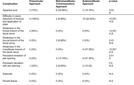
Table 5: Incidence of complications among the different approaches to the condylar neck
H.S.=Highly Significant, N.S.= Not Significant, S= significant, N.A.=Not Applicable
Indication Number (%)
Fixation of a plate after mandibular resection for cancer 12 (63.15)
Fixation of a bone graft after mandibular resection for benign
tumour 7 (36.84)
Total 19 (100)
Type of Surgical Approach Number (%)
Retromandibular-transmasseteric 29 (55.76)
Submandibular 19 (36.53)
Preauricular 4 (7.69)
Total 52 (100)
Complication Preauricular Approach Retromandibular-Transmasseteric Approach
Submandibular
Approach p-value
Apparent scar 3 (75%) 6 (20.68%) 3 (15.78%) 0.03
S Difficulty in vision,
reduction of fracture and application of plate
4 (100%) 2 (6.89%) 10 (52.63%) <0.001
H.S.
Weakness in the frontal branch of the facial nerve
2 (50%) 0 (0%) 0 (0%) <0.001
H.S. Weakness in the
buccal branch of the facial nerve
0 (0%) 2 (6.89%) 0 (0%) 0.43
N.S. Weakness in the
mandibular branch of the facial nerve
0 (0%) 0 (0%) 9 (47.36%) <0.001
H.S. Persistent limitation of
jaw opening 0 (0%) 4 (13.79%) 9 (47.36%) 0.01S
Persistent deviation
with jaw opening 1 (25%) 2 (6.89%) 2 (10,52)
0.5 N.S.
Sialocele 0 (0%) 0 (0%) 0 (0%) N.A.
Those patients with neurological deficit af-fecting the facial nerve were excluded from this work in order not to confuse the study’s end results. Likewise cancer patients with involvement of the submandibular lymph node by the tumour were also excluded from this study as their surgery will neces-sitate excision of the marginal mandibular. The scar formation after the retromandibu-lar-transmasseteric incision is mainly re-lated to the fact that this incision is of ne-cessity longer than that used in other ap-proaches. This comes in agreement with other studies1, 5, but it is my impression that the preauricular element of the reto-mandibular-transmasseteric incision is not cosmetically important. This is because the anterior part of it is in the skin crease ante-rior to the ear lobe, and its posteante-rior part is hidden by the later structure. It is the retro-mandibular extension that can leave an ob-vious scar, but I tried to keep it to minimum by meticulous closure of the wound and early removal of the stitches. As to the is-sue of access, our experience is in agree-ment with others that the preauricular ap-proach to the condylar neck is very unsatis-factory for reduction of condylar fracture or placement of a plate for fixation3. It is also my impression, like that of others that this approach is probably, useful only for the upper half of the condylar neck for condy-lotomy9
.
The submandibular approach of-ten necessitates wide stripping of the mas-seter muscle off the ramus and forceful tis-sue retraction to access the condylar neck. This can be traumatic to the tissues and causes limitation of jaw opening for periods longer than that experienced after the use of other approaches. Using this approach would also enforce oblique insertion of the plates and screws which may render the final results of reduction of condylar frac-ture, in cases of trauma, unsatisfactory10. The retromandibular-transmasseteric ap-proach, on the other hand, has proved very adequate for all surgeries on the condylar neck. My experience in this matter comesDiscussion in agreement with the experience of
oth-ers4, 5. The marginal mandibular branch is
most often injured in the submandibular, whereas the preauricular approach often causes injury to the temporal and zygo-matic branches. The retromandibular-transmasseteric approach, however, ap-pears safer to the marginal mandibular branch of the facial nerve from the sub-mandibular approach, but can cause injury to the buccal branch of the facial nerve.
The preauricular approach to the condylar neck is probably adequate for accessing the upper half of the condylar neck, but not for controlling a fracture or applying a plate. The submandibular approach alone is ade-quate for approaching the lower half of the neck, but again less than adequate for re-duction of a fracture or application of plate fixation. Finally, the retromandibular-transmasseteric approach to the condylar neck is very satisfactory as an approach to the condylar neck. Furthermore, this ap-proach causes least trauma to the facial nerve and parotid tissue.
10.Marker P, Nielsen A, Lehmann-Bastian H. Frac-tures of the mandibular condyle. Part 2: Results of treatment of 348 patients. Br J Oral Maxillofac Surg 2000; 38:422-6.
11. Ellis E, McFadden D, Simon P .surgical compli-cations with open treatment of mandibular condy-lar process fractures. J Oral Maxillofac Surg 2000; 58(9):950-8.
12.Downie JJ, Devlin MF, Carton ATM, Hislop WS. Prospective study of morbidity associated with open reduction and internal fixation of the frac-tured condyle by the transparotid approach. Br J Oral Maxillofac Surg 2009; 47:370-3.
Conclusion
References
1. Haug RH, Assael LA. Outcomes of open versus closed treatment of mandibular subcondylar frac-tures. J Oral Maxillofac Surg 2001; 59:370-5. 2.Dolwick MF, Kretzschmar DP. Morbidity associated
with the preauricular and perimeatal approaches to the temporo-mandibular joint. J Oral Maxillofac Surg 1982; 40:699-700.
3.Chen CT, Lai JP, TungTC, Chen
YR.Endoscopically assisted mandibular sub-condylar fracture repair. Plast Reconstr Surg1999; 103:60-5.
4.Vesnaver A, Gorjanc M, Eberlinc A, Dovsak DA, Kansky AA. The periauriculartransparotid ap-proach for open reduction and internal fixation of condylar fractures. J Craniomaxillofac Surg 2005; 33:169-79.
5.Wilson AW, Ethunandan M, Brennan
PA,Transmassetericantero-parotid approach for open reduction and internal fixation of condylar fractures. Br J Oral Maxillofac Surg S 2005; 43:57-60
6.Schneider M, Erasmus F, Gerlach KL, Kuhlisch E, Loukota RA, Rasse M. Open reduction and inter-nal fixation versus closed treatment and mandibu-lomaxillary fixation of fractures of the mandibular condylar process: a randomized, prospective, multicenter study with special evaluation of frac-ture level. J Oral Maxillofac Surg 2008; 66:2537-44.
7.Langdon JD. Parotid surgery. In Langdon JD, Patel MF, Ord RA, Brennan PA. Operative oral and maxillofacial surgery, 2nd ed. London: Hodder Arnold, an Hachette UK Company; 2011. P. 390. 8.Davis BA, Anson BJ, Budinger JM, Kurth LE.
Sur-gical anatomy of the facial nerve and parotid gland based upon a study of 350 cervicofacial halves. Surg Gynecol Obstet 1956; 102:385-412.