UDC 577.175
The mechanism of VeGf-mediaTed
endoThelial cells surViVal and proliferaTion
in condiTions of unfed-culTure
T. V. NIkolaIeNko, V. V. NIkUlINa, D. V. SheleST, l. V. GarmaNchUk Educational and Scientific Centre “Institute of Biology”,
Taras Shevchenko National University of Kyiv, Ukraine; e-mail: nikolaenkotetiana@yandex.ua
The mechanisms of VEGF-mediated effects on endothelial cells during cancer development and pro-gression is not clear. In present study the biological effects of VEGF, VEGF-rich culture medium of peritoneal macrophages from mice with Lewis lung carcinoma were studied on MAEC cell line under conditions of unfed culture. We have shown that VEGF increased cell proliferation by the 5th day of culturing vs control and anti-VEGF-treated cells. This effect was associated with increased consumption of glucose and NO production by the 2nd day while decreased – on the 5th day of cell culturing. VEGF-mediated NO production was dependent on ca2+ ions. Block of Ca2+-channels (LaCl
3) had more pronounced inhibitory effect vs chelator of Ca2+ ions (EDTA). It was shown that peritoneal macrophages are the main suppliers of VEGF at tumor angiogenesis, as evidenced by the data obtained on model system of endothelial cells synchronized in G0/G1 phase.
K e y w o r d s: VEGF, nitric oxide, angiogenesis, eNOS, EDTA, LaCl3.
T
he endothelial lining of blood vesselspro-vides a barrier for the exchange of nutrients and is itself actively involved in the local control of vascular homeostasis. Physiologically,
processes of proliferation and differentiation of en -dothelial cells are determined and regulated by a variety of biologically active molecules. The vascu-lar endothelial growth factor (VEGF) is a potent and critical inducer of angiogenesis. Signal transduction involves binding to tyrosine kinase receptors and re-sults in endothelial cell proliferation [1].
VEGF affects the basic functional characteris -tics of endothelial cells, such as adhesion, prolifera-tion, migraprolifera-tion, intracellular mitogenic signal trans-mission, intercellular contacts, vascularization and neovascularization [2]. All these processes are not dependent on the presence of growth factors in the environment, but also on the trophic substrates. Glu-cose is an essential metabolic substrate of all mam-malian cells for energy demand.
Glucose is taken up into cells by energy-in-dependent transportation down its concentration gradient, which is mediated by glucose transporter proteins. Transport of glucose across the plasma
membrane of mammalian cells is the first
rate-limi-ting step for glucose metabolism and is mediated by facilitative glucose transporter (GLUT) proteins [3].
GLUT-1 is glucose transporter which is expressed in endothelial cells.
VEGF plays a key role in angiogenesis and
there are only limited studies of the effects of VEGF
on endothelial cell amino acid and glucose transport. VEGF enhances microvascular permeability and modulates Ca2+ signaling in endothelial cells and causes an approximately threefold increase in
2-de-oxyglucose uptake and a fivefold increase in GLUT1
transcript [4].
Glucose transporter is also a proliferation-re-lated gene and its expression is associated with the activation of signal transduction mechanisms, its expression should also be induced when a growth factor binds to its receptor. VEGF exerts its
mito-genic effects in various types of tumor cells, in part,
by inducing the expression of genes whose products are required for endothelial cells proliferation and cancer progression. Given evidence that VEGF may act as a survival factor in tumor, the ability of VEGF to regulate endothelial cell glucose transport in con-junction with angiogenesis may serve to ensure
ad-equate substrate delivery and blood flow during tu -mor progression [5].
Angiogenesis is initiated by vasodilatation, a NO-mediated process. Nitric oxide (NO) is a
diffusib le gas that is produced from L-arginine in
the large number of tissues by the NO synthase (NOS) family of enzymes. There are three isoforms of NOS: nNOS (type I), iNOS (type II), and eNOS (type III). There is mounting evidence demonstrating the interaction between NO and VEGF. It has been convincingly demonstrated that NO may be involved in endothelial cell proliferation, migration, protease
release, increased vascular permeability and effects
important for initiation of angiogenesis [6].
Both inflammation and angiogenesis are exacer bated by increased production of chemokines/ cytokines, growth factors, proteolytic enzymes, lipid mediators and prostaglandins. Initiation and progres-sion of cancer are also closely linked to
angiogen-esis. Infiltration of macrophages is a dramatic and common feature of inflammation, angiogenesis and
cancer, and has been recently highlighted in an at-tempt to develop novel strategies for treating cancer [7]. By release of secretory products the activated
macrophages have the capability to influence each
phase of the angiogenic process, such as alterations of the local extracellular matrix, induction of en-dothelial cells to migrate or proliferate, and
inhibi-tion of vascular growth with formainhibi-tion of differenti -ated capillaries [8].
The aim of the present work was to study
bio-logical effects of VEGF on endothelial cells in the
conditions of unfed culture. materials and methods
All experiments were performed in compliance with the “Guide for the Care and Use of Experimen-tal Animals” approved by the Committee for control of maintenance and use of experimental animals of ESC Institute of Biology at Taras Shevchenko Na-tional University of Kyiv.
Model systems. Mouse aortic endothelial cell
line (МАЕC) isolated from mouse aorta and spon -taneously immortalized in culture was used [9, 10].
МАЕС preserves the main biological properties of
normal endotheliocytes, including the ability to dif-ferentiation and formation of procapillary structures in vitro. MAEC was incubated in DMEM medium (Sigma, USA) supplemented with 10% FBS (Sigma, USA), 2 mM L-glutamine and 40 mg/ml gentamicin. Cell lines were cultured under the standard
condi-tions at 37 °С in humidified atmosphere with 5%
CO2.
Mononuclear phagocyte fraction of peritoneal exudate of mice with Lewis lung carcinoma at vari-ous stages of tumor growth was obtained by
stand-ard procedure of Pietrangeli [11]. Macrophages were incubated under standard conditions at 37 °C, 100% humidity and 5% CO2 for 4 h and then their cultural medium was added to MAEC culture.
Test agents. After adaptation of cells to cul-ture medium test-agents were added: VEGF (vascu-lar endothelial growth factor), obtained previously
[12], anti-VEGF (monoclonal antibody specific for
VEGF), condition medium from peritoneal mac-rophages from mice with Lewis lung carcinoma at various stages of tumor growth, chelator of Ca2+ (EDTA) and blocker of Ca2+-channels (LaCl
3). Proliferative assay. The number of living cells was determined in wells using MTT-colorymetric test [13] and cell counts were performed using a trypan blue dye after incubation with agents. The biochemical essence of this method is based on the fact that mitochondrial dehydrogenases of living cells are capable of cleaving tetrazolium rings with formation of insoluble purple crystals (formazan). MTT (20 µl) was added to the culture medium 4 h before the termination of the cells incubation in
or-der to achieve the final concentration of 0.6 mM.
Formazan crystals formed after the incubation with MTT were dissolved in 100 µl of dimethylsulfoxide. The plate was analyzed on the spectrophotometer at 540 nm.
Flow cytometry. Apoptotic level and distribu-tion of cells in phases of cell cycle were assessed by
cytofluorimetry [14]. Cytofluorimetry was carried
out on the instrument FACS Calibur (Becton Dickin-son, United States). Special mathematical program Mod Fit LT 2.0 (BDIS, United States) for Macintosh computers was used for acquisition and data
analy-sis. Narrowband filter 585/42 nm was used in order to measure the fluorescence of PI.
Determination of glucose by glucose-oxidase method. The level of glucose in the incubation me-dium of endothelial cells was determined using a standard set based on glucose-oxidase reaction
which we modified for culture medium of cells. Ini -tial cell concentration was about 1×10⁵ cells/ml in the sample volume of 200 µl. Determination was performed according to the protocol of the manufac-turer “Felicit-Diagnostics” (Ukraine) [15].
Determination of the nitrite anion concentra-tion. The content of NO was determined by colori-metric assay using the Griess reagent [16]. Sodium nitrite solution 0.2-10 µM was used for calibration.
culture medium [17]. The procedure for determining the concentration of VEGF was carried out in 96-well plates with immobilized antibodies. The level of VEGF was determined by the calibrating curve, which was built as the absorbance dependence on the concentration of VEGF.
Statistical analysis. Experimental data were analysed using descriptive methods, by Student’s t -criterion and by the method of nonlinear regression.
results and discussion
Angiogenesis is known as a multi-step process which consists of remodeling of extracellular ma-trix, proliferation and migration of endothelial cells, changes in metabolism of endothelial cells, tube formation and blood vessels stabilization [18]. It is regulated by a well-orchestrated balance between pro- and anti-angiogenic factors. Since VEGF plays
a key role in angiogenesis, we tested its effect on en
-dothelial cells (MAEC). At the first step of the study
the cultivation of endothelial cells with exogenous VEGF and anti-VEGF was conducted within 5 days. The cultivation of endothelial cells was conducted
during five days for modeling 2 systems of growth -
exponential (cells proliferate actively) and stationary (most of the cell is in G0/G1 phase) ones. Their ef-fects on cell survival and changes in the metabolism of glucose as the main energy substrate were deter-mined. The cultivation is conducted under condi-tions of unfed culture and determination of glucose and counting of live/dead cells using trypan blue dye was carried out at every stage.
VEGF stimulates cell proliferation and
increa-ses the number of cells 1.6 and 2 times on the fifth
day compared with control and anti-VEGF, respec-tively (Fig. 1). In addition, the number of dead cells during the period of the experiment is the lowest under the influence VEGF of about 4.0 ± 0.2% (p < 0.05) and 6.0 ± 0.4% (p < 0.05) compared with the control and anti-VEGF, respectively. Thus,
VEGF showed a significant proliferative, angiogenic effect on endothelial cells and is a factor of cell sur -vival.
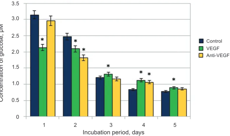
The level of glucose consumption by cells changes depending on the duration of cultivation
and the influence of agents. VEGF increased glucose
consumption by cells 1.5 times and 1.4 times com-pared with the control and anti-VEGF, respectively,
(Fig. 2) on the first day of incubation. However, on
other days the opposite situation was observed, the
number of cells was increased significantly, while
Fig. 1. The influence of VEGF and anti-VEGF on proliferation of endothelial cells
250
N
um
be
r o
f c
ell
s, 1
0
3
Incubation period, days
200
150 100
50
0 1 2 3 4 5
VEGF Control
Anti-VEGF
glucose consumption by cells was reduced. This ef-fect may be explained by stimulation of glutamine transport from culture medium by VEGF and its involvement in metabolic pathways of cells. The transport of amino acids (including L-glutamine) is a Na+-dependent process, so activation of L-glu-tamine transport via the Na+-dependent system was dependent on de novo protein and RNA synthesis, and L-glutamine supplementation protected endothe-lial cells from oxidant injury which promotes cell survival [19]. In one of the few studies focused on endothelial cell metabolism measurements of the maximum catalytic activities of the major metabolic pathways demonstrated that, in addition to glucose and fatty acids, glutamine represents an important fuel for these cells. In particular, the activity of glu-taminase is about 20-fold higher in endothelial cells than in lymphocytes (known to exhibit high rates
of glutaminolysis). These data confirm that the en -dothelial cells like tumor cells can produce biosyn-thetic intermediates via the TCA cycle independent
of coupling by OXPHOS machinery [20].
The angiogenic and inflammatory effects of
VEGF can be mediated by NO, which is produced by VEGF-activated eNOS in endothelial cell. It has been reported that VEGFR-2 plays a major role in angio-genesis, and its autophosphorylation leads to eNOS activation [21]. NO-production during 5 days of cul-tivation of endothelial cells under the conditions of
unfed culture was determined and its specific effects were identified. As it can be seen in Fig. 3 VEGF stimulates NO-production 2.3 times on the first day
and 3.5 times on the second day compared with the
control. Activating effect of anti-VEGF relative to
Fig. 2. The influence of VEGF and anti-VEGF on consumption of glucose by endothelial cells; M ± m, n = 5, * P < 0.05 relative to control
3.5
C
on
ce
nt
ra
tio
n o
f g
lu
co
se
, μ
M
Incubation period, days
2.0
1.5
1.0
0.5
0
1 2 3 4 5 3.0
2.5
VEGF Control
Anti-VEGF
Fig. 3. The influence of VEGF and anti-VEGF on NO-production by endothelial cells. Calculation is conducted according to the culture medium volume and the number of cells; M ± m, n = 5, * p <0.05 relative to control
pmol
N
O2
-/c
el
l
Incubation period, days
40
30
20
10
0
1 2 3 4 5 50
VEGF Control
Anti-VEGF
medium on trophic substrates and humoral factors which are contained in the serum.
Nitric oxide demonstrated multidirectional ac-tion, because the increase of its concentration under
the influence of VEGF significantly stimulated the
proliferation of endothelial cells, while its increase
under the influence of anti-VEGF leads to increasing
the number of apoptotic cells. Nitric oxide showed
proapoptotic effect in relation to the endothelial cells under the influence of anti-VEGF, as evidenced by
the increasing number of apoptotic cells compared
with the control and under the influence of VEGF. So, the effect of nitric oxide on endothelial cells de -pends on its concentration in the incubation medium and on microenvironment conditions.
The activity of eNOS by blocking Ca2+
-chan-nels was determined to confirm the data on eNOS
involvement in production of nitric oxide and
speci-ficity of its activation under the influence of VEGF in
can be activated by receptor-dependent and -inde-pendent agonists as a consequence of an increase in the concentration of free Ca2+ and the association of a Ca2+/calmodulin (CaM) complex with the enzyme
[22]. For this purpose EDTA (100 μM) and LaCl3 (1 μM) were used that bind and block the entry of calcium into cells. The most pronounced effect of
VEGF and anti-VEGF were found on the second and
fifth days of cultivation, so they were selected for the experiment. Although a significant stimulation of production of nitric oxide under influence of VEGF on the second and the fifth days under influence of
anti-VEGF is shown, EDTA and LaCl3 significantly
reduce the level of NO (Fig. 4).
In addition, both agents have different effect on
production of nitric oxide by endothelial cells. LaCl3
has more pronounced inhibitory effect, because it se -lectively binds with Ca2+-channels.
It is known that macrophages are supposed to play a key role in inflammatory and tumor
angiogene sis. The recruitment and infiltration of
tumor-associated macrophages in the tumor micro-environment activates them to support the malig-nant progression of cancer cells [7]. Therefore the next stage of our study was aimed to identify the angiogenic properties of the medium of cultivation macrophages derived from animals with Lewis lung carcinoma in various stages of tumor growth.
The peritoneal macrophages was obtained from mice with Lewis lung carcinoma and cultured under
Fig. 4. The influence of EDTA (A) and LaCl3 (B) on NO-production by endothelial cells. Calculation is con-ducted according to the culture medium volume and number of cells; M ± m, n = 5, * p < 0.05 relative to control
A
8
pmol
N
O2
-/c
el
l
Incubation period, days
5
4 3
2
0
2 5 7
6
Control VEGF Anti-VEGF
1
B
8
pmol
N
O2
-/c
el
l
Incubation period, days
5 4
3 2
0
2 5 7
6
1
Control VEGF Anti-VEGF
standard conditions. Culture medium was collected and added to the culture of endothelial cells, the level of VEGF was determined.
The increase of VEGF production on the 7th day about 2 times, 19th day – 4.5, 25th day – 4.7 was ob-served as compared to intact group (Fig. 5). Since the 19th and 25th days are characterized by intensive development of tumors and metastasis, the hypoxic areas are formed in this period of time. This pro-cess induces macrophages to increase production of VEGF for improving the access of oxygen and nutrients in these areas.
The 15.0 ± 0.4% (p < 0.05) increase of glucose consumption by endothelial cells is shown,
com-pared to the intact group under the influence of incu -bation medium of macrophages, on the 19th and 25th
days. This indicates an intensification of metabolic
processes in cells associated with the transition to the synthetic phase of the cell cycle.
Endothelial cells of the control group passed to S+G2/M by 5.32 ± 0.34% (p < 0.05), while the number of cells stimulated by incubation medium of peri toneal macrophages on the 19th and 25th days which passed to S+G2/M increased by 52.82 ± 3.25% and 49.72 ± 2.82% (p < 0.05), respectively (Fig. 6). The highest level of cells in S+G2/M observed on the 7th day of tumor growth and is 36.32 ± 1.25%. Thus,
Fig. 5. The level of VEGF production by peritoneal macrophages of mice at the different stages of tumor growth of Lewis lung carcinoma; M ± m, n = 5, * p < 0.05 relative to intact group
Fig. 6. The distribution of phases of the cell cycle by MAEC after incubation with macrophages of mice at dif-ferent stages of growth of Lewis lung carcinoma; M ± m, n = 5, * p < 0.05 relative to intact group
A
m
ou
nt o
f V
EG
F, n
g/
m
l
Stage of tumor growth
50
40
30
20
0
Intact group 7 days 19 days 25 days 10
80 100
60
%
Phase of the cell cycle
40
20
0
G0/G1 S G2/M
Control
7 days Intact group
19-20 days 24-26 days
Lewis lung carcinoma may be traced, that indicates the involvement of macrophages in tumor angioge-nesis.
In conclusion, the study detected a relationship of glucose uptake and production of nitric oxide
un-der the influence of VEGF and anti-VEGF by en -dothelial cells in conditions of unfed culture. Thus activation of glycolysis is characteristic not only of tumor cells but also of endothelial ones. Nitric oxide demonstrated multidirectional action on endothelial
МеханізМи VEGF-опосередкованого
виживання та проліферації ендотеліальних клітин в уМовах довготривалого культивування без заМіни середовища
Т. В. Ніколаєнко, В. В. Нікуліна, Д. В. Шелест, Л. В. Гарманчук
ННЦ «Інститут біології», Київський національний університет імені Тараса
Шевченка, Україна;
e-mail: nikolaenkotetiana@yandex.ua
Механізми VEGF-опосередкованих ефектів на ендотеліальні клітини під час розвитку та прогресії раку досі залишаються повністю нез’ясованими. Досліджено біологічні ефекти VEGF, VEGF-збагаченого середовища культиву
-вання перитонеальних макрофагів мишей з кар
-циномою легенів Льюїс на лінію ендотеліальних клітин МАЕС за довготривалого культивування без заміни середовища інкубації. Показано, що VEGF підвищував проліферацію клітин на 5-й день культивування в порівнянні з контролем та клітинами, що оброблялися анти-VEGF. Цей ефект був пов’язаний з підвищенням споживан
-ня глюкози і продукції NO на 2-й день та зни
-женням – на 5-й день культивування клітин. VEGF-опосередкована продукція NO залежала від Ca2+. Блокатор Са2+-каналів (LaCl
3) виявив
вираженіший інгібувальний ефект в порівнянні з хелатором Са2+ (ЕДТА). Показано, що
перитонеальні макрофаги є одними з головних постачальників VEGF в умовах ангіогенезу пух
-лини, про що свідчать одержані дані на модель-ній системі ендотеліоцитів, синхронізованих у
G0/G1 фазі.
К л ю ч о в і с л о в а: VEGF, оксид азоту,
ангіогенез, eNOS, EDTA, LaCl3.
МеханизМы VEGF-опосредованного
выживания и пролиферации эндотелиальных клеток в условиях длительного
культивирования без заМены среды
Т. В. Николаенко, В. В. Никулина, Д. В. Шелест, Л. В. Гарманчук
УНЦ «Институт биологии», Киевский национальный университет имени
Тараса Шевченко, Украина;
e-mail: nikolaenkotetiana@yandex.ua
Механизмы VEGF-опосредованных эффек
-тов на эндотелиальные клетки при развитии и прогрессии рака до сих пор остаются полно
-стью невыясненными. Исследованы биологиче
-ские эффекты VEGF, VEGF-обогащенной среды культивирования перитонеальных макрофагов мышей с карциномой легких Льюис на линию эндотелиальных клеток МАЕС в условиях дли
-тельного культивирования без замены среды ин
-кубации. Показано, что VEGF повышал проли
-ферацию клеток на 5-й день культивирования по сравнению с контролем и клетками, которые об
-рабатывались анти-VEGF. Этот эффект был свя
-зан с повышением потребления глюкозы и про
-дукции NO на 2-й день и снижением – на 5-й день культивирования клеток. VEGF-опосредованная продукция NO зависела от Ca2+. Блокатор Са2+
-каналов (LaCl3) проявил более выраженный ин
-гибирующий эффект по сравнению с хелатором Са2+ (ЭДТА). Показано, что перитонеальные ма
-крофаги являются одними из главных постав
-щиков VEGF в условиях ангиогенеза опухоли, о чем свидетельствуют полученные данные на модельной системе эндотелиоцитов синхрони
-зированных в G0/G1 фазе.
К л ю ч е в ы е с л о в а: VEGF, оксид азота,
references
1. Arutyunyan I, Fatkhudinov T, Kananykhina E, Usman N, Elchaninov A, Makarov A, Bolsha-kova G, Goldshtein D, Sukhikh G. Role of VEGF-A in angiogenesis promoted by umbilical cord-derived mesenchymal stromal/stem cells: in vitro study. Stem Cell Res Ther. 2016; 7: 46. 2. Szabo E, Schneider H, Seystahl K, Rushing EJ,
Herting F, Weidner KM, Weller M. Autocrine
VEGFR1 and VEGFR2 signaling promotes survival in human glioblastoma models in vitro and in vivo. Neuro oncol. 2016. pii: now043.
3. Yeh WL, Lin CJ, Fu WM. Enhancement
of glucose transporter expression of brain endothelial cells by vascular endothelial growth factor derived from glioma exposed to hypoxia. mol pharmacol. 2008; 73(1): 170-177.
4. Mann GE, Yudilevich DL, Sobrevia L. Regulation of amino acid and glucose transporters in endothelial and smooth muscle cells. Physiol Rev. 2003; 83(1): 183-252.
5. Ezhilarasan R, Mohanam I, Govindarajan K, Mohanam S. Glioma cells suppress hypoxia-induced endothelial cell apoptosis and promote the angiogenic process. Int J oncol. 2007; 30(3):
701-707.
6. Babaei S, Stewart DJ. Overexpression of
endothelial NO synthase induces angiogenesis in a co-culture model. Cardiovasc Res. 2002;
55(1): 190-200.
7. Ono M. Molecular links between tumor
angiogenesis and inflammation: inflammatory
stimuli of macrophages and cancer cells as targets for therapeutic strategy. cancer Sci.
2008; 99(8): 1501-1506.
8. Khan MA, Assiri AM, Broering DC. Complement and macrophage crosstalk during process of angiogenesis in tumor progression. J Biomed Sci. 2015; 22: 58.
9. Rodrigues SF, Granger DN. Blood cells and endothelial barrier function. Tissue Barriers.
2015; 3(1-2): e978720.
10. Bastaki M, Nelli EE, Dell'Era P, Rusnati M, Molinari-Tosatti MP, Parolini S, Auerbach R,
Ruco LP, Possati L, Presta M. Basic fibroblast
growth factor-induced angiogenic phenotype in mouse endothelium. A study of aortic and microvascular endothelial cell lines. Arterioscler Thromb Vasc Biol. 1997; 17(3): 454-464.
11. Pietrangeli CE, Skamene E, Edelson PJ,
Kongshavn PA. Measurement of 5'-nucleotidase
in mouse peritoneal macrophages in listeriosis. Infect Immun. 1981; 32(3): 1206-1210.
12. Garmanchouk LV, Pyaskovskaya ON,
Solya-nik GI. Influence of pro-angiogenic cytokines on
proliferative activity and survival of endothelial cells. Biopolym Cell. 2010; 26(3): 187-193.
13. Mosmann T. Rapid colorimetric assay for cellular growth and survival: application to proliferation and cytotoxicity assays. J Immunol Methods.
1983; 65(1-2): 55-63.
14. Nicoletti I, Migliorati G, Pagliacci MC, Grignani F, Riccardi C. A rapid and simple method for measuring thymocyte apoptosis by
propidium iodide staining and flow cytometry.
J Immunol Methods. 1991; 139(2): 271-279. 15. Nikolaienko Т, Petruk N, Shelest D,
Garman-chuk L. Influence of VEGF, EGF and their
anta gonists on proliferative activity and glucose consumption by endothelial cells. Bull. T. Shevchenko Nat. Univ. Kyiv. 2015; 1(69): 36-38. 16. Green LC, Wagner DA, Glogowski J, Skipper PL,
Wishnok JS, Tannenbaum SR. Analysis of
nitrate, nitrite, and [15N]nitrate in biological
fluids. anal Biochem. 1982 126(1): 131-138.
17. Conn G, Soderman DD, Schaeffer MT, Wile M, Hatcher VB, Thomas KA. Purification of a
glycoprotein vascular endothelial cell mitogen from a rat glioma-derived cell line. proc Natl acad Sci USa. 1990; 87(4): 1323-1327.
18. Carmeliet P, Jain RK. Principles and mechanisms
of vessel normalization for cancer and other angiogenic diseases. Nat Rev Drug Discov. 2011;
10(6): 417-427.
19. Dvorak HF. Tumor Stroma, Tumor Blood Vessels,
and Antiangiogenesis Therapy. cancer J. 2015;
21(4):237-243.
20. Polet F, Feron O. Endothelial cell metabolism and tumour angiogenesis: glucose and glutamine as essential fuels and lactate as the driving force. J Intern med. 2013;273(2): 156-165.
21. Kimura H, Ogura T, Kurashima Y, Weisz A, Esumi H. Effects of nitric oxide donors on
vascular endothelial growth factor gene induction. Biochem Biophys Res Commun. 2002;
296(4): 976-982.
22. Fleming I, Fisslthaler B, Dimmeler S, Kemp BE, Busse R. Phosphorylation of Thr(495) regulates Ca(2+)/calmodulin-dependent endothelial nitric oxide synthase activity. Circ Res. 2001; 88(11):
E68-E75.