Cerebral Blood Flow Studies in Stroke*
JOHN
STIRLING MEYER, M.D.
Professor
and Chairman, Department of
Neurology,
Baylor College
of Medicine,
Houston, Texas
I will discuss here some work we are doing in our laboratory on the measurement of cerebral blood flow (CBF). We are using the gamma camera, which is connected to a computer, to measure regional CBF. We inject 133Xe into the carotid
artery and, by using multiple probes, measure the clearance from multiple regions of the head. The adv.antage of the gamma camera is that it has ex-cellent resolution and has common probe charac-teristics. It may be thought of in terms of multiple probes, although it has a single crystal, and it achieves the desirability of multiple regional CBF recordings confined essentially to one hemisphere be-cause of the depth characteristics of the collimation.
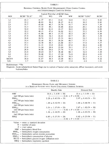
The formula for height-over-area analysis in the clearance for fast and slow flow ( or the first and second slopes of the curve, Fl and F2) is handled by the computer which gives an automatic write-out. ln addition, having injected the xenon, which washes out fairly rapidly over an interval of 12 minutes, we then inject with a nondiffusable isotope, tech-netium. By use of a different formula, this gives us the regional cerebral blood volume, so that in the same patient, we have both the regional cerebral blood flow and the regional cerebral blood volume on the computer write-out. Table 1 is a typical record that comes out of the automatic print-out; it has the advantage of being rapid, taking place while the patient is being examined. It comes out objectively with a minimum amount of error, the program being highly reliable, and gives you both the regional cerebral blood flow (ROI) and the mean hemispheric flow (RCBF "H/ A"), which is in good agreement with hydrogen clearance using an entirely different methodology, as well as the standard
* This is an edited transcription of a lecture presented
by Dr. Meyer, February 7, 1974, at the Medical College
of Virginia, Richmond.
MCV QUARTERLY 10(3): 123-127, 1974
deviation. It gives you the fast or so-called gray flow (FG), the white flow (FW), the so-called weight of gray matter (WG), the weight of white matter (WW), a regional flow calculated by an-other method (RCBF "LOG"), and the regional cerebral blood volume (RCBF), which is of con-siderable use because, knowing blood flow, blood volume, and cerebrospinal fluid pressure, you can say a great deal about the amount of blood and parenchyma present in the region under study and about the various pressure-tissue-flow relationships. For our Jl1etabolic studies, we feel that it is less traumatic and highly reliable to pass the catheter up into the lateral sinus via the brachia! vein; thi.s is done under visualization of the fluoroscope in the cardiac catheterization laboratory. I say it is highly reliable, because one can inject a little dye at the time of placement of the catheter, from which much can be learned. About 20% of patients have abnormalities of the venous system, and there is no question that, with the blind puncture or modifi-catio~s of it, the needle often finishes up sampling blood that was certainly not coming from the brain, which accounts for some of the errors in methods. To summarize some of the data-it will be found that the earlier the patient is studied after
a stroke, the greater is the reduction of CBF. I would like to point out that CBF is reduced not only on the diseased side but also on the healthy side (Table 2). We called it diaschisis and supposed that it was due to the release, following stroke, bilaterally or perhaps from the brain stem, of some neurotransmitter, and it was suggested that it might be serotonin or some related substance. Judging from pur current studies, it now appears that indeed there are neurotransmitter releases in a unilateral stroke and that these neurotransmitters, bilaterally, include norepinephrine, serotonin, and C-AMP; these reduce cerebral blood flow and decrease
TABLE 1
REGIONAL CEREBRAL BLOOD FLOW MEASUREMENTS USING GAMMA CAMERA AND lNTERTECHNIQUE COMPUTER SYSTEM
Typical Print-Out of RCBF Data
ROI RCBF"H/A" FG WG FW WW RCBF "LOG" RCBV 1,4 42.2 41. 57 65.1 14.81 34.9 32.2 9.62 2,3 35.2 48.93 38.2 28.27 61.8 36.2 8.95
2,4 33.7 43.88 46.3 19.70 53.7 30.9 7.58
2,5 32.5 37.52 49.1 18.96 50.9 28.1 6.29
2,6 32.0 33.69 61.0 16.27 39.0 26.9 6.09
3,3 32.2 46.13 47.8 19.62 52.2 32.3 8.30
3,4 32.5 41.98 49.5 17.73 50.5 29.7 6.49
3,5 27.6 38.04 34.3 19.81 65.7 26.1 5.59 3,6 26.8 50.74 35.5 25.35 64.5 34.3 5. 21 4;3 35.4 53.48 30.3 30.26 69.7 37.3 7. 36 4,4 35.4 52.30 44.3 25.99 55.7 37.6 6. 37
4,5 37.1 51. 31 33.0 19.81 67.0 30.2 4.90 4,6 23.9 31.24 29.9 25.84 70.1 27.5 4.77
5,4 26.9 49.68 29.0 23.76 71.0 31. 3 5.59 5,5 24.6 59.84 10.7 28.68 89.3 32.0 4.50
6,4 19.4 30.68 25.l 17.24 74.9 20.6 4.05
6,5 22.1 33.82 43.5 40.35 56.5 23.2 4.15
MEAN 30.0 41. 87 39.5 23.08 60.5 30.4 6.22
S.D. 5.7 13.04 13.0 6.27 13.0 4.6 1. 61
Radioisotope: 133Xe
Diagnosis: Acute subarachnoid hemorrhage due to rupture of basilar artery aneurysm, diffuse vasospasm, and acute
hydrocephalus
TABLE 2
HEMISPHERIC BLOOD FLOW AND METABOLIC INDEXES
IN A SERIES OF PATIENTS WITH ACUTE UNILATERAL CEREBRAL lSCHEMIA
HBF
(ml/100 gm brain/min) HMI0 ,
(ml/100 gm brain/min) HMico,
(ml/100 gm brain/min) HMior
(ml/100 gm brain/min) HG:O
(ml/100 gin brain/min) HRQ
Values = mean ± standard deviation N = number of cases
P = t test values
HBF = hemispheric blood flow
Healthy Side Diseased Side 34.6 ± 3.4 (N = 30) 32.4 ± 3.3 (N = 32)
f--- -- - - -10.01 < p < 0.02- - - ----<
2.28 ± 0.42 (N = 25) 2.07 ± 0.45 (N = 31) 1.84 ± 0.52 (N = 24) 1.88 ± 0.48 (N = 31) 3.54 ± l.17(N = 25) 2.87 ± 1.02(N = 30) f--- - - -- -10.02 < p < 0.05- - - ----<
1. 58 ± 0. 54 (N = 25) 1. 46 ± 0. 63 (N = 30)
0.80±0.17(N = 24) 0.92±0.23(N = 31)
1 - - - - -- - -10.02
<
p<
0.05- - - ----<HMI0, = hemispheric oxygen consumption
HMico, = hemispheric carbon dioxide production HMI01 = hemispheric cpnsumption of glucose
MEYER: CEREBRAL BLOOD FLOW STUDIES
tabolism bilaterally, despite the fact that one has only a unilateral stroke (Fig. 1).
One notices that with the passage of time, CBF improves and after about three weeks, the flow in the nonischemic hemisphere returns to normal. The quantitative reduction inflow also correlates with the size of the infarct, as assessed clinically, as well as with the degree of EEG change, particularly if it is a cortical infarct; the correlation is a little more difficult if it is a subcortical infarct. There are corre-lations, to some extent, with other assessments of the degree of involvement such as of the brain stem. Bi-lateral reduction of oxygen and glucose consumption and C02 production are noted, as well as blood flow reduction, although there is a greater reduction on the diseased side than on the healthy side, the respi-ratory quotient (RQ) is normal.
It should be noted that a hemisphere having a major infarct with hemiplegia still consumes appre-ciable oxygen-more than one would think, consid-ering the neurological deficit. Also, abnormal me-tabolism was occurring on both sides (Table 3). There was a release of free fatty acids and inorganic phosphate, as well as serotonin and norepinephrine, from the infarcted brain; this has now been con-firmed from cerebrospinal fluid. Inorganic phosphate from the infarcted brain appears only in the first 14 days of stroke and tends, with the recovery process, to revert to no essential A-V difference.
I want to mention a little about studies with
DIASCHISIS - -
-IMPAIRED NEUROTRANSMITTER
VASCULAR OCCLUSION (ARTERIAL OR VENOUS)
CARDIAC ARRESl
ANOXIC ANOXIA
TRAUMA, TUMOR, elc.
FUNCTION J \ REOUCEO CEREBRAL
125
J
O
•WPCRfMIA INC~EASED INCREASED GLUCOSE CONSUMPTION? RELEASE SEROTONI~ TISSUE pO{
COMA - RECOYERY- -'lllH IN(REASED- LACIATE- LOCAL ANAEROBIC GLYCOLYSIS
p!~~~~~;;---y ~ COUA!ERAL FLOW PRODUCllON (P)ASTEUR EFFECT)
/ "H;200\ K. NaCl FUHHER ANOXIA
'PUMPS· FAIL DEPRESSED GLUCCSE
" " _/CONSUMPTION
~ DfCR(ASEO -ENERGY PRODUCTION
the use of intravenous glycerol and its effects on CBF and metabolism. We are interested in glycerol because not only did it bring about a significant reduction in intracranial pressure in our stroke patients, after intravenous treatment, but the pa-tients also showed a significant clinical improve-ment. We were unable to show this with mannitol and, for this reason, think that perhaps glycerol had some metabolic benefits along with its hyper-osmolar effect. It is a very nice investigative tool for manipulating the cerebral metabolism as well. An infusion of glycerol increases cerebral blood flow, but ( and this was a big surprise to us) it decreases the oxygen consumption. We have 30 more patients now and this has become highly sig-nificant. The C02 production also goes down, and
the glucose consumption (in a mixed group of patients) has a tendency to increase, which was
TABLE 3
~-Hydroxybutyrate (mg/di) Glutamate
(mg/di) Triglyceride
(mg/di)
HEMISPHERIC METABOLISM IN ACUTE UNILATERAL CEREBRAL fSCHEMIA
Arterial Concentration
Arteriocerebral Venous Difference Healthy Side Diseased Side
4.36±4.47(N = 13) +I.04±0.73*(N = 6) +0.27±0.32*(N = 13) 0.001 < P < 0.005, -- -- - !
3.98 ± 2.21 (N = 9) +0.25 ± 0.25* (N = 7) +0.21 ± 0.15(N = 8)
87.5 ± 38.5 (N = 13) +I.3 ± 10.6 (N = 9) +I.5 ± 7.0 (N = 13)
1.153 ± 1.765(N = 8) +0.121 ± 0.237(N = 5) -0.208 ± 0.217* (N = 8)
2. 34 ± 0.49 (N = 14) - 0.13 ± 0.23 (N = 6) -0.15 ± 0.12* (N = 14)
Concentration in CSP
N.E.
N.E.
N.E.
N.E.
N.E. Free Fatty Acid
(mM/L) Inorganic
Phosphate (mg/di) Serotonin (µg/dl)
12.48 ± 4. 35 (N = 18) l.92± 3.19(N = 12) - 0.23 ± 1.92(N = 18) 6.10± 4.86*(N
> - - - - --10.02 < P < 0.05* -- - -...
* = statistically significant difference from normal Values = mean ± standard deviation
N = number of cases
N.E. = not examined
126
EFFECT OF IV GLYCEROL ON rCBF
STEADY STATE PoCOz. 38
AFTER 500ml of Glycerol PoCOz 36.5
A CASE OF ACUTE CEREBRAL INFARCTION
HBF : 33.8 ml/lOOgmlmin
HBF : 36.8 ml/lOOgm/min
significant in diabetics and insignificant in
non-diabetics. We have postulated that we were
im-proving metabolism other than the usual oxidative
metabolism; possibly, we were recoupling uncoupled oxidative metabolism. We also considered that
glycerol may provide another source of energy, which
may tend to improve cerebral energy production as
well as the membrane integrity and function. The
EEG improves regularly and the improvement
ap-pears within two hours during continuous recording.
There is an increase in central venous pressure, as
might be anticipated, due to the hyperosmolar effect
of an infusion of 500 cc of a substance that is
drawing fluid into the circulating blood volume. Since
the intracranial venous pressure is a mean between
the central venous pressure and the cerebrospinal fluid (CSF) pressure, then essentially, there is an
increase followed by a decrease in the intracranial
venous pressure. The mean arterial pressure is
in-creased also by a substance that increases a
cir-culating blood volume. Regional CBF and blood
volume studies of the effects of glycerol on the
ischemic area of the middle cerebral artery occlu-sion show redistribution of blood with an increase
in blood flow and blood volume in the infarcted
zone (Fig. 2). Often, if there is a marked hyperemia
on the border zone, there is a redistribution of blood
in the infarcted zone, with reduction on the bordering
zone. The mean hemispheric flow goes up regularly.
Our data show a significant reduction, after the use
of glycerol, in the release of free fatty acids and/or
inorganic phosphates from the infarcted brain. We
believe that glycerol is combining with free fatty
acids to form triglycerides. Inorganic phosphate may
be taken up by ADP to form ATP. It could also
be that the phospholipids are being resynthesized.
Norepinephrine and serotonin in the CSF of patients with acute stroke are elevated; it seemed
likely that, in cerebral ischemia, the reduced tissue
P02 might be interfering with the synthesis of
neurotransmitters, such as norepinephrine and
sero-tonin and that the release of these might interfere
with neuronal function. We measured C-AMP in
the cerebrospinal fluid of these patients and found
that it was significantly increased, actually much
more significantly than serotonin and norepinephrine.
If these neurotransmitters were disordered in the
brain and were causing trouble, we wondered
whether, with the use of adrenergic blockades such
as propranolol or phenoxybenzamine, one could
show an effect similar to that of glycerol, and
indeed, this is what happened. The introduction of
propranolol shows the same reduction of oxygen
consumption, C02 production, and glucose
utiliza-tion. This reaction makes sense because
norepineph-rine is known to stimulate glycolysis and oxidative
consumption and has been shown to be released
in infarcted brain and to stimulate the release of
free fatty acid. This finding supported our view that the release of these neurotransmitters is very
important in cerebral infarction and may enhance
the symptoms. We found also that the release of
fatty acids was improved by the infusion of
pro-pranolol, which makes sense because norepinephrine
stimulates the release of free fatty acids from fat
stores and lipids. Inorganic phosphate, likewise,
tended to be reduced and the consumption of
tri-glyceride, increased. Phenoxybenzamine, which is
another a-adrenergic blocker and blocks serotonin
as well as norepinephrine without having any effect whatsoever on hemispheric blood flow, reduced
oxy-gen consumption and C02 production in the same
pattern that we found with both propranolol and
glycerol. The use of phenoxybenzamine tended to
reduce the release of free fatty acids and enhance
the uptake of inorganic phosphate and triglycerides
by the brain.
After about the 14th day, the serotonin
dis-appears, as does the C-AMP, from the cerebrospinal
fluid in patients with acute cerebral infarction; the
serotonin change can be correlated with the cerebral
blood flow. As the serotonin disappears, CBF
in-creases, which supports the view that serotonin is
MEYER: CEREBRAL BLOOD FLOW STUDIES
of CBF. Other vasoconstrictive substances, such as
epinephrine or norepinephrine, angiotensin, or
prostaglandins may exist, which we have not
mea-sured yet.
C-AMP is the second messenger for virtually
all the neurotransmitters of the central nervous
sys-tem, certainly for serotonin and norepinephrine.
We found that there was similar elevation of
C-AMP in cerebral venous blood when compared
to the arterial blood in the steady state before
glycerol. After giving glycerol, there was a
reduc-tion in the cerebral venous C-AMP, an inhibition of
the release of C-AMP from the infarcted brain.
An elevation of C-AMP in the steady state was
noted in the cerebrospinal fluid of patients with
cerebral infarction as well. When glycerol is given
intravenously, there is a reduction of the C-AMP
in the cerebrospinal fluid.
This consideration of the relationship of
neuro-transmitters not only to cerebral infarction but also
to subarachnoid hemorrhage is, to my mind, the
127
most promising area of investigation over the next
decade. There is also a quantity of evidence that
neurotransmitters play a large part in spasm fol-lowing subarachnoid hemorrhage and in the
dis-turbance of neurological function in that situation.
Following subarachnoid hemorrhage, some
remedi-able medical problems arise. Apart from clipping
of the aneurysm, which the neurosurgeons are able
to do, there is the problem of communicating hydro-cephalus, which is extremely common in about
40% of patients. It can be discerned by the method
of determining regional CBF and doing a spinal
tap. If you note an increase in cerebral blood flow
with removal of 25 cc of spinal fluid, you know you have a problem with communicating hydro-cephalus. This is because autoregulation is disturbed.
This increase will not occur when a spinal tap is
done on a normal person who does not have
com-municating hydrocephalus. Finally, one can give
glycerol and reduce the brain edema in patients