Ardeshir Moayeri1, Maryam Nazm Bojnordi2,3, Sara Haratizadeh2, Amir Esmaeilnejad-Moghadam2, Rafieh Alizadeh4, Hatef Ghasemi Hamidabadi2,5*
Research Paper:
Transdifferentiation of Human Dental
Pulp Stem Cells Into Oligoprogenitor Cells
Introduction: The nerve fibers in central nervous system are surrounded by myelin sheet
which is formed by oligodendrocytes. Cell therapy based on oligodendrocytes and their precursors transplantation can hold a promising alternative treatment for myelin sheet repair in demyelinating diseases.
Methods: Human Dental Pulp Stem Cells (hDPSCs) are noninvasive, autologous and easy available source with multipotency characteristics, so they are in focus of interest in regenerative medicine. In the present study, hDPSCs were differentiated into oligoprogenitor using glial induction media, containing Retinoic Acid (RA), basic Fibroblast Growth Factor (bFGF), Platelet-Derived Growth Factor (PDGF), N2 and B27. The differentiated Oligoprogenitor Cells (OPCs) were evaluated for nestin, Olig2, NG2 and O4 using immunocytochemistry. Also, the expression of nestin, Olig2 and PDGFR-α gens (neuroprogenitor and oligoprogenitor markers) were investigated via RT-PCR technique.
Results: The results indicate that glial differentiation medium induces the generation of
oligoprogenitor cells as revealed via exhibition of specific glial markers, including Olig2, NG2 and O4. The expersion of nestin gene (neuroprogenitor marker) and Olig2 and PDGFR-α genes (oligoprogentor markers) were detected in treated hDPSCs at the end of the induction stage.
Conclusion: hDPSCs can be induced to transdifferentiate into oligoprogenitor cells and respond
to the routinely applied regents for glial differentiation of mesanchymal stem cells. These data suggest the hDPSCs as a valuable source for cell therapy in neurodegenerative diseases.
A B S T R A C T
Key Words:
Dental pulp stem cells, Regenerative medicine, Oligoprogenitor cells, Mesenchymal stem cells
Article info:
Received: 25 Nov. 2016
First Revision: 23 Dec. 2016
Accepted: 31 Mar. 2017
1. Department of Anatomy, Faculty of Medicine, Ilam University of Medical Sciences, Ilam, Iran.
2. Department of Anatomy & Cell Biology, Faculty of Medicine, Mazandaran University of Medical Sciences, Sari, Iran.
3. Molecular & Cell Biology Research Center, Department of Anatomy & Cell Biology, Faculty of Medicine, Mazandaran University of Medical Sciences, Sari, Iran. 4. ENT and Head & Neck Research Center and Department, Hazrat Rasoul Akram Hospital, Iran University of Medical Sciences (IUMS), Tehran, Iran. 5. . Immunogenetic Research Center, Department of Anatomy and Cell Biology, Faculty of Medicine, Mazandaran University of Medical Sciences, Sari, Iran.
* Corresponding Author:
Hatef Ghasemi Hamidabadi, PhD
Address: Department of Anatomy, Faculty of Medical Sciences, Mazandaran University of Medical Sciences, Sari, Iran.
Tel:+98(11) 33543080
E-mail: h.ghasemi@mazums.ac.ir
Citation: Moayeri, A., Nazm Bojnordi, M., Haratizadeh, S., Esmaeilnejad-Moghadam, A., Alizadeh, R., & Ghasemi
Hamidabadi, H. (2017). Transdifferentiation of Human Dental Pulp Stem Cells Into Oligoprogenitor Cells. Basic and Clinical Neuroscience, 8(5):387-394. https://doi.org/10.18869/NIRP.BCN.8.5.387
:
: https://doi.org/10.18869/NIRP.BCN.8.5.387
Use your device to scan
and read the article online
1. Introduction
yelin sheet disruption or degeneration causes severe neurological dysfunc-tion as well as disability in patients with demyelinating diseases such as
Multiple Sclerosis (MS)
(Delilovic-Vranic, 2005). Because oligodendrocyte forms the my-elin sheet, oligodendrocyte-based cell therapy is sug-gested as a promising alternative therapy for myelin
repair of demyelinated nerves (Baumann & Pham-Dinh,
2001; Blakemore & Franklin, 2007). The cell type can-didate for transplantation is very important because of ethical issuses, tumorigenic activity and possible
rejec-tion of transplanted cells (Patani & Chandran, 2012;
Nazm bojnordi, Movahedin, Tiraihi, Javan, & Ghasemi Hamidabadi, 2013, Schippling, Heesen, Zander, & Mar-tin, 2008). Mesenchymal Stem Cells (MSCs) are con-sidered as a feasible source that overcome these
limita-tions (Haratizadeh, Nazm Bojnordi, Niapour, Bakhtiari,
Ghasemi Hamidabadi, 2016; Nazm bojnordi. Ghasemi, & Akbari, 2015). They are multipotent stem cells with the neurogenic potential that make them good candidates
for different types of nervous tissues (Chopp & Li, 2002;
Pluchino & Martino, 2005).
Human dental pulp stem cells are multipotent stem cells that can be considered as a new noninvasive autologous
source for MSCs (Ghasemi et al., 2017; Nagatomo et al.,
2006; Sloan & Smith, 2007). They show neural charac-teristics like neurons and can be easily collected from
dental tissues (Huang, Chen, Lin, Shieh, & Chan 2008;
Gronthos, Mankani, Brahim, Robey, & Shi, 2000). These properties nominate hDPSCs as an appropriate cell source for neuroregenerative medicine. Up to date, the multipotency potential of hDPSCs to generate dif-ferent linages such as osteogenic, adipogenic as well as
neurogenic lines have been investigated (Sloan & Smith,
2007; Gronthos S et al., 2002). Although differentia-tion of hDPSCs into neuron cell type has been reported
(Chang, Chang, Tsai, Chang, & Lin, 2014; Chun, Soker, Jang, Kwon, & Yoo, 2016), the in-vitro oligodendrogen-esis potential of these cells and assessment of mature specific markers of differentiated cells have been over-looked. In this regard, previous research studies showed that transplantation of differentiated cells has more effec-tive than engraftment of undifferentiated stem cells. To this end, we planned to access the oligoprogenitor cells that are more restricted to the glial lineage.
Here, we differentiated hDPSCs to oligodendrocyte progenitor cells under appropriate conditions in vitro and evaluate the generation of oligoprogenitor cells using the
expression of nestin (neuroprogenitor marker) and
spe-cific glial markers, i.e. Olig2, NG2, O4 and PDGFR-α
genes by immunocytochemistry and RT-PCR techniques.
The expression of these glial specific markers has not been evaluated in previous reports. Glial differentiation potential of MSCs has been demonstrated previously by using different induction protocols and various chemical inducers. According to standard protocol for glial differentiation of MSCs, the aim of this research was to improve the induction technique for in vitro differentiation of hDPSCs into oligoprogenitor cells using retinoic acid and growth factors such as basic fibroblast growth factor, platelet-derived growth factor, N2 and B27. The finding of this study suggest the hDPSCs as an alterna-tive stem cell source usable in oligodendrogenesis in vitro for treatment of demyelinating diseases.
2. Methods
2. 1. Extraction and culture of hDPSCs
Human dental pulp tissue were collected from healthy third molar teeth of clients referring to a dental clinic af-filiated to Mazandaran University of Medical Sciences, Sari, Iran. Pulp tissue were minced and digested using mechanical and enzymatic digestion with trypsin 0.25% (Gibco, USA) enzyme. After centrifuge of tissue pieces, the supernatant was removed and cultured in medium then incubated in DMEM/F12 supplemented with 15% FBS, streptomycin /penicillin, and L-glutamine. Growth and morphological features of cells were monitored ev-ery 2-3 days via inverted microscope.
2.2. Multilineage differentiation of hDPSCs
The multipotency of hDPSCs was investigated by their dif-ferentiation into adipocyte and osteoblast according to ad-ipogenic and osteogenic differentiation protocols. Alizarin Red and Oil Red O staining were respectively used for eval-uation of osteogenic and adipogenic activity of treated cells.
2.3. Flow cytometry
The immunophenotypic detection of mesenchymal stem cell markers i.e. CD90, CD44, CD105 and hema-topoietic stem cell markers, i.e. CD34 and CD45 was performed by flow cytometry technique.
2.4. Differentiation of hDPSCs
We used preinduction and induction according to glial differentiation protocol for mesenchymal stem cells
medium, containing FBS 5% and retinoic acid (Sigma Aldrich), 1M for 4 days. In the induction stage, the cells were incubated in DMEM/F12 medium in the presence 5 ng/mL platelet-derived growth factor (Sigma Aldrich) and 10 ng/mL basic fibroblast growth factor (Sigma Al-drich) for 8 days.
2.5. MTT Test
Viability of isolated cells was carried out by Methyl Thi-azolyl Tetrazolium (MTT) in day 4 and 12 (preinduction and induction stages). First of all, 4×10⁴ cells were trans-ferred to all 6-well plate sinks. Then the cells were cul-tured in the incubator under standard conditions of tem-perature and humidity. After incubation, the medium was removed and replaced with 50 μL of Dimethyl Sulfoxide (DMSO), then placed on a shaker for 5-10 min to agitate and dissolve the formazan crystals. Absorbance at 570 nm was measured in a Cytofluor 4000 plate reader (PerSep-tive Biosystems, Framingham, Massachusetts, USA). All experiments were performed in three replicate wells.
2.6. Immunocytochemistry analysis
At the end of induction stage, the cells were harvested
for evaluation of glial specific markers i.e. Olig2, NG2 and
O4 to confirm glial differentiation of hDPSc. Also, nestin marker was examined at the end of preinduction stage.
Cells in each group were fixed in 4% paraformalde-hyde (pH=7.4) for 30 min at Room Temperature (RT). Fixed cells were permeabilized with 0.2% Triton X-100 for 10 min followed by three washes with PBS then were blocked by 10% goat serum for 30 min. Primary antibod-ies, including mouse anti-Olig2 monoclonal antibody (abcam) (1:200), mouse anti-NG2 monoclonal antibody (abcam) (1:200), mouse anti-O4 monoclonal antibody (abcam) (1:300) that are specific markers for OPc were applied. The following day, the cells were washed twice with PBS and incubated with the appropriate secondary antibody; Fluorescein Isothiocyanate (FITC) second-ary antibody IgG (1:1000) for 1 hour at room tempera-ture. After washing with PBS, cells were mounted with 4,6-diamidino-2-phenylindole (DAPI)/PBS (1:1000) for 1 min and images were captured with an Olympus phase.
2.7. RT-PCR
At the end of induction stage, hDPSCs were evaluated
for the expression of nestin, Olig2 and PDGFR-α genes.
RNX-Plus Kit (Fermentas) was used for RNA extraction followed conversion of extracted RNA to cDNA by the cDNA Synthesis Kit (Ferments). PCR reaction was done
by adding 50 ng of cDNA for 35 cycles following dena-turation for 45 seconds at 95°C, annealing for 45 seconds at 58°C, and elongation for 30 seconds at 72°C. Primer sequences of nestin gene (neuroprogenitor marker)
eval-uated using the 5′-GGAGTCCTGGATTTCCTTCC-3′
and 5′-GCCCTGACCACTCCAGTTT-3. Primer
se-quences of Olig2 gene (oligoprogenitor marker)
evalu-ated using the 5′-GCTGCGTCTCAAGATCAAC-3′
and 5′-AGTCGCTTCATCTCCTCCA-3′ and primer
sequence of PDGFR-α gene (marker for
oligoprogeni-tor) evaluated using the
5′-GTGGGACATTCATTGCG-GA-3′ and 5′-AAGCTGGCAGAGGATTAGG-3′. Primer
sequences of β-actin gene (Internal control) evaluated
using 5′-GACTTCGAGCAAGAGATGG-3′ and
5′-GA-CAGCACTGTGTTGGCGTA-3′.
2.8. Statistical analysis
The statistical analysis was performed with SPSS 13.0 software applying 1-way Analysis of Variance (ANO-VA) followed by Tukey post hoc test. P less than 0.05 was considered significant. Each point represents the av-erage of three separate experiments
3. Results
3.1. Characterization of human dental pulp stem cells
The isolated hDPSCs had elongated shaped at the onset of culture. They had the ability of colony formation, with a high proliferation and adherence activity that filled the flask bottom and then subcultured. Subcultured cells exhibited flattened and fibroblastic morphology. These phenotype confirm the mesenchymal characteristics of
isolated hDPSCs (Figure 1). The isolated stem cells were
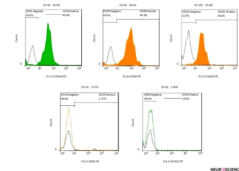
positive for the mesenchymal antigens CD 44, CD 90, CD 105 using flow cytometry. Also they had a negative tendency to CD34, CD45 i.e. hematopoietic markers
(Figure 2). Multilineage differentiation of hDPSCs was investigated their MSC characteristic. Osteogenic differ-entiation was confirmed by production of calcium
de-posits via Alizarin Red staining (Figure 3a). Adipogenic
differentiation was confirmed by Oil Red O staining of
lipid droplet (Figure 3b).
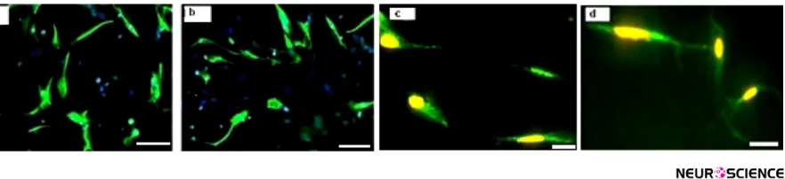
3.2. Differentiation of hDPSCs into OPCs
appeared during differentiation process i.e. spindle like
hDPSCs are converted to branched shape OPCs (Figure
4). The results of immunoflourcent staining showed that
the preinduced cells expressed nestin that is considered as a neuroepithelial marker. The mean percentages of immunoreactive cells to this marker was 69.3%±1.54%
(Figure 5a). Also, the immunostaining of induced cells indicated the expression of specific glial markers such as
Olig2, NG2 and O4 at the end of differentiation protocol
(Figure 5 b, c, d). The mean percentages of immunore-active cells to mentioned markers were 47.5%±5.19%, 42.7%±3.49% and 39.63%±1.93%, respectively.
3.3. Survival rate of hDPSCs during differentia-tion protocol
MTT results showed that the viability of the cells at
preinduction stage in the test and control groups, were
Figure 1. Human Dental pulp stem cells culture (×100).
(a) Primary culture after 12h culture; (b) After 4th passage culture.
Figure 2. Flow cytometric analysis of hDPSCs.
Cells after fourth passage were strongly immunopositive to specific surface markers of mesenchymal stem cells; CD44, CD90, CD105 but didn’t express CD34 and CD45 which are specific markers to hematopoietic stem cells.
FL 1-H CD44-FITC
100 101 102 103 104 100 101 102 103 104 100 101 102 103 104
100 101 102 103 104
100 101 102 103 104
FL2-H CD90-PE FL2-H CD105-PE
FL2-H CD34-PE FL1-H CD45-FITC
CD 44 95.4%
CD 34 1.57% CD 45 1.05%
CD 90 94.5% CD 105 93.6%
Coun
t
0
0 0
0 0
Coun
t
Coun
t
Coun
t
Coun
t
CD44 Negative
4.61%
CD34 Negative
98.4% CD45 Negative99.0%
CD90 Negative
4.61% CD105 Negative6.37%
CD44 Positive
95.4%
CD34 Positive
1.57% CD45 Positive1.05%
CD90 Positive
respectively, 94.71%±0.26% and 96.13%±0.58%. The percentages of viable cells in the treated group at the induction stage was lower than in the control group. A significant decrease was seen in cell proliferation rate
following induction stage (P<0.05) (Figure 6).
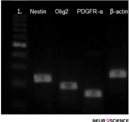
3.4. RT-PCR
RT-PCR results proved the expression of nestin gene
(neuroprogenitor marker) and Olig2 and PDGFR-α
genes (oligoprogentor markers) in treated hDPSCs at the end of the induction stage, while no gene expression was
detected in untreated hDPSCs (Figure 7).
4. Discussion
The neurogenic activity of mesenchymal stem cells in culturing with the presence of neural inducers have
been evaluated in previous literature (Black &
Wood-bury, 2001; Jiang et al., 2010). Also, the neural crest origin of hDPSCs proposes them as an alternative source for neuroglial cell population for therapeutic
strategies in neuroregenerative medicine (Yalvac et al.,
2009; Gervois et al., 2015). But a few studies investi-gated the in vitro differentiation potential of hDPSCs to oligoprogenitor cells.
Figure 3. Possibility of Multilineage differentiation of Dental pulp stem cells (×100). (a) Alizarin Red staining that shows osteogenic differentiation of hDPSC.
(b) Oil Red O staining for detection of lipid droplets which confirms adipogenic differentiation.
Figure 4. Differentiation human Dental pulp stem cells into oligoprogenitor cells. Neuroprogenitor cells after preinduction
phase (a). Further cultivation, lead to generation of OPC at the end of induction stage (c). Scale bars 10 μm.
So we proposed the culturing of hDPSCs in the pres-ence of glial inducers, e.g. bFGF, PDGF, N2 and B27 similar to standard glial differentiation protocols for MSC or BMSCs which can generate oligoprogenitor cells. They can be candidates for the treatment of
demy-elinating diseases (Liu et al., 2007). The multilineage
dif-ferentiation of hDPSCs into adipocytes and osteocyte cells was confirmed with specific staining respectively; Oil Red O and Alizarin Red staining. This result is in agreement with previous reports about the multipotency of hDPSCs as well as MSC.
Our findings showed that glial generation of hDPSCs is associated with the generation of neuroprogenitor cells at the first step of differentiation. After exposure to inducers, hDPSCs were converted from proliferation phase to differentiation stage and acquired a phenotype resembling neuroprogenitor cells. Furthermore, the pre-induced cells were immunopositive to nestin while no expression of this marker was detectable in untreated hDPSCs. These data confirm the previous reports about glial generation associated with nestin that utilized as
neuroprogenitor markers during neurogenesis
(Wislet-Gendebien, 2005; Gu et al., 2015; Bojnordi et al., 2017).
The cells were treated with PDGFF, bFGF, N2 and B27 following preinduction stage. They completed glial dif-ferentiation and were immunopositive to specific glial
markers. The fluorescence staining for Olig2, NG2 and
O4 indicates the immune positive reaction of OPCs to
these glial markers. The percentages of Olig2, NG2 and
O4 positive cells were significantly higher in this
ex-periment treated group compared to hDPSCs cultured in control media. Addition of inducers such as PDGFF,
bFGF, N2 and B27 can promote differentiation of hDP-SCs into oligodendrocyte progenitors.
Furthermore, RT-PCR results proved the expression of
nestin, Olig2 and PDGFR-α genes (oligoprogentor
mark-ers) in treated hDPSCs at the end phase of the induction protocol. Several reports have demonstrated the active role of these factors in oligodendrogenesis as well as
my-elination. The expression of Olig2 after induction phase
is in agreement with the findings of Liu et al., 2007 and
Copray et al., 2006. This helix-loop transcription factor has important role in glial fate map in oligodendrogen-esis process. Similar results were reported that showed
Figure 7. RT-PCR detection of Nestin (220 bp), Olig2 (192), PDGFR-α (124 bp) and β-actin (237 bp) in the differentiated
hDPSCs group.
The expersion of these gene were detectable in induced cells. L=DNA ladder.
Figure 6. The comparison between the mean viability rates at pre-induction (a) and induction (b) stages or in days (D) D4 (a) and D12 (b) (P<0.05).
a: significant decrease with test group in D4.
Viability Test
Viable
Cells (%)
100
98
96
94
92
90
88
86
D4 D8
Test
Control
the generation of mesenchymal stem cells into oligoden-drocytes is accompanied with overexpression of specific
glial genes (Ni et al., 2010; Sanchez-Ramos et al., 2000).
The results of MTT test showed a decrease in cell prolif-eration rate following induction stages that confirms the differentiation of hDPSCs following exposure to glial in-duction media instead of proliferation activity.
hDPSCs are multipotent cells with high proliferation rate as well as plasticity for differentiation to various cell types. In total, hDPSCs can transdifferentiate to oli-goprogenitor cells after two steps differentiation proto-col. The glial inducers include RA, PDGFF, bFGF and EGF as well as the ommition of serum, totally alter the cells microenvironment and induce differentiation of hDPSCs to oligoprogenitor cells.
Our results proved the differentiation capacity of hDP-SCs to oligoprogenitor cells that exhibit special glial mor-phology and express the special oligoprogenitor markers investigated by immunocytochemistry technique. It con-cludes that differentiation of hDPSCs into oligoprogeni-tor cells depends on addition of glial inducers which are currently used for MSC or BMSC. This finding supports the therapeutic use of hDPSCs as an alternative source for generation of glial cells for the clinical repair of de-myelinating diseases. However, more studies are neces-sary to be done to evaluate the complete differentiation of hDPSCs to fully functional oligodendrocytes.
Acknowledgments
This project was funded by the grants from Ilam Uni-versity of Medical Sciences, Ilam, Iran (No. 951018-88).
Conflict of Interest
The authors declared no conflicts of interest.
References
Baumann, N., Pham-Dinh, D. (2001). Biology of oligodendrocyte
and myelin in the mammalian central nervous system. Physi-ological Reviews, 81(2), 871–927. PMID: 11274346
Black, I. B., & Woodbury, D. (2001). Adult rat and human bone
marrow stromal stem cells differentiate into neurons. Blood Cells, Molecules, and Diseases, 27(3), 632–636. doi: 10.1006/
bcmd.2001.0423
Blakemore, W. F., & Franklin, R. J. M. (2000). Transplanta -tion op-tions for therapeutic central nervous system
re-myelination. Cell Transplantation, 9(2), 289–294. doi: 10.1177/096368970000900214
Bojnordi, M. N, Azizi, H., Skutella, T., Movahedin, M., Pour
-abdolhossein, F., Shojaei, A., et al. (2017). Differentiation of
spermatogonia stem cells into functional mature neurons characterized with differential gene expression. Molecular Neurobiology, 54(7), 5676-82. doi: 10.1007/s12035-016-0097-7
Chang, C. C., Chang, K. C., Tsai, S. J., Chang, H. H., & Lin, C. P.
(2014). Neurogenic differentiation of dental pulp stem cells to
neuron-like cells in dopaminergic and motor neuronal induc -tive media. Journal of the Formosan Medical Association, 113(12), 956–965. doi: 10.1016/j.jfma.2014.09.003
Chopp, M., & Li, Y. (2002). Treatment of neural injury with
marrow stromal cells. The Lancet Neurology, 1(2), 92–100. doi: 10.1016/s1474-4422(02)00040-6
Chun, S. Y., Soker, S., Jang, Y. J., Kwon, T. G., & Yoo, E. S. (2016).
Differentiation ofhuman dental pulp stem cells into
dopamin-ergic neuron-like cells in vitro. Journal of Korean Medical Sci-ence, 31(2), 171. doi: 10.3346/jkms.2016.31.2.171
Copray, S., Balasubramaniyan, V., Levenga, J., De Bruijn, J., Liem, R., & Boddeke, E. (2006). Olig2 overexpression induces
the in vitro differentiation of neural stem cells into mature oli-godendrocytes. Stem Cells, 24(4), 1001–10. doi: 10.1634/stem
-cells.2005-0239
Delilovic-Vranic, J. (2005) Multiple sclerosis therapy. Medical Ar-chives, 59, 191–195.
Gervois, P., Struys, T., Hilkens, P., Bronckaers, A., Ratajczak, J., Politis, C., et al. (2015). Neurogenic maturation of human den -tal pulp stem cells following neurosphere generation induces morphological and electrophysiological characteristics of functional neurons. Stem Cells and Development, 24(3), 296–311. doi: 10.1089/scd.2014.0117
Ghasemi Hamidabadi, H., Rezvani, Z., Nazm Bojnordi, M., Shirinzadeh, H., Seifalian, A. M., Joghataei, M. T., et al. (2017). Chitosan-intercalated montmorillonite/poly(vinyl alcohol) nanofibers as a platform to guide neuronlike differentiation of
human dental pulp stem cells. ACS Applied Materials & Inter-faces, 9(13), 11392–11404. doi: 10.1021/acsami.6b14283
Gronthos, S., Brahim, J., Li, W., Fisher, L. W., Cherman, N., Boyde, A., et al. (2002). Stem cell properties of human dental
pulp stem cells. Journal of Dental Research, 81(8), 531–535. doi: 10.1177/154405910208100806
Gronthos, S., Mankani, M., Brahim, J., Robey, P. G., & Shi, S.
(2000). Postnatal human dental pulp stem cells (DPSCs) in vitro and invivo. Proceedings of the National Academy of Sciences, 97(25), 13625–30. doi: 10.1073/pnas.240309797
Gu, P., Qiu, F. C., Han, R., Zhang, Z., Dong, C., Zhang, L.N et al. (2015). Efficient differentiation of neural stem cells induced by
the rat bone marrow stromal cells. International Journal of Clini-cal and Experimental Medicine, 8(5), 6713–24. PMID: 26221209
Haratizadeh, S., Nazm Bojnordi, M., Niapour, A., Bakhtiari, M., Ghasemi Hamidabadi, H. [Improvement of neuroglial dif -ferentiation from human dental pulp stem cells using CSF (Persian)]. Journal of Mazandaran University of Medical Sciences, 26(140), 1-14.
cells from a supernumerary tooth. Journal of Oral Pathology & Medicine, 37(9), 571–574. doi: 10.1111/j.1600-0714.2008.00654.x
Jiang, J., Lv, Z., Gu, Y., Li, J., Xu, L., Xu, W., et al. (2010). Adult rat mesenchymal stem cells differentiate into neuronal-like phe -notype and express a variety of neuro-regulatory molecules in vitro. Neuroscience Research, 66(1), 46–52. doi: 10.1016/j.neu
-res.2009.09.1711
Liu, Z., Hu, X., Cai, J., Liu, B., Peng, X., Wegner, M., et al. (2007).
Induction of oligodendrocyte differentiation by Olig2 and
Sox10: Evidence for reciprocal interactions and dosage-de -pendent mechanisms. Developmental Biology, 302(2), 683–693. doi: 10.1016/j.ydbio.2006.10.007
Nagatomo, K., Komaki, M., Sekiya, I., Sakaguchi, Y., Noguchi, K., Oda, S., et al. (2006). Stem cell properties of human peri -odontal ligament cells. Journal of Periodontal Research, 41(4), 303–310. doi: 10.1111/j.1600-0765.2006.00870.x
Nazm Bojnordi, M., Ghasemi, H. H., & Akbari, E. (2015). Remy -elination after lysophosphatidyl choline-induced demyelina-tion is stimulated by bone marrow stromal cell-derived oli-goprogenitor cell transplantation. Cells Tissues Organs, 200(5), 300–306. doi: 10.1159/000437350
Nazm Bojnordi, M., Movahedin, M., Tiraihi, T., Javan, M., & Ghasemi Hamidabadi, H. (2013). Oligoprogenitor cells de -rived from spermatogonia stem cells improve remyelination in demyelination model. Molecular Biotechnology, 56(5), 387– 393. doi: 10.1007/s12033-013-9722-0
Ni, W. F., Yin, L. H., Lu, J., Xu, H. Z., Chi, Y. L., Wu, J. B., et al.
(2010). In vitro neural differentiation of bone marrow stromal cells induced by cocultured olfactory ensheathing cells. Neuro-science Letters, 475(2), 99–103. doi: 10.1016/j.neulet.2010.03.056
Patani, R., & Chandran, S. (2012). Experimental and therapeutic
opportunities for stem cells in multiple sclerosis. International Journal of Molecular Sciences, 13(12), 14470–91. doi: 10.3390/ ijms131114470
Pluchino, S., & Martino, G. (2005). The therapeutic use of stem
cells for myelin repair in autoimmune demyelinating disor-ders. Journal of the Neurological Sciences, 233(1-2), 117–119. doi: 10.1016/j.jns.2005.03.026
Rodriguez, M. (2007). Effectors of demyelination and remyelina -tion in the CNS: Implica-tions for multiple sclerosis. Brain Pa-thology, 17(2), 219–229. doi: 10.1111/j.1750-3639.2007.00065.x
Sanchez-Ramos, J., Song, S., Cardozo-Pelaez, F., Hazzi, C., Sted
-eford, T., Willing, A., et al. (2000). Adult bone marrow stromal
cells differentiate into neural cells in vitro. Experimental Neurol-ogy, 164(2), 247–256. doi: 10.1006/exnr.2000.7389
Schippling, S., Heesen, C., Zander, A., & Martin, R. (2008). Stem
cell transplantation in multiple sclerosis. Journal of Neurology, 255(S6), 43–47. doi: 10.1007/s00415-008-6008-8
Sloan, A., & Smith, A. (2007). Stem cells and the dental pulp: po -tential roles in dentine regeneration and repair. Oral Diseases, 13(2), 151–157. doi: 10.1111/j.1601-0825.2006.01346.x
Wislet-Gendebien, S., Hans, G., Leprince, P., Rigo, J. M., Moonen, G., & Rogister, B. (2005). Plasticity of cultured mesenchymal
stem cells: switch from nestin-positive to excitable
neuron-like phenotype. Stem Cells, 23(3), 392–402. doi: 10.1634/stem
-cells.2004-0149
Yalvac, M., Rizvanov, A., Kilic, E., Sahin, F., Mukhamedyarov, M., Islamov, R., et al (2009). Potential role of dental stem cells in