Okezie Obasi Kanu, Chinenye Nnoli1, Omodele Olowoyeye2, Omotayo Ojo, Christopher Esezobor1, Adekunle Adeyomoye2, Olufemi Bankole, Chinyere Asoegwu, Edamisan Temiye1
Department of Surgery, 1Paediatrics and ²Radiodiagnosis, College of Medicine, University of Lagos and Lagos University Teaching Hospital, Idi-Araba, Lagos, Nigeria
Infantile subdural empyema: The role of brain
sonography and percutaneous subdural tapping in
a resource‑challenged region
Introduction
The use of brain sonography or trans‑fontanelle
ultrasound scan (TFUSS) in the diagnosis of childhood intracranial pathologies has been documented in the literature just like the use of subdural taps for the treatment of subdural empyema (SDE). Subdural tap was used both for diagnosis[1‑5] and treatment[3,5,6] of this condition but
has been largely abandoned since the advent of modern imaging techniques. Computerized tomographic (CT)
scans and magnetic resonant imaging (MRI) are better
diagnostic tools for intracranial conditions and have
become available in many centers across the world including ours. CT scan is the most common diagnostic tool used in the management of SDE and has helped to reduce mortality.[7‑11] But despite the availability of these diagnostic tools, they are not affordable to all patients who visit the hospital in most parts of Sub‑Saharan
Africa. Children with subdural empyema (SDE), whose
parents cannot afford to have an urgent CT scan, nor bear the financial burden of the surgical treatment, might
deteriorate leading to fatal outcome. Brain sonography being far cheaper than CT or MRI is therefore useful in such patients with patent anterior fontanelle.
Patient selection
Patients who fall into the above category who presented with features of postmeningitic intracranial suppuration, in whom diagnosis of SDE were made by TFUSS and treated using only Subdural taps were included in this study. The study period was from February 2006 to
August 2008.
Address for correspondence:
Dr. Okezie Obasi Kanu, Department of Surgery, Lagos University Teaching Hospital, PMB 12003, Surulere, Lagos, Nigeria. E‑mail: daddykay143@gmail.com
Access this article online Quick Response Code:
Website:
www.ruralneuropractice.com
DOI:
10.4103/0976‑3147.139978 ABSTRACT
Background This study explored the outcome of children with patent anterior fontanelles who were treated with trans‑fontanelle ultrasound scan (TFUSS), which is more affordable and available than CT scan and MRI in the diagnosis of childhood intracranial pathologies and treatment of subdural empyema, in developing countries. Patients and Methods: Seventeen infants with post‑meningitic subdural empyema, diagnosed using trans‑fontanelle ultrasound alone and treated with subdural tapping over a 31‑months period, were studied. Results: Eleven patients presented with grades II and III Bannister and William grading for level of consciousness in intracranial subdural empyema. Aspirate from 7 (41.2%) patients were sterile. The most common organisms isolated were Streptococcus faecalis 3 (17.6%), Haemophilus Influenza 2 (11.8) and Staphylococcus aureus 2 (11.8), multiple organisms were isolated in three of the patients. Ninety‑four percent (94%) of the patients had good outcome. Five subjects developed hydrocephalus, one patient had a recurrence of subdural empyema, four patients had residual hemiparesis, two of the four patients had speech difficulties, while one patient (~6%) died. Conclusion: While CT and MRI remain the gold standard for investigating intracranial lesions, transfontanelle ultrasonography is adequate for diagnosis of infantile subdural empyema in resource‑challenged areas. Percutaneous subdural tap is an affordable and effective therapy in such patients with financial challenges.
Key words: Cranial sonography, subdural empyema, subdural taps, transfontanelle ultrasonography
All patients who had subdural empyema and whose parents were poor and could not afford the cost of CT and whose diagnosis were made using brain sonography (TFUSS) and subsequently treated by subdural tap were included in this study. These patients had been diagnosed and treated for meningitis or currently on treatment when they were referred to the Neurosurgical team. The Otorhinolaryngologist assessed
the patients for ear, nose and throat (ENT)‑related causes
of intracranial suppuration.
Patients who had CT bran scan were excluded.
Controls
This is a prospective observational study. We considered that a control with patients who had CT scan and TFUSS was not necessary to access accuracy of diagnosis since the patients would be treated by subdural tap, which
confirmed the diagnosis.
Patients and Methods
The biodata of the patients were collected. The presenting clinical features, the location of the empyema, total volume of aspirate, bacteriology of samples, treatment outcome and residual deficits were documented. Diagnosis was made by cranial sonography alone.
All patients were treated using a sterile 18‑guage plastic
cannula with metal trocar. All taps were performed by aseptic technique. The use of plastic cannulae made it possible to remove almost all subdural collection in one procedure using a single tap with multiple syringes while the cannula remained in place. Patients with bilateral subdural empyema had the procedure repeated on the
contralateral side at the same sitting.
A repeat TFUSS was done within 24 h to re‑evaluate and confirm complete removal of the subdural collection.
Intravenous antibiotics were continued for 2 to 4 weeks
post‑drainage depending on clinical status. The therapy
was continued and completed with oral antibiotics subsequently.
The duration of follow‑up was 1 to 3 years.
Results
Age and gender
There were 17 patients in this study: 10 males, 7 females.
Age range was from 2 months to 14 months with a mean
age of 8 months.
Etiology
All the patients in this study were cases of post‑meningitic
intracranial suppuration. Five patients had bilateral subdural collection while the rest where unilateral. The average volume of empyema aspirated was
34.3 ml (20‑48 ml).
Clinical presentation
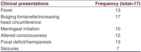
The most common clinical presentation was bulging anterior fontanelle with increased occipitofrontal circumference (OFC) as observed in all patients. Other clinical presentations were fever, meningeal irritation, altered consciousness focal deficits and seizures as shown in Table 1.
Bannister and William[10] grading of level of consciousness
was Grade I and II in 12 patients while 5 patients presented below grade III and IV [Table 2].
Therapy
A total of 18 procedures were performed in the 17 patients. One patient had a recurrence of subdural empyema which was re‑drained. All the patients received
a combination of intravenous antibiotics to ensure broad spectrum cover. All the patients had received antibiotics for treatment of meningitis before referral for neurosurgical intervention. Antibiotics were changed or
continued based on sensitivity pattern and/or clinical
improvement especially when the aspirate was sterile. Four patients additionally received intraventricular Vancomycin prescribed by the pediatric neurologist. All the seven patients who presented with seizures received
anti‑convulsant therapies and were seizure free at 1 year.
Five of the patients developed hydrocephalus of
post‑infective type and subsequently treated with
Table 1: Common clinical presentations
Clinical presentations Frequency (total=17)
Fever 13
Bulging fontanelle/increasing
head circumference 17
Meningeal irritation 10
Altered consciousness 12
Focal deficit/hemiparesis 13
Seizures 7
Table 2: Bannister and William grading for level of consciousness in intracranial subdural empyema[10]
Grade Frequency (total=17) Description
I 5 Awake and alert
II 7 Drowsy and disoriented
III 4 Responsive to stimuli
endoscopic third ventriculostomy (ETV), three of whom
failed and had ventriculo‑peritoneal shunt insertion
with good outcome. There was residual hemiparesis in four patients which necessitated physiotherapy and all resolved at 6 months post therapy. Two of the four
patients had speech difficulties and were managed by
the speech therapist.
Bacteriology
Aspirates were subjected to microscopy, culture and sensitivity (MCS) studies. The organisms isolated and their frequencies are shown in Table 3. The aspirate was sterile in seven patients (41.2%). The most common organism isolated was Streptococcus faecalis (17.6%). Haemophilus
Influenza and Staphylococcus aureus ranked second. There were multiple organisms isolated in three of the patients. All the aspirates had glucose level less than 20 mg% and white
blood cell (WBC) differentials with polymorphs ≥75%.
Outcome [Table 4]
Using the Henk W. Mauser[12] grading system for morbidity of survivors, 94% of the patients (Grade A
and B) had good outcome with only minor disabilities where present [Table 4].
Speech has improved in the two patients with residual
speech deficit undergoing speech therapy. Only one
patient (~6%) died.
Discussion
The purpose of this study is to demonstrate the usefulness of both sonography and subdural taps in the
early diagnosis and treatment of subdural empyema
especially in resource‑challenged regions where the medical insurance is ineffective yet and diagnostic delays
could increase the morbidity and mortality of SDE in such conditions. It has been shown in other series in the literature that outcome is favorable when diagnosis is made on time followed by early intervention (surgical
evacuation and antibiotics therapy). Mortality often
results from delay in referral[13] or delay in diagnosis and
commencement of therapy.[13‑16] The cost of brain CT scan is about $350 ‑ $400USD equivalent while transfontanelle
sonography costs about $20USD. Though CT and MRI
have been proven to be more effective in diagnosis,
patients still have to bear the cost of these investigations. In most developing countries the Gross Domestic
Product (GDP) is low; health insurance schemes are nonexistent or at best ineffective. In a situation where
patients and relations are poor and hardly have enough for the diagnosis and subsequent surgical therapy, there will be a delay in making diagnosis and patients might deteriorate, leading to increased morbidity or even fatal outcome. A patent fontanelle and a cheap TFUSS that
confirms diagnosis would have reduced the time of
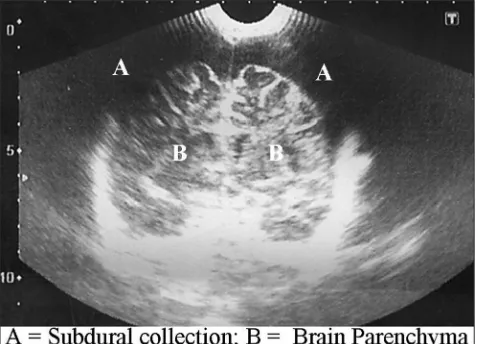
diagnosis. The added advantage of using sonography is that it could be repeated severally without the risk of radiation associated with the use of CT brain scan. Figure 1 is a typical sonography of one of the patients with bilateral subdural collection.
In our study, it was possible to accurately diagnose the presence of subdural empyema in all patients.
The history of previous or on‑going meningitis with
the clinical features enumerated helped to clinch the diagnosis.
The use of percutaneous subdural tap as described above made it possible to evacuate the subdural collection
Table 3: Bacteriology spectrum
Micro‑organism Frequency
Sterile 7
Staphylococcus aureus 2
Escherichia coli 3
Klebsiella pneumoniae 1
Streptococcus faecalis 3
Pseudomonas aeruginosa 1
Anaerobes 2
Haemophilus influenzae 2
*Multiple organisms were found in three patients
Table 4: Henk W Mauser grading system for morbidity of survivors of intracranial subdural empyema[12]
Grade Frequency (percentage) Description
A 14 (82) Survival without or with a minor, not disabling focal deficit
B 2 (12) Survival with not disabling seizures and with or without a minor focal C Nil (0) Survival with severe disability
and this was carried out effectively. Burr holes and
craniotomies have been used in older patients and
though some reports favor craniotomy;[9,17‑19] there is still no consensus on the method of surgical evacuation and this varies with age and the stage of the abscess.[20]
Percutaneous aspirations through an open fontanelle and twist drill have been used as minimally invasive techniques with satisfactory results.[4,5,21‑23] Most authors favor early drainage and antibiotics therapy to reduce morbidity, clinical deterioration and mortality.[9,18,19] Antibiotics therapy alone was not adequate for treatment
when neurologic deficits have already set in.[18,24,25] The aspirates were sterile in 7 patients (41.2%) and
the commonest organism isolated was Streptococcus faecalis (17.6%). Curless[3] recorded negative culture
stains in some of his patients who had H. Influenza
meningitis. Eighteen of 37 cultures performed by Mauser
and Tulleken[12] were sterile. These results may be due
to the fact that antibiotics previously administered for the treatment of meningitis interfered with the culture yield, but the low glucose level and high polymorph
WBC differentials in these patients suggest ongoing
bacterial activity.
Only one patient had a recurrence which was treated with a repeat tap. This patient was seen at Bannister and Williams’[10] grade IV level of consciousness at the time of
transfer from a private facility. The parents did not accept transfer to a tertiary hospital till the level of consciousness
dropped significantly. She had a prolonged course and
later died. This is the only mortality recorded in our series (~6%). Mortality in the literature is closely related to delay in referral or delay in treatment.[7,8,10,11]
Outcome in our study compares favorably with Jacobson and Farmer[5] though most of their six patients
were managed with burr holes. Curless[3] on the other
hand reported that repeated subdural taps produced satisfactory results in eight of nine patients in his series. Diagnoses in these patients were done using CT scan in some and subdural taps in others. The patient populations being small would not allow objective
comparison of the effect of imaging modalities on overall
outcome but certainly CT scan is superior to TFUSS used in this study. However, Farmer and Wise[26] lost four
of their eight patients in their series and this may be
attributable to delayed diagnosis due to lack of CT scan.
There is no record of TFUSS being used in any of their patients.
Most residual deficits resolved satisfactorily at 6 months
to one year. Patients with hydrocephalus had endoscopic third ventriculostomy (ETV) or ventriculoperitoneal
shunt (VPS) when the ETV failed or cisterns were significantly scarred. Anticonvulsant therapy was however continued for a period of 2 years in those who had seizures though they were seizure free at 1 year. Two
out of 9 patients reviewed by Curless[3] had recurrence
of subdural empyema. One of his patients had severe psychomotor retardation and another patient had
sustained speech problems 2 years after infection. One patient was lost to follow‑up. There was no record of
hydrocephalus in his series. We believe however that the presence of hydrocephalus is an indication of the severity of the infective process and not a complication of treatment.
It is not possible to compare the present result with the group managed with CT scan and burr holes because the local data presently consists of adult patients in whom subdural empyema were managed with burr holes and craniotomy, and the etiology is varied.
Conclusion
While CT and MRI remain the gold standard for investigating intracranial lesions, and will be deployed
where available, trans‑fontanelle ultrasonography is
adequate in making diagnosis of infantile subdural
empyema especially in resource‑challenged areas.
Subdural empyema in children with patent anterior fontanelle can be satisfactorily treated with percutaneous subdural taps with low recurrence rates and low mortality rates. This saves cost and surgical time.
Majority of culture results are organism‑negative and broad spectrum antibiotics are effective in providing
satisfactory antibacterial cover. The effectiveness of intrathecal antibiotics and mode of delivery for SDE requires consideration and evaluation in subsequent studies.
References
1. Feigin RD, Dodge PR. Bacterial meningitis: Newer concepts of pathophysiology and neurologic sequelae. Pediatr Clin North Am 1976;23:541‑56.
2. Smith MH, Dormont RE, Prather GW. Subdural effusions complicating bacterial meningitis. Pediatrics 1951;7:34‑43.
3. Curless RG. Subdural empyema in infant meningitis: Diagnosis, therapy, and prognosis. Childs Nerv Syst 1985;1:211‑4.
4. Woodhall BL. Osteomyelitis and epi‑, extra‑, and subdural abscesses. Clin Neurosurg 1966;14:239‑55.
5. Jacobson PL, Farmer TW. Subdural empyema complicating meningitis in infants: Improved prognosis. Neurology 1981;31:190‑3.
6. Pattisapu JV, Parent AD. Subdural empyemas in children. PediatrNeurosci 1987;13:251‑4.
8. Zimmerman RD, Leeds NE, Danziger A. Subdural empyema: CT findings. Radiology 1984;150:417‑22.
9. Smith HP, Hendrick EB. Subdural empyema and epidural abscess in children. J Neurosurg 1983;58:392‑7.
10. Bannister G, Williams B, Smith S. Treatment of subdural empyema. J Neurosurg 1981;55:82‑8.
11. Luken MG 3rd, Whelan MA. Recent diagnostic experience with subdural
empyema. J Neurosurg 1980;52:764‑71.
12. Mauser HW, Tulleken CA. Subdural empyema. A review of 48 patients. Clin Neurol Neurosurg 1984;86:255‑63.
13. Weinman D, Samarasinghe HH. Subdural empyema. Aust N Z J Surg 1972;41:324‑30.
14. Mauser HW, Van Houwelingen HC, Tulleken CA. Factors affecting the outcome in subdural empyema. J NeurolNeurosurg Psychiatry 1987;50:1136‑41.
15. Hitchcock E, Andreadis A. Subdural empyema: A review of 29 cases. J Neurol Neurosurg Psychiatry 1964;27:422‑34.
16. Bradley PJ, Shaw MD. Subdural empyema management of the primary source. Br J Clin Pract 1984;38:85‑8.
17. Anagnostopoulos DI, Gortvai P. Intracranial subdural abscess. Br J Surg 1973;60:50‑2.
18. Coonrod JD, Dans PE. Subdural empyema. Am J Med 1972;53:85‑91.
19. Cowie R, Williams B. Late seizures and morbidity after subdural empyema. J Neurosurg 1983;58:569‑73.
20. Pathak A, Sharma BS, Mathuriya SN, Khosla VK, Khandelwal N, Kak VK. Controversies in the management of subdural empyema. A study of 41 cases with review of literature. Acta Neurochir (Wien) 1990;102:25‑32.
21. Ellis SS, Austin TL. Subdural empyema. Am J Dis Child 1978;132:1147. 22. Mahapatra AK, Bhatia R. Salmonella intracerebral and subdural
abscess‑report of two cases. Postgrad Med J 1987;63:373‑5.
23. Mahapatra AK, Bhatia R, Banerji AK, Tandon PN. Subdural empyema in children. Indian Pediatr 1984;21:561‑7.
24. Black P, Graybill JR, Charache P. Penetration of brain abscess by systemically administered antibiotics. J Neurosurg 1973;38:705‑9. 25. Khan M, Griebel R. Subdural empyema: A retrospective study of
15 patients. Can J Surg 1984;27:283‑5, 288.
26. Farmer TW, Wise GR. Subdural empyema in infants, children and adults. Neurology 1973;23:254‑61.
How to cite this article: Kanu OO, Nnoli C, Olowoyeye O, Ojo O,
Esezobor C, Adeyomoye A, et al. Infantile subdural empyema: The role of brain sonography and percutaneous subdural tapping in a resource-challenged region. J Neurosci Rural Pract 2014;5:355-9.
Source of Support: Nil. Conflict of Interest: None declared.
Staying in touch with the journal 1) Table of Contents (TOC) email alert
Receive an email alert containing the TOC when a new complete issue of the journal is made available online. To register for TOC alerts go to
www.ruralneuropractice.com/signup.asp.
2) RSS feeds
Really Simple Syndication (RSS) helps you to get alerts on new publication right on your desktop without going to the journal’s website. You need a software (e.g. RSSReader, Feed Demon, FeedReader, My Yahoo!, NewsGator and NewzCrawler) to get advantage of this tool. RSS feeds can also be read through FireFox or Microsoft Outlook 2007. Once any of these small (and mostly free) software is installed, add
![Spiro [2 3′]oxindole spiro [3 3′′]oxindole 4[p methylbenzyl]pyrrolidizine](data:image/gif;base64,R0lGODlhAQABAIAAAP///wAAACH5BAEAAAAALAAAAAABAAEAAAICRAEAOw==)