M
AŁGORZATAK
RZYSTEK−K
ORPACKA1, M
AŁGORZATAM
ATUSIEWICZ1,
K
RZYSZTOFG
RABOWSKI2, D
OROTAD
IAKOWSKA2, D
OROTAB
OEHM1,
I
RENAK
USTRZEBA−W
ÓJCICKA1, T
ERESAB
ANAŚ1Immunoenzymatic Method
for Midkine Determination in Serum
Immunoenzymatyczna metoda oznaczania midkiny w surowicy krwi
1 Department of Medical Biochemistry Silesian Piasts University of Medicine in Wrocław, Poland
2 Department of Gastrointestinal and General Surgery Silesian Piasts University of Medicine in Wrocław, Poland
Adv Clin Exp Med 2006, 15, 2, 247–252 ISSN 1230−025X
ORIGINAL PAPERS
Abstract
Background.Midkine is a secreted protein originally engaged in neurogenesis and recently reported to be impli− cated in tumorigenesis. Midkine expression, both at the level of protein and mRNA, is elevated in various types of cancer. A potential significance of midkine level determination as a marker of tumour presence and progression is intensively studied. However, at the time when presented studies were conducted, no commercial ELISA kits for the assessment of serum midkine level were available.
Objectives.Developing a sensitive method for midkine determination in serum with application of commercially available antibodies and chromogenic substrate. The other goal of presented studies was practical evaluation of invented assay in serum samples from apparently healthy subjects and establishment of the reference range and test sensitivity.
Material and Methods.The studies were conducted on serum samples from 43 apparently healthy blood donors. In the described assay, double antibody sandwich indirect ELISA (DASI−ELISA) with two policlonal antibodies against recombinant human midkine raised in different hosts, was applied. For the enhancement of test sensitivity, the authors used biotin−streptavidin amplification system. Horseradish peroxidase and tetramethylbenzidine were applied for colour development.
Results.The sensitivity of the assay was 3–4 pg of midkine per ml of serum. The median midkine value, deter− mined for the studied group, was 141 pg/ml and mean ± SD was 220 ± 320.6 pg/ml. The upper normal range limit, determined as 82ndpercentile, was 541 pg/ml.
Conclusions. The invented ELISA proved to be a sensitive method for determination of serum midkine concen− tration and can be applied in studies on the serum level of this circulating growth factor in various physiological and pathological conditions (Adv Clin Exp Med 2006, 15, 2, 247–252).
Key words: midkine, ELISA, tumour marker, growth factor.
Streszczenie
Wprowadzenie. Midkina jest peptydem wydzielniczym zaangażowanym przede wszystkim w neurogenezę. W ostatnich latach wykazano jednak udział midkiny w karcynogenezie, a jej zwiększoną ekspresję zarówno na po− ziomie białka, jak i mRNA potwierdzono w wielu typach nowotworów. Podjęto więc badania nad możliwościami zastosowania midkiny jako markera obecności i progresji choroby nowotworowej. Mimo tak intensywnie prowa− dzonych doświadczeń, w czasie gdy prowadzono badania własne, nie były dostępne komercyjne testy pozwalają− ce na oznaczenie stężenia midkiny w surowicy.
Cel pracy.Opracowanie czułej metody immunoenzymatycznej pozwalającej na oznaczanie stężenia midkiny w su− rowicy, wykorzystującej komercyjnie dostępne przeciwciała. Opracowany test poddano ocenie praktycznej, usta− lając zakres referencyjny i czułość przez oznaczenie stężenia midkiny w grupie honorowych krwiodawców.
Materiał i metody.Badania przeprowadzono na surowicach pozyskanych z krwi 43 honorowych krwiodawców bez widocznych objawów chorób. W teście wykorzystano technikę immunoenzymatycznej detekcji typu sandwich
Midkine is a secreted, heparin−binding protein of great functional diversity, strongly expressed during embryogenesis [1]. Together with pleiotro− phin, midkine constitutes a distinct family of hepa− rin−binding growth factors not related to fibroblast growth factor (FGF) family. Midkine is, first of all, implicated in neurogenesis as it exhibits neurite− promoting activity, promotes survival and migra− tion of neurons and induces synapse formation at neuro−muscular junction [2]. Besides, it has been shown to promote fibrinolytic activity of vascular endothelial cells as well as neutrophil, macro− phage and osteoblast migration [2]. In studies con− ducted by Yu et al. [3] midkine suppressed cyto− toxic activity of amyloid β−peptide while Calle− baut et al. [4] demonstrated that this chemokine inhibits HIV infection. It has also been found that this growth factor plays an angiogenic role in car− cinogenesis [5]. Midkine, which is, in some tissues under physiological conditions, expressed at low level, has been overexpressed in tissue sections from lung [6], breast [7], gastrointestinal [8], thy− roid [9] and brain tumours [10]. The midkine− induced enhancement of tumour cells resistance to anti−cancer drugs has also been observed [11].
Midkine proved to be useful as a prognostic marker. Its overexpression has been correlated with worse prognosis in patients with neuroblas− toma [12], urinary bladder carcinoma [13] and glioblastoma [14] and has been connected with tumour progression in case of prostate [15] and colon carcinomas [16]. Most of the reports deal with midkine at mRNA level or protein expression in tissue sections. Recently, the studies on serum midkine levels in various cancers and its correla− tion with tumour histopathological features, like type, size, grade and ability to form metastasis are in progress [17–20]. Although two sandwich ELISA tests for determination of serum midkine have been already described [17, 21], still there is no commercial ELISA kit available.
Materials and Methods
Serum Samples
Sera of apparently healthy blood donors (n = 43) were kindly provided by Regional Centre
of Blood Donation and Therapeutics in Wrocław, Poland. Age and sex distribution of blood donors included in the presented studies is summarized in Table 1. Samples were kept frozen (–25°C) until
examination. Prior to assay, the serum samples were brought to room temperature and mixed completely. The authors used sera ten times dilut− ed with 0.05% Tween−20 in PBS. All samples were run in duplicates. Local Medical Ethics Committee approved the project presented in this paper.
Enzyme−Linked Immunoassay
The authors applied the double antibody sand− wich indirect ELISA (DASI−ELISA) method, where, in one test, two antigen−specific antibodies, raised in different hosts are used. In the described assay, rabbit anti−human recombinant midkine polyclonal antibodies were applied as capture anti− bodies while goat anti−human recombinant mid− kine polyclonal antibodies conjugated with biotin were used as detection antibodies. The wells of flat−bottom microtiter plate (Nunc MaxiSorp) were coated with 50 µl of anti−human midkine polyclonal rabbit antibodies (Gentaur, Belgium, cat. no.: 5479–100, lot no.: P8119) at concentra− tion of 4 µg/ml in 50 mM carbonate buffer, pH 9.6 by overnight incubation. All incubation steps, unless otherwise stated, were conducted at room temperature on microplate rotation table at 100 rpm. After intensive washing with five changes of 300 µl of washing buffer (0.05% Tween−20 in PBS), the wells were blocked with 250 µl of buffered SuperBlock Protein with 0.05% Tween−20 (Pierce,
Table 1. Age and sex distribution of blood donors includ−
ed in the studies
Tabela 1. Charakterystyka badanej grupy pod względem
wieku i płci
Sex Age n
(Płeć) (Wiek)
Female 25–40 9
(Kobiety) > 40 1
Male 25–40 22
(Mężczyźni) > 40 11
Wyniki.Czułość opracowanego testu została wyliczona na 3–4 pg/ml. Mediana stężenia midkiny w grupie refe− rencyjnej wyniosła 141 pg/ml, zaś średnia 220 ± 320,6 pg/ml. Górna granica zakresu normalnego, definiowana ja− ko średnia + odchylenie standardowe, została wyliczona na 541 pg/ml.
Wnioski.Opracowano czuły test do oznaczania stężenia midkiny w surowicy, który można wykorzystać w bada− niach nad sekrecją tego czynnika wzrostu w warunkach fizjologicznych i patologicznych (Adv Clin Exp Med 2006, 15, 2, 247–252).
USA, cat no.: 37545, lot no.: FA66348 and cat. no.: 28320, lot. no.: GC94248, respectively) in a manner suggested by manufacturer. After wash− ing, 50 µl of serum samples or standards were added and incubated for six hours. The emptied and washed wells were next filled with 50 µl of biotinylated goat anti−human midkine polyclonal antibodies (RnD Systems, USA, cat no.: BAF258, lot no.: UT01) at concentration of 0.5 µg/ml in PBS with 0.05% Tween−20. After overnight incu− bation at 4°C, the amplification and detection sys− tem of 50 ml of 0.2 µg/ml streptavidin conjugated with horseradish peroxidase (Jackson Immuno− research, USA, cat. no.: 016−030−084, lot. no.: 63794) in PBS with 0.05% Tween−20 was applied and allowed to react for 30 minutes. The wells were next washed thoroughly with six changes of washing buffer and subsequently 50 µl of the sub− strate solution (1−Step ultra TMB−ELISA, Pierce, USA, cat. no.: 34028, lot no.: FD801109) was added for 30 minutes incubation in the dark. The colour development was stopped by acidification with equal volume of 2 M H2SO4. The amount of
oxidized 3,3’,5,5’−tetramethylbenzidine was mea− sured on a microplate colorimetric reader (Multi− scan MS, Labsystems) with 450 nm filter. Recombinant human midkine (Peprotech, USA, cat. no.: 450−16, lot. no.: 119190−B) dissolved in ten times diluted human serum depleted of mid− kine by affinity chromatography served as a stan− dard. The references were used in the concentra− tion range of 10–2000 pg/ml. Ten times diluted midkine−deprived serum served as a test sample blank in order to control the non−specific interac− tions of sera with a plate. Additionally, a reagent blank (a negative control of the test) was prepared by replacing serum samples with serum diluent (0.05% Tween−20 in PBS).
Midkine−Depleted Serum
Midkine−depleted human serum was obtained by affinity chromatography procedure on heparin coupled to Sepharose column as described by Muramatsu et al. [21]. Serum samples from five healthy individuals were pooled together and serum in total volume of 5 ml was passed, in 20 mM Tris− HCl buffer (pH = 7.5) with 0.2 M NaCl, through 1 ml Hi−Trap Heparin HP column (Amersham Biosciences, Sweden). Collected unbound fraction was then dialyzed against PBS with 0.05% Tween− 20 and concentrated to the initial volume of 5 ml by centrifugation at 5,500 rpm, 4°C using vivaspin 20 ml concentrator with 3,000 MWCO PES mem− branes (Vivascience, Germany).
Estimation
of Non−Specific Interactions
15 out of 43 studied sera were tested for the presence of non−specific interactions of sera com− ponents with a plate. In the assay, randomly cho− sen serum samples were accompanied by individ− ual controls, where PBS with 0.05% Tween−20 was added instead of detection antibody.
Calculation of Test Results
The mean values from two measurements were calculated for test samples, standards, test sample blanks and reagent blanks. The absorbance of the test sample blank (ten times diluted mid− kine−deprived serum) was subtracted from stan− dards and examined samples. Absorbancies of standards, obtained in this way, were plotted against their concentration on log−log scale. Linear regression analysis was applied to construct the best−fitted standard curve. From the reference curve, the measured values of tested samples were converted into concentrations in picograms per millilitre (pg/ml) and multiplied by serum dilution factor.
Results
The optimal signal was achieved with coating antibody concentration of 4 µg/ml in carbonate buffer, pH = 9.6 and concentration of detection antibody of 0.5 µg/ml. The biotin−steptavidin detection and amplification system was applied with optimal streptavidin concentration of 0.2 µg/ml. Different blocking systems were tested and the lowest signal to noise ratio combined with the shortest time required for blocking step was obtained with SuperBlock Protein from Pierce with 0.05% Tween−20. The background was sig− nificantly lowered by the addition of Tween−20 in concentration of 0.05% to antibody and strepta− vidin diluents. When incubation time and temper− ature regime were obeyed, the absorbancies of mean test sample blank varied between tests from 0.118 to 0.124 and their SD from 0.001 to 0.003, while those of reagent blank from 0.052 to 0.054 and SD from 0.001 to 0.003.
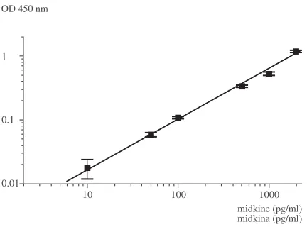
correlation coefficient ≥0.996. A typical reference curve, with standard deviations from the mean val− ues, is presented in Figure 1.
The optimised test was used for quantification of serum midkine levels of 43 apparently healthy subjects to establish reference range of normal serum midkine concentration. The median value of serum midkine level was 141 pg/ml while mean value was 220 ± 320.6 pg/ml. The midkine values for individual serum samples of blood donors are presented in Figure 2, while the distribution of midkine levels in normal serum is summarized in Figure 3. Most of the examined cases were in the subrange from non−detectable to 100 pg/ml, fol− lowed by 100–200 pg/ml. The upper normal range limit, determined as 82ndpercentile, was 541 pg/ml.
The sensitivity of the assay was 3–4 pg of midkine per ml of serum.
Non−specific interactions of sera components with a plate were estimated and the mean absorbance of individually established controls for randomly selected 15 cases was 0.121 ± 0.008, which is a comparable value with those obtained for test sample blanks (from 0.118 ± 0.001 to 0.124 ±0.003 in different assays).
Discussion
The serum midkine assay presented in this paper utilizes, in one test, two kinds of polyclonal
anti−human midkine antibodies, in contradistinc− tion to midkine ELISA described by Muramatsu et al. [21], where only rabbit anti−human midkine antibodies were used for antigen capture and detection. Application of only one type of antibod− ies results in low test sensitivity as the same type of epitopes is recognized in capture and detection steps. To improve sensitivity of this method, the authors used detection system with fluorescent substrate, which, however, requires fluorescence microplate reader. Compared to one antibody sys− tem, sandwich ELISA with antibodies from two different species exhibits much higher sensitivity, as various antigen epitopes are utilized. Moreover,
Fig. 1. Standard curve of the concentration of recom− binant human midkine suspended in diluted serum. The mean values of two measurements with standard deviations are presented on log−log scale. The referen− ce curve was fitted by linear regression with correla− tion coefficient of 0.997
Ryc. 1. Krzywa standardowa stężenia ludzkiej midki− ny zawieszonej w rozcieńczonej surowicy krwi. War− tości średnie dwóch pomiarów z odchyleniem standar− dowym zaprezentowano na skali logarytmicznej. Krzywą wzorcową dopasowano za pomocą regresji liniowej ze współczynnikiem korelacji 0,997 OD 450 nm
midkine (pg/ml) midkina (pg/ml) 0.01
10 100 1000
0.1 1
Fig. 2. The midkine concentration in serum samples of individual blood donors in the descending sorting. In the serum samples 28–43 midkine concentrations were non−detectable
Ryc. 2. Stężenie midkiny w surowicy krwi pochodzą− cej od indywidualnych krwiodawców uporządkowano malejąco. Stężenie midkiny w próbkach 28–43 było niewykrywalne
midkine (pg/ml) midkina (pg/ml)
serum samples próbki surowicy krwi 0
200 400 600 800 1000 1200 1400 1600
5 10 15 20 25 30 35 40
Fig. 3. Distribution of serum midkine levels of appa− rently healthy individuals (n = 43); nd – non−detectable
Ryc. 3. Rozkład częstości stężeń midkiny w surowicy krwi osób uznanych za zdrowe (n = 43); nd – wartość niewykrywalna
midkine (ng/ml) midkina (ng/ml) frequency
częstość
0 5 10 15 20
nd–0.1 0.1–0.2 0.2–0.3 0.3–0.4 0.4–0.5 0.5–0.6 0.6–0.7 0.7–0.8 0.8–0.9 0.9–1.0 >
as pointed out by Ikematsu et al. [17], this type of ELISA can be used with colorimetric system of antigen detection. In his improved midkine assay, rabbit and chicken anti−midkine antibodies were used. Detection antibodies were directly labelled with horseradish peroxidase and tetramethylbenzi− dine was utilized as a substrate. Application of chicken antibodies increased diversity of recog− nized antigen epitopes. However, their availability only on order is highly disadvantageous. The goal was to develop ELISA test equally sensitive but utilizing commercially available antibodies. The authors propose application of antibodies against human recombinant midkine from rabbit and goat, both of which are offered by main antibody manu− facturers. In the assay, detection antibodies were biotin−conjugated and streptavidin coupled to horseradish peroxidase was used for antigen detec− tion and signal amplification. The assay is linear in tested range of 10–2,000 pg of midkine per millil− itre of serum, which is a better result than obtained by Muramatsu (50–10,000 pg/ml) [21]. Although, Ikematsu et al. [17] did not give the details con− cerning the test sensitivity, comparison of standard curves implies that the assay is even slightly more sensitive than the one described by them. It is probably due to the application of biotin−strepta− vidin amplification system in presented method. The sensitivity of presented test, calculated as mean of noise signal absorbancies plus three times standard deviation (mean + 3 SD), was estimated
for 3–4 pg of midkine per ml of serum. The authors used serum depleted of midkine, by the means of affinity chromatography, as a standard solution in order to mimic the composition of serum samples. This, besides application of “sand− wich” system of ELISA with saturating concentra− tions of capture antibody and both separate block− ing step and non−ionic detergent as a diluent, min− imalized the effect of non−specific adsorption to the plate. The absorbancies of individual controls for randomly chosen sera were found to be com− parable with that of midkine−deprived serum. It validated the application of serum depleted of midkine as test sample blank.
The range of reference midkine concentrations is similar to that reported by Ikematsu et al. [17], although in the presently studied group the authors observed some individuals with midkine level ele− vated above 500 pg/ml (Figure 2). The value of 500 pg of midkine per millilitre of serum was established by Ikematsu [17] as a cut−off value and although in the case of described test most of the measured midkine levels were below 200 pg/ml, 541 pg/ml corresponded to 82ndpercentile.
The newly developed method for immunode− tection of midkine in serum proved to be sensitive and reliable. The further investigation on its appli− cation in the studies on potential use of midkine as a marker of tumour presence and progression are under way.
Acknowledgements
We wish to thank dr Ryszard Kozłowski and Mrs Małgorzata Nóżka from Regional Centre of Blood Donation and Therapeutics in Wroclaw, Poland for supply of sera.
References
[1] Kadomatsu K, Huang R−P, Suganuma T, Murata F and Muramatsu T:A retinoic acid responsive gene MK found in the teratocarcinoma system is expressed in spatially and temporally controlled manner during mouse embryogenesis. J Cell Biol 1990, 110, 607–616.
[2] Muramatsu T: Midkine and Pleiotrophin: Two Related Proteins Involved in Development, Survival, Inflammation and Tumorigenesis. J Biochem 2002, 132, 359–371.
[3] Yu GS, Hu J, Nakagawa H:Inhibition of b−amyloid cytotoxicity by midkine. Neurosci Lett 1998, 254, 125–128.
[4] Callebaut C, Nisole S, Briand JP, Krust B, Hovanessian AG:Inhibition of HIV infection by the cytokine mid− kine. Virology 2001, 281, 248–264.
[5] Choudhuri R, Zhang HT, Donnini S, Ziche M, Bicknell R:An angiogenic role for the neurokines midkine and pleiotrophin in tumorigenesis. Cancer Res 1997, 57, 1814–1819.
[6] Garver RI Jr, Chan CS, Milner PG:Reciprocal expression of pleiotrophin and midkine in normal versus malig− nant lung tissues. Am J Respir Cell Mol Biol 1993, 9, 463–466.
[7] Garver RI Jr, Radford DM, Donis−Keller H, Wick MR, Milner PG:Midkine and pleiotrophin expression in normal and malignant breast tissue. Cancer 1994, 74, 1584–1590.
[8] Aridome K, Tsutsui J, Takao S, Kadomatsu K, Ozawa M, Aikou T, Muramatsu T:Increased midkine gene expression in human gastrointestinal cancers. Jap J Cancer Res 1995, 86, 655–661.
[9] Kato M, Maeta H, Kato S, Shinozawa T, Terada T:Immunohistochemical and in situ hybridization analyses of midkine expression in thyroid papillary carcinoma. Mod Pathol 2000, 13, 1060–1065.
[11] Qi M, Ikematsu S, Ichihara−Tanaka K, Sakuma S, Muramatsu T, Kadomatsu K:Midkine rescues Wilms’ tumor cells from cisplatin−induced apoptosis: regulation of Bcl−2 expression by midkine. J Biochem 2000, 127, 269–277.
[12] Nakagawara A, Milbrandt J, Muramatsu T, Deuel TF, Zhao H, Cnaan A, Brodeur GM: Differential expres− sion of pleiotrophin and midkine in advanced neuroblastomas. Cancer Res 1995, 55, 1792–1797.
[13] O’Brien T, Cranston D, Fuggle S, Bicknell R, Harris AL:The Angiogenic Factor Midkine Is Expressed in Bladder Cancer, and Overexpression Correlates with a Poor Outcome in Patients with Invasive Cancers. Cancer Res 1996, 56, 2515–2518.
[14] Mishima K, Asai A, Kadomatsu K, Ino Y, Nomura K, Narita Y, Muramatsu T, Kurino T: Increased expres− sion of midkine during the progression of human astrocytomas. Neurosci Lett 1997, 233, 29–32.
[15] Konishi N, Nakamura M, Nakaoka S, Hiasa Y, Cho M, Uemura H, Hirao Y, Muramatsu T, Kadomatsu K:
Immunohistochemical analysis of midkine expression in human prostate carcinoma. Oncology 1999, 57, 253–257.
[16] Ye C, Qi M, Fan Q−W, Ito K, Akiyama S, Kasai Y, Matsuyama M, Muramatsu T, Kadomatsu K:Expression of midkine in the early stage of carcinogenesis in human colorectal cancer. Br J Cancer 1999, 79, 179–184.
[17] Ikematsu S, Yano A, Aridome K, Kikuchi M, Kumai H, Nagano H, Okamoto K, Oda M, Sakuma S, Aikou T, Muramatsu H, Kadomatsu K, Muramatsu T:Serum midkine levels are increased in patients with various types of carcinomas. Br J Cancer 2000, 83, 701–706.
[18] Shimada H, Nabeya Y, Tagawa M, Okazumi S, Matsubara H, Kadomatsu K, Muramatsu T, Ikematsu S, Sakuma S, Ochiai T:Preoperative serum midkine concentration is a prognostic marker for esophageal squamous cell carcinoma. Cancer Sci 2003, 94, 628–632.
[19] Shimada H, Nabeya Y, Okazumi S, Matsubara H, Kadomatsu K, Muramatsu T, Ikematsu S, Sakuma S, Ochiai T: Increased serum midkine concentration as a possible tumor marker in patients with superficial esophageal cancer. Oncol Rep 2003, 10, 411–414.
[20] Obata Y, Kikuchi S, Lin Y, Yagyu K, Muramatsu T, Kumai H, Tokyo Research Group on Prevention of Gastric Cancer:Serum midkine concentrations and gastric cancer. Cancer Sci 2005, 96, 54–56.
[21] Muramatsu H, Song X, Koide N, Hada H, Tsuji T, Kadomatsu K, Inui T, Kimura T, Sakakibara S, Muramatsu T:Enzyme−linked immunoassay for midkine and its application to evaluation of midkine levels in de− veloping mouse brain and in sera from patients with hepatocellular carcinomas. J Biochem 1996, 119, 1171–1175.
Address for correspondence:
Małgorzata Krzystek−KorpackaDepartment of Medical Biochemistry Silesian Piasts University of Medicine Chałubińskiego 10
50−368 Wrocław Poland
tel.: +48 071 784 13 85 fax: +48 071 784 00 85
e−mail: [email protected]
Conflict of interest: None declared
Received: 13.06.2005 Revised: 1.07.2005 Accepted: 19.07.2005
Praca wpłynęła do Redakcji: 13.06.2005 r. Po recenzji: 1.07.2005 r.