E
WAD
WORNICZEK1, E
WAM
RÓZ1, J
ACEKS
KAŁA2, A
NNAP
RZONDO−M
ORDARSKA1,
A
GNIESZKAG
OJ2, M
AŁGORZATAB
ORTNICZUK2Trimethoprim Resistance in Escherichia coli
Strains Isolated from Patients
with Urinary Tract Infection in 1989–1994
Oporność na trimetoprim wśród szczepów
Escherichia coli
izolowanych od pacjentów z zakażeniem układu moczowego
w latach 1989–1994
1 Department of Microbiology, Silesian Piasts University of Medicine in Wrocław, Poland 2 Department of Genetics, Institute of Genetics and Microbiology, University of Wrocław, Poland
Adv Clin Exp Med 2007, 16, 1, 35–42 ISSN 1230−025X
ORIGINAL PAPERS
© Copyright by Silesian Piasts University of Medicine in Wrocław
Abstract
Introduction. Trimethoprim (Tmp), alone or in combination with sulfamethoxazole (co−trimoxazole), is com− monly used for the treatment of urinary tract infections (UTI). In Poland, Tmp has been used in monotherapy since the late nineties.
Objective.In this study the level of resistance to Tmp and the prevalence of dfrgenes among E. colistrains iso− lated from urine in the period of 1989–1994 was investigated.
Material and Methods.Five hundred fifty−seven E. colistrains isolated from patients with significant bacteriuria were studied. E. coliC600K12 and E. coliJ53K12 were used for the conjugational transfer of the resistance deter− minant of Tmp−resistant E. coli(donor strains) and recipients. The disc−diffusion method was performed to detect antibiotic resistance. The MICs of the antibiotics were determined by an agar dilution method. PCR was used to determine dfr genes encoding for dihydrofolate reductases.
Results. Of the 557 E. coliisolates, 15% were resistant to trimethoprim (MIC ≥4 mg/l). Of this group, 72% of the strains exhibited high−level resistance to Tmp (MIC > 1024 mg/l). Most of them were additionally resistant to three or more other antibiotics and resistance to doxycycline was dominant. Tmp resistance was transferred on conjuga− tive plasmids from 55% of the donor strains to recipient E. coli. Co−transfer of various other resistance determi− nants with Tmp resistance was observed, streptomycin resistance being the most common. A gene determining Tmp resistance, dfrA1, was the most prevalent in the E. coliisolates (91%). The remaining E. colistrains (9%) pos− sessed the dfrB2gene.
Conclusions.Most E. colistrains were highly resistant to Tmp (MIC > 1024 mg/l). Tmp resistance was mediated by conjugative plasmids containing dfrgenes. Type I dihydrofolate reductase (DHFR I) was the main enzyme responsible for high−level resistance to Tmp in the studied E. colistrains. Despite the late introduction of Tmp (a single agent) into therapy, the ubiquitous use of co−trimoxazole has caused the development of resistance to Tmp among E. coli(Adv Clin Exp Med. 2007, 16, 1, 35–42).
Key words: E. coli, trimethoprim, resistance, dfr, DHFR.
Streszczenie
Wprowadzenie.Trimetoprim (Tmp), pojedynczo lub w połączeniu z sulfametoksazolem (kotrimoksazol), jest po− wszechnie stosowany w leczeniu zakażeń dróg moczowych (z.u.m.).
W Polsce trimetoprim wprowadzono do monoterapii pod koniec lat 90.
Cel pracy.Oznaczenie poziomu oporności na Tmp oraz występowania genów dfrwśród szczepów E. coliizolo− wanych z moczu w latach 1989–1994.
Urinary tract infection (UTI) caused byEsche− richia coli is a frequent disease in hospitals as well as in ambulatory patients. The susceptibility pat− terns of antibiotics for this pathogen vary in differ− ent parts of the world [1]. Trimethoprim (Tmp), in combination with sulfonamides, has been success− fully used in the treatment of UTI. Since 1972 it has also been available as a single agent in many countries [2–4]. In Poland, however, Tmp alone was introduced into clinical use at the end of the nineties. Its use in conjunction with sulametoxa− zole (co−trimoxazole), introduced into therapy in 1969 [5], seems to be preferable to Tmp alone.
Tmp selectively inhibits bacterial dihydrofo− late reductase (DHFR), which catalyses the con− version of dihydrofolate to tetrahydrofolic acid, and this affects the biosynthesis of DNA. Several different mechanisms of Tmp resistance have been identified in bacteria, but the most common is the production of an additional plasmid−encoded Tmp−resistant DHFR [6–9]. Tmp resistance due to mutational changes in the intrinsic chromosomal DHFR confers low or intermediate levels of resis− tance, while a plasmid−borne DHFR usually medi− ates resistance to a high concentration of Tmp. dfr
genes expressing Tmp−insensitive DHFR are localized on transferable elements, the latter being efficiently spread on plasmids, transpozons, and integron cassettes. Among Gram−negative bacte− ria, insensitive DHFR spreading on plasmids seems to be of the greatest clinical significance [8–11].
The epidemiology of Tmp−resistant dfrgenes in Poland is not well known since, for many years, Tmp alone was not usually tested against the microorganisms. Sensitivity testing in Polish hos− pital laboratories was performed for co−trimoxa− zole rather than for Tmp alone. In this paper, a fol− low−up study of Tmp resistance among E. coli
strains isolated from patients in the period from 1989 to 1994 and analyzed for the distribution of the dfrA1, dfrA5, dfrA15, dfrA15b, dfrA16,
dfrA16b, dfrB1, dfrB2, and dfrB3genes encoding
DHFR types I, V, XV, XVb, XVI, XVIb, IIa, IIb and IIc, respectively is described. Additionally, the resistance to other drugs ‘linked’ with Tmp resis− tance was determined.
Material and Methods
Bacterial strains
During the 5 years between 1989 and 1994, 557 consecutive E. coliisolates that were a factor in significant bacteriuria were collected. The strains were isolated from out− and inpatients of clinics of the Medical University in Wrocław. Repeated samples were excluded.
The recipient strains used in conjugation experiments were E. coli C600K12 (TmpS, NaR,
lac–, tre–, leu–, the–) and E. coli J53K12 *(TmpS,
NaS, RfR, lac+, pro–, met–), obtained from the Insti−
tute of Immunology and Experimental Therapy PAN (IIET), Poland (Na−nalidixic acid, lac−lac− tose, tre−trehalose, leu−leucine, the−thiamine, Rf− rifampicin, pro−proline, met−methionine, S−sensi− tive, R−resistant, (–) negative, (+) positive.). For control plasmids, the E. coliNCTC 50535, 50536, and 50515 strains were used. Bacteria were grown in Nutrient Broth/Agar or in Brain Heart Infusion broth (BHI) (Biomed, Poland) and identified to the species level by biochemical tests (ID 32E, bioMerieux, France). Iso−Sensitest medium (Oxoid, UK) was used for susceptibility testing (supplemented with lysed horse blood 5% v/v) when the MIC for Tmp was determined [12], Luria−Bertani (LB) medium (Difco, USA) ws used for isolation of plasmids and Falkow minimal medium for the conjugation experiments [16].
Plasmids
The plasmids used as a source of positive con− trol DNA for PCR were pFE872 for dfrA1 gene [13] and pWZ820 for dfrB2gene [14].
Wyniki. Spośród 557 badanych szczepów E. coli15% było opornych na Tmp (MIC 4 mg/L). W grupie tej 72% szczepów wykazywało wysoki poziom oporności (MIC > 1024 mg/L). Większość z nich była dodatkowo oporna na 3 lub więcej antybiotyków z wyraźną przewagą oporności na doksycyklinę. U 55% szczepów oporność na Tmp była przekazywana na plazmidach w procesie koniugacji do komórek biorcy. Wraz z opornością na Tmp przeka− zywane były markery oporności na inne leki, głównie streptomycynę. Gen dfrA1warunkujący oporność na Tmp stwierdzono u większości badanych szczepów E. coli(91%). Pozostałe szczepy E. coli(9%) zawierały gen dfrB2.
Wnioski. Większość badanych szczepów E. coli wykazywała wysoki poziom oporności na Tmp (MIC > 1024 mg/L). Za oporność odpowiadały koniugacyjne plazmidy z genami dfr. Reduktaza dihydrofolianu typu I (DHFR I) była głównym enzymem odpowiedzialnym za wysoką oporność na Tmp u badanych szczepów. Mimo późnego wprowadzenia Tmp (pojedynczego leku) do terapii, powszechne stosowanie w lecznictwie kotrimoksazolu spowo− dowało wyraźny rozwój oporności na Tmp u pałeczek E. coli(Adv Clin Exp Med. 2007, 16, 1, 35–42).
Susceptibility Testing
The disc−diffusion technique was applied using commercial discs with the following antimi− crobial agents: trimethoprim, doxycycline, specti− nomycin, amikacin, gentamicin, streptomycin, ka− namycin, ampicillin, piperacillin, carbenicillin, ceftazidime, cefuroxime, cefotaxime, and chlor− amphenikol [15]. The minimal inhibitory concen− tration (MIC) was determined by an agar dilution method [12]. The concentration of the antibiotics ranged from 4 to 1024 mg/l. High−level resistance to Tmp was defined as MIC > 1024 mg/l.
Resistance Transfer
Experiments
In conjugation studies, selected E. coli strains which were highly resistant to Tmp were used as donor strains. They were mated with the recipient
E. coli C600K12 strain. The resulted Tmp−resistant transconjugants were then mated with a second type of recipient, E. coli J53K12 (confirmation of trans− fer of resistance determinants). All mating experi− ments were performed as follows: overnight cul− tures of donor and recipient strains diluted 10 times with BHI broth were incubated at 37°C and 28°C. After the incubation period, the donor and recipient strains were mixed in the ratio 1:1 in the same broth and incubated for 24 h at 37°C and 28°C. From each overnight culture, 0.1 ml was spread on the selec− tive Falkow agar supplemented with growth factors for the recipient and antibiotics from the spectrum resistance determined by the plasmid carried by the donor. After the incubation period, the number of colonies was counted. The colonies were purified three times on an appropriate selective medium. The frequency of transfer of Tmp resistance was expressed as the number of transconjugant cells in relation to the number of donor cells [16].
DNA Isolation
Bacterial strains were incubated for 20 h at 37°C with constant agitation (196 r.p.m.) in LB medium (1% yeast extract, 1% bactotryptone, and 0.5% sodium chloride) containing 500 µg/ml of Tmp. Plasmid DNA was extracted from the 3−ml culture with a Plasmid Mini kit (A&A Biotech− nology, Gdynia, Poland) according to the manu− facturer’s instructions.
Molecular Typing
PCR was performed in a volume of 25 µl con− taining a 1−µl DNA sample, 2.5 µl of PCR buffer and 2.5 U of Taq polymerase (Biotools Labs,
Madrid, Spain), 0.2 mM of each dNTP and 50 pmol of both forward and reverse primers. The reactions were carried out in a DNA Engine PTC− 200 (JM Research, USA) thermocycler, compris− ing 30 cycles of 1 min at 94°C, 1 min at 65°C, 1 min at 72°C, and a final extension of 5 min at 72°C. The primers IAF (5’−GTGAAACTATCAC− TAATGG−3’), IAR (5’−TTAACCCTTTTGC CAGATTT−3’), IIF (5’−GATCGCCTGCGCAA− GAAATC−3’), and IIR (5’−AAGCGCAGCC ACAGGATAAAT 3’), described previously by Navia et al. [17], were used. The pair of primers IAF/IAR results in an expected PCR product of 474 bp for the dfrA1, dfrA5, dfrA15, dfrA15b,
dfrA16, and dfrA16b genes, while the primers IIF/IIR yield a band of 141 bp for the dfrB1, dfrB2, and dfrB3 genes [17]. For the final dfrgenes iden− tification, the amplicons were digested by the restriction enzyme TasI (Tsp509I, Fermentas, Lithuania) and the resulting RFLP (Restriction Fragments Length Polymorphism) pattern was compared with data published by Navia et al. [17]. Whole PCR products and restriction frag− ments were visualized by electrophoresis on 1.3% or 2% agarose (Basica LE, Prona, Spain) gels with ethidium bromide (0.25 µg/ml) using standard procedures [18].
Results
Trimethoprim Resistant Isolates
The results showed that from 557 E. coli
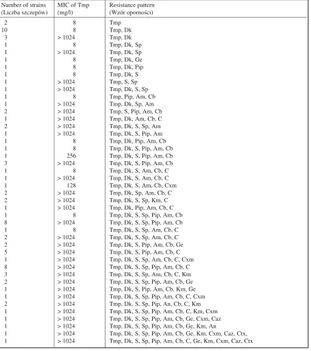
strains isolated during the period of study, 15% (86) were resistant to Tmp. Among them, 72% (62) exhibited high−level resistance (MIC > 1024 mg/l). The remaining strains showed resistance to Tmp with MIC of 4–1024 mg/l (Table 1).
Resistance to Other Drugs
All (86) strains resistant to Tmp (MIC ≥4 mg/l) were also tested for their resistance to other antibio− tics and chemotherapeutics. The variety of resistance patterns for E. coliTmpR(Tmp−resistant) isolates is
shown in Table 1. Resistance to doxycycline (93%) was the most common. Over 70% of TmpR strains
were found to carry resistance to ampicillin. More strains presented resistance to carbenicillin (69%), piperacillin (54%), and chloramphenicol (48%) than to kanamycin (13%) and gentamicin (12%). Resi− stance to streptomycin and spectinomycin was noted in 71% and 53% of the strains, respectively. Less fre− quent was resistance to cefuroxime (8%) and ceftazi− dime (6%). Only 2% of the TmpRstrains showed re−
Transfer of Trimethoprim
Resistance
For conjugational crosses, 55 highly TmpR
strains were selected as donors. Seven isolates
resistant to one of the selection markers were rejected. In the case of 30 (55%) donor strains, resistance to Tmp was successfully transferred to recipients. No further transfer of resistance deter− minants was observed, even when conjugation Tabela 1. Wzory oporności na antybiotyki 86 szczepów E. coli(TmpR)
Table 1. Antibiotic resistance patterns in the 86 E. coli(TmpR) isolates
Number of strains MIC of Tmp Resistance pattern (Liczba szczepów) (mg/l) (Wzór oporności)
2 8 Tmp
10 8 Tmp, Dk
3 > 1024 Tmp, Dk
1 8 Tmp, Dk, Sp
1 > 1024 Tmp, Dk, Sp
1 8 Tmp, Dk, Ge
1 8 Tmp, Dk, Pip
1 8 Tmp, Dk, S
1 > 1024 Tmp, S, Sp
1 > 1024 Tmp, Dk, S, Sp
1 8 Tmp, Pip, Am, Cb
1 > 1024 Tmp, Dk, Sp, Am
2 > 1024 Tmp, S, Pip, Am, Cb
1 > 1024 Tmp, Dk, Am, Cb, C
2 > 1024 Tmp, Dk, S, Sp, Am
1 > 1024 Tmp, Dk, S, Pip, Am
1 8 Tmp, Dk, Pip, Am, Cb
1 8 Tmp, Dk, S, Pip, Am, Cb
1 256 Tmp, Dk, S, Pip, Am, Cb
3 > 1024 Tmp, Dk, S, Pip, Am, Cb
1 8 Tmp, Dk, S, Am, Cb, C
1 > 1024 Tmp, Dk, S, Am, Cb, C
1 128 Tmp, Dk, S, Am, Cb, Cxm
2 > 1024 Tmp, Dk, Sp, Am, Cb, C
2 > 1024 Tmp, Dk, S, Sp, Km, C
1 > 1024 Tmp, Dk, Pip, Am, Cb, C
1 8 Tmp, Dk, S, Sp, Pip, Am, Cb
8 > 1024 Tmp, Dk, S, Sp, Pip, Am, Cb
1 8 Tmp, Dk, S, Sp, Am, Cb, C
2 > 1024 Tmp, Dk, S, Sp, Am, Cb, C
2 > 1024 Tmp, Dk, S, Pip, Am, Cb, Ge
5 > 1024 Tmp, Dk, S, Pip, Am, Cb, C
1 > 1024 Tmp, Dk, S, Sp, Am, Cb, C, Cxm
8 > 1024 Tmp, Dk, S, Sp, Pip, Am, Cb, C
3 > 1024 Tmp, Dk, S, Sp, Am, Cb, C, Km
2 > 1024 Tmp, Dk, S, Sp, Pip, Am, Cb, Ge
1 > 1024 Tmp, Dk, S, Pip, Am, Cb, Km, Ge
1 > 1024 Tmp, Dk, S, Sp, Pip, Am, Cb, C, Cxm
2 > 1024 Tmp, Dk, S, Sp, Pip, An, Cb, C, Km
1 > 1024 Tmp, Dk, S, Sp, Pip, Am, Cb, C, Km, Cxm 1 > 1024 Tmp, Dk, S, Sp, Pip, Am, Cb, Ge, Cxm, Caz 1 > 1024 Tmp, Dk, S, Sp, Pip, Am, Cb, Ge, Km, An
1 > 1024 Tmp, Dk, S, Sp, Pip, Am, Cb, Ge, Km, Cxm, Caz, Ctx, 1 > 1024 Tmp, Dk, S, Sp, Pip, Am, Cb, C, Ge, Km, Cxm, Caz, Ctx
Tmp – trimetoprim, Dk – doksycyklina, Sp – spektynomycyna, Am – ampicylina, Cb – karbenicylina, C – chloramfenikol, S – streptomycyna, Ge – gentamycyna, An – amikacyna, Km – kanamycyna, Pip – piperacylina, Cxm – cefuroksym, Caz – ceftazydym, Ctx – cefotaksym.
was repeated at 28°C. These strains required mobi− lization the X+factor (E. coliC20, F+ (IIET, PAN)).
Three of these strains transferred Tmp resistance via mobilization. All transconjugants obtained were highly resistant to Tmp (MIC > 1024 mg/l). The frequency of conjugational transfer varied from 2.4 × 10⁻⁶ to 4.4 × 10⁻¹ transconjugants per
donor. In most cases the transfer frequency amounted to 10–4– 10–3.
Linkage of Trimethoprim
Resistance with Resistance
to Other Antibiotics
For all transconjugants highly resistant to Tmp, resistance profiles (MIC) for other drugs were determined. More than 80% of transconju− gants showed a linkage of Tmp and streptomycin resistance. However, co−transfer with spectino− mycin resistance was shown only in 32% of the transconjugants. Determinants expressing resis− tance to ampicillin, doxycycline, carbenicillin, and piperacillin were found in (rates for transconju− gants obtained from matings with recipients a) E. coliC600K12 and b) E. coliJ53K12) (a)87–b)79%,
84–76%, and 71–68% of the transconjugants, respectively. Lower co−transfer with Tmp was demonstrated for chloramphenicol (21–18%), gen− tamycin (21–16%), and kanamycin (11–13%). Resistance to 3 cephalosporines was co−trans− ferred to less than 6% of the transconjugants. None of the TmpR transconjugants exhibited resistance
to amikacin.
Characterization
of Tmp
RPlasmids
Most of the donor strains (91%) were shown to harbor plasmids ranging in size from 1 to 60 kb. Although in three strains plasmids were not visu− alized, the transfer of resistance to Tmp and other drugs was successful. In 70% of the isolates, large plasmids (50–60 kb) were detected. This plasmid was transferred to 42% of the transconjugants in the first conjugation mating and to 40% in the sec− ond. The most commonly observed resistance pat− terns which correlated with a transfer of the above plasmid were Tmp−Dk−S, Tmp−Pip−Am−Cb, and Tmp−Dk−S−Pip−Am−Cb. The remaining transcon− jugants possessed 1–5 small plasmids in the size range of 1–7 kb. In several multiresistant transcon− jugants, no plasmids were visualized.
Genes Conferring
Tmp Resistance
The presence of dfr genes was confirmed by PCR in E. coliclinical isolates and in transconju− gants obtained from conjugational crossing with the second recipient, E. coliK12J53 (Figures 1–3). Genes detected in all transconjugants correlated well with those detected in the donor strains. Most (30/33) of the strains harbored the gene dfrA1and only 2/33 strains possessed the gene dfrB1. The genes dfrA5, dfrA15, dfrA15b, dfrA16, dfrA16b,
dfrB2, or dfrB3 were not found in the studied strains. One strain expressed Tmp resistance encoded by an unidentified gene.
Fig. 1. Specific amplification of selected fragments of dfrgenes. Electrophoretic separation of amplicons obtained with the following primer pairs and DNA: lane (1) primers IAF/IAR and control plasmid pFE872 (dfrA1); (2) IAF/IAR and control plasmid pWZ820 (dfrB2); (3) negative control for PCR with IAF/IAR primers; (4) primers IIF/IIR and control plasmid pFE872 (dfrA1); (5) IIAF/IIAR and control plasmid pWZ820 (dfrB2); (6) negative control for PCR with IIAF/IIAR primers; (7) IAF/IAR and DNA from clinical isolate 383; (8) IAF/IAR and DNA from clinical isolate 917; (9) IAF/IAR and DNA from transconjugant J383/1/1; (10) IAF/IAR and DNA from transconjugant J917/1/29; (11) IIF/IIR and DNA from clinical isolate 907; (12) IIAF/IIAR and DNA from transconjugant J907/1/9. M: molecular weight marker (pUCMix8, Fermentas). The arrows indicate the expected amplicon size of 474 bp and 141 bp [17]
Ryc. 1.Specyficzna amplifikacja wybranych fragmen− tów genów dfr. Elektroforeza amplikonów uzyskanych z użyciem następujących par starterów i matrycowych DNA: ścieżka (1) – startery IAF/IAR i plazmid kon− trolny pFE872 (dfrA1); (2) – IAF/IAR i plazmid kon− trolny pWZ820 (dfrB2); (3) – kontrola negatywna re− akcji PCR ze starterami IAF/IAR; (4) – startery IIF/IIR i plazmid kontrolny pFE872 (dfrA1); (5) – IIAF/IIAR i plazmid pWZ820 (dfrB2); (6) – ujemna kontrola reakcji ze starterami IIAF/IIAR; (7) – IAF/IAR i DNA ze szczepu klinicznego nr 383; (8) – IAF/IAR i DNA ze szczepu klinicznego nr 917; (9) – IAF/IAR i DNA z transkoniuganta nr J383/1/1; (10) – IAF/IAR i DNA z transkoniuganta nr J917/1/29; (11) – IIF/IIR i DNA ze szczepu klinicznego nr 907; (12) – IIAF/IIAR i DNA z transkoniuganta nr J907/1/9; M – marker masy (pUCMix8, Fermentas). Strzałkami za− znaczono wielkość spodziewanych amplikonów 474 bp i 141 bp [17]
1 2 3 4 5 6 M 7 8 9 10 11 12
474 bp
Discussion
Trimethoprim−resistant bacteria have been iso− lated worldwide since the combination of trime− thoprim and sulfonamides was introduced for clin− ical treatment. Resistance to this chemotherapeutic in Gram−negative bacteria and a variety of mecha−
nisms responsible for it have been widely reported [19–25]. In Poland, Tmp alone has been registered for clinical use since 1998. Co−trimoxazole, how− ever, has been extensively used for the treatment of various infections, including UTI, during the last 30 years.
Results of these studies show that 15% of E. coliisolated from UTI in the Wrocław area were resistant to Tmp. These data are comparable to those seen in 1998 in Rovaniemi (14%), Turku (16%), and Helsinki (19%) in Finland [20]. The first screening work on Tmp resistance in our geo− graphical region was conducted by Złotorzycka et al. on E. coliisolated from urine and fecal samples [data not published]. The report demonstrated heightened resistance from 0% in 1977–1979 to 1% in 1980–1981 and 1.2% in 1983. Compared with these data, in the years 1989–1994 a 15−fold increase of Tmp resistance in E. coli strains is observed. These findings therefore indicate a slow development of trimethoprim resistance to the year 1994. This incidence is much lower than that seen in India [26], Chile [27], and Taiwan [28], where by the end of the eighties more than 40% of isolates were resistant to Tmp. This variable resis− tance may reflect the consumption and control of drugs in particular countries. Our results also show a high proportion (72%) of high−level resistance to this agent in the studied strains. Similarly, Brumfitt et al. [29] reported an increase in highly Tmp resistant E. coliisolates from 22% in 1973 to 91% in 1981. Heikkilä et al. [20] reported high− level Tmp resistance in more than 90% of strains in Finland and Tsakris et al. [22] of 89% in Greece at the beginning of the nineties. This increasing tendency may suggest plasmid/transpozon−medi− ated Tmp resistance dissemination.
Sixty percent (33/55) of the strains studied could have transferred Tmp resistance determi− nants via conjugation, indicating the presence of R−plasmids. Similar proportions (more than half of the strains) are reported by Tsakris et al. [30] and Mayer [31]. Lin−Li Chang reported 66% of trans− ferable high−level Tmp resistance in 1987, but at the same time emphasized a decreasing tendency to 53.7% in 1989, suggesting an influence of non− transferable plasmids or transpozons [28].
In bacteria, genetic changes concerning resis− tance to antimicrobial agents have been rapid. Intensive, multiple, and sometimes prolonged application of drugs usually leads to the selection of multiresistant strains. Such strains may carry linked determinants of antibiotic resistance [32]. The present authors observed that along with resis− tance to Tmp, many other determinants were co− transferred. This result probably reflects the wide spectrum of antibiotics used in Poland. The most Fig. 2. Electrophoresis of amplikons obtained with
primers IAF/IAR and digested by TasI enzyme. Lanes 1–8: clinical isolates number 654, 716, 893, 917, 16, 294, 975, and 28, respectively. M: molecular weight marker (pUCMix8, Fermentas). The arrows indicate the 236 bp and 163 bp restriction fragments typical for the dfrAI gene [17]
Ryc. 2. Elektroforeza amplikonów uzyskanych parą starterów IAF/IAR i trawionych enzymem restrykcyj− nym TasI. Ścieżki 1–8 odpowiednio szczepy kliniczne numer 654, 716, 893, 917, 16, 294, 975 i 28. M – mar− ker masy (pUCMix8, Fermentas). Strzałkami zazna− czono fragmenty restrykcyjne o wielkości 236 bp i 163 bp charakterystyczne dla genu dfrA1 [17]
1 2 3 4 5 6 7 8 M
236 bp 163 bp
Fig. 3. Electrophoresis of amplicons obtained with primers IIAF/IIAR and digested by TasI enzyme. Lanes 1–4: clinical isolates and their transconjugants 907 and J907/1/9, 223, and J223/1/5, respectively. M – molecular weight marker (pUCMix8, Fermentas). The arrows indicate the 78 bp and 63 bp restriction frag− ments typical for the dfrB1 gene [17]
Ryc. 3. Elektroforeza amplikonów uzyskanych parą starterów IIAF/IIAR i trawionych enzymem restryk− cyjnym TasI. Ścieżki 1–4 odpowiednio szczepy kli− niczne i ich transkoniuganty 907 i J907/1/9 oraz 223 i J223/1/5. M – marker masy (pUCMix8, Fermentas). Strzałkami zaznaczono fragmenty restrykcyjne o wiel− kości 78 bp i 63 bp charakterystyczne dla genu dfrB1 [17]
1 M 2 3 4
frequent in Tmp−resistant transconjugants was the linkage with resistance to streptomycin, ampi− cillin, doksycycline, and piperacyllin. This may follow (except streptomycin) their role in the treat− ment of urinary tract infections. Amyes indicated that Tmp−resistant bacteria often had linked plas− mid genes conferring both trimethoprim and ampi− cillin resistance [33]. Thus, ampicillin might play the role of a powerful selector for plasmid dfr
genes in bacterial populations. Resistance to gen− tamicin and kanamycin was co−transferred less frequently than for streptomycin and spectino− mycin. None of the transconjugants was resistant to amikacin, which may reflect their low usage compared with other aminoglycosides during the study period. Similar rates were observed for chlo− ramphenicol and cephalosporines.
In the most of the trimethoprim−resistant trans− conjugants, variable plasmids were present. They differed in their molecular mass and antibiotic resistance patterns. However, additional studies are needed to determine their relationship.
Here, the prevalence of dfrgenes in E. coliuri− nary isolates was investigated. These data showed
that dfrA1 was the most prevalent gene, while
dfrB2was detected in only a few strains. The pres− ence of dfrA5, dfrA15, dfrA15b, dfrA16, dfrA16b, and dfrB2or dfrB3genes in the studied strains was not observed. Similar results, i.e. the prevalence of type I DHFR (dfrA1) in E. coli, have been report− ed in other parts of the world [22, 3–36]. The pre− sent authors believe that this is the first report on the distribution of trimethoprim resistance genes in Gram−negative rods in Poland. The results sug− gest that the plasmid−borne dfrA1gene has spread efficiently among E. coli from this geographical region, probably under the selective pressure of the ubiquitous use of Tmp in combination with sulfamethoxazole. In this study it was not deter− mined whether dfrA1 gene was a component of Tn7 or was integrated in gene cassettes. The high percentage of transconjugants resistant to both trimethoprim and streptomycin may suggest, how− ever, the presence of transpozon Tn7 as a mediator of Tmp resistance in the studied strains. Further studies will be conducted for the exact calculation of trimethoprim resistance gene dissemination in hospital settings in Poland.
References
[1] Yu HS, Lee JC, Kang HY, Ro DW, Chung JY, Jeong YS, Tae SH, Choi CH, Lee EY, Seol SY, Lee YC, Cho DT:Changes in gene cassettes of class 1 integrons among Escherichia coliisolates from urine specimens collect− ed in Korea during the last two decades. J Clin Microbiol 2003, 4, 5429–5433.
[2] Amyes SGB, Doherty CJ, Wonnacott S: Trimethoprim and co−trimoxazole: a comparison of their use in respi− ratory tract infections. Scan J Dis 1986, 18, 561–566.
[3] Lacey RW:Do sulphonamide−trimethoprim combinations select less resistance to trimethoprim than the use of trimethoprim alone? J Med Microbiol 1982, 15, 403–427.
[4] Lacey RW, Lord VL, Gunasekera KW, Leiberman PJ, Luxton DEA: Comparison of trimethoprim alone with trimethoprim−sulphamethoxazole in the treatment of respiratory and urinary tract infections with particular refer− ence to selection of trimethoprim resistance. Lancet 1980, i, 1270–1273.
[5] Coque TM, Singh KV, Weinstock GM, Murray BE: Characterization of dihydrofolate reductase genes from trimethoprim−susceptible and trimethoprim−resistant strains of Enterococcus faecalis. Antimicrob Agents Chemother 1999, 43,141–147.
[6] Steen R, Sköld O: Plasmid−borne or chromosomally mediated resistance by Tn7 is the most common response to ubiquitous use of trimethoprim. Antimicrob Agents Chemother 1985, 27, 933–937.
[7] Huovinen P: Increases in rates of resistance to trimethoprim. Clin Infect Dis 1997, 24, S63–66.
[8] Lee JC, Oh JY, Cho JW, Park JC, Kim JM, Seol SY, Cho DT: The prevalence of trimethoprim−resistance−con− ferring dihydrofolate reductase genes in urinary isolates of Escherichia coliin Korea. J Antimicrob Chemother 2001, 47, 599–604.
[9] Huovinen P, Sundström L, Swedberg G, Sköld O: Trimethoprim and sulphonamide resistance. Antimicrob Agents Chemother 1995, 39, 279–289.
[10] White PA, Rawlinson WD:Current status of the aaaA and dfrgene cassette families. J Antimicrob Chemother 2001, 47, 495–502.
[11] Yu HS, Lee JCH, Kang HY et al.: Prevalence of dfrgenes associated with integrons and dissemination of dfr A17 among urinary isolates of Escherichia coliin Korea. J Antimicrob Chemother 2004, 53, 445–450.
[12] Towner KJ: Classification of transferable plasmids conferring resistance to trimethoprim isolated in Great Britain. FEMS Microbiol Lett 1979, 5, 319–321.
[13] Fling ME, Richards C: The nucleotide sequence of the trimethoprim−resistance dihydrofolate reductase gene har− boured by Tn7. Nucleic Acids Res 1983, 11, 5147–5158.
[14] Zolg JW, Hanggi UJ, Zachau HG:Isolation of a small DNA fragment carrying the gene for a dihydrofolate reductase from a trimethoprim resistance factor. Mol Gen Genet 1978, 164, 15–29.
[16] Kurowska E, Cebrat S, Lachowicz TM:Transformation of Escherichia coliby R404 factor DNA and proper− ties of the transformants. Acta Microbiol Polon 1976, 26, 3.
[17] Navia MM, Ruiz J, Sanchez−Cespedes J, Vila J:Detection of dihydrofolate reductase genes by PCR and RFLP. Diagn Microbiol Infect Dis 2003, 46, 295–298.
[18] Sambrook J, Fritsch EF, Maniatis T:Molecular Cloning: a Laboratory Manual. Cold Spring Harbor Laboratory Press, Cold Spring Harbor, NY, USA, 1989, 2ndedn., 9.34–9.51.
[19] Donnan PT, Wei L, Steinke DT, Phillips G, Clarke R, Noone A, Sullivan FM, MacDonald TM, Davey PG:
Presence of bacteriuria caused by trimethoprim resistant bacteria in patients prescribed antibiotics: multilevel model with practice and individual patient data. BMJ 2004, 324, 1297.
[20] Heikkilä E, Renkonen O−V, Sunila R, Uurasmaa P, Huovinen P:The emergence and mechanism of trimetho− prim resistance in Escherichia coli isolated from outpatients in Finland. J Antimicrob Chemother 1990, 25, 275–283.
[21] Grape M, Farra A, Kronvall G, Sundström L:Integrons and gene cassettes in clinical isolates of co−trimoxa− zole−resistant Gram−negative bacteria. Clin Microbiol Infect 2005, 11, 185–192.
[22] Tsakris A, Johnson AP, Legakis NJ, Tzouvelekis LS:Prevalence of the type I and type II DHFR genes in trimetho− prim−resistant urinary isolates of Escherichia colifrom Greece. J Antimicrob Chemother 1993, 32, 665–671.
[23] Haider K, Chatkaemorakot A, Kay BA, Talukder KA, Taylor DN, Echeverii AP, Sack DA:Trimethoprim resistance gene in Shigella dysenteriae 1 isolates obtained from widely scattered locations of Asia. Epidemiol Infect 1990, 104, 219–228.
[24] Adrian PV, Thomson CJ, Klugman KP, Amyes SGB: Prevalence and genetic locations of non−transferable trimethoprim resistant dihydrofolate reductase genes in South African commensal faecal isolates. Epidemiol Infect 1995, 115, 255–267.
[25] Huang DB, Jiang ZD, Ericsson CD, Adachi J, Dupont HL: Emergence of trimethoprim−resistant Escherichia coli in healthy persons in the absence of prophylactic or therapeutic antibiotics during travel to Guadalajara, Mexico. Scan J Infect Dis 2001, 33, 812–814.
[26] Young H−K, Jesudson MV, Koshi G, Amyes SGB:Trimethoprim resistance amongst urinary pathogens in South India. J Antimicrob Chemother 1986, 17, 615–621.
[27] Urbina R, Prado V, Canelo E: Trimethoprim resistance in enterobacteria isolated in Chile. J Antimicrob Chemother 1989, 23, 143–149.
[28] Chang LL, Chang SF, Chow TY, Wu WJ, Chang JC: The distribution of the DHFR genes in trimethoprim− resistant urinary tract isolates from Taiwan. Epidemiol Infect 1992, 109, 453–462.
[29] Brumfitt W, Hamilton−Miller JMT, Wool A: Evidence flora slowing in trimethoprim resistance during 1981 – a comparison with earlier years. J Antimicrob Chemother 1983, 11, 503–509.
[30] Tsakris A, Johnson AP, George RC, Mehtar S, Vatopoulos AC: Distribution and transferability of plasmids encoding trimehoprim resistance in urinary pathogens from Greece. J Med Microbiol 1991, 34, 153–157.
[31] Mayer KH, Fling ME, Hopkins JD, O’Brien TF: Trimethoprim resistance in multiple genera of Enterobacteriaceaeat U.S. Hospital: spread of the type II dihydrofolate reductase gene by a single plasmid. J Infect Dis 1985, 151, 783–789.
[32] Steinke DT, Seaton RA, Philips G, MacDonald TM, Davey PG:Prior trimethoprim use and trimethoprim−resis− tant urinary tract infection: a nested case−control study with multivariate analysis for other risk factors. J Antimicrob Chemother 2001, 47, 781–787.
[33] Amyes SGB:The success of plasmid encoded resistance genes in clinical bacteria: an examination of plasmid− mediated ampicillin and trimethoprim resistance genes and their resistance mechanisms. J Med Microbiol 1989, 28, 73–83.
[34] Murray BE, Alvorado T, Kim K−H:Increasing resistance to trimethoprim−sulphamethoxazole among isolates of Escherichia coliin developing countries. J Infect Dis 1985, 152, 1107–1113.
[35] Steen R, Sköld O: Plasmid−borne or chromosomally mediated resistance by Tn7 is the most common response to ubiquitous use of trimethoprim. Antimicrob Agents Chemother 1985, 27, 933–937.
[36] Heikkilä E, Siitonen A, Jahkola M, Fling M, Sundström L, Huovinen P: Increase of trimethoprim resistance among Shigellaspecies, 1975–1988: analysis of resistance mechanisms. J Infect Dis 1990, 161, 1242–1248.
Address for correspondence:
Ewa Dworniczek
Department of Microbiology, Wrocław Medical University, Chałubińskiego 4,
50−368 Wrocław
Conflict of interest: None declared
Received: 17.10.2006 Revised: 3.01.2007 Accepted: 12.01.2007
Praca wpłynęła do Redakcji: 17.10.2006 r. Po recenzji: 3.01.2007 r.